Proteomic and genomic characterisation of venom proteins from Oxyuranus species
81
0
0
Full text
(2) 95. Chapter 4 Isolation and characterisation of venom proteins. Chapter 4 Isolation and characterisation of a serine protease from O. s. scutellatus. 4.1 INTRODUCTION. The abundance and varying antigenicity of the lower molecular weight proteins reported in Chapter 3 suggested these proteins may provide an interesting area of research. In addition to the numerous small proteins, 0. s. scutellatus proteins form large complexes affecting coagulopathy (Walker, et al,. 1980; Speijer, et al., 1986; Lalloo, et al., 1995b; Kini, et al., 2001).. Differing isomers of these proteins and. peptides may hold the key that may help to describe clinical variations encountered after differing Oxyuranus species envenomation.. Due to the varying coagulopathy's. encountered between Oxyuranus envenomations, the precedent of large, complex enzymes effecting these coagulopathy's and the ability to identify specific proteins within the venom, selected large proteins were isolated and characterised.. 4.1.1 Coagulant Enzymes. Haemostasis is a balance of two opposing forces: clot formation and dissolution. Correct clotting is essential at the site of a wound in order to maintain haemostasis, however clotting away from the site of a wound must be minimised in order to prevent life-threatening thrombotic events. Blood clotting, which occurs through the production of fibrin resulting from the activation of prothrombin to thrombin, involves a cascade of biochemical events that can be initiated intrinsically or extrinsically. The intrinsic pathway requires the clotting factors VIII, IX, X, XI, and XII, prekallikrein, calcium ions and high molecular-weight kininogen. Each of these constituents leads to the conversion of factor X (inactive) to factor Xa (where 'a' signifies active). Factor V, calcium ions and phospholipids secreted from platelets are required for the conversion of prothrombin to thrombin which in turn leads to the production of fibrin (Figure 4.1). The extrinsic pathway is initiated at the site of injury (eg snake bite) in response to the release of tissue factor (factor III), and occurs at factor Xa (the point where the intrinsic.
(3) Chapter 4 Isolation and characterisation of venom proteins. 96. and extrinsic pathways meet). Tissue factor is a cofactor in the factor VIla-catalysed activation of factor X. Factor VIla, a serine protease, cleaves factor X to factor Xa in a manner identical to that of factor IXa of the intrinsic pathway (Davie, et al., 1991; King, 2001). Many species of the Australian elapids show a strong coagulant activity (Chester and Crawford, 1982; Joseph and Kini, 2002; Joseph, et al., 2002). These contain factor Xalike, and to a lesser extent factor Va, plus prothrombin activating enzymes (Suttie and Jackson, 1977; Rosing and Tans, 1988; Mann, et al., 1990; Davie, et al., 1991; King, 2001) which are responsible for most of the disruption of blood chemistry and haemorrhage seen clinically (DIC or defibrinogenation) (Sutherland, 1976; King and Smith, 1991; Lalloo, et al., 1997), see Figure 4.1. One group of therapeutically useful venom proteins that cause haemostatic reactions, which have been highly characterised in the venoms of true vipers (Viperinae) and pit vipers (Crotalinae), are the serine proteases. All have shown to contain highly effective serine proteases and act on different steps of the blood coagulation cascade (Ouyang, et al. 1992; Serrano, et ai., 1995; Markland, 1998a and b; Pirkle, 1998; Stocker, 1998;. Same1, et ai., 2002). Some of these venoms can specifically hydrolyse fibrinogen to release fibrinopeptide A, fibrinopeptide B or both (Hofmann and Bon, 1987;Lollar, et ai., 1987; Markland, 1998a). Some affect other substrates, such as kininogen (Komori. and Nikai, 1998; Nikai and Komori, 1998), plasminogen (Zhang, et ai., 1995; Zhang, et ai., 1997) and protein C (Stocker, 1994). It has been reported the variety of functions. this family of venom proteases undertakes may, again, be due to gene duplication and an accelerated evolution which acquired these special functions (Deshimaru, et ai., 1996; Wang, et ai., 2001). Therapeutic venom proteases are used in areas such as antithrombotics to facilitate tissue oxygenation, prevent arterial embolism (Chang, et ai., 1995) and myocardial infarction (Farid, et ai., 1989).. The venom of 0. s. scutellatus contains a potent converter of prothrombin to thrombin in the absence of all other known clotting factors (Denson, 1969) and it has been demonstrated the venom also possesses an activator of Factor VII (Nakagaki, et ai., 1992). The multimeric prothrombin activator purified from 0. s. scutellatus was. named scutelarin (EC 3.4.21.60) (Walker, et ai., 1980 Speijer, et ai., 1986)..
(4) 97. Chapter 4 Isolation and characterisation of venom proteins. Figure 4.1: Diagrammatic representation of principle ways snake venom interacts with human haemostasis. (Illustration copyright © 2005 reproduced with permission of Dr. Julian White).. Facllitates activatio,1 c::...~ Inhibits acllvalion Transformation to active form OISINlEGRINS Venom component causlng activation Of direct action. (mSINrEGRINS) Venom component causing inhibition. EXTRINSIC SYSTEM. Tissue Factor Ca++ FactorVll. INTRtNS1C SYSTEM. Factor Xlla. Factor XIII. PLATELET AGGREGATION INHIBITION BY ENDOTHELIAL CELLS PRO$TACYCLIN ENHANCE PROTE!N C ADENOSINE ACTIVATION PROTEASE NECTIN ENHANCEATIU SEROTON!N. «. ~.....;lII--""" /'~...".-~ ~=.:.;.:;~.,::;; /~~,......,.. ~.".."..~"" /.......".""~. :e. ~~~~~~~~~~~~~~~~~~~~~~~.
(5) Chapter 4 Isolation and characterisation of venom proteins. 98. is activator is approximately 260 kDa and is composed of four subunits: A 110 kDa subunit showing similar enzymic activity to Factor Va, an 80 kDa subunit similar to Factor X and two disulphide linked polypeptides, of which one or both contain the active site of 30 kDa each.. Scutelarin is classed as a Group C prothrombin activating. protein, requiring calcium and phospholipids for optimal activity. These proteins are also present in species of Pseudonaja ( Masci, et a!., 2000; Williams, et al., 1994; Masci, et al., 1988; Stocker, 1994).. Neither the structure nor sequence of the. prothrombin activator from Oxyuranus has been reported.. 4.2 RESULTS 4.2.1 N-terminal sequence Proteins of 0. s. scutellatus whole venom were separated on reducing SDS-PAGE and the N-terminal sequences are summarised in Table 4.1.. Table 4.1: N-terminal sequence of four protein bands from O. s. sClltellatlls. Band BI B2 B3 B4. Size(kDa). Query Sequence. Hit Sequence. -110 -100. ?(Y)QLREYR ?EPLPLLT ?LPAKTTF ?LLNF ANLIE?. no matches of known proteins 2003 pseutarin EPLPLL no matches of known proteins OS 1= secretary phospholipase. -75 peak 4 GF. 4.2.2 Serine Protease Activity Sequence data of one protein, B2, a 300 kDa protein under non-reducing conditions and 100 kDa under reducing conditions, showed a close match to a characterised serine protease from Xenopus laevis (GenBank accession no. AAB96905). Whole venom of 0. s. scutellatus and 0. microlepidotus were tested for activity using chromogenic p-. nitro aniline substrates S-2288, S-2222 and S-2266 (Chromogenix). O. s. scutellatus showed the highest activity with S-2288, a broad range serine protease substrate (Figure 4.2).. Whole venom from 0. s. scutellatus was fractionated by size exclusion. chromatography (SEC), and each fraction assayed for serine protease activity using S2288. Most serine protease activity was found in peak one (Figure 4.3)..
(6) 99. Chapter 4 Isolation and characterisation of venom proteins. Figure 4.2: Protease activity profile from OS and OM whole venom using chromogenix substrates. Whole venom from OS and OM were assayed for protease activity using chromogenic substrate S2288 (1.74mM -Ile-Pro-Arg-pNA), S2222 (2.03mM -Ile-Glu-Gty-Ara pNA) and S2266 (2.07mM -Val-LeuArg-pNA). Increase in absorbance at A405 is seen on the left over time in seconds. 8.2©BBfS^`.-.. .. .. .:. :1:F... o.etoosE. EC. Figure 4.3: Specific activity of OS whole venom fractionated by size exclusion chromatography.. Percent specific activity of SEC fractions compared to whole venom specific activity y when assa ed for serine. protease activity at A 405 (S2288).. % Speci fi c. r.9N , N 0. 0 a.. Whole venom. activty minus whole venom specific activity 20 40 60 80. - 2. Peak 1. 5>0. o 0. 0 IL. Peak 2 Peak 3 Peak 4 Peak 5 Peak 6. 5 11. 7 2 2. 4.2.3 Stabilit y from fractionated venom Most of the serine protease activity eluted in peak 1, this fraction contained both B2 and scutelarin. To establish if these proteins underwent autolysis, degradation of this fraction under varying handling conditions was established using electrophoretic analysis prior to the isolation of B2. Stability experiments were mimicked from Chapter 3 and in protocols Chapter 2. Time. Samples of peak one were incubated at room temperature for up to 24 hours. No difference in electrophoretic patterning of protein banding above 80 kDa was detected, while those proteins below 30 kDa became very faint (gel not shown), similar to the patterning seen in pH studies..
(7) Chapter 4 Isolation and characterisation of venom proteins. 100. Temperature Incubation of peak one samples at differing temperatures was visualised on SDSPAGE. Banding patterns were seen to be identical to the control up to 80°C, whereas at 100°C the protein precipitated.. Repetitive freeze/thaw cycles did not affect. electrophoretic mobility's of the protein fractions (results not shown). pH. Differences between treated samples and control samples were detected when samples were stored at pH 6.0. A broad range of proteins, of 6.5 kDa, 10 to 14 kDa, 20 to 30 kDa and 45 kDa were very faint or non-existent compared to the control at pH 7.6. Though some protein recovery did appear at around 45 kDa with samples at pH 6.0 treated with PMSF or benzamidine. Contrasting to this, samples at pH 9.0 showed similarity to the control samples with the exception of an extra band at 14 kDa. Proteins appeared to be stable when stored at pH 9.0 (Figure 4.4).. 4.2.4 Two dimensional gel electrophoresis. The variety and number of proteins present in peak one was demonstrated using 2DE. The 2DE images of the SEC fractions demonstrated a broad distribution of molecular masses and pI. The major bands observed in SEC peak one at 120, 100, 80, 30 and 14-6 kDa were separated, these smaller proteins migrated to a pI between 5 and 7 (Figure 4.5).. 4.2.5 Isolation and characterisation of 300 kDa protein. The fractionated venom was stable under a wide variety of storage conditions. The serine protease activity seen in peak one from SEC could be due to the scutelarin (with factor Xa-like serine protease) or to B2. These proteins needed to be separated to establish their individual activity..
(8) 101. Chapter 4 Isolation and characterisation of venom proteins. Figure 4.4: Effect of pH 6.0 and 9.0 to OS peak one. Peak one of OS whole venom, fractionated on size exclusion chromatography, was exposed to pH 6.0 and 9.0. Samples were separated on 15% SD- PAGE and visualised using silver stain.. I-. C. U. U. n 0. G. +. a. x 4_ 0.. x 0.. x Cy. s x x 0. 0.0.. a.. 1 ug at room temperature C: Control, sample on ice C pH 7.6: Sample not pH treated, diluted to copy pH samples pH 6: sample exchanged to pH 6 pH 6+Ben: Sample at pH 6+ 1 mM Benzamidine pH 6 +PMSF: Sample at pl-1 6 + 1 mM PMSF pH 6 + P. inhib: Sample at pH 6 + Bacterial Protease Inhibitor Cocktail mixture (2ml/20m1) pH 9 as above pH 6 samples. Figure 4.5: Resolution of proteins in peak 1 by two dimensional gel electrophoresis. Peak one (8 fig) was loaded onto an I lcm Amersharn JEF strip pH 3-10. These 11 cm focusing strips would not fi t on the BioRad mini protean II system, therefore were cut and run on two gels with a protein marker (M, BioRad) and 8 pg of peak one (P 1). •. KD. ,I. PH10>. MPI. a. -^^. 200. I ,. 116 97. r,. +is,r^F. 66 45. 31. TTT. 21 14. 6. \. ^• -. -.
(9) Chapter 4 Isolation and characterisation of venom proteins. 102. Chromatography Scutelarin and B2 could not be separated by either size exclusion or ion exchange chromatography. Both of these methods of protein fractionation did not adequately isolate B2. Affinity chromatography using a benzamidine HiTrap fast flow column (Amersham) was used to separate these proteins (Figure 4.6). Whole venom (Figure 4.7) and peak one from SEC (Figure 4.8) were eluted from this column.. Peak one. loaded onto the affinity column resulted in the isolation of a 300 kDa protein; B2.. Characterisation of an isolated 100 kDa protein Storage and handling parameters of B2 were determined. Optimal buffering of the protein was checked through activity studies using S-2288. A variety of buffers were used within the working conditions for the p-nitroaniline substrate and the highest protein activity was detected using 50mM HEPES (pH 7.6) + 20mM NaCI, and this was used in subsequent assays (Figure 4.9). The proteinase was active between pH 5 and 11, with the highest activity at pH 8 (Figure 4.10). Exposure of the proteinase to extreme pH's from pH 3 to 4 resulted in abolition of enzyme activity. Activity was detected at O'C to 50'C, the highest rate of activity seen at 30'C (Figure 4.11). Abolition of activity was seen above 60'C to 80'C. B2 degraded rapidly over several hours at room temperature and also proved to degrade over two freeze thaw cycles, and this was reflected by a faint or absent protein band on SDS-PAGE (results not shown). The specific activity or fold purification did not indicate B2 was highly active (Table 4.2).. This assumption was reflected by Michaelis-Menten kinetics revealing a. sigmoidal curve (Figure 4.12). Activities of B2 separated via ion exchange were also low (Table 4.3). Possible activators and inhibitors for B2 were investigated to indicate its molecular properties (Table 4.4). An activator of the protein was not found at this time, therefore further sequence of this protein was sought..
(10) Chapter 4 Isolation and characterisation of venom proteins. 1 03. Figure 4.6: Affini ty chromatography profile of whole venom. Whole venom (20 µg) was loaded onto a benzamidine affinity column and run at Iml/min at 4°C. Peak two seen in this trace was the protein flow through (F) not binding to the column. Peak 3-5 is a IM NaCl wash (NaCI), he salt gradient. seen as the black line, and was used to remove nonspecific binding. Peak 6 and 7 are the proteins eluting (E) from the column using glycine. Not shown in this trace are later column washes to remove bound proteins using Guanidine (G). Peak one bound to this column and gave an identical pattern, with a lower absorbance in the flow through. Elution of Peak one (SEC) from the Benzamidine column isolated the B2 protein. All chromatography was analysed using BioLogic software (BioRad) as per gel filtration. FracSons. 1 3 4 5 6 76 6C 12 14 18 100-0 00350. 90.0. 0.0300. 00-0. 18 20 22 24 26 28 30 32 34 35 38 40 42 44 40 48. 8ufler8 4$f. 40f 0.0250. F lowthrough (F). 35(. 2. 70.0. 30f 60.0. 0.0200. 50.0. 0.0150. 40.0. 2.0100. 251 20f. 30.0. 151. Eluent (E). I MNaCI was (NaCI). 10f. 20.0. 0.0050. 2 0000. f. 3. 5. 4. 50.. 8. 6.5. 0.0 00:20:00. 00:10:00. ni 6m m. Hr:Min:Sec. A ll. Figure 4.8: Peak I bound and eluted from a Benzamidine column. Fractions separated by SDS-PAGE (12 %). W V=uhole venom. Figure 4.7: Whole venom bound and eluted from a Benzamidine column. Fractions were separated by SDS-PAGE (15 %). WV=whole venom (a) M kDa. F. WV. NaCI. NaCI. E. (b). G. M. WV. P1. F. NaCl. -. lll^l^^ 116/9. ,r. 7. F a^. 45. 31 .r.. 14. 6. r^,f. 'iii e. - ., --. s.. ,. E.
(11) Chapter 4 Isolation and characterisation of venom proteins. 104. Figure 4.9: The effect of differing buffers to the activity of isolated B2 In a final volume of 100 ).11, 10 mM ofS-2288 and 0.001 mg/ml of enzyme, varying components were investigated in an attempt to maximise enzyme activity. All future work and assays was undertaken in 50mM HEPES (PH 7.6) + 20mM NaCl.. I) 50mM HEPES (pH 7.6) + 100mM NaCI 2) 50mM HEPES (pH 7.6) + 20mM NaCI 3) 50mM HEPES (pH 8.3) + I OOmM NaCI 4) 50mM HEPES (pH 8.3) + 20mM NaCI 5) 50mM HEPES (pH 8.3)+0.5% BSA+20mM NaCl 6) 50mM HEPES (pH 8.3) + 0.5% BSA + 130mM NaCI 7) 50mM HEPES (pH 7.6) + 0.5% BSA + 20mM NaCI 8) 50mM HEPES (pH 7.6) + 0.5% BSA + 130mM NaCl 9) 50mM HEPES (pH 7.6) + 0.5% BSA + 20mM NaCI + ImM CaCI + lui Vesicle 1O)50mM HEPES (pH 7.6) + 0.5% BSA + 20mM NaCI + ImM CaCI + 10ui Vesicle. AU'Min. o. 0.002. 0.004. 0.006. 0.008. Figure 4.10: Effect of pH upon enzyme activity. Bar graph reflecting the specific activity (units/mg) of B2 was measured when exposed to various pH.. Specific Activity. 0. 0.005. 0.01. 0.015. 0.02. 31: 5. 5 6. J:. c... 7. 7. oSeries1 i: 8. 9. 11. 9, 10, 11. Figure 4.11: Effect of temperature upon enzyme activity.. '"g. o. Bar graph reflecting the Specific activity (units/mg) ofB2 when exposed to various temperatures..
(12) 105. Chapter 4 Isolation and characterisation of venom proteins. Table 4.2: Purification Table for B2 *U=Activity was determined by pNa release. One unit corresponds to the amount of enzyme, which converts 1 Ilmole of substrate per min at 37'C under standard conditions (Chromogenix S-2288) Purification Step. Total Activity (Units*). Specific Activity (units*/mg). Purification (fold). % Yield. Whole Venom Gel Filtration: Peak one. 480,000 7900,000. 3,800 2600,000. I 677. 100 1,625. Benzamidine Flow through 1M Wash Eluent. 200,000 233,000 11,000. 180,000 360,000 224,000. 46 92 58. 40 48 3. Table 4.3: Specific activity of OS fractions from ion exchange Peak one from SEC was separated on ion exchange chromatography. Fractions were exchanged into 25mM HEPES+ 20mM NaCI and assayed for activity using S-2288 (10mM) Column Anion Peak one. Details. Specific Activity (units/mg). SEC. 0.034. Flow through Gradient Cation. B2 Scutelarin. 0.001 0.042. Peak one Flow through Gradient peak 1 Gradient peak 2. Scutelarin B2. 0.042 0.080 nil activity 0.03.
(13) 106. Chapter 4 Isolation and characterisation of venom proteins. Figure 4.12: Kinetics curve of B2 and comparative catalytic rates Kinetics conducted using isolated B2 and S-2288 substrate. The rate obtained for the venom protein is compared to the established rates of other enzymes. 7e-7.,.-------------------, 6e-7. 5e-7. I. 4e-7. ~ ~. 3e-7 2e-7. 0.0006. 0.0008 •. 0.0010. Col 1 vsCol2. 0.0012. 0.0014. 0.0016. 0.0018. 0.0020. 0.0022. [SIM. I. Enzyme Acetylcholinesterase Carbonic Anhydrase Catalase Fumerase Scutelarin (OS prothrombin activator, spiejer) B2 (OS 300kDa) S2288 B2 (OS 300 kDa) S2266. K.,,/K m (M"s-') I.5xlO' 8.3xlO' 4xl08 1.6xl0 7xl0 5.8xI0'. Km(M). K.,,, (s"). 9.5xlO· ' 1.2x10⁻² 2.5xI0-2 5xlo-' 3x101.8xlO· L3xlO·2. l.4xIO' IxIO' IxIO' 8xl0 2 8.2xlO'. V""' (M"min"). 7.lxI0·9 fmin/mg 9.1xlO·. Note: all rates for B2 are Ko.s and not K Catalytic Rates of Enzymes (voet and Voet pg 353). Table 4.4: Effect of various compounds on the activity of purified B2 The effect of various compounds on the activity of purified B2 was expressed as the % specific activity compared to the controL An "*,, indicates where the solution precipitated when this compound was added.. Inhibitors PMSF lOmM ImM Benzamidine 1mM .5mM 50uM 10uM NEM 10mM Iodoacetamide 10mM ImM DTT 10mM ImM 1,10-phenonitroline 10mM ImM EDTA 10mM. % activity 10 68 8 18 50 100 17 60 70 17 100 40 90 22. Inhibitor/Activator EDTA ImM EGTA ImM TritonX-100 1% Tween 20 BSAO.5% SDS 1% CaCllOmM CaCllmM Mg 10mM MglmM Mn 10mM MnlmM Zn *10mM ImM. % Activity 100 100 90 95 130 1 50 100 40 100 50 100 300 200.
(14) Chapter 4 Isolation and characterisation of venom proteins. 107. 4.2.6 Determination of the amino acid sequence Further sequence data from B2 through MS and later N-terminal sequence was obtained to establish any similarities to characterised proteins. Mass spectrometry (MS). Isolated B2 was sent to APAF for MSIMS sequencmg. The resulting 5 peptide sequences, summarised in Table 4.5, required manual interpretation and the data was used to interrogate the public NCBInr database using the protein BLAST search program (http://www.ncbi.nlm.nih.gov/BLAST/). No confident matches were obtained using the sequences. Peptide four was used to produce an oligonucleotide primer for the isolation of B2 from the 0. s. scutellatus cDNA library. MS scans can be found in appendix IV. Table 4.5: Peptides sequenced from mass spectrometry (Sample RW300S) Peptides sequences ofB2 from mass spectrometry (Sample RW300S). Amino acids in brackets could not be discerned. Peptide no. 1. 2. 3. 4. 5.. Peptide sequence YF[L/llVSNPGR AA[L/l]F AD[LlI]SPAE[LlI]R TW[L/l][L/l][L/l][QIK]R AV[F/M][FIM1Q[QIK]R (AVFFQ[Q/K]R) [L/l]NSHAG[L/l]V[LlI]PR. N-terminal sequence. Further N-terrninal sequence of B2 was undertaken attempting to produce a long 20 to 30 amino acid sequence. This sequence would be used for database mining and for use as a 'megaprimer' to isolate B2 from the OS cDNA library. The resulting sequence, AING??ATCPQ??Y, did not return any database matches.. 4.2.7 peR Due to the lack of sequence matches for this protein, the DNA sequence of B2 was pursued. Oligonucleotide primers were derived from peptide sequences to isolate this protein from an 0. s. scutellatus cDNA library through PCR.. In addition to this,.
(15) 108. Chapter 4 Isolation and characterisation of venom proteins. protein expression of the cDNA library was probed with isolated antibodies for B2 from ".. the taipan antivenom.. PCRI Oligonucleotide primers were produced from MS peptide 4 AVFFQQR (forward and complementary) and matched with T3 and T7 respectively.. Two broad protein bands. from both PCR resulted in 300 and 500 bp doublets. All sequences (10 for the 300 bp and 10 for the 500 bp band) obtained for these PCR products were different. One of the clones (Clone no. RW6) matched, with 88% accuracy, a short neurotoxin 6 precursor from Pseudonaja textilis (brown snake, GenBank accession no. Q9W7J7), and is described in Chapter 5. Nineteen of the 20 sequences obtained from this PCR did not match any of the sequence data held thus far for B2. One sequence matched, with 90% accuracy, peptide four. Subsequently, the 0. s. scutellatus cDNA library was probed using this DNA sequence. Sequences obtained from this, again, did not match any sequences for B2. Sequences obtained were for HSP 70 (2 kb), myosin (800 bp), OS2 (800 bp) and other sequences of 2.5 kb, 1.5 kb and 1 kb did not have any matches in public databases. All sequences are described in Chapter 5 (RW6) or Appendix VII.. PCR2 Due to the non-specific results from PCR 1, new primers for MS peptide four and. the. N-termina1. sequence. were. produced using inosine and a nested PCR. Figure 4.13: PCR products from nested PCR. PCR products (20 Ill) from nested PCR were visualized on a 1% agarose gel. M. protocol. The resultant bands were strong and clear (Figure 4.14). Isolation, cloning. 10kb. and sequencing of these PCR products did. marker. not contain any of the N-termina1 or MS peptide sequences of B2, nor did the sequences conclusively match DNA or protein sequences on public databases. Each of these bands were used to probe the cDNA library, no plaques isolated using these DNA probes matched any B2. 1500 1000 800 600. 400 200. PCR.
(16) Chapter 4 Isolation and characterisation of venom proteins. 109. N-tennina1 or MS data sequences. Internet searches using BLAST matched DNA or translated sequences with 10 to 60% accuracy. From the 1000 bp band these matches were; hypothetical protein, POL protein, CD80, lipoprotein receptor related protein or no matches in the BLAST database. peR 3. N-tenninal sequence was obtained with the goal to isolate B2 using a 'megaprimer', consisting of 10 to 20 amino acids. Unfortunately, the N-tenninal sequence was neither clean nor long. tenn i-v).. Primers were synthesised to reflect part of the sequence received (N. With numerous specific primers (summarised in Figure 4.15), nested PCR. was used to isolate a specific fragment from B2.. Despite the previous success with. PCR, and a complete replacement of all PCR reagents and primers, none of the primers would work with the cDNA library. Positive controls worked with established clones in pBS, though not in the library.. Figure 4.14; Primers specific for B2. Primer sequences were deduced and optimised using MacVector then synthesized by PROLIGO. N term i-v MS4 B2 Nterm cDNA MS4RC. 4.2.8 Isolation of B2 using specific antibodies. Whole venom controls showed the "B2" antibodies bound strongly to B2 and to proteins smaller than 14 kDa (results not shown). To how see this result translated in the cDNA library, this isolated antibody was used to screen the 0. s. scutellatus cDNA expressed with IPTG.. Over 98 % of plaques were positive for this antibody. (approximately 200 plaques), therefore, this technique was not pursued further..
(17) Chapter 4 Isolation and characterisation of venom proteins. 110. 4.3 DISCUSSION. 4.3.1 N-terminal sequence and 2DE Physical data was obtained from the largest proteins visualised under reducing conditions within the venom. Two of the three subunits were assumed to be part of the described prothrombin activator, the first 110-120 kDa band, denoted B1 was assumed to be the factor V subunit while the third 80 kDa band (B3), assumed to be the FX component (Speijer, et a!., 1986). The second 100 kDa protein band (B2), under nonreducing conditions was 300 kDa, suggested this protein formed a trimer of three subunits. Separation of this protein by ion exchange chromatography indicated it was not part of the prothrombin activator. A fourth protein from gel filtration fraction three of 14 kDa (B4), thought to be OSl, was sequenced to check the accuracy of protein preparation, BLAST searches and assumptions made of the protein bands. Clean sequences of the N-terminal sequences were returned for all four protein bands, which was surprising due to the number of protein 'trains' seen on 2DE. This suggested the proteins represented in 2DE (Figure 3.13) as trains of spots are homologues or isoforms of the same proteins, with differences due possibly to variations of the levels of glycosylation. This also indicated B2 was composed of three identical subunits. The peptide mass data were matched against theoretical peptide mass and sequence of all known proteins in NCBI and SWISS-PROT databases. The identification parameters included peptide mass tolerance, number of peptides matched, modifications and protein sequence coverage of identified peptide sequences. Protein identification was based on using five or more matching peptides. The 120 kDa band or B1, thought to be similar to factor V, did not match any proteins until an mRNA clone was excised for the 0. s. scutellatus cDNA library matching the N-terminal sequence. In February 2002, Rao described a factor V subunit of P. textilis that matched the DNA sequence seen in O. s. scutellatus (Rao and Kini, 2002)(see Chapter 5). The 80 kDa (B3), also did not share homology with peptides in the BLAST database. This is not surprising as there is little venom protein data within public databases. The 14 kDa band 4, according to literature (Lambeau, et al., 1990) should have been OS 1 and proved to be so, exhibiting 100% identity to OS lover the.
(18) Chapter 4 Isolation and characterisation of venom proteins. III. area sequenced. The N-tenninal sequence of the protein named B2 (300 kDa non reduced SDS, 100 kDa reducing SDS) was observed to be common with many BLAST matches.. These matches were narrowed by searching for this sequence at the N-. tenninal region of other proteins. This search returned one hit. B2 showed 100% homology with the N-tenninal region of a previously identified serine protease from Xenopus laevis (African Clawed Frog). Further investigation revealed the N-tenninal. area of the Xenopus sequence was outside the conserved region of the described serine protease. Therefore an investigation of B2 was required to ascertain if this protein was a 300 kDa serine protease. 4.3.2 Serine protease activity. Whole venom of all 0. s. scutellatus and 0. microlepidotus were assayed for broad serine protease activity, factor Xa activity and glandular kallikreins and factor Xla using Chromogenix substrates S-2288 and S-2222 and S-2266, respectively. Assays were undertaken using fresh venom and the venom stored for several months with no difference in assay results (see methods Chapter 2). Fractions from gel filtration were also stored at -20°C and showed no variation in electrophoretic patterning or activity over subsequent months (results not shown). Whole venom from 0. s. scutellatus showed the highest serine protease activity and 0. microlepidotus showed strong factor Xa activity. As it was serine protease activity that was sought,. o. s.. scutellatus whole venom was fractionated to ascertain which. fractions contained serine protease activity. Fractions from SEC showed the first peak fractionated from whole venom contained the highest serine protease activity. The remaining fractions contained low or no serine activity (Figure 4.3). This was expected, as subunits from the 0. s. scutellatus prothrombin activator (scutelarin) are known to contain serine-like proteases. protease. protein.. Unknown at this time was if B2 was also a serine. Thus the prothrombin activator needed to be separated from the 300 kDa.
(19) 112. Chapter 4 Isolation and characterisation of venom proteins. 4.3.3 Stability. It was initially hypothesised that the integrity and stability of venom components would be adversely affected by dilution, freeze thawing cycles and changes in storage temperatures. Results from electrophoretic patterns demonstrated that the same venom proteins would predictably degrade with the majority of the major protein banding remaining intact, regardless of treatment. A potential problem with stability assays using fractionated venom samples was the dilution of the samples. Stability studies undertaken with whole venom in Chapter 3 reflected that degradation could occur through protease activity after dilution. Because of this, inhibitors were used in parallel with the stability studies. Initial stability studies of peak one from SEC under varying handling and storage conditions reflected the whole venom stability study undertaken.. Peak one from SEC was placed at room. temperature for extended periods of time showing no change in electrophoretic patterning until after 24 hours, after this time minor band patterns between 20 to 30 kDa become very faint, similar banding patterns were seen within the pH study (Figure 3.2). Protein recovery was seen at 45 kDa by the use of inhibitors Benzamidine< PMSF< Protease inhibitor and may have been due to a prevention of serine protease activity. Proteins between 20 to 30 kDa were not recovered by inhibitors, indicating another mechanism was occurring at pH 6.0 or protein degradation.. Degradation of these. protein bands were consistent between whole venom and peak one.. Importantly,. throughout all stability studies the protein B2 showed consistency throughout the studies. This protein would now be isolated. As a point of interest, peak one did not degrade in the same manner at pH 6.0 as pooled fraction one from 0. s. scutellatus (FI: pooled SEC peak one and two, Chapter 3). At pH 6.0, neither SEC peak one, two (results not shown) or whole venom showed this degradation. It could be suggested that the interaction of proteins from peaks one and two combined allowed the mechanics of an acid protease mechanism to be present. Further work not encompassed within this thesis will confirm or reject this hypothesis..
(20) Chapter 4 Isolation and characterisation of venom proteins. 113. 4.3.4 Isolation of th~e serine protease (B2) from O. s. scutellatus Chromatography Isolation of B2, the 300 kDa serine protease, was not achieved by SEC (Chapter 3). due to a number of large proteins eluting within the same fraction. Ion exchange of SEC peak one separated scute1arin (120, 80 and 30 kDa) from B2. The prothrombin activator eluted on the salt gradient of anion exchange, the 300 kDa (B2) protein eluted in the flow through, with smaller proteins eluting within each of these fractions. B2 showed little activity when assayed with S-2288, and it was unknown at this time if these proteins were associated with this protein. Alternatively, when SEC peak one was eluted from a cation exchange column, scutelarin eluted in the flow through as B2 eluted in 25% 1M NaC1 along with a 14 kDa protein. The serine protease activity of these fractions revealed the B2 fraction was now active. The activity of B2 eluted from anion exchange, with the addition of varying concentrations of salt from 25 mM up to 100 mM, was not comparable to rates seen from the cation exchange (25% NaC1). This indicated the presence of Na+ was not the cause of the differences of activity (Dang, 1997). It is possible the activity was due to the lower pH of the cation exchange buffer at pH. 6.0, the Chromogenix substrate activity was not optimal at pH 6.0 and therefore was not pursued.. A more probable explanation for the differences in activity between ion. exchange is indicated in 2DE. The pI of the 120 kDa, 100 kDa, 80 kDa and 30 kDa proteins overlap in the pI range between 4.5 to 6, due to multiple isomers. It is possible the active subunits from scute1arin were contaminating the other fractions eluting from ion exchange causing the changes in activity seen. This would be possible due to the differing binding and elution profiles of the ion exchange columns. Whereas the anion exchange separated proteins from pI 4.9 to 4.6 cation exchange did not show the same separation with protein separation between 6.9 and 8.7 shown (Appendix V and VI). Affinity chromatography. Assuming B2 to be a serine protease with a differing Ki to scute1arin, separation of these proteins was undertaken using affinity chromatography. Whole venom and peak one from SEC were loaded onto the column and bound proteins were eluted using a low.
(21) Chapter 4 Isolation and characterisation of venom proteins. 114. pH. Seven protein bands eluted from the column when both whole and peak one venom samples were loaded, including B2. Stringent salt washes and elution of bound proteins from SEC peak one eluted one protein; a 300 kDa protein, B2. The other proteins eluting in the extended salt wash (Figure 4.7 and 4.8).. Some bands of 100 kDa were. also within the 10 ml salt wash, possibly reflective of a mixture of protein subunits and the carbohydrate moieties attached as indicated by the Concanavalin A staining in Chapter 3 and the visualisation of protein trains seen in 2DE in Figure 3.13. Further characterisation utilising deglycosylation of the proteins and re-running the twodimensional gels or affinity chromatography would resolve this question. B2 had been isolated and elution from the affinity column revealed the three 100 kDa subunits of B2 bound benzamidine with more affinity than scutelarin.. Subsequent. assays for serine protease activity showed both the salt wash and eluent active for serine protease activity. It could be argued that any activity seen in the eluent may have been due some 30 kDa active subunits of the prothrombin activator, which may be bound to the benzamidine column and not be visualised by silver stain, giving B2 false activity. The scutelarin subunits have been shown to be inactive on their own, requiring calcium to catalyse activity.. Assays were undertaken with and without calcium. Although. sluggish, the 100 kDa (B2) protein did confer activity without calcium. Little activity was seen in the salt wash fractions without calcium. From this it was assumed the 30 kDa subunit was not a factor in the activity seen ofB2. These steps resulted in a highly purified end product with a modest but workable yield.. 4.3.5 Characterisation of B2. Varying buffer conditions were used to reflect the varying enzyme requirements reflected in 0. s. scutellatus literature such as calcium and phospholipids (vesicles) required for scutelarin and differing salt concentrations required of enzymes indicated by Chromogenix. Protease activity was greatest in 50 mM HEPES (PH 7.6) + 20 mM NaCl and showed an average physiological working range for pH and temperature. The isolation of B2 revealed it degraded rapidly.. Storage at -20°C for over 1 week,. or greater than two freeze-thaw cycles resulted in the loss of activity and protein integrity when separated using SDS-PAGE and visualised with silver stain.. This.
(22) Chapter 4 Isolation and characterisation of venom proteins. 115. confirmed the opinion held in Chapter 3 comparing protein-banding patterns to protein degradation. The degradation of B2 was not reflected in fractionated samples or whole venom, where these samples were stable for over 12 months (results not shown). The degradation of isolated B2 may have been due to a lack crowding agents required to maintain folding or to prevent aggregation (Berg, et aI., 1999). Assays using BSA, glycerol or Tween 80 did not change activity or prevent the degradation of B2. Dilutions of both whole venom and peak one to the same concentration of B2 did not initiate the rapid degradation of B2. The stability noted with whole and fractionated venom may be due to an inherent inhibitor within the venom, a factor absent within the affinity isolated protein. Although not undertaken at this time, further stability studies should encompass the identification of SEC fractions or specific proteins (especially in peak one) within the venom that may prevent the degradation ofB2. Purification ofB2 and activity assays did not indicate the 300 kDa protein was highly active. This lack of activity may have been due to sample degradation, yet kinetics suggested this protein underwent allosteric control (Figure 4.12). The sigmoidal curves obtained from assays using B2 were not reflected in assays undertaken using peak one from SEC or fractions containing scutelarin, which resulted in a hyperbola. The broad range serine protease substrate S-2288 gave a Ko.5 1.8x10⁻³M and kcatlko.5 7x10⁴ M-Is- I and k cat(S-I) 8.2 x 10 Isec- I. The S-2266 gave a Ko.5 1.3x10⁻²M and kcat/ko.5 5.76x10 1 MIS-I. No activity was seen for Chromogenix substrate S-2222. These rates were far lower than those seen of scutelarin. As B2 bound tighter to the benzamidine column this may suggest B2 has higher substrate specificity than scutelarin, yet this was not demonstrated. Either the broad range serine substrate was not optimal for B2 or perhaps the allosteric activation observed with the kinetics was real. A possible activator for this protein was sought, or, if an activator could not be found inhibitors affecting the activity ofB2 may divulge a clue its molecular properties (Table 4.4).. The protein was strongly inactivated by the serine/trypsin protease inhibitor. benzamidine and higher concentrations of PMSF. Inhibition was also observed for metal chelator 1, 10-phenonitroline although high concentrations were required for an effect. Both alkylating reagents NEM and Iodoacetamide and reducing agent DTT appeared to modify enzyme activity, high concentrations of these were also required. Non-ionic detergents such as Triton X-100 and Tween 20 had a negligible effect on.
(23) Chapter 4 Isolation and characterisation of venom proteins. 116. enzyme activity. In contrast, SDS effectively prevented any activity of the protein. BSA showed to have a small effect on the enzyme activity. Proteinase activity was insensitive to the bivalent cations Mg++ and Mn++ and Ca++, while the addition of Zinc increased enzyme activity drastically. This was found to be due to a precipitate forming when zinc was added to the assay. The decrease in activity at 10 mM may be due to crowding of the protein or a decrease in the flexibility of the protein, thus activity. An increase in Na+ concentrations did not confer increased activity. Further sequence data was sought through MS to see if internal sequence would match any known serine proteases and give an idea of a possible physiological role for this venom protein. 4.3.5.1 Sequencing To further characterise the isolated protein, rather than N-terminal sequence, internal peptide sequence data was sought through mass spectrometry (MS).. Internal MS. sequence may match proteins within public databases and give an indication of the family this protein may belong. Six peptides were retrieved from the glycosylated B2 through MSIMS.. The sequences required manual interpretation and the data was used. to interrogate the public NCBInr database using the protein BLAST search program (http://www.ncbi.nlm.nih.govIBLAST/). No confident matches were obtained using the sequences. It was stated by AP AF with reasonable confidence that peptide 4 contained two phenylalanines (FF), but this could not be guaranteed. The second last amino acid is Lysine and Glutamine, as the Lysine may be a tryptic misc1eavage. Although MS has become a central element for proteomic analysis (Siigur, et al., 1998; Sun, et al., 1999; Kamiguti, et al., 2000), mass mapping relies on databases of known peptide sequences (such as predicted trypsin digest fragment sequences of known proteins)(Wilkins, et al., 1999). This approach can be successfully applied for organisms where genomes of which are fully sequenced and annotated, or for proteins having sequences well conserved for cross species reference (Wang, et al., 2003). This approach to sequencing specific Oxyuranus venom proteins was not productive due to the paucity of knowledge of venom proteins and the low representation of venom proteins within public databases. This problem has also been described within other Elapid research (Kinter and Sherman, 2000; Jonsson, 2001; Kazmi and Krull, 2001; Fry.
(24) Chapter 4 Isolation and characterisation of venom proteins. 117. et al., 2003a). If the entire protein were subjected to MS, without sequence similarities. to align the MS peptides against the sequence would not be able to be assembled. The nucleotide sequence of B2 was pursued using 0. s. scutellatus cDNA library using primers derived from the N-terminal and MS data using peR.. 4.3.5.2 peR Oligonucleotide primers were chosen from protein sequences that gave the least number of codon usage. Inosine was later used to increase the concentration of primers due to this codon usage variation. It was unknown if the three subunits were translated as one 100 kDa peptide, associating to form a trimer, or, if all three protein subunits were coded on one cDNA producing a 300 kDa protein. Therefore the expected size of a peR product for B2 using the N-terminal sequence at one end of a cDNA clone and T7 at the other was between 1.5 to 6 kbp. Glycosylation of the mature protein also hindered an estimation of the correct protein size, thus an estimation of DNA base pairs. Deglycosylation of the protein was attempted, though the reagents did not work, from this all peR products over 1 kbp were potentially the target DNA sequence. This size variation was compounded through the use of mass spectrometry peptides for peR primers. As it was unknown where in the protein the MS peptides were sequenced, therefore the size of the product was ultimately unknown. All peR products from 100 bp to 6 kbp may have contained the target DNA sequence. Nevertheless, peR was pursued using variations in peR reagents and cycling parameters to increase specificity, while variations of specific primers were used to isolate a DNA sequence containing one or more of the N-terminal or MS peptide sequences once translated. This approach was not successful. The minimal sequence data and unknown protein size could mean the target DNA sequence may have been isolated but not recognised.. It is possible the peR products isolated and sequenced. were DNA segments between the N-terminal or MS peptides. The inability to find the target DNA sequence after probing the cDNA library with peR products from nested peR (peR 2) was a disappointment. It is not understood why so many non-specific. proteins were isolated. It is possible there are multiple conserved regions through out.
(25) Chapter 4 Isolation and characterisation of venom proteins. 118. the venom peptides, a possibility that could be attributed in the results seen using antivenom later. Alternatively, perhaps the cDNA encoding the B2 protein was not present in the cDNA library.. Described in Chapter 5, the largest protein band (B1) was shown to be. the factor V subunit of scutelarin. This protein band appears, through electrophoretic patterning, to be a prominent band similar to the bands at 100 kDa (B2) and 80 kDa (B3). This 120 kDa protein comprised 10% of the 115 clones isolated from the 0. s. scutellatus cDNA library. Surprisingly other highly expressed characterised proteins. were also not found ego the subunits of taicatoxin (Possani, 1992) or any factor X-like subunits (Fohlman, Eaker et al. 1976). As it is known 0. s. scutellatus species contain the major proteins mentioned above, perhaps there is some, as yet undiscovered, regulation of expression of the differing venom toxins, causing an abundance of some mRNA sequences and a rareness of others.. 4.3.6 Antivenom binding studies to isolate B2. Antibodies bound to B2 were isolated from taipan monovalent antivenom (CSL). These antibodies were used to probe an expressed cDNA library and were also bound to 0. s. scutellatus whole venom separated on reducing 15% SDS-PAGE as a control. Antibody binding was visualised using DAB and showed bands at 100 kDa and a large, broad band at 6 to 15 kDa. These antibodies, while binding strongly to B2 also bound to lower molecular weight proteins. This was of concern as these proteins make up to 80% of the venom components, decreasing the probability of isolating B2. Expression of the cDNA library was continued, unfortunately the resultant isolated B2 taipan antibody reflected the scenario of the control, and approximately 98% from 200 plaques counted bound the isolated antibody..
(26) Chapter 5 Sequences of 0. s. scutellatus venom toxins. 119. Chapter 5. Sequences of O. s. scutellatus venom toxins 5.1 INTRODUCTION. Snake venoms contain a large number of biologically active substances, and much work has been conducted with the most abundant and lethal proteins of Oxyuranus. However, there has been little work conducted in regard to increasing a broad general base of understanding of this venom and its constituents. It is from a general base of understanding from which interesting or novel proteins molecular interactions may become apparent. Chapter 4 introduced the use of a venom gland cDNA library from 0. s. scutellatus in an attempt to isolate a novel protein identified from whole venom. This cDNA library was expressed and probed using taipan monovalent antivenom (CSL) to discover the antigenic nucleotide and potential peptide sequences that were present within whole venom.. 5.2 RESULTS. The sequences obtained from the 5' and/or 3' ends of isolated antigenic clones were analysed and homologous sequences identified using the nucleotide (BLASTN) and protein (BLASTX) programs.. The identification, isolation, sequencing and. identification of open reading frames and subsequent putative peptides of these sequences are described in sections 2.2.2.12 and 13). Many of the clones analysed revealed significant similarity with sequences from different sources (Table 5.1, for sequences see Appendix VIII). A variety of venom proteins matching, for example, polymerases, carboxylases, dehydrogenases and myosin sequences have been found within venoms, belying their assumed origins (Nawarak, et al., 2003). Therefore, the venom or non-venomous origins or designated roles of many proteins could not be allocated through sequence homologies..
(27) Chapter 5 Sequences of 0. s. scutellatus venom toxins. 120. The majority of sequences isolated shared homology with proteins used for protein folding or as chaperones (protein disulphide isomerases (PDI) and HSP 70 and 90). The translation of some nucleotide sequences also required the use of vertebrate mitochondrial codon usage to obtain an ORF. These sequences shared homology with cytochrome C and NADH dehydrogenase. For a minority of sequences an ORF could not be deduced regardless of the codons used. Of the clones isolated from the cDNA library, 17% of the nucleotide sequences from a total of 115 clones were identified and showed homology with characterised toxins. Due to the large number of nucleotide sequences and implied proteins, thirty clones were chosen to be fully sequenced. These clones were chosen based on the results of BLAST searches of their 5' -sequences or matches of interest. A group of eight sequences were selected for discussion within this chapter based upon observed toxic components from Oxyuranus and elapid envenomation (Table 5.2). Four of the putative peptide sequences shared identity with PLA2 and pre-synaptic neurotoxins. These are likely isomers of taipoxin subunits and OS2.. Two of the. sequences putatively encode the p-taipoxin subunit and the OS 1 peptide with one amino acid residue difference to established peptide sequences. Potentially, two new postsynaptic short-chain neurotoxins have also been identified. One of these sequences showed homology with 0. scutellatus post-synaptic neurotoxins, toxin 1 and 2 and may have been encoded by two different genes. The final sequence represents a class of post-synaptic toxins not previously described within Oxyuranus. V component of scutelarin is also described (Table 5.2).. The complete factor. The full nucleotide and. putative translated sequences of these clones are included in an annexure at the end of this Chapter.. This. lS. the. first. time. these nucleotide. sequences. have. been reported..
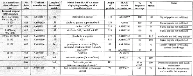
(28) Chapter 5 Sequences of 0. s. scutellatus venom toxins. 121. Table 5.1: Summary of mRNA sequences from O. sClltellatlls venom gland cDNA library and the sequences they share homology with. Summary of sequences an 0. scutellatus venom gland cDNA library and their alignment to sequences to which they share homology. Database comparisons were carried out using the National Centre for Biotechnology Information website (www.ncbi.nlm.nih.gov). The nucleotide (BLASTN), translated and protein sequence (BLASTX) databases were mined while searching for homologous sequences. Nucleotide sequences of the 0. s. scutellatus clone are listed as the clone reference no. and/or the name of the peptide to which the sequence shared homology. This is followed by the length of the nucleotide sequence, GenBank accession number and the size of the potential mature peptide are shaded. Sequences with the highest homology to these sequences are listed by description, size and their GenBank accession number. To the right of the GenBank accession number is the sequence coverage of the 0. s. scutellatus sequence to sequences from the BLAST database expressed as a percentage, and is followed by the matching residues within the sequence, again listed as a percentage. Due to the paucity of toxin nucleotide sequences, most matches were made using the BLASTX database. The nucleotide sequences are included in Appendix VIII. (ME: Clone isolated using mass excision) O. s. selltellatlls clone reference. no.. No. base pairs. . Length of GenBank accession no. . translated . Drotein. Match from BLAST database displaying homology to O. s. selltellatlls clones. Length of protein. BLAST match accession no.. % Sequence coverae:e. % Match. Notes. 182. Beta taipoxin subunit. 118. AF332697. 100. 100. Signal peptide not published. .Toxins & serpent DNA matches .. R 15, R24 mega: RW 18,19,20 22 R 120. 625. AY691659. 151. similar to gamma taipoxin subunit. 133. P00616. 100. 94. Signal peptide not published. R 125. 634. AY691661. 148. similar to OS2. 119. AAB33760. 100. 94. Signal peptide not published. R56/Ron4,. 615. AY691662. 147. similar to OS2, I aa diff to R 125. 119. AAB33760. 100. 95. Signal peptide not published. RW 26, 27, 28,22 R 132. 467 621. AY691658 AY691660. 146 155. Similar to a taipoxin. 119 118. AAB33760 AAB33759. 100 100. 86.5 100. a taipoxin and OS2 velY similar Signal peptide not published. R 121. 687. AY691664. 84. 83. AAL54894. 100. 90. 121&147 similar for two stop eodons then diverge. 81. 90. 468. AY691664. 84. AAD08813 As above. 100. R 147. post-synaptie neurotoxin (Naja sputatrix), shOlt neurotoxin (Lapemis hardwickii) As above. R27. 806. AY691663. 144. natriuretic peptide (0. seutellatus). 35. P83228. 100. 100. 144. Natriuretie peptide (Mieurus corallin natriuretic) Post synaptic neurotoxin (pseudonaja textilis). 185. 100. Q9WYJ7. 33 to 41%* 100. 690. AY691657. I. Well4/R4. 806 RW6. 445. AY691665. 78. OSI. 79. 70 -. Dependent on amino acid match or similarity Possible there are other proteins coded within this sequence I.
(29) Chapter 5 Sequences of 0. s. scute1!atus venom toxins. O. s. sClite/lattls. clone reference no.. No. base pairs. R5. 3317. GenBank accession no.. Length of translated protein NoORF. 122. Match from BLAST database displaying homology to O. s. sClltellatlls clones. Length of protein. BLAST match accession no.. % Sequence coveral!e. % Match. Notes. cobratoxin (Naja atra). Y13399 AJ416991 AF54466I D13522 D31777. 2 2 8.5 7 3. 87 87 89 93 84. Many ORF Genomic DNA match. Gene (Elaphe obsolete) Atractcospis microlepidosi Trimeresurus Flavoviridis. 2387 bp 2358 bp 535 bp 469 bp 301 bp. PLA2 (BunJ<arus multisinctus). R8. 1928. NoORF. Rll. 1501. NoORF. R51. 657. Elaphe bimaculata. 812 bp. AF236671. 60. 84. genomic DNA. 1251. NoORF. Vipera ammodytes (Boy B) Bungarus multisinctus PLA2 inhibitor (Elaphe quadrivirgata) PLA2 gene (Laticauda colubrine). 4606 bp 1203 bp 2433 bp 4304 bp. AF332697 AJ25 122 AB060638 AB062448. 1.5 0.1 105 0.8. 93 93 97 91. Genomic DNA matches all in the same area of all sequences.. 1460. Pseutarin (pseudonaja textilis). 1460. AA038805. 100. 97. 1460. FV (Homo sapiens). 2251. PI2259. 100. 50. 531. POI (Gallus gallus). 515. P09102. 100. 666. HSP 70 (Gallus gallus). 634. AAP37959. 100. 78 to 88 97. 406. EF2 (Gallus gallus). 858. AAA87587. 47. 98. EF 2 (Homo sapiens). 357. PI3639. 100. 98. EF2 (Rattus lIorvegicus). 342. NP058941.1. 100. 98. CGI protein (Homo sapiens). 224. Genomic DNA Many ORF ManyORF !. R65 (incomplete). ,. Complete FV. ,,'. 4725. AY69 I 656. 4725 PDI. 2495. AY691666 ". HSP70. 2340. AY69J667. ". EF2. 1315. AY691668. Clone complete. CGI protein. 682. AY691669. 182. NP861974. 81. 98. Clone complete. 2.19 gene. 763. AY69 I 670. 165. MemberA (Homo sapiens). 230. AAH08912. 71. 90. Clone complete. Bet 3. 757. AY69 1670. 182. BET 3 (Homo sapiens) BET 3 homologue (Mus muselus). 180. NP055223.1. 100. 97. Polyposis-R 117! TB2 Cytochrome C subunit IV (RI44). 772. 214. Polyposis (Homo sapiens) TB2 (Homo sapiens) Cyt C (IV) (Meleagris gallopavo) Cyt C (IV) (Bos taurus). 180 185 198 171 169. NP038746.1 NP005660 AAA6635I AAQ I 4274 AA93149. 100 100. 97 71. 700. AY691674. 180. -. ORF. + signal 100 100. 71 64.
(30) 123. Chapter 5 Sequences of 0. s. sClilellalus venom toxins. O. s. sClitellafliS clone reference no. RI9. Match from BLAST database displaying homology to O. s. scutellatus clones Polydenylate binding protein Ribonuclease protein. Length of protein 126. Gallus gallus C-tenn end. 1308. Archease (Rattus norvegicus) (Mus musculus) (Gallus gallus). 2013. NBM. No. base pairs 1504. Length of translated protein 171. GenBank accession no. AY691673. RW21 811. R26 R36 R42 ME R46,RW32 ME R50 ME R54 ME. NoORF. 1851 ,. HSP90 ME. I. AY02468. 2609. 5' AY02457 3'AY702458. 2523. ,. ". EFt I,. Oncogene. 1433. Disease resistance protein (R 31) G prot binding (R32) Ca ATPase (R 122) Golgi assoc (R62). 1333. Lecithin Retinol (R20) Carboxypepsidase ". 1305 797. 5' AY702460 3' AY702461 AY691672 5' AY702469 3' AY702470 AY702462. 680. HSP 90 (Mus musculus). 680. HSP 90 (Homo sapiens). 112. EF I (Bas taurus). XP216347 BAB26341 BX930134. 42. Notes. 94. + signal peptide ? polycistronic. 46. 80. Many ORF, some overlap. 81. Many ORF; DNA match. 2099 bp. CR354147 BX537527 AADI7298 AK025505. 1-1110 43. Genomic DNA +55. 2326. ---. ". AY702464. 2567bp/ 725aa 181. Internal could not be sequenced. NM007355. 93. 96.5. BAC5675.1. 61. 94. 21. 100. 634. NP036473. 31. 100. Ca transportingATPase (fast twitch skeletal muscle) Gallus ~allus Golgi associated (Homo sapien). 994. A32792. 12. 91. 900. S35342. 16. 78. 667. (Mus musculus). 228. NP076113. 100. 68. 247. Carboxypepsidase A2 (pancreatic (Homo sapiens). 417. NPOOl860. 59. 80. 207. .. XPI42222. NP009298 P42684. +-. 123. ORF, matches mRNA Many ORF, several match 2 seq join. 1182. -+. ,. ManyORF. B Abelson oncogene(Homo sapiens) Tyrosine Kinase(Homo sapiens) Lecithin retinol or disease resistance protein G protein binding (homo sapien). AY702463. 740. 167 146 1070 bp. % Match. NBM. 623. 2523. Gallus gallus Homo sapiens Endocrine regulator (Homo sapiens). % Sequence coveraec 100. 1948 bp. BLAST match accession no. AAHOl716 XP352454 AAGOl716 Bx934062. Clone not complete.
(31) Chapter 5 Sequences of 0. s. seutellatus venom toxins. O. $. scutellatlls clone reference no. Carboxylate Riborphorin (R45RW3J) Protoadherin. B Actin. c. No. base pairs. 903 1439. GenBank accession no. AY702465 5'AY702474 3' AY702475. Length of translated protein 214 343. Match from BLAST database displaying homology to O. s. sCllte/latlls clones Carboxvlate synthase (Homo sapiens) Mus musculus Homo sapiens Homo sapiens. Length of protein 501 608 608 932. BLAST match accession no. NP115981.1 AAHI6080 NP00294I 09Y5HO. % Sequence coveral!e 43 56.5 56.5. 6/0 Match. Sigmodon hispidus Gallus f(allus Skeletal (Homo sapiens). 375 375 377. AALl6942 CAA25004 NPOOI091.1. 69 60. 100 100 100. Myosin regulatory light chain-(Homo sapiens) Felis calus Superfast myosin heavy(Felis calliS). 173. NP002468. 100. 91. 1945. AAK00755 BABI5219. II. 85.5. Ribonuclease HI Ige(zebra fish) and Xenopus laevis Cytochrome C oxidase subunit IV ( Bos taurus). 308. AAH46061. 70. 78. 169. AAA30461.1. 100. 796. AY702473. 259. A actin. 726. AY702472. 242. myosin light. 594. AY702471. 177. Myosin heavy. 664. AY702466. 221. Ribonuclease HI sub Cytochrome C subunit IV Mitochondrial sequences Cytochrome C subunit 1 Cytochrome C subunit II Cytochrome C subunit III NADH dehydrogenase (RW7)R40 MallyORF Wei 12, R2 (PCR). 877. AY702467. 218. 700. AY70256. 1273. Wei 16-R6 (PCR). 1247. -. 700. 124. 180. 66 Intemalnot sequenced. 744. AY691675. ." 247. Dinodon semicarinallis. 533. NP008421.1. 46. 86. 669. AY691676. 223. 228 192. 665. AY691677. 221. Dinodon semicarinatlls Laepemis hardwickii Dinodon semicarinallis. NP008422.1 AAL55554 NP008425.1. 100 100 85. 83 90 94. BAA33030 NP008428. 100 32. 81 85. WeI 1024/3. 744. R 131. 741. 91 142. Dinodon semicarinatlls Dinodon semicarinatlls. 261 95 445. Notes. + signal. Clone complete. Incomplete, N term seq starts 70bp into seq. Complete Incomplete, C term would not sequence Incomplete, Two ORF, two different proteins on one seouence. Similar matches to R 65. Similar to R 65 matches. NBM. Intemal not sequenced. NBM. NoORF. NBM. Good sequence, no ORF. I.
(32) Chapter 5 Sequences of 0. s. seutellatus venom toxins. 125. -. O. s. sClltella/lIs. clone reference 110.. R 138, 139 -. Surface protein R 148. No. base pairs 141. -. Match from BLAST database displaying homology to O. s. sClltel/allls clones NBM. -. NBM. GenBank accession no.. 451 .......................... Length of translated protein. Length of protein. BLAST match accession no.. 0/0. 0/0. Sequence coverage. Match. Notes. i.
(33) Chapter 5 Sequences of 0. s. scutellatus venom toxins. 126. Table 5.2: Summary of clones isolated from the O. sClItellatlls venom gland cDNA library sharing homology with characterised toxins. Summary of complete cDNA clones isolated from 0. scutellatus venom gland selected for analysis compared to homologous published sequences (below. sha4ed).. <D: undertaken using MacVector software, - not undertaken. Toxin or O. s. sClitellatlls. MW (Da). pI. Residues. No. Cysteine residues. Comment. Reference. 5.2.1 Nucleotide sequences of O. s. scutellatus The analysis of the cDNA nucleotide sequences, (summarised in Annex 1 of this chapter) using MacVector software was undertaken to identify eukaryotic promoter sequences. Some sequences contained a larger 5' -upstream region than others. This may have possibly been due to the lack of a secondary structure within the mRNA. Homology searches indicated the Oxyuranus nucleotide sequences shared homology with the nucleotide sequences from other Elapid species.. These species included. Austrelaps superbus, Notechis scutatus, Laticauda semijasciata, Pseudonaja textilis, Bungarus jlaviceps and Ophiphagus hannah.. These comparisons indicated the. sequences were conserved across active sites and conformed to structural restrictions described within the specific toxin groups (data not shown).. Comparisons of the. Oxyuranus nucleotide sequences sharing homology with pre- and post- synaptic.
(34) Chapter 5 Sequences of 0. s. scutellatus venom toxins. 127. neurotoxins revealed point mutations in the first and second bases of codons (data not shown).. Although further analysis of nucleotide sequences would not be pursued. within this chapter, this trend confirmed the directional mutations (accelerated evolution) suggested, as seen is PLA2 (Ogawa et al., 1992), cardiotoxins (Lachumanan, et al., 1998), and neurotoxins (Afifiyan et al., 1999) instead of random mutations within. all sequences. Due to the low representation of nucleotide sequences within the Oxyuranus species, the putative peptide sequences were used for further analysis.. 5.2.2 Potential pep tides isolated from O. s. scutellatus. The analysis of the translated peptides (see Chapter 2, 2.2.2.13) indicated the sequences could be classified into distinctly different classes. Therefore, sequences were discussed within the classes to which they shared homology; pre-synaptic neurotoxins, post-synaptic neurotoxins and the Factor V component of scutelarin.. 5.2.2.1 Oxyuranus sequences sharing homology to PLAz! pre-synaptic neurotoxins. The nucleotide sequences of the cDNA clones were translated and identified using MacVector or Sequencher programs. The clones designated R24, RW26 and R120 shared homology with taipoxin subunits, whereas clone R132, R125 and R56 shared homology with OS 1 and OS2. These sequences and homologies were summarised in Annex 1 at the end of this Chapter, Figures 5.10 to 5.19. The Oxyuranus clones demonstrated high homology with the PLA2 pre-synaptic neurotoxins from other elapid species, displayed in Figures 5.2 and 5.3. High sequence homology was reflected over the active and calcium-binding sites with 90 to 100% sequence identity (Figure 5.1). These peptides could also be segregated into group IA and group IB phospholipases based on these homologies.. This suggests these. sequences may be isomers of the characterised Oxyuranus peptides and evolutionary derivatives of the other homologous Elapid sequences. Interestingly, the signal peptides of these sequences were also highly conserved (Figure 5.4). Additionally, the. 0.. s.. scutellatus putative peptides shared similar SIze,. hydrophobicity and cysteine positions, thus potentially structure, to the characterised.
(35) Chapter 5 Sequences of 0. s. scutellatus venom toxins. 128. Oxyuranus peptides to which they shared homology. The pIs were also similar. The. putative protein sequences of RW26, R56 and R125 were basic. The peptide encoded by RW26 showed the highest homology with the basic a-taipoxin subunit (80%), OS2 (74%) and. ~-taipoxin. (67%) peptide sequences. Similarly, the basic peptides R56 and. R125, differed by only one amino acid, and shared homology with OS2 (93%), ataipoxin (83%), RW26 (81.4%) and. ~-taipoxin. (65%).. In contrast, the putative peptide sequences ofR120 and R132 were acidic. These shared close alignment with the pancreatic group IB peptides, y-taipoxin and OS1. R120 shared the closest homology with the y-taipoxin chain (94%), and contained a propeptide, pancreatic loop and a potential carbohydrate moiety. Whereas R132, although it did not contain a propeptide or potential carbohydrate binding site, contained a pancreatic loop and differed by only one amino acid from OS 1. These sequences contained larger hydrophobic areas at the N- and C-terminals and differient charges compared to the other peptides described (RW26, R125 and R56), indicating they may have different binding properties. Regions outside the active and calcium-binding sites displayed a similarity of approximately 50% with the C-terminus (30 aa) showing 65% similarity. Despite the high homology of the PLA2 catalytic and calcium binding domains, few residues suggested for toxicity (approx 13%, Gll o and Arg43 ) within other genera were present within the Oxyuranus sequences (see introduction and Table 5.3). A preliminary pair-wise, neighbour joining alignment of the putative isolated Oxyuranus peptides and homologous sequences was undertaken to display the. relationships between the sequences (Figure 5.5 and 5.6). Data sets of the Oxyuranus putative peptide sequences were bootstrapped (x 10,000) to check how robust the clusters were (Appendix VII)..
(36) Chapter 5 Sequences of 0. s. scutellatus venom toxins. 129. Figure 5.1: Pairwise alignment of putative O. sClltellatlls peptides. The group IA and IB (y-taipoxin, R120, OSl, R132) peptide and putative peptide sequences of O. scutellatus sequences using the ClustalW program (Higgins et at., 1994) on the EMBL-EBI site (http://www.ebi.ac.uk/). The conserved cysteine residues are indicated (*). The variant amino acids are displayed as symbols denoting the degree of conservation observed in each column: "*,, means that the residues in that column are identical in all sequences in the alignment. ":" means that conserved substitutions have been observed. "." means that semi-conserved substitutions are observed. Gaps (-) have been inserted for optimal alignment. The first box outlines the PLA2 calcium binding domain from amino acid residues 25 to 33, the second box outlines the active site from amino acid residues 42 to 52. A pancreatic loop is described within group 1B peptides (y-taipoxin, R120, OSl, R132) from amino acid residues 62 to 66; (N/S/K) (YNILIP) (LiM/QN/P) (MIP/LlV) (SIN/D) and a propeptide is seen at the N-terminal of some of these peptides. The pairwise alignment used the parameters: aln w/numbers, PHYLIP tree type, distance correction off and ignore gaps off.. IA. IB. R56 R125 OS2 a-taip_ RW26 b-talp R24 g-Taip:= R120 OSl R132. R56 R125 OS2 a-taip_ RW26 b-taip_R24 g-Talp R120 OSl R132. ". 1 1 --------NLVQFGFMIKCANG --------NLVQFGFMIKCANG --------NLAQFGFMIRCANG --------NLLQFGFMIRCANR --------NLVQFGFMIKCANR --------NLVQFGKMIECAIR SELPQPSIDFEQFSNMIQCTIP SEIPQPSLDFEQFSNMIQCTIP --------SLLNFANLIECANH --------SLLNFANLIECANH. ... :*. : * *". "". 0 YGCYCGK YGCYCGK YGCYCGK YGCYCGK YGCYCGP YGCYCGK YGCYCGP YGCYCGP YGCYCGK YGCYCGK ******. ". 30 GSGTPVDELDR GSGTPVDELDR GRGTPVDDLDR GSGTPVDDLDR GSGTPIDDLDR -SGTPVDDLDR GSGTPIDDLDR GSGTPIDDLDR GRGTPLDDLDR. ". 50 52 52 52 52 52 51 60 60 CHVHDDCY 52 52 ***:*:*** *: . **:** CQVHDECY CQVHDECY CQVHDECY CQVHDECY CQVHDQCY CQVHDECY CKTHDECY. ". 70 80 6* GEAEKRLGCSPFVT-----LYSWKCYGKAPSCN-TKTDCQRFVCNCDAKAAECFARSPYQ GEAEKRLGCSPFVT-----LYSWKCYGKAPSCN-TKTDCQRFVCNCDAKAAECFARSPYQ GEAEKRLGCSPFVT-----LYSWKCYGKAPSCN-TKTDCQRFVCNCDAKAAECFARSPYQ GEAVRRFGCAPYWT-----LYSWKCYGKAPTCN-TKTRCQRFVCRCDAKAAECFARSPYQ DEAVRRYRCSPYLT-----MYSWKCYGTAPSCN-SKTQCEGFVCNCDAKAAECFARSPYQ AEAEK-HGCYPSLT-----TYTWECRQVGPYCN-SKTQCEVFVCACDFAAAKCFAQEDYN AEAGKLSACKSVLSEPNNDTYSYECNEGQLTCNDDNDECKAFICNCDRTAVTCFAGAPYN AEAGKLSACKSVLSEPNNDTYSYECNEGQLTCNDDNDECKAFICNCDRTAVTCFAGAPYN GEAEKLPACNYLMSSPYFNSYSYKCNEGKVTCTDDNDECKAFICNCDRTAAICFAGATYN GEAEKLPACNYLMSSPYFNSYSYKCNEGKVTCTDDNDECKAFICNCDRTAAICFAGATYN *: : : * *: *: *. 1tO R56 R125 OS2 a-taip RW26 b-taip R24 g-TaipR120 OSl R132. SRPVSHYM SRPVSHYM SRSPLDYT SRPVWHYM SRPVSHYM RRPALDFM GSECLAYM GESCLAYM. KKNWNIN--TKARCK 119 KKNWNIN--TKERCK 119 KKNWNIN--TKARCK 119 NSNWNIN--TKARCR 119 PSNRNIN--TKERCK 119 PAHSNIN--TGERCK 117 DDLYNIG--MIE-CHK 133 DLNYNIG--MIEHCK 133 DENFMISKKYNDICQ 127 DENFMISKKRNDICQ 127. 106 106 106 106 106 104 120 120 112 112.
(37) Chapter 5 Sequences of 0. s. scutellatus venom toxins. 130. Figure 5.2: Sequence alignment of chosen group IA peptides.. Group IA 0. scutellatus cDNA clones shared alignment with Laticauda laticaudata (GenBank Accession no. BAB72249), Austrelaps superbus (GenBank Accession no. AAD56557) and Notechis scutatus (GenBank Accession no. X14043). The peptide sequence of Oxyuranus scutellatus a-taipoxin subunit (GenBank Accession no. P006l4), p-taipoxin subunit (GenBank Accession no. P006l5) and OS2 (GenBank Accession no. AAB33760) are included. The nucleotide and signal peptide of these sequences are unknown at this time. Amino acids sharing similarities are shaded and the common sequence is included beneath the shaded text. Gaps (-) have been inserted for optimal alignment. The first box outlines the PLA2 calcium binding domain from amino acid residues, the second box outlines the active site. Page 132. Figure 5.3: Sequence alignment of chosen group IB peptides.. Group IE 0. scutellatus cDNA clones shared alignment with Notechis scutatus (GenBank Accession no. AAB34l22), Lapemis hardwicki (GenBank Accession no. AAL55555), Pseudonaja textilis (GenBank Accession no. AF144319), Laticauda semifasciata (GenBank Accession no. AB078348) and Austrelaps superb us (GenBank Accession no. AAD564l0), These clones shared the highest homology with the Oxyuranus scutellatus 8-taipoxin subunit (GenBank Accession no. P00616), and OSI (GenBank Accession no. AAB33759). The nucleotide and signal peptides of these sequences are unknown. Amino acids sharing similarities are shaded and the common sequence is included beneath the shaded text. Gaps (-) have been inserted for optimal alignment. The first box outlines the PLA2 calcium binding domain from amino acid residues, the second box outlines the active site. Page 133.
(38) 131. Chapter 5 Sequences of O. s. scutellatus venom toxins. Figure 5.2: Clustal alignment of chosen peptides within group IA PLA2•. Formatted Alignments. Notechis scutatus Austrelaps superbus Laticauda laticaudata Beta taipoxin Alpha taipoxin RW26 OS2 R56 R125. IY. 10. 30. 20. YK~. f~ Y. " Y. IH ;H M. P A H L L V L L A V C V S L L G A A S. I P P L P L N L. Notechis scutatus Austrelaps superbus Laticauda laticaudata Beta taipoxin Alpha taipoxin RW26 OS2 R56 R125. Notechis scutatus Austrelaps superb us Laticauda laticaudata Beta taipoxin Alpha taipoxin RW26 OS2 R56 R125. Notechis scutatus Austrelaps superbus Laticauda laticaudata Beta taipoxin Alpha taipoxin RW26 OS2 R56 R125. P . . TLYSWKCYGKAP.CN.KT. 130. CQRFVC. 140. 150. Notechis scutatus Austrelaps superbus Laticauda laticaudata Beta taipoxin Alpha taipoxin RW26 OS2 R56 R125. CDAKAAECFARSPYQ. KNWNINTK. RCK8a.
(39) Chapter 5 Sequences of 0. s. scutellatus venom toxins. 132. Formatted Alignments 10 Austrelaps superbus Laticaudai semifasciata Laepemis hardwickii Notechis scutatus Pseudonaja textilis OS1 R 132 Gamma taipoxin R120. I!'!I H ;li~'im£jj!f~IL~it~V~~%~~'(;l:l],n!t'G]T.& A R H 1~~JIl!M~175!;,\'IB~llijXlllM4i~~~S~~Fzi'Gl S. M Austreiaps superbus Laticaudai semifasciata Laepemis hardwickii Notechis scutatus Pseudonaja textilis OS1. R 132 Gamma taipoxin. R120. 20. iSE. ;S. PAHLLVLLAVCVSLLGAS. E E. E L.
(40) 133. Chapter 5 Sequences of O. s. scutellatus venom toxins. Figure 5.4: Alignment of the signal peptides isolated from an O. s. scutellatus cDNA library and the homology with other Elapid species. A) Pre-synaptic neurotoxins i) Pre-synaptic neurotoxin signal peptides from clones isolated R24((3): MHPA1 ILLVLLAVCVSLLGASDIPPLPL.NLVQ MHPAHLLVLLAVCVSLLGASDIPPLPLNLVQ RW26.(tx) R 125(0S2) WMAHLLVLLAVCVSLLGASDIPPLPLN R 132 (OS1) MHPAHLLVLLAVCVSLLGAARIPPLPI-SLLN MHPAHLLVLLAVCVSLLGSSELPQPSLDFEQF R 120 (y) I) Homology ofpre-synaptic neurotoxin signal peptides 0. scutellatus R120 (y-taipoxin) 0. scutellatus (a,¢, OS2) 0. scutellatus R132 (OS 1) B. multicinctus (Kondo, 1982) A. Laevis (Ducance, 1988) L.semifasciara (Fujimi,2000) A. superbus (Singh, 2000). M HPAHL LVLLA VCVSL LG - S M HPAHL LVLLA VCVSL LGASD IPPLPL M HPAHL LVLLA VCVSL LGAAR IPPLPL M YPAHL LVLSA VCVSL LGA.\N IPP', PL M 'i PAHL LVLLA VCVSL LGASD IPPLPL M NPAHL LVLLA VCVSL LGASA IPPLPL M YPAHL LVLLA VCVSL LGASD IPPI.IPL -27. -i6. -21. -11. -6. -1. Table 5.3: Annotation of suggested toxin residues within Elapidae pre-synaptic neurotoxins. Annotation of toxin residues from O. scutellatus pre-synaptic and putative neurotoxins compared to the suggested residues necessary for toxicity within Elapidae. The numbering incorporated within this table does not include the residues encompassing the pancreatic loop of the group l B peptides, as the many toxic' residues are numbered using the group 1A (toxic) peptides that do not include a pancreatic loop. Areas of high homology are coloured in blue. After residue 53 the numbering becomes difficult due to the numerous insertions and deletions Sites suggested causing activity/ toxicity within pre-synaptic neurotoxins 30G. 30(3. 31 W 43R. a taip. RW26 (a). 0 taip. y taip. R120. OSI. (r). R132 (OSI). OS2. R56 (OS2). R125 (OS2. Notes. I uxi,. Nontoxic. Nontoxic. Nontoxic. 30G. 30G. 30G. 30G. 30(3. 30G. 30(3. 30(3. =0G. 31 K. 31 P. 31 K. 31 P. 31 P. 31 K. 31 K. 31 K. 31 K. 31 K. -1 3. It. 43R. 43R. 43R. 43R. 43 it. 43R. 43R. 43R. 43R. 46 K. 46 Q. 46 Q. 46Q. 46 K. 46 K. 46 1-i. 46 H. 46 Q. 46 Q. 46 Q. 50 D. 50 E. 50Q. 50 E. 50 E. 50 E. 50D. 50 D. 50 E. SUE. 50 E. 54G. 54 E. 54E. 54E. 54E. 54E. 54E. 54E. 54E. 54E. 54E. 57. 57 R. 57k. 57 K. 57K. 57 K. 57 K. 57 K. 57 K. 57 K. 57 K. 58K. 58 R. 58 R. 58 H. 58 L. 58 L. 58 L. 58 L. 58 R. 58 R. 58 R. 63 Wor F 64Y. 63 P MY. 63 P 64Y. 63 P 64S. 63 P 64V. 63 P 64V. 63 P 64L. 63 P 64. 63 P 64F. 63 P 64F. 63 P 64F. 69W. 69 S. 69 V. 69 W. 69 N. 69 N. 69 Y. 69 Y. 69 S. 69 S. 69 S. 70. 70 W. 70 W. 70 Y. 70 Y. 70 Y. 70 Y. 70 W. 70W. 70 W. 70 W. 74 N. 74 Q. 74 G. 74 G. 74 E. 74 E. 74 I3. 74 E. 74 G. 74 G. 74 G. No N in vicinity. 900. 90 C. 90C. 90C. 90 C. 90 C. 90C. 90C. 90C. 90C. 90C. AllDat93. 94 G. 94 A. 94 A. 94 A. 94 V. 94 V. 94 A. 94 A. 94 A. 94 A. 94 A. Used residue 97. Th.'. All toxic peptides contain W . 70.
(41) Chapter 5 Sequences of 0. s. scutellatus venom toxins. 134. Figure 5.5; Alignment of O. s. scutellatus pre-synaptic PLA2s and putative O. s. scutellatlls amino acid sequences. The predicted protein sequences of the mature, processed peptides were aligned by using Clustal. The phylogenetic relationships of the proteases were assessed by the neighbour joining method using the ClustalW program (http://www.ebi.ac.uk. Higgins, 1994). Trees were generated and robustness was assessed by bootstrap analysis using 10,000 replicates; clades with more than 80% support are depicted. Actual bootstrap phylogeny and values can be viewed in Appendix VII. The branch lengths shown to the right of the peptide labels are proportional to the amount of inferred evolutionary change. This clustering was mimicked when additional elapid PLA 2 sequences were included within the analysis (Figure 5.6). Figure 5.5 indicated the Type IB (y-taipoxin, OSl R120, R132) and IA peptides evolved differently, with type IB Oxyuranus clones and subsequence putative pep tides an older lineage .. . - - - - - - - - - - - - - - b . t a i p : 0.36574 1...-_ _ _ _ _ _ _ _ _..,OS1:0.00000. R132: 0,00000 ........_ - - RW26: O:n271. 1A. ?----a.tsiP; 0.07585 05Z: 0.05Z35. l. R5d: 0.00321. R125: 0.00534. g.l'alp: 0.0'165& R120; 0.01763.
Figure


+7


Related documents
Comments This can be a real eye-opener to learn what team members believe are requirements to succeed on your team. Teams often incorporate things into their “perfect team
Date of Birth Driver’s License Number State Marital Status Number of Dependents.. Corporation/LLC/Partnership Name Federal Tax Identification Number (EIN) D &
Meets expectations -- student demonstrates sufficient knowledge, skills, and abilities or does what is expected of students in collegiate business programs.. Does not meet
It is possible that a segment is stored at multiple peers in the set cover, in this case the requesting client can pick a peer based on some other criteria such as delay or number
• Our goal is to make Pittsburgh Public Schools First Choice by offering a portfolio of quality school options that promote high student achievement in the most equitable and
Unfortunately student loans didn’t cover all my school expenses so I started getting credit cards to buy the things I needed for school and to pay school-related bills. I worked
Make changes to section on agreements to reflect emphasis of agreements in new
However, USAID reserves to require a CDC/ILB evaluation under Section H of the test as a condition of approval... For the CDC/ILB evaluation, a test kit must meet the
