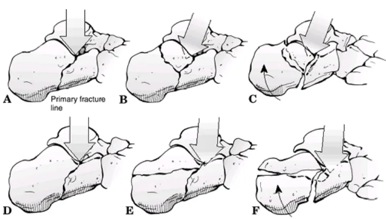
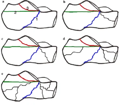
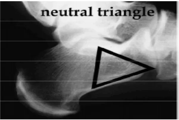
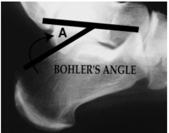
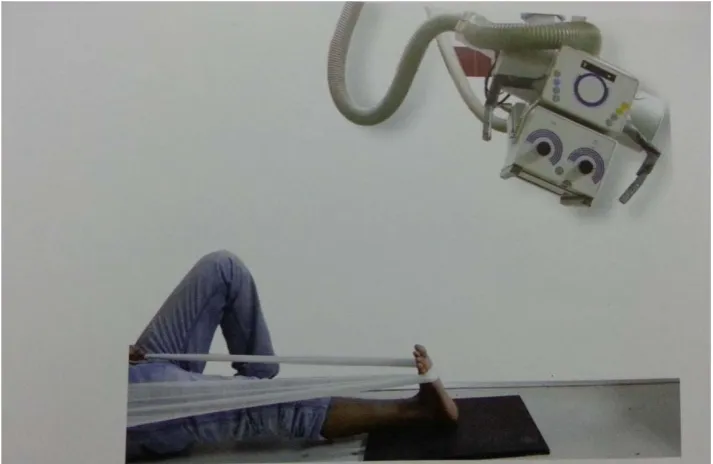
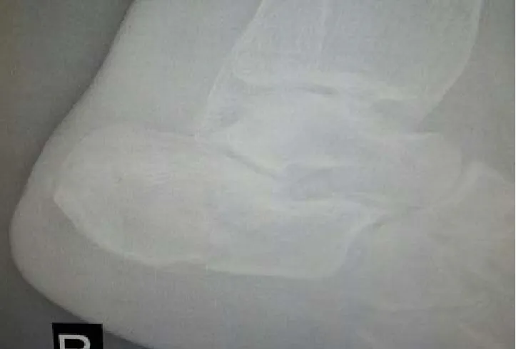
A comparative study between locking compression plate and non locking compression plate in the treatment of intraarticular calcaneal fracture
126
0
0
Full text
Figure
+7
Outline
Related documents