A DISSERTATION ON
“ COMPARING THE DIAGNOSTIC ACCURACY OF CT AND USG IN THE DIAGNOSIS OF ACUTE APPENDICITIS ”
Submitted to
THE TAMIL NADU Dr.M.G.R.MEDICAL UNIVERISTY
CHENNAI
In Partial Fulfilment of the Regulations
For the Award of the degree
M.D. DEGREE BRANCH VIII
RADIODIAGNOSIS
MADRAS MEDICAL COLLEGE,
CHENNAI.
BONAFIDE CERTIFICATE
Certified that this dissertation is the bonafide work of
Dr.G.GEETHA on “COMPARING THE DIAGNOSTIC
ACCURACY OF CT AND USG IN THE DIAGNOSIS OF ACUTE APPENDICITIS” during her M.D.RADIODIAGNOSIS course from March 2014 to August 2014 at the Madras Medical College and Rajiv Gandhi Government General Hospital, Chennai – 600003.
PROF, Dr D.RAMESH, M.D.R.D
ASSOCIATE PROFESSOR BARNARD INSTITUTE OF RADIOLOGY
MADRAS MEDICAL COLLEGE & RAJIV GANDHI GOVERMENT GENERAL HOSPITAL, CCHENNAI – 600 003
PROF.Dr.N.KAILASANATHAN, M.D.R.D
HEAD OF THE DEPARTMERNT BARNARD INSTITUTE OF
RADIOLOGY
MADRAS MEDICAL COLLEGE &
RAJIV GANDHI GOVERNMENT GENERAL HOSPITAL, CHENNAI -600 003
PROF.Dr. K.VANITHA, M.D.R.D,
DMRD,DRM,DHA DIRECTOR,
BARNARD INSTITUTE OF RADIOLOGY
MADRAS MEDICAL COLLEGE & RAJIV GANDHI GOVERMENT GENERAL HOSPITAL, CCHENNAI – 600 003
DR.R.VIMALA, M .D
DEAN,
MADRAS MEDICAL COLLEGE &
RAJIV GANDHI GOVERNMENT GENERAL HOSPITAL, CHENNAI -600 003
DECLARATION
I, certainly declare that this dissertation titled, “COMPARING THE DIAGNOSTIC ACCURACY OF CT AND USG IN THE DIAGNOSIS OF ACUTE APPENDICITIS”, represent a genuine work of mine. The contribution of any supervisors to the research are consistent with normal supervisory practice, and are acknowledged.
I, also affirm that this bonafide work or part of this work was not submitted by me or any others for any award, degree or diploma to any other university board, neither in India or abroad. This is submitted to The Tamil Nadu Dr.MGR Medical University, Chennai in partial fulfilment of the rules and regulation for the award of Master of Radiodiagnosis Branch VIII
Date :
Place: Chennai Dr.G.GEETHA
ACKNOWLEDGEMENT
I would like to express my deep sense of gratitude to the Dean,
Madras Medical College and PROFESSOR DR.K.VANITHA, Director,
Barnard Institute of radiology, MMC & RGGGH, for allowing me to
undertake this study on “COMPARING THE DIAGNOSTIC
ACCURACY OF CT AND USG IN THE DIAGNOSIS OF ACUTE APPENDICITIS”
I was able to carry out my study to my fullest satisfaction, thanks to guidance, encouragement, motivation and constant supervision
extended to me, by my beloved Head of the Department PROFESSOR
DR .N.KAILASANATHAN. Hence my profuse thanks are due for him.
I would like to express my deep gratitude and respect to my guide PROFESSOR DR.D.RAMESH whose advice and insight was invaluable to me. This work would not have been possible without His guidance, support and encouragement.
I am also extremely indebted to PROFESSOR DR.S.BABU
PETER for his valuable suggestions, personal attention, constructive cricticism during my study.
My sincere thanks to PROFESSOR DR.S.KALPANA for her
practical comments and guidance especially at the inception of the study
and I also wish to thank PROFESSOR DR .K.MALATHY for her
I am bound by ties of gratitude to my respected Assistant
Professors, Dr.Manimegala.E, Dr.Geetha.K, Dr.Chezhian.J,
Dr.S.Anbumalar, Dr.M.S.Shyamala, Dr.S.Saranya, Dr.Balan.M.P in general, for placing and guiding me on the right track from the very beginning of my career in Radiodiagnosis till this day.
I am fortunate to have my fellow postgraduate colleagues
Dr.R.Rajalakshmi, Dr.P.K.Latha, Dr.Komalavalli, Dr.Sivakumar,
Dr.Iyengaran for their invaluable suggestions, relentless help for shouldering my responsibilities. Simply words cannot express its depth for their unseen contributions. My lovable thanks to my parents and my husband for their moral support.
I would be failing in my duty if I don’t place on record my sincere
thanks to those patients who in spite of their sufferings extended their fullest co-operation.
TABLE OF CONTENTS
SI.
NO TITLE
PAGE NO
1 INTRODUCTION 1
2 REVIEW OF LITERATURE
HISTORY OF APPENDIX
ANATOMY OF APPENDIX
HISTOLOGY OF APPENDIX
PATHOPHYSIOLOGY OF APPENDIX
DIAGNOSTIC IMAGING
X-RAY USG CT 3 7 10 11 25
3 AIMS AND OBJECTIVE 59
4 METHODOLOGY 63
5 CASES 66
6 STATISCAL ANALYSIS 80
7 OBSERVATION AND DISCUSSION 97
8 RESULTS
9 CONCLUSION 107
10 BIBLIOGRAPHY
11 ANNEXURE
ETHICAL COMMITTEE CERTIFICATE
CONSENT FORM
PROFORMA
PLAGIARISM
LIST OF ABBREVIATION
CRP - C reactive protein
WBC - White blood count
PPV - Positive Predictive value
NPV - Negative Predictive value
PR - Perforation Rate
NAR - Negative Appendectomy Rate
ED - Emergency department
HPE - Histopathology
CT - Computed Tomography
USG - Ultra sonogram
No - Number
n - Number of case
ABSTRACT
AIM OF THE STUDY:
The Aim of the study was to evaluate the accuracy of CT and USG in the diagnosis of
acute appendicitis in patients who are taken for appendectomy on clinical basis
To calculate the sensitivity ,specificity positive predictive and negative predictive value
of CT and USG
METHODOLOGY
Patients who were admitted in the surgical emergency ward with clinical findings
and symptoms suspected of appendicitis .A total study sample of 100 was selected
USG PROTOCOL
A routine USG was done in SONOSCAPE machine for the upper abdomen and
pelvis using a 3-5–MHz convex transducer to rule out alternative abnormalities related
to solid organs and to rule out free fluid.Then graded compression and colour Doppler
sonography of the right lower quadrant giving attention to the site of maximal
tenderness was performed using a linear transducer.
CT PROTOCOL
Examinations were performed on a MDCT performed using a 4-slice C scanner (
TOSHIBA ) at 120 kVp and 100 mAs; a pitch of 1 was used. CT of the lower abdomen
and pelvis, from the xiphoid to the pubic symphysis, was performed with 80 mL of
non-ionic contrast material Iohexol 350 (Omnipaque 350) was injected through a 18-gauge
cannula placed in the volar aspect in the cubital vein at a flow rate of 4 ml/s and delay of
50 sec.
Axial reconstructions from the raw data were done at 3 mm thick, at 1.5-mm
increments were obtained. The second data set was reformatted coronal at a thickness of
RESULT
From the study it is concluded that CT is more sensitive ,specificity
,PPV,NPV. Hence the CT investigation is more accuracy than USG in diagnosing cases
of appendicitis.
CONCLUSION
Evaluating a case of appendicitis is mainly clinical ,depending on the clinical
scores and signs. But there is increase in the negative appendectomy rate on depending
only on clinical findings .
Usually USG is the first primary techniques ,considering its easy availability,
low cost and reproducible with no radiation But it has its own pitfalls ,being operator
dependent .
CT on the other hand is more specific than USG and hence could rule out
appendicitis .
Most of the studies including our study has shown that CT has more sensitivity,
specificity ,Negative predictive value and Positive predictive value in diagnosing
appendicitis.
Weighing the cost versus the radiation and the real need to rule out appendicitis
,and the dire need in search of alternate diagnosis should be considered before deciding over
which imaging modality to choose.
But CT without doubt has definitely more diagnostic performance than USG in
1
Vague abdomen pain is the most commonly encountered
symptom in the emergency department at any hospital. It may be
associated with vomiting, fever and diarrhoea but the most distressing
symptom is the pain. As the pain threshold varies from person to person
the severity of the disease could not be evaluated taking, only this
symptom into account.
The various cause of the abdomen pain may vary from benign to
life threatening disease. Diagnosing and treating the condition in time is
in the hands of the surgeons or the physician who handle them. Time is a
very important factor as any delay may lead to grievous consequences
like perforation , and may lead to morbidity and in some case also
mortality. Hence timely diagnosis is crucial and remains a challenge to
the people in medical field.
Appendicitis is the most common cause of abdomen pain in
patients admitted at the emergency department. Diagnosing this in young
male patient is mostly straight forward, but the same becomes a problem
2
This is mainly due to the reason that number of gynaecological
problems in women can present with abdominal pain mimicking
appendicitis. So it becomes a real challenge to exclude the diagnosis in
women more than diagnosing a positive case of appendicitis.
Problems also arise in extremes of age because of the delay in
seeking medical care, or difficulty in obtaining history and it also
becomes a mountain moving task in performing an accurate physical
examination in these patients.
The timely diagnosis and intervention of acute appendicitis is
important due to the fact of its grave complication like perforation. As
the increase rate of perforation also increase the morbidity and
mortality rates, the first few hours of timely intervention is very
crucial.
Some surgeons are in favour of early laparotomy, even if there is
no definite diagnosis of appendicitis, taking into account only the
clinical findings .This is done mainly to minimize the risk of appendiceal
3
HISTORY OF APPENDICITIS1
Appendicitis is a common and frequently made diagnosis . History
of appendicitis was made and written in the past two generations.
Hippocrates has given description of a picture similar to that
matched, like present appendix of appendicitis with perforation , in his
writing title “The Epidemics”:
“The woman who lodged at the house of Tisamenas has a
troublesome attack of iliac passion , acute abdominal pain and
distension ,much vomiting ;could not keep her drink; pain about the
hypochondria, and pain also in the lower part of belly ;not thirsty
;became hot; extremities cold throughout with nausea and insomnolency;
urine scanty. Nothing could do her any good. She died”
The appendix was first depicted in western medicine by
Leonardo Da Vinci in his drawings. Vesalius in 1541 depicted appendix
and listed the central cause of appendicitis as due to a fecolith or a
inspissated ball of stool that obstructs the appendiceal lumen.
The function of the appendix was not entirely made out in the
fifteenth century. It was recognised as an organ attached to the gut with
4
variability of presentation led the Natural Philosophers like Darwin to
classify the appendix as vestigial, and harmless organ that could be safely
ignored.
Berengaria Carpi, surgeon gave the first description of this
structure. He quoted that the organ was empty inside ,measuring 3
inches, present at the end of caecum .He made his findings in the early
fifteenth century in 1522.
Twenty-one years later, the findings of Berengaria was
augmented by the writings and description by Versalis, who gave
several illustrations about the structure of appendix. Much confusion
existed between the caecum and the appendix.Versalis insisted to call
it vermiformis a “ blind ending pouch”. Fallopius in 1561, compared
appendix to a worm like structure.
Anders Celsius in year 1744 quoted in his writings :
"Distemper seated in the large intestine, particularly affecting that
part, where I mentioned the caecum to be, accompanied by violent
inflammation and vehement pains, particularly in the right side" .He
5
Jacopo Berengaria Carpi was the first who found that the pain
in right lower quadrant was due to appendix.
The three coats of appendix along with the mucous glands,the
meso-appendix the peritoneum fold adjacent to the appendix, in this
region was described at the start of nineteenth century.
The mucous membrane of appendix was found by Gerlach in
1847.He also found that these mucous membrane, function as a valve to
occlude the appendiceal lumen.
In 1711 Lorenz Heister described the blackened stump of an acute
gangrenous appendix in his dissection .The appendix was first removed
in a planned operation by Dr. Lawson Tait in the year 1880.
In 1886, Reginald H. Fitz of Boston gave a clear picture that the,
inflammation of the right iliac fossa, the “fons et origomali” was the
vermiform process of the caecum. He was the first to use the term
“appendicitis” in his article . Now the word appendix is universally used.
The three classical sign of pain in the right lower quadrant with
fever and chills, and peritonitis was contributed by McBurney in
6
maximum tenderness at the junction of a line drawn from umbilicus
to anterior superior iliac spine.
Dr.Deaver says, “So many times does it appear that acute
observers stumbled on the very threshold of the discovery that the
original lesion in these conditions was in the vermiform appendix, that it
seems scarcely credible that for less than forty five years have we had
any adequate knowledge of appendicitis.”
Perforated appendix was closed by suture in the year 1887 by
Sand and revised later in 1888 by Treves. Since 1890, the history of
appendicitis has been one of refinement in the technique and the
diagnosis. Today we have a multiplicity of signs and symptoms, that
7
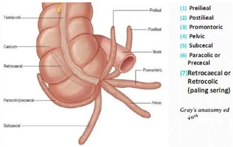
ANATOMY OF APPENDIX
The vermiform appendix is a tubular structure from the postero
medial portion of caecum. It is a blind ending tubular structure. It is
situated inferior to the ileco caecal junction. The length varies from 7.5
to 10mm.
The base of the appendix lies in a constant position. The base is
formed by the confluence of the taenia coli. Base of the appendix is
roughly deep to the McBurneys point. Localised pain and guarding at
this point is the most important physical examination finding for the
diagnosis of appendicitis
While the base of the appendix is essentially constant the free
end of the appendix or the tip of the appendix is found in various
position. And this different location of the appendix sometimes lead to
false negative diagnosis at USG imaging .The position also influence
the clinical finding2.
The position may be retrocaecal ,post and pre ileal ,pelvic,
8
Fig: 1 Various position of appendix
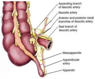
The appendix is suspended by a fold of peritoneum which is a
part of the mesentry of the terminal ileum and gets attached to the
caecum and proximal part of the appendix. This is called the
mesoappendix and contains the appendicular artery, a branch of
ileocolic artery. The ileocolic and the right colic drains the appendix to
9
[image:20.595.171.479.134.390.2]
Fig:2 Arterial Supply of appendix
The lymphatic drainage is via the ileocolic node along the
superior mesenteric to celiac and end in cisterna chyli. Nerve supply is
through T10 spinal segment which also explains the pain that is
10
HISTOLOGY OF APPENDIX
There are 5 layers from inner to outer.They are
The mucosa,
Lamina propria,
Sub mucosa,
Muscularis, and
Adventitia.
FIG:3 HISTOLOGY PICTURE OF APPENDIX
11
It has no digestive glands or secretory ducts, which confirms the
vestigial nature of the organ with no digestive function. It has a role in
immunity, which is suggested by the presence lymphoid aggregations in
the sub mucosal layer. The aggregates are responsible for the immense
inflammatory response in case of acute appendicitis. However loss of
this organ does not endanger the immune system of an individual
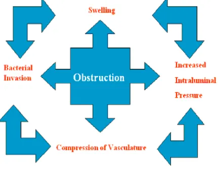
PATHOPHYSIOLOGY OF APPENDIX
Appendicitis is mainly due to obstruction of the appendicular
lumen. The obstruction may be due to foreign body, crohns disease
,parasite infection, gastroenteritis, upper respiratory tract infection,
fecolith and lymphoid hyperplasia.
Within the obstructed lumen there is increase in the mucous
secretion and hence, there is increase in the intraluminal pressure
causing distension of the appendix.
Mucosal edema and ulceration occurs with overgrowth of
bacteria. With increase in luminal pressure there is venous obstruction
and vascular congestion of the appendix extending up to the serosal
12
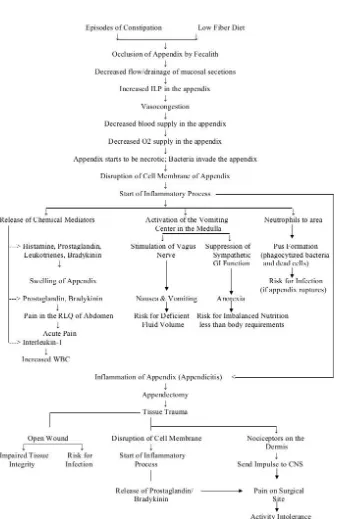
The increase in pressure also stretch and stimulate the nerve
endings of the visceral efferent which is perceived by the patient as
periumblical or epigastric pain.
When the inflammation spread to the peritoneum the pain shifts to
the right lower quadrant. Venous congestion and stasis may cause
thrombosis which results in gangrene of the appendix.
At the end stage due to tissue ischemia the appendix get infarcted
and perforated.
Rupture of appendicitis may cause the inflammatory process to
spread, with inflammatory thickening of the adjacent bowel loop, or
abscess and collection at the ruptured site.
These features leads to generalised peritonitis. Sometimes the
collection gets walled off by the greater omentum and bowel loops
causing a phlegmatous mass.
13
[image:24.595.102.533.199.537.2]
FIG:4 CYCLIC CHANGES IN APPENDICITIS
14
Fig:5 Representative algorithm of pathophysiology of Appendicitis
15
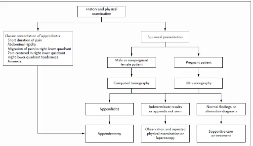
History and physical examination
The diagnostic cornerstone in the evaluation of acute abdomen
pain is history taking and physical examination. Combination of
various signs and symptoms may support the diagnosis.
Three signs most predictive of acute appendicitis4,8
The right lower quadrant pain
Abdominal rigidity
Migration of pain from the periumbilical region to the right lower
quadrant
The duration of pain contribute to an important predictor5,8 .
Misdiagnosis is most common, among women due to gynaecological
problems like pelvic inflammatory disease, ruptured ovarian follicle,
and ectopic pregnancy6,8 and mimics like gastroenteritis, urinary tract
infection.
Predictors of pelvic inflammatory disease7,8
1. history of vaginal discharge,
2. urinary symptoms,
3. tenderness outside the right lower quadrant
16
Acute appendicitis is a clinical diagnosis .Most of the surgeons
and physician depends on various clinical scoring system for the
accurate diagnosis of appendicitis. Among the various scoring system
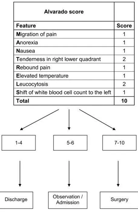
ALVARADO scoring is commonly used in practice.
[image:27.595.176.404.218.569.2]
FIG:6 ALVARADO SCORE (ref:Alvarado et al 94)
17
The ALVARADO Score (MANTRELS)
Alvarado published clinical score for appendicitis in the year
1986. He compared suspected patients with common clinical and
laboratory findings with the pathologically proven acute appendicitis.
Eight criteria were chosen to be included in the diagnostic score.
Most predictive and prevalent was the right lower quadrant pain and
a left Shift of WBC count .
Each criteria was given 1 point .Right lower quadrant pain and
leucocytosis was given 2 points each reaching a total of 10.The score
was applied to adults and children , with an age ranging from 4 to 80
18
An Alvarado Score of ≥7 was considered high risk for appendicitis
with sensitivity of 81% and a specificity of 74%94,95.
[image:29.595.147.468.225.533.2]
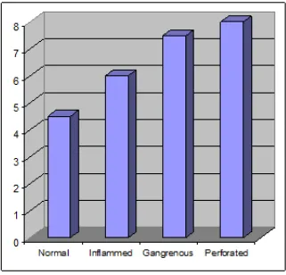
FIG: 7 The mean ALVARADO score of different categories of
inflamed appendix are compared with each other and the p value
19
20
Laboratory testing
Routine investigation of the patients admitted with right
quadrant pain includes the laboratory investigations like complete
blood count , c-reactive protein , the urine routine and urine culture
examination .
The investigation of female patient under the age group of 25-45
years or the reproductive age group includes the β-HCG (Human
Chorionic gonadotropins ) level in order to exclude ectopic pregnancy.
The inflammation of appendix may cause hematuria, pyuria which
may be similar to the presentation, in patients with urinary tract
infection .Studies have shown such patients to be about 10%9.Hence
routine urine examination is important to rule out UTI.
Nearly 70-90% of patients of acute appendicitis have an
elevated neutrophil count .It has poor specificity for diagnosing acute
appendicitis10-14.
WBC has been found to be elevated in acute appendicitis
which may be due to the mural inflammation of the appendix. Studies
have also shown that the WBC count correlates with the severity of
21
CRP is an acute phase reactant that has similar role as that of
WBC in appendicitis 15.There has been a reported sensitivity of 40-90%
and specificity of 27-90%16 in the diagnosis of appendicitis.
Another study shows that WBC was found to differentiate normal
appendix from the early inflamed appendix, than the CRP level .
Amalesh et al17 quoted “ The accuracy of CRP for diagnosing
acute appendicitis is low and that CRP levels are not useful when
deciding on surgery”.
Ortega-Deballon et al18 concluded “That CRP level is the most
useful laboratory parameter in terms of diagnosing acute appendicitis
and that CRP levels strongly correlates with inflammation severity of
the inflamed appendix ”.
CRP levels were found to be more accurate when there is more
severe , an increase in inflammation like that of gangrenous or perforated
appendix .Studies have shown , the correlation of CRP level with CT
findings and also could predict the probability of the patient going for
22
OBSERVATION AND LAPAROSCOPY
Diagnostic laparoscopy has mainly found its advantage in cases
that shows equivocal findings ,where the surgeons are in dilemma of
relying on the imaging techniques or the diagnostic laparoscopy. The
end point is to reduce the unnecessary appendectomy19.
It is of major use in female patient were many gynaecological
problems may mimic appendicitis in 10-20%20,21.These patients warrant
some active measures to rule out appendicitis or to favour an alternate
diagnosis.
Diagnostic laparoscopy comes into issue, when the surgeons are
not in favour of surgery and also reluctant to keep the patients in
observation. Both the decision is a double edged sword, were the risk of
perforation is more in positive cases and increase, in the rate of
unnecessary appendectomy 18 in false negative case.
The practice of observation has reduced the negative
appendectomy without increasing the perforation rate22-24 .Any diagnostic
23
Diagnostic laparoscopy has the advantage of27
Rapid and accurate diagnosis
Reduce the rate of unnecessary laparotomy
28
Additional caecal and colonic lesion are identified
Disadvantage of diagnostic laparoscopy 27
Invasive Procedure
Increased expenditure and cost
Hof et al29 quoted “Laparoscopy is the gold standard for
diagnosis of patients with suspected acute appendicitis ”. Acute
appendicitis can be diagnosed by laparoscopy in early stages .It also
lowers the threshold for appendectomy30.
Garbarino and Shimi et al31 “Routine use of Diagnostic
laparoscope in women significantly reduced the negative appendectomy
rate to 5%”
Lim et al.32 “Use of Diagnostic laparoscope changed the
24
Limitation of Diagnostic laparoscopy is that it could not be
compared with the gold standard , no tissue excision is done as it is a
diagnostic procedure and hence no specificity or sensitivity calculated.
Diagnostic laparoscopy has the high specificity of 95% as
compared to CT and ultrasound of 72 and 63% respectively and PPV of
85%-100%.Women has specificity of 95% in laparoscope compared to
72% in CT and 63% in USG27.
With the improved diagnostic accuracy of ( CT) computed
tomography, early use of CT has reduced the overall cost and use of
hospital resources33than the observation strategy.
Being a invasive procedure diagnostic laparoscopy also have the
added disadvantage with approximately, a 5 percent rate of
complications, which in most cases are associated with the use of a
25
DIAGNOSTIC IMAGING IN ACUTE APPENDICITIS
Acute abdomen pain is the most common symptom we encounter
in most of the emergency department. The abdominal pain is attributed
to many cause, of which the appendicitis occupies within the first few of
the cause. Evaluating a case of appendicitis is mainly clinical ,depending
on the clinical scores and signs.
But there is increase in the negative appendectomy rate,
depending only on clinical findings . And also in patients with atypical
and equivocal clinical findings surgeons are in favour of imaging
modalities for arriving at a diagnostic conclusion ,rather than to keep the
patient in observation.
As the later practice of observation has lead to increase in the
percentage of perforation rate, here comes the major role of the imaging
techniques like CT and USG.
Considering the imaging technique, there comes a question which
is the best or which is the first modality to be considered. Usually USG is
the first primary technique recommended considering it’s easy
26
But it has its own pitfalls, being operator dependent, highly
depending on the skill and experience of the radiologist who does the
scan. And also other factors like the built of the patient, and the various
position of the appendix , makes it difficult for the scanning radiologist
to visualise the appendix .
Sometimes USG also gives a equivocal findings were in we are
forced to switch over to CT or other modalities. CT on the other hand is
more specific than USG and hence could rule out appendicitis .Both the
imaging technique could give an alternate diagnosis if appendicitis is
ruled out.
Literature shows many studies that have debated over the best
modality for diagnosing acute appendicitis. Most of them come up with
more or less the same results. Both the technique have definitely reduced
the rate of negative appendectomy in recent years.
Weighing the cost versus the radiation and the real need to rule out
appendicitis ,and the dire need in search of alternate diagnosis should be
considered before deciding over which imaging modality to choose.
27
ROLE OF XRAY IN THE DIAGNOSIS OF APPENDICITES
With the advent of newer techniques like CT and USG X ray
has outdated, in the diagnosis of appendicitis ,but it confirms the presence
of appendicolith in 80-100% which is indicative of an appendicitis,
mostly perforated one.
X ray is also of use in the differential diagnosis of renal stone,
crohn's disease, ileocaecal tuberculosis, intussusceptions , and
malrotation of the gut34. Four out of five patients with false-positive
radiographs for acute appendicitis have other conditions like ,ruptured
ovarian cyst, leaking carcinoma of the caecum, or a low-lying inflamed
gallbladder.
This emphasis the fact that radiology reflects all diseases
affecting the right lower quadrant, the commonest being acute
appendicitis. Abdominal X-ray is neither sensitive nor specific for
appendicitis but can provide clues to an alternate diagnosis or clue in
favour of appendicitis.
Ellis34 recommends plain x-ray films of the abdomen in all
cases acute abdomen. Brooks and Killen have listed these radiological
28
RADIOLOGICAL FEATURES IN ABDOMINAL X-RAY
i) Air-fluid levels localised to the caecum and/or terminal small
bowel are indicative of localised inflammation in the right
lower quadrant of the abdomen.
ii) Localised adynamic ileus ,gas in the caecum, ascending colon
and terminal ileum.
iii) Increased soft-tissue density in the right lower quadrant.
iv) Blurring of the right flank stripe.
v) Appendicolith, the calcified concretions in the appendix with
typical laminated densities in the right lower quadrant
vi) Alteration of the psoas outline and blurring of its distal third.
vii) Gas-filled appendix, a rare but valuable sign.
viii) Extra luminal gas or free gas in the peritoneal or retroperitoneal
space.
ix) Deformity of the caecum.
29
ULTRASOUND IN THE DIAGNOSIS OF APPENDIX
USG is a simple procedure that can be done. It is a non-invasive
technique and it is also cost effective and easily available even at
primary centres.
It was introduced by Puylaert in the year 1986 which was
nearly ten decades after Fitz published his paper on acute appendicitis.
Ultrasound is used as the first diagnostic modality, followed by
CT scan of the abdomen, if only the ultrasound is negative or
equivocal.35-38 It also avoids excessive radiation.
The common technique used is the graded compression. This has
the advantage of displacing gas filled bowel loops between the
abdominal walls. This helps in better visualization of the appendix free
from the intestinal loops . Lean patients have higher rates of detection of
appendicitis with USG.39-41
30
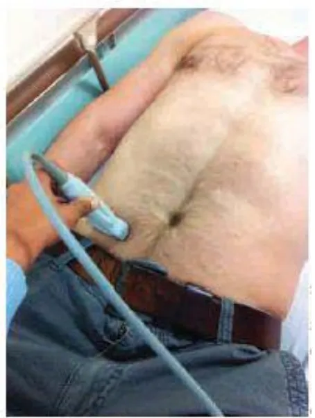
“The patient should be placed in the
supine position for the ultrasound
examination, and a high-frequency
linear array transducer should be
applied to the anterior abdominal wall
[image:41.595.198.425.168.469.2]over the area of maximal tenderness”
FIG:9 VARIOUS METHODS OF GRADED COMPRESSION
31
Limitations in visualising normal appendix
Various factors like obesity and position of the
appendix may limit the normal visualisation of appendix .Various
USG techniques helps the radiologist in such cases .Patients may
be put in left lateral or a posterior manual technique ,may help in
visualising the appendix in case of the appendix being retrocaecal
in position.
Sometimes the ascending in the right iliac fossa
may mislead the scanning radiologist .These bowel loops may
also sometimes appear as a non peristaltic loop. At, times like
these ,added techniques like posterior manual compression or the
left lateral decubitus would be of use.
Posterior manual compression is done with
additional compression given to the patient’s back in an anterior
direction by keeping a hand in the posterior of the trunk in the right
lumbar region.
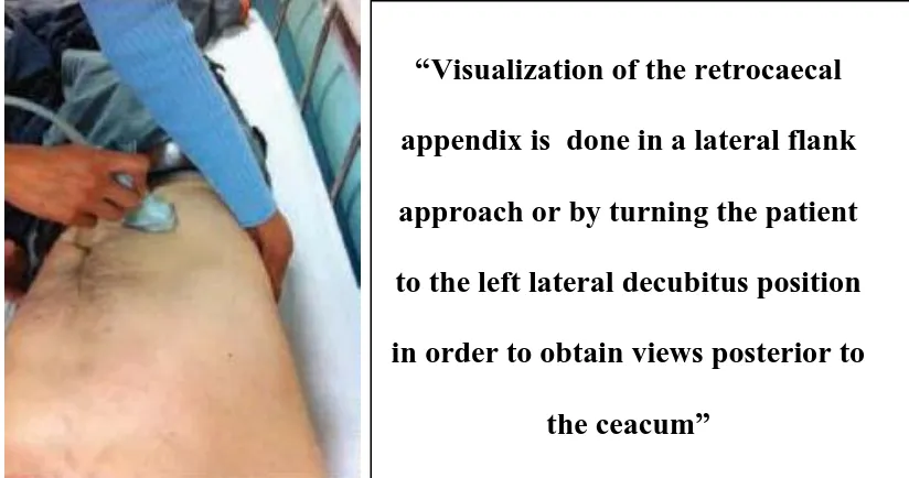
Lateral decubitus position is used to visualise the
region posterior to the caecum ,and hence in visualisation of the
32
“Posterior manual compression is
performed by placing one hand on
the patient's back, applying forced
compression in the antero medial
direction added to graded
compression with the transducer on
the anterior abdominal wall”
‘
“Visualization of the retrocaecal
appendix is done in a lateral flank
approach or by turning the patient
to the left lateral decubitus position
in order to obtain views posterior to
[image:43.595.99.511.457.674.2]the ceacum”
FIG:10 Posterior manual compression
33
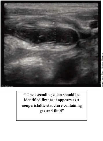
FIG:12 Ascending colon mimicking appendix
“ The ascending colon should be identified first as it appears as a nonperistaltic structure containing
34
NORMAL VISUALISATION OF APPENDIX IN USG
FIG :13 Longitudinal scan
FIG:14 Target sign in transverse scan
“ Longitudinal axis that measures greater than 6 mm in diameter and lacks
peristalsis”
35
Inflamed appendix appears as
A Aperistaltic
B Blind loop
C Non-compressible
D Diameter greater than 6 mm
FIG: 15 PICTURE OF AN INFLAMMED APPENDIX
The inflamed wall of the appendix appears laminated.
Sometimes appendicolith may be seen. This appendicolith are nothing but
36
appendix. They are seen in USG as a white echogenic structure which
gives a post acoustic shadowing.
Appendicolith is a contributory factor in the diagnosis of
appendicitis. Other additional findings can be identified that may give a
clue to the diagnosis. These include the caecal wall thickening and the
periappendiceal fat stranding.
A very good and experienced radiologist could even find these
minor details that may lead us to the diagnosis of appendicitis
Main clue to the diagnosis may come from the patient himself.
Typical patients with appendicitis will have right iliac fossa
tenderness,which the patient may localize. The most tender point shown
by the patient could be picked up by the radiologist as the probe
tenderness.
Additional use of colour Doppler may clinch the diagnosis of
appendicitis. The colour Doppler in the diagnosis of acute appendicitis
was first presented by LimHK and Quillin SP. The findings in Doppler is
the presence of peripheral increase in vascularity of the appendix.
This is due to the fact of the increased flow in the inflamed wall
37
the radiologist performing the scan to look for the wall of appendix, as
the disappearance of Doppler signal, in other wise an inflamed appendix,
is that it is going for gangrene or perforation.
It is important to mention these findings so that it alerts the
operating surgeon to make an urgent decision to operate the patient , as
the perforated appendix ,in itself has grave complication leading to long
term morbidity and mortality if ignored.
Appendicitis presents in most atypical manner, with many
disease process mimicking it. It is so atypical that even an experienced
surgeon may remove normal appendix. Surgeon’s upper limit of negative
appendectomy rate is 20%. This is done in order to avoid the
unnecessary complication of perforated appendix in case of delay.
Hence there should be a balance between negative appendectomy
and perforation rate .Ultrasound has come a long way and is now
routinely recommended by the referring physician or the surgeon to
diagnose a case of appendicitis in the most atypical and equivocal case.
Puylaert introduce the graded compression technique and reported
a sensitivity of 89% and specificity of 100%. Lots of studies which came
38
A meta-analysis by Doria lists “sensitivity of ultrasound as 88%
and 83% and its specificity as 94% and 93%, for children and adults,
respectively”.42
Many studies were done comparing the usefulness of ultrasound in
the diagnosis of appendicitis. One study compared the diagnosis of
appendicitis in two groups with one group, was diagnosed of
appendicitis with only clinical findings and the other with help of
ultrasound.
It was found the group one who were mainly diagnosed on the
clinical basis had 93% sensitivity and hence had many false positive
cases. Depending on only this value it was found that at least 10 more
patients were taken for surgery, for no reason or cause, with just clinical
basis findings only.
The second group of patient who were diagnosed on only the
USG findings had sensitivity of 81%, where in few patient who needed
surgery were left untreated as patients were misdiagnosed as normal.
This is due to the low sensitivity of USG which might lead to the
complication of perforation. So if only USG findings were taken into
39
untreated leading to morbidity. All studies pin point that any imaging
findings is never to override the clinical judgment.
But the picture changes when the specificity is taken into account
as a USG shows a specificity of 95% while that of clinical diagnosis
44%. This shows that greater number of false positive was present in the
patients who were clinically diagnosed. These patients were to undergo
unnecessary procedure of appendectomy. The procedure itself has its
own complication. This number of false positive is not acceptable in any
of the clinical diagnosis.
Appendix being a vestigial organ allows the acceptability of
unnecessary surgery to a certain extend but this could not be the case in
other grave disease .But on the other hand ultrasound has 95% of
specificity thereby reducing the unnecessary operation.
Both NAR and PR were also low in the second group who
underwent USG. There was a statistical significant drop in NAR from
25% in first group to 7.4% in the second Group. The perforation rate
symmetrically decreased from 15.6% to 15% in group one and two
40
different to other studies that show PR rate to increase with decrease in
NAR42.
Some studies did not take into account the gangrenous appendix
into perforation, hence this falsely gave a low PR rate. Gangrenous
appendix is more or less and definitely has higher a probability, to go in
for perforation, if timely intervention is not carried out. So a study could
do no justice if it does not takes the gangrenous appendix into
account.
As seen earlier bringing the USG as the diagnostic work up for
acute appendicitis, both NAR and PR has decreased which very well
shows the reciprocal relation of NAR and PR. Hence adding ultrasound,
decrease the negative appendectomy rate without increasing the
perforation rate.
Study by Stefan pug et al showed a decrease in NAR from 36.6%
to 3.2 with use of ultrasound. Negative appendectomy and PR both being
an adverse outcome, both could be added to get total adverse outcome
without taking into account their mutual relationship. It was found that
41
picture of the use of ultra sound in the diagnostic work up of acute
appendicitis.
Though the importance of ultrasound in equivocal cases are
helpful, because of its false positive and negative values it must not be
allowed to override the clinical acumen.
Hence for good clinical outcome ,combining the ultrasound and
clinical findings should be done. Some studies show that clinical
Alvarado score of 8 would need no ultrasound findings to diagnosis and
these patient were taken for surgery without subjecting the patient for
ultrasound.
At the other extreme clinical score of 4, patients were not taken for
surgery, only on the basis of ultrasound finding. The usefulness mainly,
lay in the clinical score of 4 –8. Within this intermittent score the
clinician and surgeon find it difficult to decide on ,with only the clinical
findings and also in case of equivocal clinical diagnosis.
Added value is present when the ultrasound could pickup
additional findings that clinch the alternate diagnosis for abdomen pain
42
Some of the works on USG using graded compression by
Terasawa and co workers43 showed an overall “sensitivity 0.86%
Specificity 0.81% PPV – 84% NPV – 85%”.
Meta analysis in Korea 44showed “sensitivity of 86.7% and
specificity of 80% and reported accuracy of ultrasound to be 86% -
96%” .
Advantages of USG
Safe in pregnancy
No risk of radiation exposure
Short scan time
No need for contrast
Non invasive
Easily performed in small children
Added benefit of diagnosing other alternate cause of abdominal
43
Though its usefulness has been well described it has its own
disadvantage and pit falls
First and the fore most is that it is an operator depended, hence the
final diagnosis also depends on the experience of the radiologist,
performing the scan.
Individual skill is important45
It is inferior to other imaging techniques like CT , in sensitivity
It has low negative predictive value it could not confidently
exclude the diagnosis of appendicitis
Difficult in female population because of overlap of symptoms46-50.
Difficulty in getting adequate good graded compression in obese
patient and in patients who had previous abdominal surgery
Sometimes the location of the appendix also leads to misdiagnosis
Most of the false positive is due to non-visualizations or only the
44
While positive ultrasound findings have a relatively high
positive-predictive value, identification of a normal appendix is sometimes
difficult.
Excellent results have been achieved at select centres. No
visualization of the appendix, being reported to have a
negative-predictive value of 90% 51.
Graded-compression USG remains our first-line method. It can
be performed at any time, regardless of specific patient’s preparation.
But in some equivocal cases subsequently they should undergo
Computed Tomography assessment 52,53. However it is non-invasive ,non
ionising, less expensive and also repeatable.
CT AND ITS ROLE IN DIAGNOSING ACUTE APPENDICITIS
There is an increasing surge for using CT in diagnosing
appendicitis .It has an excellent sensitivity , specificity and accuracy in
the preoperative diagnosis of acute appendicitis .The benefit of CT is
still controversy .There are greater number of patient who are subjected
to CT imaging and were still not operated.
Improved CT technology ,its wide spread availability and the trend
45
dependent ,there has been increasing use of CT technique. CT is good
in excluding the diagnosis of appendix and also added benefits of giving
an alternative diagnosis.
Various CT techniques are in use including
Unenhanced Helical CT57-59.
Targeted are focused appendiceal techniques using rectal
contrast54-56
IV enhanced CT
IV with oral or without oral contrast61,62
Low dose CT
IV with caecal air insufflations60
There is always debate over which technique is appropriate or good
The use of IV technique has its own disadvantage listed,
Allergic reaction to contrast63
Cost related
Extravasations of contrast material64
Tissue injury due the above leakage
46
Use of oral contrast68has as the added disadvantage of
Patient discomfort.
Increase in the scan time and also waiting time.
Some case if the contrast do not reach the caecum – the imaging
becomes a total failure.
Advantages of oral contrast65
When ceacum and ileum fills with contrast, appendix is visualized
well behind the background of contrast.
On the pre – text of the appendix filling with contrast appendicitis
could be ruled out.
Many studies favour ,and some have found no difference in
accuracy rate on using oral contrast. Anderson et al66and Keyzer etal67
quoted “No difference in sensitivity, specificity, positive predictive value
47
Unenhanced CT
Unenhanced scan decrease the time of scanning as there is no
need for oral contrast .It eliminates the risks associated with iv contrast.
Ege et al concluded that Unenhanced CT has a “ sensitivity of 96%,
specificity of 98%, positive predictive value of 97%, and negative
predictive value of 98%”69. Heaston et al. showed a “sensitivity of 84%
and a specificity of 92%”70 for unenhanced CT.
Non – focused Technique
Non – focused Technique gave a high diagnostic accuracy when
larger population sample were used with average prevalence of acute
appendicitis. This is the most commonly used CT technique .
Rao et al used and reported cases with use of oral and colon
contrast with prevalence of 53%54 of acute appendicitis with diagnostic
accuracy of 98%55”. This is based on the routine body imaging technique
used in early days. It uses both IV and oral contrast.
It has the advantage of finding both normal and inflamed
appendix with added advantage of finding extra appendiceal pathology.
Though helical CT with iv or oral or only rectal or other combination is
available this non-focused technique is widely used due to the fact that
48
Focused technique or the Appendiceal CT
Appendiceal CT is a focussed CT Technique and is advised for
patient when the clinician suspect acute appendicitis to be the only cause
for the patient’s pain. Helical Scanning with 5 mm collimation and 5mm
thickness is used.
Upper abdomen is left out covering only 15 cm of the lower
abdomen and the upper pelvis centered at the tip of the caecum. Small
rectal catheter is used to instill contrast into the colon with average
volume of 900 ml of contrast. No iv or oral contrast is used in this
technique. The scan time is complete in 20 – 30 minutes .
Negative was reported if the contrast filled the lumen or the
lumen is filled with air .Reported positive if the appendix is enlarged > 6
mm and if the appendix is not opacified or filled with contrast.
Positivity is given if specific signs like arrow head and cecal bar sign is
present. Appendicolith is another positive sign of appendicitis.
The main disadvantage is that other alternate diagnosis may be
missed as the entire abdomen is not covered in the scan. But this
technique can confidently confirm or exclude the diagnosis of acute
49
Rhea et al quoted “Focused appendiceal CT may lower both fixed
and variable cost in caring the patient with appendicitis”72 .
Rho et al “Focused technique reduces the use of hospital
resource”73
Fefferman et al reported high “sensitivity (97%), specificity
(93%), positive predictive value (90%), and negative predictive
value(98%) 71” in focussed technique.
The highest ,a CT accuracy for diagnosing acute appendicitis is
also from this technique of about 93 to 98%.As only limited section is
covered, the radiation dose to the patient is also minimal with reduced
exposure and cost. This technique also reduces the appendiceal
perforation rate from 22 to 14% and the negative appendectomy rate from
20 to 7%73.
Focussed techniques depend on expert interpretations and may
not always provide an alternate diagnosis for pain in patients with acute
symptoms. Imaging every patient with suspected appendicitis may be
impractical at many centres , because helical CT facilities and on-site
50
Low dose protocol
Taking into account the radiation from standard dose, CT low dose
protocol with no use of iv or oral contrast was used. This technique may
be adequate for diagnosing acute appendicitis . It is in the hands of the
radiologist to bring a change. Many studies based on low does CT are
done
KeyZer at al quoted “ No difference in sensitivity and specificity
value in diagnosing acute appendicitis on using standard does and
simulated low does” 67
Seo et al after having made studies with low does technique and
came up with the same results.
Contradicting KeyZer et al, studies have shown compromise in
low dose technique like
Alternate diagnosis and finding normal appendix
Loss of reader confidence
51
But still noise reducing post processing algorithm can be used to
increase the diagnostic accuracy in low does technique. This kind of
improvement in post processing will decrease the noise and increase the
image quality. The next issue in low does technique is the explanation of
alternate diagnosis, in case that had been reported negative for
appendicitis.
To be reported as false positive it had to be “ un equivocal
diagnosis of the disease with no differential diagnosis”. CT scans to be
reported as true negative “ the image must give either an alternate
diagnosis or must report it has normal findings”.
CT has been increasingly incorporated in most institution because
of high accuracy rate, an easy available range at present time. It has the
advantage of decreasing the NAR without increasing the perforation rate
CT CRITERIA FOR DIAGNOSIS OF ACUTE APPENDICITIS
The primary diagnostic criteria for acute appendicitis is
visualization of a
Thickened and distended appendix width >6 mm
Mural thickening and enhancement and
Wall thickening of appendix >2mm
52
Secondary diagnostic criteria are
Appendicolith,
Periappendiceal abscess,
Small-bowel obstruction,
Pericaecal inflammation
Target appearance - Concentric inflammatory thickening of
appendix
Presence of air both in intralumen and extralumen
The sensitivity and specificity of a pelvic and abdominal CT scan
are 94 percent and 95 percent, respectively 43.
The additional benefit of CT is that alternative diagnoses are
made in up to 15 percent of patients 74
A definitive CT diagnosis of acute appendicitis can be ruled out
if there is air or contrast in the appendiceal lumen
If rectal contrast is given two signs help in identifying
53
The caecal bar sign
The contrast filled caecum is seen distinctly due the interface
created by the inflammatory soft tissue thickening at the base of the
appendix.
The arrow head sign79
It is the contrast filling in the caecum, with the arrow pointing to the
point of occlusion in the appendix. It is not seen all the films. Thin
section will better depict this sign in CT. And it is also a necessary pre
requisite that the caecum must be well distended with contrast.
Caecal apical thickening.
Though both CT and USG have a synergistic value ,many
radiologist are in favour of CT, as they are more confident in
interpreting CT than sonography.80
Imaging techniques in suspected acute appendicitis have definitely
results in fewer unneeded laparotomy.(74,75,76)
Routine imaging is ,cost-effective and would also result in less delay
54
Effect of CT imaging on false positive
Surgically Accepted False Positive and Negative appendectomy
rate among the surgeons is 20% 82which has dramatically decreased in the
recent years by the liberal use of preoperative imaging technique like CT
and USG.
The False Positive rate is more in females compared to men due to
the overlap of gynaecological symptoms which is as high as 42% while
many studies have shown reduction in the above rate with increased use
of imaging. Some large scale studies have shown no improved clinical
outcome81.
Various studies have shown that there has been increase in use of
CT by the physicians and surgeon, as the first line imaging modality.
There is a decline in the USG imaging. However USG may play its role
in some diagnosis, mainly in female patients like fibroid, ovarian cyst
and pelvic inflammatory disease.
And also as the CT usage has increased, so is the decrease in the
appendiceal perforation with statistical significance of p < 0 .001.
There is also a significant decrease in the false positive diagnosis
55
Negative Appendectomy – Effect of Imaging
NAR was defined “as the portion of pathologically normal
appendices removed surgically in patients suspected of having acute
appendicitis”. Literature shows that 15-25% of such normal appendix
was removed82,83.
The need to reduce the unnecessary appendectomy is due the fact,
to avoid the risk of surgical complication and the cost. But it itself is
double edged sword. Surgeons have the upper limit of negative
appendectomy rate of 20%84. This is to avoid the negative and grave
consequence of delayed diagnosis and perforation.
The diagnostic accuracy of clinical findings is about 80%85. This
my fall to 60% to 68% percent in women population due to the overlap
of the gynaecological symptoms84-86. There has been an increase in
diagnostic accuracy to above 83% to 98% percent if in addition to the
clinical findings the imaging findings from CT and ultrasound are
combined73,75,88. There has been marked increase in the clinical outcome
56
Studies have show there has been significant decrease in NAR
value in women who have gone with preoperative imaging. One such
study have shown the overall sensitive of CT 96% and PPV (Positive
predictive value) 96% and correctly diagnostic in 89%. Same studies
showed ultrasound sensitive to be 86% and PPV 95% with correct
diagnosis in 79%90.
Prior studies have reported NAR of 5 to 16 % in men and 11 to
34% in women87. The most common misdiagnosis in women is the
pelvic inflammatory disease which is the major cause of increase in
negative appendectomy rate in women.
The studies also showed a decrease of about 27% in the negative
appendectomy rate some 34% to 7% in CT and to about 8% with USG
imaging90.
“Rao et al” showed a significant (P<0.001) decrease in NAR for
women from 35% to 11% in CT imaging89. Studies showed low NAR
value in males and boys regardless of preoperative imaging.
Coming to the perforation rate, literature shows perforation rate
of 14-31% Patients who underwent CT imaging had higher perforation
57
the time of CT imaging may be the cause of increased perforation rate in
the study group that undergo CT Examination.
Karakas et al reported “ PR of 54% in children who underwent
CT to PR of 20% with no imaging done”91
,possibly due to delay in
imaging
Most of the surgeons depend on the imaging technique, only when
clinical findings are equivocal. Perforation rate and NAR are inversely
relative, in that any increase in negative appendectomy rate, usually
decrease the PR and decrease the number of study people who are kept
under observation.
Studies also suggested that more than the in hospital stay the delay
from the patients side play a major role in the perforation rate and that
the high perforation rate is unrelated to the imaging technique performed.
Another study showed that the preoperative CT has significant
decreased in the NAR in age group of < 45 years in women, but did not
have any effect in male and women in > 45 years . The study has the
58
Raman et al showed that with increase in the percentage of
patients who undergoes CT image from 18.5 to 94.2% ( P< .00001) ,NAR
decreased from 16.72 – 8.7% with statically significant p value <
0.000189,92.
“Rhea at al” showed a decrease in NAR from 20 to 7% while Rao
et al quoted “11 to 5% CT imaging showed false positive of 1.7 to 10%
and false negative of 0 to 2.4%”89.
Another study by “Raja et al” showed with increase use in CT
from 1% to 97.5% (P < 0.0001), NAR decrease from 23% to 1.7% (P <
0.0001) with female rate decreasing from 29.8% to 1.6% and male rate
decreasing from 15.5 to 1.8 both having P Value of < 0.0001 which was
59
AIM AND OBJECTIVE
To subject the patients admitted in emergency department
suspected of acute appendicitis on clinical grounds ,to imaging
technique ,both CT and USG.
To calculate the sensitivity ,specificity ,positive predictive
value and negative predictive value for both CT and USG
having the histopathology findings as gold standard.
To find the diagnostic accuracy of both the imaging
60
RATIONALE FOR THE STUDY
Acute appendicitis is mostly, clinically diagnosed disease where
the surgeons or the physician depends mostly on the clinical scores and
physical examination and physical signs.
But there is increase in the negative appendectomy rate,
depending only on clinical findings .So the surgeons favour the use of
imaging technique like CT and USG ,if not in all cases ,at the least in
atypical and equivocal ones where there is a need to rule out or confirm
the diagnosis of acute appendicitis
Literature shows many studies that have debated over the best
modality for diagnosing acute appendicitis. Most of them come up with
more or less the same results.
USG is a non invasive ,cheap ,readily available technique with no
need for contrast .But however it has its own limitation being operator
depended ,highly depending on the skill and experience of the
radiologist who scan. And also other factors like the built of the patient
and the various position of the appendix ,makes it difficult for the
scanning radiologist to visualise the appendix..CT on the other hand has
the limitation of ionising radiation, but it also has the benefit of
definitely ruling out appendicitis or confirm it because it has more
61
Both the USG and CT has the advantage of alternate diagnosis if
the diagnosis of appendicitis is ruled out. Both the technique have
definitely reduced the rate of negative appendectomy in recent years.
Hence adding the imaging modality either of the two or both,
would benefit the attending surgeon over the treatment strategy. Deciding
over which technique is the best modality, with high diagnostic accuracy
is important, to be cost effective, avoid unnecessary surgery, and the
study would answer the above doubts
Prospective observational study
Sample size-100 patient
Study period - 6 months
Study center- Institute: Rajiv Gandhi Government General
62
Inclusion criteria.
This is a Prospective observational study conducted in Patient
who was admitted in the emergency department at Rajiv Gandhi
Government Hospital from march 2014 to august 2014 with symptoms
of acute abdomen pain and clinical findings highly suspicious of
appendicitis.
Main criteria was to take into account patients who have undergone
both the imaging techniques of CT and USG.
The criteria was to select patients who had both imaging done and
were taken for surgery on clinical findings
This study protocol was approved by the ethical committee of the
institutions and the departmental review board and institutional
informed consent guidelines were observed
Exclusion criteria
Patient with inflammatory focus like mesenteric adenitis found
through initial USG screening and history
63
Patients who were in need of immediate surgery and no time for
imaging modality.
Non consenting patient.
Patients who had only one imaging done or no imaging done were
excluded
METHODOLOGY
Subject:
Patients who were admitted in the causality surgical emergency ward
within the age group of 15-45 who presented with clinical findings and
symptoms of acute appendicitis like right iliac fossa pain ,fever and
vomiting were enrolled in the study. A total study sample of 100 was
selected The clinical history regarding present history was taken in the
prescribed proforma. Informed consent was obtained from each
participating patient and the protocol was approved by the institutional