UNIVERSITY OF SOUTHERN QUEENSLAND
Development and Characterisation of a
Chemical Film Actinometer with a Large
Dynamic Range for Measurements of Solar
Ultraviolet Exposure
A Dissertation Submitted by
R.A. Lester, BSc(Hons)
For the Award of
Master of Philosophy
iii
Declaration
The research contained in this dissertation is the full documentation of the research
results that were published as
Lester, RA, Parisi, AV, Kimlin, MG, & Sabburg, J. 2003, ‘Optical properties of
poly(2,6-dimethyl-1,4-phenylene oxide) film and its potential for a long-term solar
ultraviolet dosimeter’, Physics in Medicine and Biology, vol. 48, pp. 3685-98.
I declare that this dissertation is comprised entirely of my own research (except
where due acknowledgement is made), and no part of this dissertation has been
written by another person. To the best of my knowledge, the work presented here is
original and has not been published elsewhere other than in the reference stated
above. This dissertation contains no material that to any substantial extent resembles
work previously submitted for any other award at the University of Southern
Queensland or any other educational or academic institution. All contributions to this
dissertation and the research within are explicitly acknowledged.
Rick Lester
Signed ...
iv
Acknowledgements
I wish to acknowledge the various contributions made to this project by a number of
members of staff of the Faculty of Sciences at the University of Southern
Queensland.
My Principle Supervisor, Dr Alfio Parisi (now Assoc Prof) initiated the concept for
the project, provided supervision and training in a number of areas relating to
ultraviolet physics, assisted with the assembly of experiments, and contributed
substantially to the proof reading and editing of documents relating to this project.
Dr Jeff Sabburg made valuable contributions to experimental design and provided
advice on error analysis, and also contributed to proof reading and editing. As an
Associate Supervisor, Dr Michael Kimlin (now Assoc Prof) provided guidance for
the casting aspect of the pilot study and also assisted in proof reading and editing
during the early stages of the project. Prof John Mainstone, also an Associate
Supervisor, contributed to project discussions and proof reading, and Dr Brad Carter
took on the role of Associate Supervisor during the final stage of the project.
Dr Tania van den Ancker and Dr Ray Marshall provided essential workspace in the
chemistry laboratory, assisted with the chemistry equipment required for the casting
of chemical films, and also contributed to discussions relating to the photochemical
nature of various actinometer materials.
Mr Graham Holmes provided technical assistance and provided a valuable support in
equipment maintenance and repairs throughout the project, and also played a pivotal
role in problem solving related to experimental logistics. Mr Pat McConnell also
assisted with experimental apparatus.
Mr Oliver Kinder designed and constructed a number of precision mechanisms
required for experimental work, and provided much needed technical assistance with
these and other experimental devices, as well as repairs and maintenance. Although
Mr Ken Mottram retired before the beginning of this project, I would like to thank
v
In addition, the Faculty of Sciences at the University of Southern Queensland
financed the project by means of a research scholarship, and also provided the
equipment and resources necessary for this project. I am grateful to, and thank all of
vi
Abstract
Solar ultraviolet (UV) radiation arriving at the Earth’s surface is a biological
requirement for most forms of life, but also causes adverse responses in humans,
animals and plants in cases of overexposure. Many of the adverse responses are
cumulative in nature, and hence solar UV related environmental risk assessment
requires quantification of long-term exposures and large UV doses.
Dosimetry methods for quantifying solar UV radiation exposure are extremely
versatile and cost-effective compared to radiometric methods, and allow time
integrated doses to be quantified efficiently. Biodosimetry often provides a large
dynamic range, but is expensive, labour intensive and time consuming. Chemical
actinometry is a cost and labour effective alternative to biodosimetry, but is
disadvantaged for large-dose measurements by its relatively small dynamic range.
Poly(dimethyl phenylene oxide) (PPO) film was identified from the literature as a
chemical actinometer material with the potential to reduce the labour and costs
involved in the quantification of large solar radiation doses by means of a larger
dynamic range. A fabrication technique for PPO film actinometers was established,
and the optical properties of the actinometers were fully characterised.
The resulting actinometer provides an efficient method for quantifying either
unweighted UVB dose or biologically effective dose. The spectral response
resembles the erythemal action spectrum, and the solar erythemal calibration
function is near linear. The PPO film actinometer is therefore very well suited to
human exposure research, especially for evaluation of chronic responses, or
cumulative acute responses in which large-dose measurements are required.
The PPO film actinometer now provides an additional tool in the quantification of
solar UV radiation exposure. It has equal versatility, and similar costs, labour and
equipment requirements to the most commonly employed actinometry methods. The
larger dynamic range of PPO film however, reduces labour and costs associated with
vii
Contents
Declaration ... ii
Acknowledgements ... iv
Abstract ... vi
Contents ... vii
List of Figures ... xi
List of Tables ... xv
Chapter 1: Introduction ... 1
1.1 Introduction ... 2
1.2 Definitions and Terminology ... 4
1.3 Project Objectives ... 5
1.3.1 Literature Search for a Suitable Dosimeter Material ... 6
1.3.2 Fabrication of PPO Film ... 6
1.3.3 Investigation of PPO Film Thickness and Dose-Response ... 7
1.3.4 Quantification of the Optical Properties of PPO Film Actinometers ... 7
1.3.5 UV Radiation Calibrations ... 7
1.4 Dissertation Overview ... 7
1.5 Summary ... 8
Chapter 2: Responses of Biological Systems to Solar UV Radiation ... 10
2.1 Introduction ... 11
2.2 Molecular and Cellular Level Responses in Humans ... 12
2.2.1 DNA and Cellular Damage ... 12
2.2.2 DNA Repair Mechanisms ... 12
2.2.3 Mutation of the p53 Tumour Suppressor Gene ... 13
2.3 Organism Level Responses in Humans ... 14
2.3.1 Dermal Responses ... 14
Vitamin D Synthesis ... 14
Erythema and Sunburn... 14
Sun-Tanning ... 15
Skin Cancer ... 15
viii
2.3.3 Immunosuppression ... 19
2.4 Responses in Animals ... 19
2.5 Responses in Plants ... 20
2.6 Biologically Effective Radiation ... 21
2.7 Summary and Conclusions ... 25
Chapter 3: Variability and Distribution of Solar UV Radiation ... 27
3.1 Introduction ... 28
3.2 Astronomical and Geometrical Factors ... 28
3.3 Atmospheric Processes ... 29
3.3.1 Atmospheric Absorption and Scattering ... 29
3.3.2 Effects of Clouds ... 32
3.3.3 The Altitude Effect ... 32
3.4 Spatial Distribution ... 33
3.4.1 Latitudinal Dependence ... 33
3.4.2 Hemispherical Differences ... 33
3.4.3 Surface Albedo ... 34
3.5 Stratospheric Ozone and Total Column Ozone ... 34
3.6 Summary and Conclusions ... 36
Chapter 4: Review of the Optical Properties of UV Radiation Dosimeters ... 38
4.1 Introduction ... 39
4.2 Optical Absorbance and Response ... 40
4.3 Sensitivity ... 41
4.3.1 Exposure Sensitivity ... 41
4.3.2 Spectral Sensitivity ... 42
4.4 Dose-Response and Calibration Functions ... 43
4.5 Dynamic Range ... 45
4.6 Angular Response ... 46
4.7 Time-Irradiance Reciprocity ... 46
4.8 Temperature Dependence ... 47
4.9 Dark Reaction ... 48
ix
Chapter 5: Evaluation of Currently Available Dosimeters for
Large-Dose UV Measurements ... 50
5.1 Introduction ... 51
5.2 Biodosimetry ... 52
5.2.1 Vitamin D, Iodouracil, and DNA ... 52
5.2.2 Bacillus Subtilis ... 52
5.2.3 Uracil Thin Layer ... 53
5.3 Actinometry ... 53
5.3.1 Chemical Solution Actinometers ... 54
5.3.2 Polysulphone Film ... 54
5.3.3 Dyed Polyvinyl Alcohol and Polyvinyl Butanol Films ... 55
5.3.4 Photosensitised Polyvinyl Chloride Films ... 56
5.3.5 Poly(Dimethyl Phenylene Oxide) Film ... 57
5.4 Summary and Conclusions ... 60
Chapter 6: Physical Development and Optical Characterisation of Poly(Dimethyl Phenylene Oxide) Film Actinometers ... 62
6.1 Introduction ... 63
6.2 Equipment and Instrumentation ... 63
6.2.1 Casting Table ... 63
6.2.2 UV Radiation Sources ... 65
6.2.3 Irradiance Measurements and Calculations ... 69
6.2.4 Optical Absorbance Measurements and Calculations ... 75
6.3 Physical Development and Actinometer Fabrication ... 75
6.3.1 General Development Procedure ... 75
6.3.2 PPO Film Quality and Durability ... 76
Film Thickness ... 77
Mixing Ratio and Chloroform Evaporation Rate ... 77
6.3.3 Film Thickness Measurements ... 78
6.4 Optical Characterisation of PPO Film Actinometers ... 80
6.4.1 Effects of Mixing Ratio and Film Thickness on Dose-Response ... 80
Mixing Ratio ... 80
x
6.4.2 Spectrophotometer Measurement Wavelength ... 85
6.4.3 Effect of Spectrophotometer Beam ... 88
6.4.4 Response Reproducibility ... 88
6.4.5 Temperature Dependence ... 91
6.4.6 Artificial Source Calibration Functions ... 93
6.4.7 Time-Irradiance Reciprocity ... 96
6.4.8 Spectral Response ... 99
6.4.9 Angular Response ... 102
6.4.10 Solar Erythemal Exposure Calibration ... 105
6.4.11 Dynamic Range ... 107
6.4.12 Dark Reaction ... 108
6.5 Summary and Conclusions ... 113
Chapter 7: Conclusions ... 115
7.1 Introduction ... 116
7.2 Consolidation of Research Objectives ... 117
7.3 Further Research ... 119
7.4 Final Conclusion ... 121
References ... 122
Appendices ... 141
Appendix A – Pilot study ... 141
Appendix B – Optical wedge interferometry ... 144
Appendix C – Paired t-tests for spectrophotometer beam effect on PPO film ... 146
Appendix D – Analysis of variance test for temperature dependence ... 147
xi
List of Figures
Figure 2.1: Incidence rates of cutaneous malignant melanoma for Australia, the United States and the United Kingdom diagnosed in males (), females (),
and both males and females (). The data for this chart were obtained from
the National Statistics Clearing House (nschdata.org), the Australian Institute of Health and Welfare (aihw.gov.au), and the Surveillance, Epidemiology, and End Results Database (seer.cancer.gov). ... 17
Figure 2.2: Solar spectral UV irradiance (a) (left axis) and erythemal spectral irradiance (b) (left axis) on a horizontal plane. The solar irradiance was measured in Toowoomba at a solar zenith angle of 44. The erythemal spectral irradiance is calculated from the product of the solar spectral irradiance and the CIE erythemal action spectrum (CIE 1987) (c) (right axis). ... 23
Figure 2.3: Action spectra for biological responses including human erythema (CIE 1987), vitamin D synthesis (MacLaughlin, Anderson, & Holick 1982), previtamin D3 synthesis (CIE 2006), actinic response (IRPA 1989), DNA damage (Setlow 1974), photoconjunctivitis (CIE 1986a), and photokeratitis (CIE 1986b). ... 24
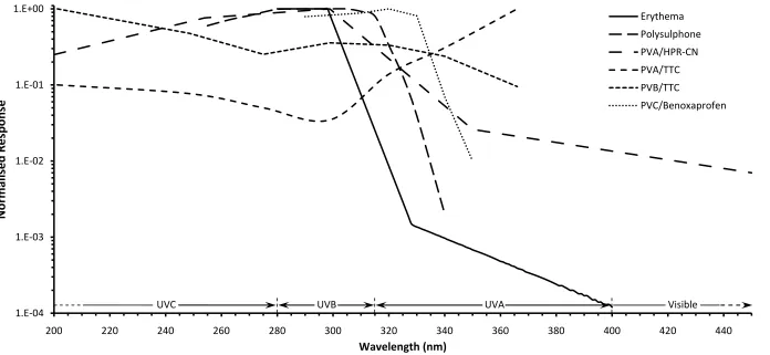
Figure 5.1: A comparison of the spectral responses of several of the actinometer materials reviewed in this chapter. These include polysulphone (Diffey 1989), PVA/HPR-CN (Abdel-Rehim, Ebrahim & Abded-Fattah 1993), PVA/TTC and PVB/TTC (Ebraheem et al. 2000), and PVC/benoxaprofen (Diffey, Oliver & Davis 1982). The erythemal action spectrum (CIE 1987) is included for reference. ... 59
Figure 6.1: The polymer film casting table employed by physicists at the University of Southern Queensland for the fabrication of various chemical films for use in UV actinometry. ... 64
xii
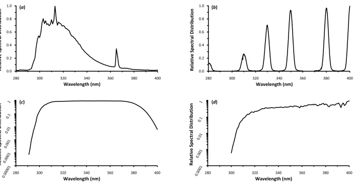
bandwidth (b), the solar UV simulator (c), and natural solar UV radiation (d). ... 67
Figure 6.3: The irradiation monochromator at the University of Southern Queensland’s physics laboratory. The lamp housing is seen toward the rear of the instrument and the monochromator is toward the front. A PPO film actinometer is held in front of the output aperture by a retort stand for irradiation with monochromatic UV. ... 68
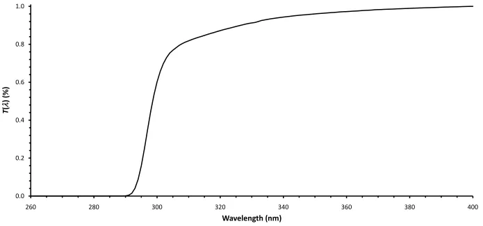
Figure 6.4: Average relative transmission spectrum T() of cellulose acetate film for an 8 h broadband exposure (272 kJ m-2) to the fluorescent UV lamp.
... 71
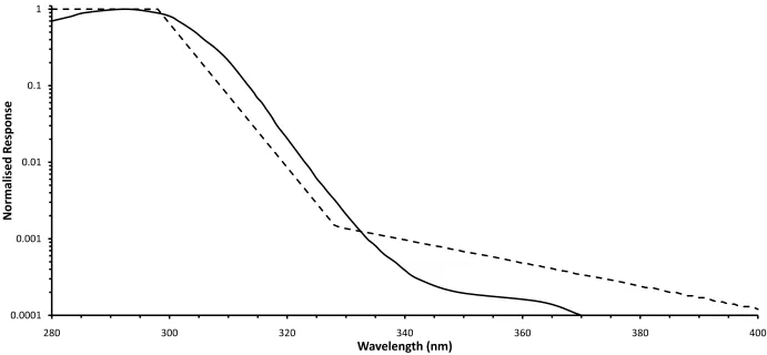
Figure 6.5: Spectral sensitivity of the Solar Light 501 UV-biometer (solid curve) normalised to the maximum sensitivity at 293 nm compared to the CIE erythemal action spectrum (dashed line) (CIE 1987). ... 74
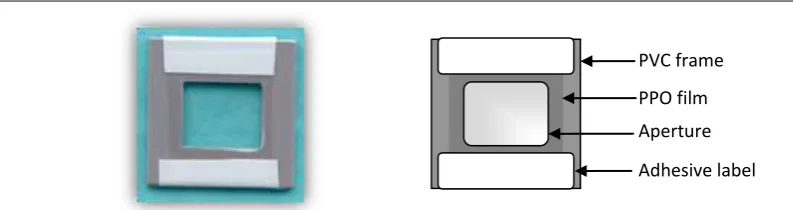
Figure 6.6: An actual size PPO film actinometer displayed on a blue background (left) and the PPO film actinometer schematic (right). ... 76
Figure 6.7: Dose-response comparison for 20 m PPO film cast using mixing ratios of 0.06 (dashed curve) and 0.12 (dotted curve). The relative difference between the responses at the two mixing ratios is given by the solid curve (right axis). The error bars represent an estimated 6% error in the response due to film thickness variations, timing errors, and lamp alignment errors. ... 82
Figure 6.8: Dose-response curves (left axis) resulting from the broadband UV exposure of PPO films of thicknesses 15 m (a), 20 m (b), 40 m (c), 50
xiii
Figure 6.9: Optical absorbance spectra of 40 m PPO film samples after exposure to broadband UV doses of 17 kJ m-2 (a), 272 kJ m-2 (b), 544 kJ m-2
(c), 1223 kJ m-2 (d), and 2310 kJ m-2 (e)... 87
Figure 6.10: Reproducibility of broadband UV doses in terms of the coefficient of variation (CV) as a function of dose (). The dashed curve represents the
least squares regression 2
1.1647 0.4618 3.1
y x x fitted to the CV data (R2 = 0.947). The calibration curve () employing the mean A(320 nm) at
each dose is included for reference. ... 90
Figure 6.11: Effect of temperature during exposure on the UV induced response of 40 m PPO film actinometers from 1.5C to 50C. The x-error bars indicate the uncertainty in temperature during the exposure, and the y-error bars are based on a 6.6% CV predicted by equation 6.7. ... 92
Figure 6.12: Fluorescent UV lamp calibration curves for broadband UV dose (), UVB dose (), UVA dose (), erythemal dose (), and photokeratitis
weighted dose (). Solid lines are scaled against the left axis while dashed
lines are scaled against the right axis. The error bars represent the CV as estimated for each A(320 nm) by equation 6.7. The error bars are identical for all calibration curves. ... 95
Figure 6.13: Reciprocity result for 40 m PPO film actinometers exposed to a broadband dose of 1.9 MJ m-2. The A(320 nm) () is plotted against the
irradiance required to administer a dose of 1.9 MJ m-2 over the given exposure
time period () (right axis). The x-error bars indicate the estimated 10% error
associated with measurements of irradiance, and the y-error bars indicate a CV
ranging from 3.3% to 4.5% predicted by equation 6.7 for each A(320 nm) response. ... 98
xiv
exposure waveband. The spectral response of polysulphone film (left axis) and the erythemal action spectrum (left axis) are included for comparison. ... 101
Figure 6.15: The angular response of 40 m PPO film actinometers in the azimuth plane () and the altitude plane () (central axis), and the cosine
error () for each angle (right axis). Three repeated measurements were made at 45 of altitude () to provide an estimate of the reproducibility of
about 4% for this experiment, as indicated by the error bars. ... 104
Figure 6.16: Solar erythemal exposure calibration curve of 40 m PPO film actinometers. The curve represents the third order cubic regression (equation 6.12). The three open circles represent the data points that occur within the region of optical saturation as they deviate substantially from the regression line. The y-error bars show the uncertainty of about 10% in the UV-biometer measurements, and the x-error bars represent the CV of the response ranging from 3% to 12.4% as estimated by equation 6.7. ... 106
Figure 6.17: Dark reaction of 40 m PPO film actinometers stored at temperatures of -16C (a), 2C (b), 24C (c), and 40C (d) after exposure to a broadband dose of 729.5 kJ m-2. The dashed curves represent power
curve regression lines for temperatures of 24C and 40C. ... 109 Figure 6.18: Dependence of the dark reaction of 40 m PPO film actinometers
on dose. The percent change in ADK(320 nm) is plotted against the A(320
nm) response induced by the broadband doses given in table 6.5. The
ADK(320 nm) was determined at post-exposure times of 22.5 h (a, ), 49.5
h (b, ), 72 h (c, ), 104 h (d, ), 176.5 h (e, ), and 493.5 h (f, ). The
xv
List of Tables
Table 6.1: Results of the feeler gauge leaf thickness measurements made using the
optical wedge interference technique to test the accuracy of PPO film thickness
measurements using this method. ... 79
Table 6.2: Response measurements of 40 m PPO film actinometers after exposure at six temperature levels. ... 91
Table 6.3: Fluorescent UV lamp irradiances for various UV wavebands and
biological weightings, and the ratio A of the irradiance of a given waveband or weighting to the broadband UV irradiance. ... 96
Table 6.4: Distances, measured irradiances and calculated exposure times used in the
time-irradiance reciprocity experiment. ... 97
1
Chapter 1
: Introduction1. Chapter 1
Introduction
1.1
Introduction
1.2
Definitions and Terminology
1.3
Project Objectives
1.4
Dissertation Overview
2
1.1
Introduction
Solar radiation is ubiquitous over the daytime side of Earth’s surface. The energy
imparted by solar ultraviolet (UV) radiation on incidence to terrestrial surfaces and
biological systems is sufficient to alter the molecular or cellular structures of many of
these materials. Such alterations may result in discolouration and degradation of
polymers, and more importantly, disease in humans, animals and plants.
Substantial evidence has been presented in the literature suggesting that many
organisms and ecosystems are perturbed by exposure to ambient levels of solar UV
radiation (Häder et al. 1998). In addition to ambient radiation stress, organisms may also be exposed to increased levels of solar UV radiation, elevated by both natural
and anthropogenic forces. These issues have been reinforced in recent years by the
growing awareness of stratospheric ozone depletion due to industrial emissions of
ozone-depleting gases into the atmosphere, and of the associated increase in solar
UV radiation at Earth’s surface (Appenzeller 1993; Herman et al. 1996; McKenzie,
Connor & Bodeker 1999). Important environmental issues are therefore, the
quantification of solar UV exposure and the assessment of its effects on organisms
and ecosystems.
Many biological responses to UV radiation are cumulative in nature. Time-integrated
exposure measurements are therefore required to assess the chronic effects of UV
radiation on the biosphere. Radiometry is frequently employed to quantify solar UV
radiation. The strength of this method is in the measurement accuracy afforded by
sophisticated electronic detectors. Exposure calculations from instantaneous
radiometric measurements however, require many measurements over the exposure
period of interest, and the greater the variability of the UV radiation source, the
greater the number of measurements required for integration such that a given level
of accuracy is maintained. Since solar radiation continually varies at the Earth’s
surface due to complex atmospheric optics and geometrical factors, the time and
labour involved in obtaining a sufficient number of instantaneous radiometric
measurements for accurate integrations of large doses is often prohibitive. Although
automated systems are becoming available, such instruments are expensive, bulky
3
An alternative to radiometry is passive dosimetry, which is well suited to UV dose
measurements since dosimeters naturally integrate over time and incorporate all
variability into a measurement. The two main branches of dosimetry are biological
dosimetry, or biodosimetry, and actinometry (also known as physical or chemical
dosimetry).
Passive UV dosimeters must ultimately be calibrated against a radiometric
instrument, and also incur imprecision related to the optical properties of the
dosimeter. Dosimetry is therefore inherently less accurate than radiometry.
Dosimetry methods however, have grown in popularity due to their low cost and
versatility compared to radiometry, as well as their ability to provide direct
measurements of time-integrated UV dose. The variability inherent in solar UV
radiation that occurs over many time-scales is captured by the dosimeter in a single
exposure measurement. This is particularly important where further UV radiation
variability occurs due the motion of the subject under measurement (e.g. Milne et al. 1999; Cockell et al. 2001). Their small size also allows significant numbers of dosimeters to be deployed simultaneously, allowing spatially resolved UV data to be
acquired over complex surfaces (e.g. Kimlin & Parisi 1999).
Biodosimetry is the leading technique in large-dose UV quantification, and provides
the largest dynamic ranges available. Biodosimeters utilise UV sensitive organisms,
or simple biological systems to gauge UV radiation dose, usually through UV
induced DNA damage (Koussoulaki et al. 1997; Bérces, Gáspár & Rontó 1999, pp.
141-2; Rontó et al. 2000). The preparation of biodosimeters, and the post-exposure analytical procedures associated with this method however, are often complex and
time-consuming. A measure of UV radiation dose by Bacillus subtilis dosimetry for
example, is determined from the proteins synthesized after incubation and staining
(Quintern et al. 1994).
Actinometry methods are available that simplify the preparation and analytical
procedures, and hence reduce the labour and costs involved. These techniques utilise
a change in optical absorbance of the actinometer material in response to UV
radiation exposure. The absorbance change can often be quantified by means of
4
The dose-capacity, or the maximum dose that can be registered by a single
dosimeter, is substantially smaller for currently employed actinometer materials
compared to biodosimetry materials. The resulting smaller dynamic range is a
fundamental limitation of the actinometry methods in current use.
The most extensively employed actinometer material for solar UV radiation
dosimetry is polysulphone (e.g. Knuschke & Barth 1996; Sydenham, Collins & Hirst
1996; Dunne 1999; Kimlin & Parisi 1999; Milne et al. 1999; Lester & Parisi 2002). Polysulphone actinometers have an operational life-span of about one day in summer
at mid-latitudes, or a dose-capacity of about 12 kJ m-2 (CIE 1992). Many chronic UV
induced biological responses are therefore difficult to quantify by actinometry since
they occur over periods of years, provoked by the large-dose equivalent of many
accumulated doses.
Large UV dose measurements employing polysulphone actinometry have been
achieved by replacing actinometers on a daily basis, and calculating the cumulative
exposure (Parisi et al. 2000). The disadvantages are however significant. A large number of actinometers are required, and the replacement procedure is time
consuming and labour intensive. Time-integration errors are also introduced. Ideally,
an actinometer material with a greater dynamic range should be employed for
large-dose measurements.
An actinometer is required that has a larger dynamic range than is currently available
to alleviate the labour, costs, and time associated with large-dose actinometry. The
focus of the research is therefore on identifying an actinometer material with a
greater dynamic range, and fully characterising the material’s optical properties to
provide a large-dose actinometer for solar UV exposure measurements.
Manufacturing costs of such actinometers must be kept to a minimum such that the
advantages of a larger dynamic range are not outweighed by fabrication costs.
1.2
Definitions and Terminology
Electromagnetic radiation (EMR) is a form of energy that propagates through space
by means of electric and magnetic fields oscillating perpendicular to each other. The
5
fields. The nature and propagation of EMR through space is described by Maxwell’s
equations (Halliday, Resnick & Walker 1997, p. 803).
The International Commission on Illumination (CIE) is an international authority of
lighting and radiation. The CIE definitions and terminology recommendations have
been adopted in this dissertation. The UV radiation waveband is defined by the CIE
as the region of the EMR spectrum that lies between the ionising-nonionising
boundary (100 nm) and the visible boundary (400 nm). The CIE also recognises the
ultraviolet-C (UVC), ultraviolet-B (UVB) and the ultraviolet-A (UVA) wavebands as
biological subdivisions of the UV waveband. These subdivisions constitute the
respective wavebands of 100 nm to 280 nm, 280 nm to 315 nm and 315 nm to 400
nm (CIE 1987).
Electromagnetic radiation in the UV waveband is most often quantified
radiometrically in terms of various radiance units. Irradiance is defined as the radiant
power per unit area incident upon a surface at all wavelengths, while spectral
irradiance is the radiant power distribution expressed as irradiance per unit
frequency. The SI unit for irradiance is W m-2 and that for spectral irradiance is
W m-2 Hz-1. The unit of W m-2 nm-1 is often used for irradiance quantification in the
UV waveband, and is the only deviation from SI units in this dissertation. Exposure
is wavelength- and time-integrated irradiance and its measurement is dose with units
of J m-2. Biologically effective UV irradiance is the spectrally determined irradiance
component of the UV waveband that causes a given biological response.
Biologically effective dose is defined as the wavelength- and time-integrated
irradiance, where the spectral irradiance is weighted by the biological response of
interest.
1.3
Project Objectives
The principal objective of this research is to reduce the labour and costs involved in
the dosimetry of large solar UV radiation doses by developing an actinometer with a
6
The steps required to meet this objective are as follows:
To identify a chemical with physical and optical properties that are suitable
for use in the dosimetry of large doses of solar UV radiation.
To develop an efficient method for the fabrication of actinometers that
utilise the material identified above.
To characterise the identified material by experimentally investigating and quantifying its optical properties that are relevant to the quantification of
solar UV radiation.
To calibrate the response of the fully characterised actinometer for UV
radiation dose measurements against a suitable radiometer, and to evaluate
the potential of the actinometer for quantification of UV exposure within
specific wavebands and for biologically effective exposure.
These objectives in detail are as follows:
1.3.1
Literature Search for a Suitable Dosimeter Material
The literature was extensively searched for chemicals that are photosensitive in the
UV radiation waveband. Information concerning the optical properties of these
chemicals was compiled and studied in order to identify one that would provide the
optimum properties for large-dose solar UV dosimetry. The factor of priority in this
search was the dynamic range, which led to further investigations of
poly(2,6-dimethyl-1,4-phenylene oxide) (PPO).
1.3.2
Fabrication of PPO Film
The literature indicates that the most cost effective and versatile methods of
actinometry employ photosensitive materials in the form of a chemical film. A cost
effective method of casting PPO into thin film was therefore required. Casting
experiments were conducted to assess the feasibility of fabricating PPO film
7
1.3.3
Investigation of PPO Film Thickness and Dose-Response
In addition to resistance to damage from normal handling, a dosimeter film for
large-dose solar UV radiation measurements must be sufficiently durable to withstand the
outdoor environmental forces over long periods of time. The film thickness that
provides sufficient durability must also have a useful dose-response. It was therefore
necessary to investigate the dose-response of PPO film as a function of film
thickness.
1.3.4
Quantification of the Optical Properties of PPO Film
Actinometers
The characteristics of a dosimeter are determined by its optical properties, and it is
these characteristics along with the calibration that determine the accuracy with
which a dosimeter measures dose. The quantification of the optical properties of PPO
film was therefore necessary to establish the accuracy of dose measurements and to
optimise the use of PPO film for solar UV actinometry. The dose-response, spectral
response, angular response, reciprocity, temperature effects, dark reaction, and
reproducibility were experimentally quantified.
1.3.5
UV Radiation Calibrations
PPO film actinometers were calibrated against a spectroradiometer and a broadband
solar erythemal radiometer. These calibrations were extended over the entire
dynamic range of the actinometer to determine the largest biologically effective UV
dose measurable by a single PPO film actinometer.
1.4
Dissertation Overview
The approach of the research presented in this dissertation is outlined as follows:
Chapter 2 provides an account of the biological effects of solar UV radiation on
humans, animals and plants, which emphasises the need to develop efficient
8
Due to the energetic nature of the UV waveband and the constantly changing UV radiation field at the surface of the Earth, solar UV radiation is technically
difficult to quantify. The atmospheric and geometrical parameters that modulate
solar UV are discussed in chapter 3.
The optical properties and theory relevant to solar UV actinometry are
reviewed in chapter 4, while the optical properties of currently available
photosensitive materials that may be employed in passive UV actinometry are
reviewed in chapter 5. The relevant optical properties are discussed in detail,
and the most suitable materials for large-dose UV radiation measurements are
evaluated.
The experimental research conducted in the development and characterisation
of the PPO film actinometer is presented in chapter 6. The experimentally
determined optical properties are analysed and discussed in the context of their
utility for high-dose measurements of solar UV radiation. The research findings
are discussed and the project is concluded in chapter 7.
1.5
Summary
Ambient levels of solar UV radiation are detrimental to many organisms including
humans. Environmental and sociological risk factors are therefore associated with
UV radiation exposure, and are enhanced by stratospheric ozone depletion.
Monitoring systems are therefore required to assess UV radiation exposure risks.
Passive UV radiation actinometry provides a cost-effective method for such
monitoring, but is limited to the measurement of relatively small doses. Large-dose
quantification can be achieved by replacement of actinometers, but introduces error
and is time and labour intensive.
The research presented in this dissertation provides a cost-effective method for
quantifying larger doses of solar UV radiation than has been possible with a single
actinometer. This allows the labour, costs, and integration errors in large-dose
measurements to be minimised, while preserving the versatility of the actinometric
methods. This is achieved by utilising PPO as an actinometer material, since this
9
actinometer materials. The manufacturing procedures for the PPO film actinometer
are developed and the physical properties and optical characteristics of the
actinometer are determined. Finally, the actinometer is calibrated for measurements
10
Chapter 2
:
Responses of Biological Systems to Solar UV Radiation 2. Chapter 2Responses of Biological Systems
to Solar UV Radiation
2.1
Introduction
2.2
Molecular and Cellular Level Responses in
Humans
2.3
Organism Level Responses in Humans
2.4
Responses in Animals
2.5
Responses in Plants
2.6
Biologically Effective Radiation
11
2.1
Introduction
Sunlight is a requirement for almost all animals and plants. Solar UV exposure
provides a catalyst for the production of vitamin-D in mammals and plants, and
plants require photosynthetically active radiation (PAR) for photosynthesis. As a
result of required sunlight exposure, the dermal tissue of plants and animals are
exposed by necessity to solar UV radiation. The ocular tissue of sighted diurnal
animals is also exposed. In many cases, the UV radiation dose necessary for
biological function is much smaller than that imposed by the Sun. For example, solar
UV doses are routinely measured at subtropical latitudes that are greater by an order
of magnitude or more than the dose required to produce sufficient vitamin-D in
humans (Holick 2004; Seckmeyer et al. 2008). Inadvertent or involuntary
overexposure to UV radiation is therefore a frequent occurrence. Such overexposures
result in a myriad of well known adverse biological responses in humans, animals
and plants.
Biological responses may occur at the molecular level, or at the macroscopic level
where large portions of tissue, or the organism as a whole is affected. Responses may
also be acute or chronic. These factors depend on the duration, flux, and spectral
distribution of the exposure, and the type of tissue exposed. In general, the
magnitude of an acute response is directly related to the UV dose, while chronic
responses are often proportional to cumulative UV dose. The accumulation of many
intermittent small doses can therefore lead to chronic responses, even when the small
doses are in themselves too small to trigger an acute response. Chronic responses
tend to continue long after the initial offending exposure has ceased, and may result
from prolonged or cumulative exposure to UVA and/or UVB radiation.
The chronic nature of many UV induced responses emphasises the need for both
long-term and cumulative solar UV dose measurements. This chapter describes the
biological responses induced by UV radiation, and provides an understanding of the
12
2.2
Molecular and Cellular Level Responses in
Humans
2.2.1
DNA and Cellular Damage
Isolated biomolecular bonds absorb UV radiation at wavelengths shorter than about
200 nm. The peak absorption of biomolecules moves toward longer wavelengths
with increasing number of conjugated single and double bonds. Purine and
pyrimidine bases of DNA contain conjugated chains or rings of such bonds (de Gruijl
2000). DNA molecules are therefore major targets for the absorption of UV
radiation. Coohill (2001, p. 93) presents the absorption spectrum of DNA and typical
proteins, showing absorption peaks of 260 nm and 280 nm for these molecules
respectively. DNA absorption increases rapidly with decreasing wavelength between
about 300 nm and 260 nm. A similar increase in absorption occurs for proteins as
wavelengths decrease from 300 nm to 280 nm. Therefore, in the solar UV radiation
waveband the maximum absorption by both DNA and proteins occurs at 280 nm.
Under most circumstances absorbed UV energy is dissipated innocuously along the
DNA structure as heat. On occasion however, new chemical bonds known as
photolesions are formed within the DNA molecule. The most prevalent forms of
DNA photolesions are the pyrimidine dimers (Petersen & Small 2001), which
constitute 98% of DNA damage. The remaining 2% occur in the form of
DNA-protein cross-linkages and DNA single-strand and double strand breaks. Of the
pyrimidine dimers, about 75% are cyclobutane pyrimidine dimers (CPDs) and 25%
are pyrimidine(6-4)pyrimidone dimers (6-4PP) (Tobin 2002, p. 186).
2.2.2
DNA Repair Mechanisms
Cells have evolved a number of efficient repair mechanisms to cope with the various
types of DNA lesions that result from UV radiation exposure. DNA lesions may be
repaired by excision-repair, photo-reactivation, recombination repair or apoptosis
mechanisms. In response to moderate DNA damage, the excision-repair mechanism
is thought to employ cell-cycle-arrest followed by DNA repair. In this process
13
DNA strand as a template. This repair mechanism appears to be activated by CPD
lesions (Lo et al. 2005). If both DNA strands contain photolesions, then there is no suitable template available and the repair mechanism fails leaving the photolesions
intact. Photo-reactivation involves the reduction of pyrimidine dimers by enzymatic
action, which occurs only in the presence of UVA radiation (Roza et al. 1991). Like
excision-repair, photo-repair is not 100% efficient and not all UV damaged cells are
repaired (Coohill 2001, pp. 99-104; Petersen & Small 2001). In the case of extensive
DNA damage, apoptosis (programmed cell death) may be induced, and is initiated
mainly by 6-4PP lesions (Crespo-Hernádez, Cohen & Kohler 2005). Recombination
repair responds to double strand breaks. A reciprocal exchange and subsequent
rejoining of the parent DNA strands occur to repair this type of lesion (Sherbet 2003,
p. 8). Thacker (2001, pp. 419-23) provides comprehensive details of recombination
repair. Photolesions that are not removed by a repair mechanism or by apoptosis
inhibit the ability of DNA to replicate correctly, leading to genetic mutations that
give rise to organism level inflictions including different types of skin cancers, and
various eye diseases (Crespo-Hernádez, Cohen & Kohler 2005).
2.2.3
Mutation of the
p53
Tumour Suppressor Gene
The immune system employs a number of tumour suppression genes that under
normal circumstances use cell cycle arrest, DNA repair, and apoptosis to prevent
cancerous cells from replicating (Ho & Li 2005). The gene known as p53 is a tumour suppressor gene that plays a significant role in responding to cell damage caused by
UV radiation. The p53 gene is critical for inducing apoptosis and regulating cell division (Gross et al. 2005, p. 576). Research has shown that the p53 gene is itself the most commonly mutated gene in humans by UV radiation (Pacifico & Leone
2007). As p53 damage accumulates with increasing exposure, increasing numbers of
abnormal cells are able to proliferate (Ackerman & Mones 2006). The p53 tumour suppression gene is frequently found in mutated form at sites of actinic keratosis
(Einspahr et al. 1997), basal cell carcinoma (D’Errico et al. 1997), squamous cell
carcinomas (Einspahr et al. 1997; Pacifico & Leone 2007) and malignant melanoma
14
2.3
Organism Level Responses in Humans
2.3.1
Dermal Responses
Vitamin D Synthesis
Typically, over 90% of the human vitamin D requirement is synthesised in the skin
through exposure to solar UV radiation. Evidence of the beneficial effects of vitamin
D has been reviewed by several authors. The results of Garland et al. (2006) and van der Rhee, de Vries and Coebergh (2006) show convincing evidence of sufficient
vitamin D status as a defence against a number of internal cancers including colon,
breast, prostate and ovarian tumours (Norval et al. 2007). Vitamin D status has also been implicated as a defence against autoimmune diseases including type II diabetes,
multiple sclerosis, rheumatoid arthritis, and inflammatory bowel diseases (Norval et
al. 2007). The importance of small (suberythemal) regular doses of sunlight therefore cannot be underestimated, and is required for general well being.
Current guidelines suggest that daily exposures of 40% of the body to 0.25 minimum
erythemal dose1 (MED) is adequate to produce a sufficient level of vitamin D for
optimum health. At mid-latitudes during summer, this translates to about 4 to 10
minutes of midday exposure to solar radiation for the fair skin population, and about
60 to 80 minutes for those with dark skin colour (Grant & Holick 2005). Since these
periods are often exceeded through outdoor occupational or recreational solar
exposure, overexposure frequently occurs.
Erythema and Sunburn
Erythema and sun-tanning are the skin’s initial responses to UV overexposure.
Erythema is an acute inflammatory response to UVB radiation that causes reddening
of the skin marking the onset of sunburn, and subsides rapidly after exposure has
ceased. UVB radiation exposure beyond erythema results in sunburn. Another more
severe type of erythema is associated with sunburn and occurs as a delayed reaction
usually several hours after the exposure. The injury resulting from sunburn is acute
1
15
and reversible as damaged skin cells are removed in the process of peeling (Swanson
2006, pp. 624-5).
Sun-Tanning
Sun-tanning is a protective response of human skin to UV radiation. UVA radiation
stimulates the secretion of pre-existing melanin from the melanocytes resulting in
acute sun-tanning. The melanocytes are stimulated by UVB radiation to create and
secrete new melanin, resulting in a chronic form of sun-tanning (Ballotti & Ortonne
2002, p. 2-4). Melanin has photochemical properties that allow the efficient
conversion of absorbed UV radiation into non-radiative forms of energy that is
harmlessly absorbed by the skin cells (Meredith & Riesz 2004). The protective
effects of sun-tanning are however limited. Melanin is capable of absorbing only
50% to 75% of incident UV radiation (Brenner & Hearing 2008), leaving the
remainder in a potentially damaging form. Furthermore, photolysis of melanin has
been implicated as a cytotoxin involved in the development of cutaneous malignant
melanoma (Wood et al. 2006; Brenner & Hearing 2008). The large cumulative UV
radiation exposures involved in maintaining a suntan for cosmetic purposes may lead
to any type of skin cancer (Jones, Moseley & Mackie 1987). Sunbathing and the use
of tanning beds are therefore high-risk activities.
Skin Cancer
Genetically injured skin cells that escape repair are able to replicate to form tumours
of the epidermis resulting in skin cancers. UV radiation in large doses is therefore a
powerful carcinogen. Skin cancers in humans occur in three main types depending on
the cells that are affected. Basal cell carcinomas (BCC) and cutaneous squamous cell
carcinomas (SCC) are nonmelanoma skin cancers (NMSC) that do not involve the
melanocytes. The vast majority (about 97%) of skin cancers are NMSC. Of these,
around 80% are BCC and 20% are SCC (Alam & Ratner 2001). BCC involves the
formation of tumours within the basal cell layer at the base of the epidermis. The
mortality rate due to BCC is low as these tumours metastasise infrequently and are
therefore not usually serious, but medical treatment is required in order to avoid
16
BCC incidence and mortality are attributable to UV radiation exposure (Lucas et al.
2006 p. 35), and from 50% to 70% of the total SCC burden is caused by UV
exposure (Lucas et al. 2006 p. 28).
The less common SCC are more serious than BCC as they are often more invasive
and metastasise more frequently at a rate of about 3% (AOCD n.d.). Cutaneous
squamous cell tumours arise in the keratinocytes of the epidermis. They often begin
as pre-malignant lesions called actinic keratosis, which themselves form in skin areas
that have been damaged by UV radiation (Bogle 2006, p. 143). Between 10% and
15% of actinic keratosis evolve into SCC. The presence of actinic keratosis also
indicates the existence of UV induced skin damage, and is therefore a significant risk
factor for any type of skin cancer (AOCD n.d.).
The most dangerous type of skin cancer is the cutaneous malignant melanoma
(CMM), which occurs in about 3% of all skin cancer cases. CMM originate in
melanocytes; the pigment producing cells within the basal layer at the base of the
epidermis. CMM have a high rate of metastasis. Non-cutaneous tumours resulting
from metastasis of the original CMM to other organs including the lungs, liver and
brain occurs frequently, in which case the disease most often becomes life
threatening. The estimated upper and lower limits of the burden of CMM caused
directly by UV radiation exposure are 50% to 90% respectively (Lucas et al. 2006,
pp. 20-1).
The risk of skin cancer depends on a number of factors including age, skin
pigmentation, chemical exposure, and genetic predisposition. The primary
aetiological factor for all types of skin cancer however, is UV radiation exposure
(D’Errico et al. 1997; Lens & Dawes 2004; Breitbart, Greinert & Volkmer 2006). The incidence of all types of skin cancer and the mortality rate from melanoma are
significantly greater in the Caucasian population compared to the dark-skin
population, and has grown substantially since the late 1970s. The global incidence of
BCC is estimated to have increased some 10% annually (Wong, Strange & Lear
2003), while the incidence of malignant melanoma has increased faster than any
other type of cancer (Lluria-Prevatt & Alberts 2005, pp. 239-40). Figure 2.1 shows
17
Figure 2.1: Incidence rates of cutaneous malignant melanoma for Australia, the United States and the United Kingdom diagnosed in males (), females (), and both males and females (). The data for this chart were obtained from the National Statistics Clearing House (nschdata.org), the Australian Institute of Health and Welfare (aihw.gov.au), and the Surveillance, Epidemiology, and End Results Database (seer.cancer.gov).
0 5 10 15 20 25 30 35 40 45 50
1965 1967 1969 1971 1973 1975 1977 1979 1981 1983 1985 1987 1989 1991 1993 1995 1997 1999 2001 2003 2005
CMM
Diagnosis
P
er 10
0,00
0
Year
Australia
United States
18
The increases in the incidence of skin cancer are due to several factors. These include
an increase in exposure of populations to solar UV radiation, through both ozone
depletion (Orellana 2002) and changes in sociological behaviour (including the use
of tanning beds) (Stulberg, Crandell & Fawcett 2004), and more frequent diagnosis
due to increased public awareness of Sun exposure and skin cancer.
2.3.2
Ocular Responses
By necessity for outdoor vision, mammalian eyes are exposed to solar UV radiation.
In humans, UV radiation is absorbed by the cornea and lens of the eye, but is not
utilised in vision. Excessive ocular exposure to UV radiation is the cause of several
different types of serious eye injury and disease. Photokeratitis and
photoconjunctivitis are temporary inflammations of the cornea and conjunctiva
respectively that result from ocular exposure. These afflictions can present severe
pain, temporary chromatic visual disturbances and temporary loss of vision. The
damage is usually reversed within about 48 hours after exposure has ceased and
permanent eye damage is rare (McCarty & Taylor 2001; WHO n.d.). Due to the
cumulative effects of UV radiation however, repeated episodes of photokeratitis
and/or photoconjunctivitis are a risk factor in the development of chronic responses
including pterygium, cortical cataracts and ocular carcinomas. Cortical cataracts
form when a small region of the eye’s lens tissue becomes opaque, and are a leading
cause of visual impairment and blindness worldwide (Thylefors et al. 1995; McCarty
& Taylor 2001). Cumulative ocular exposure to broadband or UVB radiation has
been implicated as the primary environmental risk factor in the development of
cortical cataracts (McCarty & Taylor 2001; Oriowo, Cullen & Sivak 2002). Lucas et al. (2006 p. 50) estimate that 20% of the total incidence of cortical cataract, and 5% of all cataract related disease is the result of UV exposure. Pterygium forms as a
triangular network of fleshy tissue that grows over the cornea of the eye. Pterygium
are believed to result from eye infections, chronic eye irritation from exposure to
dust, and exposure to UV radiation. 42% to 74% of the total incidence of pterygium
19
2.3.3
Immunosuppression
Human skin contains a number of cells that form part of the immune system. These
cells identify and destroy foreign agents that enter the body such as cells that have
been mutated by viral infection or radiation exposure. The destruction of such cells
results in inflammation. With the introduction of a large number of damaged cells, as
in the case of overexposure to UV radiation, the resulting inflammatory response
may interfere with other necessary functions of the skin. The body’s natural defence
mechanism therefore suppresses the immune response to allow normal function of
the skin (Longstreth et al. 1998). Such UV induced immunosuppression has been related to an increased growth rate of cancer cells (Damian et al. 2008).
2.4
Responses in Animals
UV induced diseases of dermal and ocular tissue inflict a number of animals as well
as humans. Little clinical data is available that clearly demonstrates the role of UV
exposure in these animal diseases, but some research and veterinary records indicate
that the risk factors relating to UV exposure for some diseases resemble those for
humans.
Many domestic animals including horses, livestock, and cats and dogs suffer from
skin cancers (Elwood 2004, p. 3). As for humans, UV exposure is a factor in the
aetiology of actinic keratosis in dogs and cats, and canine and feline SCC are
commonly associated with sites of actinic keratosis (Gross et al. 2005, p. 576). SCC in animals occur most frequently at anatomical sites that have fair coloured
pigmentation, or at sites that are not protected from Sun exposure by fur (Rabo,
Usman & Kolo 2000). These sites include the eyes, eyelids, ears, teats and
mucocutaneous margins (Sherman 2002, p. 216). Equine SCC is the second most
common cancer in horses (Habegger 1999). The incidence of ocular SSC in
Appaloosa and draft horses is greater closer to the equator and at higher altitude
locations, implicating solar UV exposure as an aetiological factor. Cattle are also
susceptible to SCC. In some parts of Australia, up to 6% of Hereford cattle are
inflicted with bovine ocular SCC, which has a significant economic impact on the
well-20
known precursor to infectious bovine keratoconjunctivitis (pinkeye) in cattle.
Infection is often subsequent to inflammation of the conjunctiva and cornea of the
eye due to solar UV exposure, especially in cattle lacking pigmentation of the eyelids
(Kirkpatrick & Lalman n.d.).
Melanoma is common but benign in cattle and rarely occurs in sheep. Equine
melanoma is usually less aggressive and metastasises less frequently compared to
melanoma in other animals. However, the cost of equine melanoma is high in lightly
coloured horses, with up to 80% of mature grey and white Lipizzaner, Arabian and
Percheron breeds affected. Canine melanomas account for about 7% of malignant
tumours in dogs. The prognoses for dogs, and most other animals diagnosed with
melanoma are generally grim. The incidence profile of equine melanoma (Smith,
Goldschmidt & McManus 2002), and the latitudinal incidence distribution of
melanoma diagnosed in Angora goats (Sherman 2002, pp. 216-7) suggests a likely
connection between melanoma in animals and UV exposure.
2.5
Responses in Plants
As photosynthetic organisms, plants are inevitably exposed to solar UV radiation,
which has well known detrimental effects on many susceptible species. These effects
are primarily related to genomic damage and damage of the photosynthetic system
(Fiscus & Booker 1995). The types of DNA lesions that occur in mammals occur
also in plant cells. Like mammals, plants have also evolved protective mechanisms
such as UV absorbing flavonoids (Madan & Sengupta 2000), and DNA repair
including photoreactivation and excision repair (Kimura et al. 2004). Despite these protective mechanisms, solar UVB radiation has been observed to cause measurable
DNA damage in barley (Mazza et al. 1999) and in temperate rainforest species of South America (Rousseaux et al. 1999).
In addition to genetic damage, the photosynthetic apparatus of plants may be
damaged by UV radiation. Significant reductions in photosystem II (PSII)
photosynthesis has been observed in the leaves of a number of higher plant species,
21
Vredenberg & Rodrigues 2007), bean plants (Cen & Bornman 1990), cucumbers
(Battaglia & Brennan 2000), and many others.
The consequence of DNA damage and photosynthetic inhibition due to solar UV
radiation may be a reduction in plant biomass. Examples of growth retardation due to
UVB exposure have been observed in wheat seedlings (Tian & Lei 2007) and
soybeans (Guruprasad et al. 2007).
2.6
Biologically Effective Radiation
The tissues of humans, animals and plants are complex biological structures with
spectral absorption properties that differ significantly from one tissue type to another.
The magnitude of a biological response depends therefore on both irradiance and the
wavelength of induction. In order to find a correspondence between UV irradiance in
physical units and a given biological response, a spectral weighting function known
as a biological action spectrum is required. An action spectrum for a given biological
response is derived from the relative efficiency of spectral UV irradiance in inducing
that response as a function of wavelength. If 0 is the wavelength of maximum
efficiency, then the action spectrum for the response is given by
0,
, H t A H t
(2.1)
(de Gruijl 2000) where A() is the spectral weighting function (or action spectrum), H(0,t) is the monochromatic dose administered at the wavelength of peak efficiency
for a time period of t, and H(,t) is a dose administered for the same time period but at any wavelength of interest. If A() is independent of dose, then equation 2.1 indicates that the quantity A()H(,t) is the equivalent of the dose H(,t) at the
monochromatic wavelength of 0 (de Gruijl 2000). Biologically effective irradiance
(IBE) is therefore the wavelength-integrated product of spectral irradiance of the
source and the action spectrum of interest, given by
2
1
-2 (W m )
BE
I I A d
22
where I() is the spectral irradiance of the source, A() is the action spectrum, and 1
and 2 are the upper and lower bounds of the waveband of interest (Diffey 1989, pp.
139-40; CIE 1992).
The product of solar spectral irradiance and the erythemal action spectrum is shown
in figure 2.2, and action spectra for a number of biological responses are shown in
figure 2.3. Biologically effective dose is then calculated as the time-integrated
biologically effective irradiance, or
2
1
-2
0 0
(J m )
T T
BE BE
H I dt I A d dt
(2.3)where T is the total duration of the exposure.
The minimum erythemal dose (MED) is frequently used unit for quantifying UV
radiation dose that is weighted against the erythemal action spectrum. The MED
quantifies the approximate mean minimum dose required to induce the erythemal
response in previously unexposed human skin within 24 hours of exposure. The dose
required to induce erythema depends strongly on skin type; the fairer the skin, the
lower the required dose. The MED must therefore be defined for a given skin type.
Most commonly (and for the purpose of this research), the MED is defined for type I
skin which sunburns easily and never tans. For type I skin, the MED is defined as a
23
Figure 2.2: Solar spectral UV irradiance (a) (left axis) and erythemal spectral irradiance (b) (left axis) on a horizontal plane. The solar irradiance was measured in Toowoomba at a solar zenith angle of 44. The erythemal spectral irradiance is calculated from the product of the solar spectral irradiance and the CIE erythemal action spectrum (CIE 1987) (c) (right axis).
1.E-05 1.E-04 1.E-03 1.E-02 1.E-01 1.E+00 1E-05 1E-04 1E-03 1E-02 1E-01 1E+00
290 300 310 320 330 340 350 360 370 380 390 400
R ela tiv e Spec tr al Er yth em al R es ponse Spec tr al Irr adian ce (W m -2nm -1) Wavelength (nm)
(a)
(c)
(b)
24
Figure 2.3: Action spectra for biological responses including human erythema (CIE 1987), vitamin D synthesis (MacLaughlin, Anderson, & Holick 1982), previtamin D3 synthesis (CIE 2006), actinic response (IRPA 1989), DNA damage (Setlow 1974), photoconjunctivitis (CIE 1986a), and photokeratitis (CIE 1986b).
1E-06 1E-05 1E-04 1E-03 1E-02 1E-01 1E+00
220 230 240 250 260 270 280 290 300 310 320 330 340 350 360 370 380 390 400
R
ela
tiv
e
Spec
tr
al
biologic
al
R
es
ponse
Wavelength (nm)
Erythema
Vitamin D
Previtamin D3
Actinic
DNA Damage
Photoconjunctival
Photokeratosis
25
In the case that the spectral sensitivity of a broadband detector corresponds to a given
biological action spectrum, the detector may be used to provide direct measurements
of biologically effective dose without the need for spectral measurements. Therefore,
although equations 2.2 and 2.3 depend on spectral irradiance, action spectra are also
an important aspect of broadband radiometry and dosimetry.
2.7
Summary and Conclusions
UV radiation is required by animals and plants for biological function, but due to the
significant intensity of this waveband at the Earth’s surface, UV overdose is a
common occurrence that leads to a number of adverse biological responses. In both
humans and animals, these responses include erythema and sunburn, ocular diseases,
skin cancer, and damage to DNA and the immune system. Humans are frequently
exposed both occupationally and recreationally to UV radiation. The synergy of
DNA damage, p53 mutation, and immunosuppression is most likely a contributing factor to the high incidence of skin cancer in Australia and other countries. Solar UV
radiation is also strongly implicated as the primary aetiological factor in the
development of skin cancer in domestic pets and livestock, and is known to impede
the growth of a number of plant species, many of which are commercially important.
Due to the biological requirement of UV exposure and the potential for overdose, the
quantification of this waveband at the Earth’s surface must be a priority in the
assessment of environmental health and safety. The array of chronic effects induced
by large or cumulative doses warrants the development of cost effective methods for
large-dose quantification. The measurement of UV irradiance or dose provides
limited information with regard to biological responses due to the dependence of
these responses on the spectral distribution. However, the availability of a number of
biological action spectra provides a means of quantifying biologically effective
radiation such that the effects of solar UV radiation on the biosphere can be fully
assessed.
The effects of UV radiation overdose are of primary concern to human health. Due
26
and agricultural sciences also benefit from the quantification of biologically effective
27
Chapter 3
: Variability and Distribution of Solar UV Radiation3
Chapter 3
Variability and Distribution of
Solar UV Radiation
3.1
Introduction
3.2
Temporal Variation
3.3
Spatial Distribution
3.4
Stratospheric Ozone and Total Column Ozone
28
3.1
Introduction
Solar radiation emanates from the Sun and irradiates the daytime side of Earth’s
surface via Earth’s atmosphere. Solar irradiance at the Earth’s atmospheric perimeter
varies cyclically on a global scale with diurnal and annual periods due to the Earth’s
motion and the geometry of the Earth-Sun system. Atmospheric absorption and
scattering processes result in random and seasonal fluctuations of both solar UV
irradiance and spectral UV irradiance at the Earth’s surface at micro- to
synoptic-scales. Ground level irradiance variations are therefore the net effect of the cyclical
variations at the atmospheric perimeter and sporadic effects that occur within the
atmosphere. The spatial distribution of solar UV irradiance also depends on these
geometrical and atmospheric factors. Stratospheric ozone depletion has additionally
caused an unnatural disturbance in both the latitudinal and spectral distributions of
solar UV irradiance.
The variability of solar radiation results in a complex and continually changing UV
radiation field at the Earth’s surface. This complexity imposes difficulties and
limitations in estimating UV exposures in the absence of direct measurements. The
performance and accuracy of solar UV detectors involved in direct measurements
however, are themselves influenced by the nature and variability of the UV radiation
field. It is therefore important to consider variability factors in the radiation field in
which a detector is to operate.
3.2
Astronomical and Geometrical Factors
For any fixed terrestrial coordinate, solar irradiance waxes and wanes at the Earth’s
surface through the course of a solar day due the axial rotation of the Earth. This
diurnal irradiance cycle peaks at solar noon as the Sun reaches its highest point in the
sky, and then declines to effectively zero after sunset. The Earth’s axis of rotation is
inclined at an angle of 23.5 to the normal to the plane of the ecliptic and effectively retains its orientation in space as the Earth orbits the Sun. As a result, for one half of
the Earth’s orbit one hemisphere is tilted toward the Sun while the other is tilted
away. The hemisphere tilted toward the Sun experiences spring followed by summer,
non-29
equatorial locations, the period of daylight is therefore greater in the spring/summer
hemisphere, resulting in larger solar radiation exposures of this hemisphere
compared to the autumn/winter hemisphere. During the second hal