Copyright © 2004, American Society for Microbiology. All Rights Reserved.
Interaction between Human Immunodeficiency Virus Type 1
Reverse Transcriptase and Integrase Proteins
Eric A. Hehl,
1† Pheroze Joshi,
1Ganjam V. Kalpana,
2* and Vinayaka R. Prasad
1*
Departments of Microbiology and Immunology1and Molecular Genetics,2
Albert Einstein College of Medicine, Bronx, New York
Received 19 July 2003/Accepted 22 January 2004
Reverse transcriptase (RT) and integrase (IN) are two key catalytic enzymes encoded by all retroviruses. It has been shown that a specific interaction occurs between the human immunodeficiency virus type 1 (HIV-1) RT and IN proteins (X. Wu, H. Liu, H. Xiao, J. A. Conway, E. Hehl, G. V. Kalpana, V. R. Prasad, and J. C. Kappes, J. Virol. 73:2126–2135, 1999). We have now further examined this interaction to map the binding domains and to determine the effects of interaction on enzyme function. Using recombinant purified proteins, we have found that both a HIV-1 RT heterodimer (p66/p51) and its individual subunits, p51 and p66, are able to bind to HIV-1 IN. An oligomerization-defective mutant of IN, V260E, retained the ability to bind to RT, showing that IN oligomerization may not be required for interaction. Furthermore, we report that the C-terminal domain of IN, but not the N-terminal zinc-binding domain or the catalytic core domain, was able to bind to heterodimeric RT. Deletion analysis to map the IN-binding domain on RT revealed two separate IN-interacting domains: the fingers-palm domain and the carboxy-terminal half of the connection subdomain. The carboxy-terminal domain of IN alone retained its interaction with both the fingers-palm and the connec-tion-RNase H fragments of RT, but not with the half connecconnec-tion-RNase H fragment. This interaction was not bridged by nucleic acids, as shown by micrococcal nuclease treatment of the proteins prior to the binding reaction. The influences of IN and RT on each other’s activities were investigated by performing RT proces-sivity and IN-mediated 3ⴕprocessing and joining reactions in the presence of both proteins. Our results suggest that, while IN had no influence on RT processivity, RT stimulated the IN-mediated strand transfer reaction in a dose-dependent manner up to 155-fold. Thus, a functional interaction between these two viral enzymes may occur during viral replication.
Reverse transcription is a key event in the replication cycle of human immunodeficiency virus type 1 (HIV-1) that is initi-ated after the virus enters the cell. Events subsequent to re-verse transcription include nuclear transport of the preintegra-tion complex (PIC), targeting PICs to the sites of integrapreintegra-tion, proviral integration, and finally the repair of the termini by mechanisms not yet fully understood. Reverse transcription is initiated in the cytoplasm, and the viral cDNA is synthesized in the cytoplasmic compartment. However, it is unclear if reverse transcription is completed prior to proviral integration, as pro-viral DNA containing discontinuities in plus-strand DNA has been shown to efficiently integrate in vitro. Indeed, reverse transcriptase (RT) has been detected in PICs and intracellular reverse-transcription complexes isolated from the nuclei of HIV-1-infected cells (5, 11). Thus, it is possible that RT is required for polymerization subsequent to nuclear transport, including the completion of plus-strand DNA synthesis and for repair of the single-strand gaps that remain after integration. In certain retrotransposons, exemplified by R2Bm, reverse transcription begins after endonucleolytic cleavage of the genomic DNA by integrase (IN). The RNA is copied using the
3⬘-OH terminus generated by cleavage of the genomic DNA (24), which would necessitate the retention of RT in the nu-clear PICs.
Furthermore, in avian leukosis virus (14, 30) and in human T-lymphotropic leukemia virus type 1 (33), RT and IN are parts of a single polypeptide forming the ␣ subunit of the heterodimeric␣complex. In Rous sarcoma virus, where it has been well studied, the ␣ complex retains both RT and IN activities. In addition, the recent demonstration that the ␣
complex localizes to the nucleus suggests that the RT and IN proteins are likely present together in the nuclear PICs (37). In contrast, the RT and IN proteins of murine retroviruses and of HIV are fully separated by proteolytic cleavage during virion maturation (27). The observation that RT is present in the nuclear PICs (5) in addition to within reverse-transcription complexes (11) suggests that RT may be retained via protein-protein or protein-protein-nucleic acid interactions with other viral components.
Previous studies led to the observation that the RT and IN proteins specifically interact with each other and that this in-teraction is not mediated by nucleic acid bridging (38). Here, we demonstrate that both monomeric and heterodimeric forms of RT can interact with IN, and we map the domains of inter-action on both protein partners. Attempts to assess the effect of IN on RT function showed that RT function was unaffected by IN. In contrast, in the presence of RT, the IN-mediated joining reaction was stimulated significantly, while the 3⬘-end processing was unaffected. These results suggest functional interaction of the two proteins.
* Corresponding author. Mailing address: Department of Microbi-ology and ImmunMicrobi-ology, Albert Einstein College of Medicine, 1300 Morris Park Ave., Bronx, NY 10461. Phone for Vinayaka R. Prasad: (718) 430-2517. Fax: (718) 430-8976. E-mail: [email protected]. Phone for Ganjam V. Kalpana: (718) 430-2354. Fax: (718) 430-8778. E-mail: [email protected].
† Present address: University of Maryland University College, Col-lege Park, MD 20742.
5056
on November 8, 2019 by guest
http://jvi.asm.org/
MATERIALS AND METHODS
Bacteria and plasmids.TheE. colistrain, M15::pDM1.1 (23) was used to express wild-type and mutant RTs and untagged HIV-1 RT and
hexahistidine-tagged IN, and the strain BL21 {F⫺ompT gal[dcm] [lon]hsdSB(r
B⫺mB⫺)}, an
E. coliB strain, was used to express glutathioneS-transferase (GST)-tagged wild-type and mutant HIV-1 IN proteins.
Heterodimeric and monomeric hexahistidine-tagged RT p66/p51, p66, and p51 proteins were expressed from plasmids pRT6H-PROT, p6HRT, and pRT6H51,
respectively (21). The expression plasmid encoding IN from HIV-1HxB2was
generated by ligating a 1.45-kb BamHI and SalI fragment from the yeast expres-sion plasmid pSHIN (18) into pGEX-3XPL (a gift of D. Shore, University of Geneva, Geneva, Switzerland), yielding pGEX-IN. The HIV IN expression
plas-mids pT7IN and pT7⌬IN were provided by R. Swanstrom (University of North
Carolina, Chapel Hill). GST was expressed from the vector pGEX-3XPL. The plasmid pHIV-PBS (2) was a gift of M. A. Wainberg (McGill University).
A maltose-binding protein (MBP)-IN fusion protein expression construct was prepared by inserting the IN fragment from pGEX-IN in frame with the MBP sequences in pMal c2.1 to yield the pMal c2.1-IN construct using BamHI and SalI sites.
Generation of RT and IN truncations and other mutations.Deletions in RT were generated by PCR amplification of sequences corresponding to each of the RT structural subdomains, with the borders of the domains as defined by Jacobo-Molina et al. (16). The primers used contained the restriction sites BamHI (upstream) and SalI (downstream) for cloning into the p6HRT plasmid. The domains that were subcloned included fingers and palm (FP; amino acids [aa] 1 to 242); fingers, palm, and thumb (FPT; aa 1 to 322); thumb (T; aa 242 to 322), thumb-connection-RNase H (TCR; aa 242 to 560), connection and RNase H (Conn-R; aa 322 to 560), partial connection and RNase H (C*R; aa 387 to 560), and RNase H (R; aa 422 to 560), respectively. The PCR products were cloned into vectors, and the resulting plasmids were sequenced to ensure the absence of point mutations. The various GST-IN truncation mutants were obtained as follows. The IN deletions previously described, such as the core domain (48 to
208) or IN⌬221–288 (18), were subcloned into the GST expression plasmid. The
N-terminal and C-terminal domains of IN were PCR amplified from a proviral
clone of HIVHxb2using primers containing sequences complementary to the
regions indicated immediately flanked by a BamHI site at the 5⬘end and a stop
codon immediately followed by SalI site at the 3⬘end. The IN domain constructs
used in this study were the zinc finger domain (aa 1 to 50), the core domain (aa 48 to 208), and the carboxy-terminal domain (201–288).
The GST-IN constructs containing the point mutations H12A, H16A, D116A, and F185A were all generated via site-directed mutagenesis using the QuikChange XL kit (Stratagene) as detailed in the manufacturer’s instruction. The primer pairs used for generating each of these mutations (the bases corre-sponding to the mutant codon are shown in boldface) were as follows: H12A
(5⬘-GGCCCAAGATGAAGCTGAGAAATATCACAGTAATTGGAG-3⬘and
3⬘-CCGGGTTCTACTTCGACTCTTTATAGTGTCATTAACCTC-5⬘); H16A
(5⬘-GAACATGAGAAATATGCCAGTAATTGGAGAGCAATGGC-3⬘ and
3⬘-CTTGTACTCTTTATACGGTCATTAACCTCTCGTTACCG-5⬘), D116A
(5⬘-GCCAGTAAAAACAATACATACTGCCAATGGCAGCAATTTC-3⬘ and
3⬘-CGGTCATTTTTGTTATGTATGACGGTTACCGTCGTTAAAG-5⬘), and
F185A (5⬘-GCAGTATTCATCCACAATGCTAAAAGAAAAGGGGGGAT
T-3⬘ and 3⬘-CGTCATAAGTAGGTGTTACGATTTTCTTTTCCCCCCT
AAC-5⬘). The presence of the desired mutations and the absence of the
unde-sirable mutations were confirmed by sequencing.
The mutations W235A and W235E were created using PCR mutagenesis as follows. First, IN sequences were amplified in two parts: codons 1 to 235 and 236
to 288. The larger fragment was generated using the 5⬘-FragmentUPand 5⬘
-FragmentDOWNPCR primers. The primer 5⬘-FragmentDOWNcontained one
randomized position (equal proportions of all 4 nucleotides) in a nucleotide corresponding to codon 235 (the complement of the mutant codon shown in boldface below) to generate both an Ala codon (GCG) and a Glu codon (GAG) upon PCR mutagenesis. The downstream fragment contained no mutations. The restriction sites flanking the two IN fragments were BamHI and BsmBI (for the large fragment) and BsmBI and SalI (for the small fragment). The fragments were digested with appropriate restriction enzymes and cloned into pGEX or pMal c2.1 vectors in a three-fragment ligation. Twenty-four clones were se-quenced to identify the desired mutants and to confirm the absence of undesired mutations. The oligonucleotide sequences, with the vector sequences in
lower-case letters, were as follows: 5⬘-FragmentUP, 5⬘-atcctaggatccccTTTTTAGATG
GAATAGATAAGGCCC-3⬘; 5⬘-FragmentDOWN, 3⬘-CTGTCGTCTTTAAGTG
AACNCTTTCCTGGTCGTTTCCTCTGC-5⬘; 3⬘-FragmentUP, 5⬘-CGTCTCGC
AAAGCTCCTCTGGAAAGGTGAAG-3⬘; and 3⬘-FragmentDOWN, 3⬘-CACAC
CGTTCATCTGTCCTACTCCTAATCcagctgcagtag-5⬘.
GST pull-down assay.Bacteria expressing all three forms of RT (p66, p51, and
the p66/p51 heterodimer) were induced with 1 mM IPTG (isopropyl--D
-thio-galactopyranoside) for 3 h, and the bacterial pellets were lysed with lysozyme in 50 mM HEPES (pH 7.2)–300 mM NaCl–0.1 mM phenylmethylsulfonyl fluoride
(PMSF). Precleared lysates were used for interaction studies or stored at⫺70°C
for later use. One-liter bacterial cultures expressing GST and GST-IN plasmids were pelleted and resuspended in 20 ml of buffer Y (0.2 M NaCl) consisting of 50 mM HEPES (pH 7.2), 200 mM NaCl, 1 mM EDTA, 0.5% IGEPAL CA-630
(a nonionic detergent), 0.3M aprotinin, 4M leupeptin, 2M pepstatin A,
and 0.1 mM PMSF, and the lysates were prepared by six freeze-thaw cycles followed by lysozyme treatment. Glutathione-agarose beads (G beads) were prepared by resuspending them in Tris-buffered saline (25 mM Tris-Cl, pH 7.4, 136 mM NaCl, and 2 mM KCl) followed by two washes with water and one with buffer Y (containing 50 mM NaCl). The lysates (20 ml) were then bound to 0.4 ml of G-bead suspension. The protein-bound beads were washed extensively with buffer Y (0.2 M NaCl), and the bound proteins were quantitated by resolving them on sodium dodecyl sulfate-polyacrylamide gel electrophoresis (SDS-PAGE) alongside known quantities of bovine serum albumin (BSA), followed by Coomassie blue staining.
The binding reactions were carried out by mixing 50l of G beads and bound
GST fusion proteins with 37l of RT lysate in 20 mM HEPES (pH 7.2)–120 mM
NaCl–5 mM dithiothreitol (DTT)–4 mM MgCl2–1 mM EDTA–0.5% IGEPAL
CA-630–0.3M aprotinin–4M leupeptin–2M pepstatin A–0.1 mM PMSF at
4°C with gentle agitation. The beads were collected by centrifugation, washed six
times with buffer Y to remove unbound proteins, resuspended in 2⫻SDS sample
buffer, boiled, and subjected to SDS-PAGE analysis.
Micrococcal-nuclease treatment.GST, GST-IN, or GST-IN carboxy-terminal domain fusion protein bound to G beads was washed and quantitated on SDS-PAGE using known inputs of BSA, followed by Coomassie staining the gels.
Approximately 1g equivalent of the bound proteins was resuspended in buffer
Y and supplemented with calcium acetate to a final concentration of 1 mM, followed by the addition of 1.25 U of micrococcal nuclease (25 U/ml). The reactions were carried out at 37°C for 30 min and terminated by the addition of EGTA to a final concentration of 4 mM. The RT p66 and the deletions FP, Conn-R, and C*R were similarly treated with micrococcal nuclease.
Approxi-mately 200l of induced bacterial lysate containing the RT proteins was first
supplemented with calcium acetate, followed by the addition of 5 U of micro-coccal nuclease (25 U/ml), and reactions were carried out as described above. The GST pull-down assay was carried out using nuclease-treated binding part-ners as described above, and the bound proteins were separated on SDS-PAGE and subjected to immunoblot analysis using the appropriate anti-RT antibodies to detect the RT p66 or the deletion proteins. A duplicate gel of the binding reaction was run and Coomassie stained to confirm that equal inputs of the GST proteins were used in all reactions.
Hexahistidine pull-down assay.Nine milliliters of cleared lysates containing
hexahistidine-tagged RT were incubated with 1 ml of Ni2⫹-nitrilotriacetic acid
(NTA) agarose beads and prewashed three times with a wash buffer (50 mM
Na-PO4[pH 8.0], 200 mM NaCl, 5 mM-mercaptoethanol [ME], 20 mM
imidazole, 0.3M aprotinin, 4M leupeptin, 2M pepstatin A, 0.1 mM PMSF).
The binding reactions were set up by mixing 200l of a control IN-lacking
(T7⌬IN) or IN-containing (T7IN) lysate with 100l of bound Ni2⫹beads in a
buffer containing 50 mM Na-PO4(pH 7.0), 200 mM NaCl, 5 mMME, 5 mM
imidazole, 0.3M aprotinin, 4M leupeptin, 2M pepstatin A, 0.1 mM PMSF,
and 10 mg of BSA/ml. The reaction mixtures were incubated at 4°C for 1 h with
gentle agitation, washed three times with wash buffer, mixed with 2⫻SDS sample
buffer, and resolved by SDS-PAGE.
Immunoblot analysis.Proteins resolved by SDS-PAGE were transferred to
nitrocellulose and probed with one of the anti-RT (␣-RT) monoclonal antibodies
8C4D7, 5B2B2, and 7E5E6 (31), which recognize epitopes common to both p66
and p51 polypeptides, or the polyclonal␣-HIV-1 HXB2 IN antiserum (directed
against N-terminal residues 23 to 34; provided by D. Grandgenett, St. Louis University Health Sciences Center, St. Louis, Mo.) (12).
Purification of IN protein.Purified HIV-1 IN for the processing and joining reactions was obtained by inducing 1 liter of p6H-IN in M15::pDM1.1. After the
cultures were pelleted and lysed with 20 ml of lysis buffer (50 mM NaPO4, pH
7.4, 0.1 mM EDTA, 5% glycerol, 10 mMME, 1 mM PMSF, 20 mg of lysozyme),
the lysates were supplemented with 1.16 g of NaCl, sonicated three times, and passed through a 21.5-gauge needle. The lysates were then mixed with an equal volume of lysis buffer to reduce the NaCl concentration to 0.5 M, followed by centrifugation at 35,000 rpm (Optima LE 80K; Beckman Coulter) for 30 min and
binding of the supernatants to Ni2⫹-NTA agarose. The columns were washed
on November 8, 2019 by guest
http://jvi.asm.org/
with 50 mM NaPO4(pH 6.3), 0.1 mM EDTA, 5% glycerol, 10 mMME, 1 mM
PMSF, 0.5 M NaCl, and 5 mM imidazole and eluted with 50 mM NaPO4, pH 7.4,
0.1 mM EDTA, 5% glycerol, 10 mMME, 1 mM PMSF, and 0.5 M NaCl in the
presence of an imidazole gradient. The protein was then dialyzed against 20 mM HEPES, pH 7.5–1 mM DTT–1 mM EDTA–0.5 M NaCl–20% glycerol.
3ⴕprocessing-strand transfer reactions of IN.The model DNA substrates for
IN-mediated 3⬘-end processing and joining reactions were as described
previ-ously (28). For 3⬘-end processing, a pair of DNA oligonucleotides, U5.3 (5⬘-G
GATCCGGAAAATCTCTAGCAGT-3⬘) and U5.4, (5⬘-ACTGCTAGAGATTT
TCCGGATCC-3⬘) were used. For joining reactions, U5.4 was used with the
oligonucleotide U5.5 (5⬘-GGATCCGGAAAATCTCTAGCA-3⬘), which mimics
a preclipped version of U5.3. The oligonucleotides U5.3 and U5.5 were 5⬘end
labeled and separately annealed to U5.4. For each processing and joining reac-tion, 0.1 pmol of U5.3/U5.4 or U5.5/U5.4 was used.
The 3⬘-end processing-joining reactions were performed in either IN buffer (25
mM HEPES [pH 7.2], 50 mM NaCl, 7.5 mM MnCl2, 10 mMME, 0.1 mg of
BSA/ml, 3% glycerol) or RT-IN buffer, which was optimized for RT-IN
inter-action (20 mM HEPES [pH 7.2], 120 mM NaCl, 4 mM MnCl2, 5 mM DTT, 0.1
mg of BSA/ml, 3% glycerol). The 3⬘-end processing reactions were set up by
mixing 4.68 pmol of IN with various amounts of RT ranging from a 32:1 to a 1:2 molar ratio of IN monomer to RT heterodimer. The joining reactions were set up by mixing a constant input of the IN protein (4.68 pmol) with various amounts of RT ranging from an 8:1 to a 1:1 molar ratio of IN monomer to RT het-erodimer. After the addition of both IN and RT, the reaction mixtures were preincubated at room temperature for 5 min. The IN reactions were initiated by
the addition of 1l of processing substrate (0.1 pmol of radiolabeled U5.3/U5.4)
or joining substrates [0.1 pmol of U5.4/U5.5 and 0.2g of pBluescript SK(⫹)].
The reactions were carried out for 1 h at 37°C.
Measurements of RT processivity.The effect of IN on the processive poly-merization of RT was assessed by using an RNA template generated from pHIVPBS. The in vitro transcription of pHIVPBS was carried out using an Ambion Megascript kit, which produced an RNA that corresponds to
nucleo-tides 473 to 1444 of HIVHxB2. A 28-mer DNA primer (corresponding to
posi-tions 662 to 635 of HIVHxB2), 5⬘-CGCTTTCAGGTCCCTGTCCGGGCGCC
AC-3⬘, was32P end labeled and annealed to the RNA template. The reactions,
in a total volume of 25l, were carried out as follows. A constant input of
purified HIV-1 RT (5.5 pmol) and various amounts of IN (0.17 to 22 pmol) were
incubated with the template-primer (1M) prepared as described above for 5
min at 37°C in 1⫻RT-IN reaction buffer (20 mM Tris-Cl, pH 7.0, 120 mM KCl,
and 5 mM DTT). The RT reactions were started by the addition of 5l of
cocktail containing poly(rA)䡠oligo(dT) as an enzyme trap (heparin) and
de-oxynucleoside triphosphates (dNTPs) to initiate the reaction. After incubation for 15 min at 37°C, the reactions were terminated and resolved on 5% PAGE gels under denaturing conditions.
RESULTS
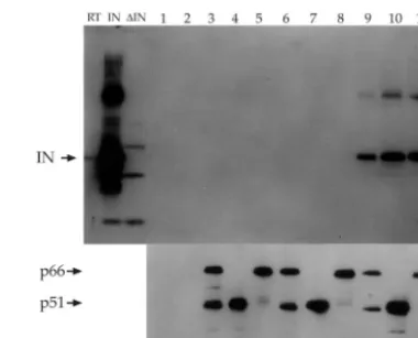
IN can bind to the heterodimeric p66/p51 and monomeric p66 and p51 RTs.It has been reported that the GST-IN fusion protein is able to interact directly with heterodimeric hexahis-tidine-tagged p66/p51 RT present in bacterial lysates (38). In that report, the authors also showed that the interaction is not bridged by nucleic acids, as demonstrated by pretreatment of both interacting partners with micrococcal nuclease prior to mixing them. In the present study, we wanted to determine if p66, which is known to exist in a homodimeric form, and p51 monomers also bind to HIV-1 IN. We expressed hexahistidine-tagged p51, p66, and p66/p51 RTs and unhexahistidine-tagged IN, all derived from HIV-1HXB2 in bacteria. Each preparation of RT was
bound to Ni2⫹-agarose beads, washed, and incubated with
bacterial lysates containing IN. After the beads were washed to eliminate unbound proteins, the bound proteins were sepa-rated by SDS-PAGE, followed by immunoblot analysis with
␣-IN antibodies. An identical sample of bound proteins sepa-rated on SDS-PAGE was transferred to nitrocellulose and probed with ␣-HIV-1 RT antibodies to ensure that the amounts of bound RT proteins in all reaction mixtures were comparable. The results indicated that each of the three forms
of RT was able to bind to IN protein from the bacterial lysates (Fig. 1, top). The presence of comparable inputs of RT pro-teins in the reaction mixtures was verified by immunoblot anal-ysis of a duplicate gel with the ␣-RT monoclonal antibody 8C4D7 (Fig. 1, bottom).
Multimerization of IN is not required for binding to RT.
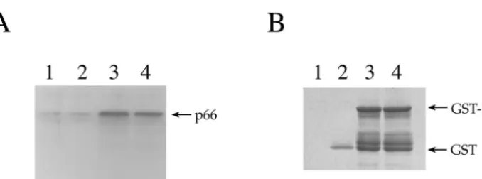
The formation of multimers of IN is known to be essential for IN function (9, 10, 12, 18, 35). Therefore, it was of interest to determine if the ability of IN to multimerize was necessary for association with RT. A multimerization-defective mutant of IN with a V260E substitution, isolated via a two-hybrid screen, was previously reported (20). This mutant exhibited a reduced ability to interact with wild-type IN both in the two-hybrid system and in vitro as a GST fusion protein but retained the ability to interact with INI1/hSNF5, indicating that it is specif-ically defective for homomeric interactions but not for hetero-meric interactions. Furthermore, viruses containing a V260E mutation were defective for replication, suggesting that IN multimerization is important for IN function. We tested the ability of GST-IN or GST-INV260E bound to G beads to interact with p66 in bacterial lysates using the same conditions described above. The results indicated that the V260E mutant of IN was able to bind to RT as effectively as wild-type IN (Fig. 2A). Thus, disruption of IN multimerization does not appear to affect the ability of IN to interact with RT, indicating that IN multimerization may not be required for this interaction.
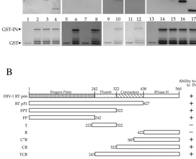
[image:3.603.323.513.67.221.2]Determination of IN-binding domain on RT.To determine the domain of RT that is necessary and sufficient to bind to IN, we expressed a panel of N- and C-terminal deletions of RT
FIG. 1. Native IN interacts with all three forms of HIV-1 RT. Hexahistidine-tagged RT heterodimer (p66/p51), p51, and p66 bound to Ni2⫹-NTA agarose beads were incubated with crude bacterial ly-sates containing native IN. The beads were washed, and the bound proteins were resolved by SDS–15% PAGE. (Top) Immunoblot probed with polyclonal␣-IN antibodies directed to N-terminal resi-dues 23 to 34. Lanes RT, IN, and⌬IN contain unreacted lysates from bacteria (RT), the IN expression plasmid (pT7IN), and the expression plasmid without the IN sequences (pT7⌬IN). Lanes 1 and 2, empty Ni2⫹-NTA beads incubated with pT7⌬IN lysate or pT7IN lysate; lanes 3 to 5, Ni2⫹-NTA beads bound to p66/p51, p51, or p66; lanes 6 to 8, Ni2⫹-NTA beads bound to p66/p51, p51, or p66 incubated with pT7⌬IN lysate; lanes 9 to 11, Ni2⫹-NTA beads bound to p66/p51, p51, or p66 incubated with pT7IN lysate. (Bottom) Parallel protein transfer blot probed with the␣-RT antibody 8C4D7 to ensure the presence of similar input RT bait protein. Lanes 1 to 11 are identical to those in panel A.
on November 8, 2019 by guest
http://jvi.asm.org/
corresponding to FP, FPT, T, TCR, Conn-R, C*R, and R (Fig. 3B). These fragments were each expressed with an N-terminal hexahistidine tag in bacteria and tested for interaction in vitro using GST-IN bound to glutathione agarose beads. The bound proteins were analyzed both by probing an immunoblot with anti-HIV-1 RT antibodies to detect the truncated RT proteins that retain binding and by Coomassie staining to assess the amount of GST-IN protein in the reactions. The results dem-onstrated that two nonoverlapping fragments bordering the polymerase domain were each able to bind to IN, indicating that there are two IN-binding sites on RT. One of the binding sites resides in the FP domain (Fig. 3A, lanes 14 to 17), and another resides in the C-terminal domain of connection be-tween aa 387 and 422 (Fig. 3A, lanes 3 to 6, and B, bottom).
Determination of RT-binding domain on IN. To map the domain of IN necessary and sufficient to bind to RT, we ex-pressed GST fusion proteins of each of the three structural domains of IN: the zinc-binding, the catalytic core, and the carboxy-terminal domains. The lysates containing each of these fusion proteins were then bound to G beads and sub-jected to interaction with the heterodimeric form of RT. The proteins bound to GST-IN fragments were resolved by SDS-PAGE and analyzed by immunoblotting them with monoclonal
␣-RT 5B2B2 antibodies (Fig. 4). The results indicate that while neither the zinc-binding domain nor the central core was ca-pable of interacting with RT, the C-terminal domain of IN could bring down RT as efficiently as the full-length IN. To confirm the results showing that the C-terminal domain is the RT-binding domain, we tested the interaction of a GST fusion of a mutant of IN with a deletion of the carboxy-terminal domain (⌬IN, retaining amino acid residues 1 to 220) with RT. The results indicated that ⌬IN was unable to bind to RT, confirming that the RT-binding site resides in the C-terminal domain of IN (Fig. 4, lane 10). Two other deletions lacking 80 and 82 residues from the carboxy terminus were also defective for RT interaction (data not shown).
Interaction between the smallest interacting fragments of RT and IN.The results described above showed that the car-boxy-terminal domain of IN can interact with full-length RT and that the FP and Conn-R fragments can both interact with full-length IN fused to GST. It was unclear whether the car-boxy-terminal domain of IN alone can bind to RT fragments encoding the FP or the Conn-R domain. Therefore, we tested
the abilities of the GST-IN and GST–C-terminal IN proteins to bind p66, Conn-R, or C*R expressed in bacteria. Our results, presented in Fig. 5, show that both GST-IN and the GST–C-terminal IN proteins can pull down p66, as well as its deletion proteins containing FP and Conn-R. Interestingly, however, C*R could be brought down by GST-IN but not by GST–C-terminal IN, suggesting that other regions of IN may be re-quired for interaction with this region of RT either directly or indirectly. Equivalent amounts of the GST-IN and GST–C-terminal IN proteins were used in these experiments, as deter-mined by Coomassie staining of a parallel SDS-PAGE gel (a 4 to 20% gradient acrylamide gel) of the same proteins used in the binding reaction (data not shown).
Interaction of minimal domains of RT and IN is not bridged by nucleic acids. Since all the structural domains involved (from both the interacting proteins), FP and Conn-R of RT and the C-terminal domain of IN, contain nucleic acid-binding domains, the question is whether the interaction observed is mediated by nucleic acid bridges. To address this issue, we performed a parallel reaction along with the above-mentioned experiment in which each interacting partner was pretreated with micrococcal nuclease prior to being adding to the in vitro binding reaction. Our results (compare Fig. 5A and B or E and F [⫹micrococcal nuclease] with C and D or G and H [⫺ mi-crococcal nuclease]) indicate that the interaction of the mini-mal domains was unaffected by micrococcal-nuclease pretreat-ment, thus confirming the absence of nucleic acid bridging between the interacting partners.
[image:4.603.130.469.64.190.2]Substitutions at the W235 residue do not prevent RT-IN interaction. In an earlier report, Ishikawa et al. stated that monoclonal antibodies directed to the C-terminal domain of IN prevented interaction between the HIV-1 RT and IN pro-teins and that the RT-IN interaction was disrupted by W235A and W235E substitutions in the IN protein (15). Therefore, we generated GST fusions of these mutant proteins and tested their abilities to pull down RT as described above. Surprisingly, both of the mutants were able to associate with RT as effi-ciently as the wild-type IN protein in the GST pull-down assays (data not shown). In evaluating the basis for this discrepancy, we noted that Ishikawa et al. used an MBP fusion of IN protein for their pull-down reactions. It is formally possible that our inability to reproduce their results was due to the use of a fusion partner here (GST) different from that used by Ishikawa
FIG. 2. Multimerization of IN is not critical to RT-IN interaction. (A) Lanes 1 to 3, empty, GST-bound, and GST-IN-bound G beads incubated with bacterial lysates containing RT p66; lane 4, GST-IN/V260E-bound G beads incubated with p66 RT lysate. The immunoblot was probed with the␣-RT antibody 5B2B2. (B) Parallel SDS-PAGE gel from the experiment shown in panel A Coomassie stained to ensure the presence of equivalent amounts of the proteins.
on November 8, 2019 by guest
http://jvi.asm.org/
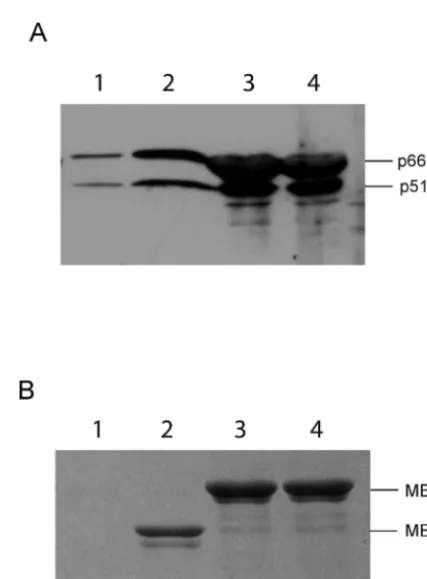
et al. (MBP). Therefore, we created MBP fusions of W235A and W235E IN mutants and used them in a pull-down assay to bring down heterodimeric RT from crude bacterial lysates, followed by analysis of the bound proteins. Our results show that MBP fusions of both of the mutant proteins bound to the RT heterodimer, indicating that these mutations do not disrupt RT-IN interaction (Fig. 6).
Mutations in conserved residues of IN do not affect RT interaction. Previous studies showed that mutations in the
conserved residues of IN, such as H12A, H16A, F185A, and
[image:5.603.87.491.150.479.2]⌬22, affect HIV replication at the level of reverse transcription. Therefore, it was of interest to determine whether these mu-tations disrupted RT-IN interaction. We performed GST-IN pull-down experiments using each of these mutant IN proteins. As illustrated in Fig. 7, our results show that none of the mutations we tested has any effect on the ability of GST-IN to pull down RT from crude lysates. These results suggest that the defect in reverse transcription exhibited by mutant viruses
FIG. 3. Mapping the IN-binding domain on HIV-1 RT. In experiments similar to those shown in Fig. 2, G beads bound to GST or GST-IN were incubated with bacterial lysates containing various truncation mutants of RT and washed, and the bound proteins were resolved on duplicate SDS-PAGE gels. The proteins on one gel were transferred to nitrocellulose and subjected to immunostaining with␣-RT antibodies, and the second gel was stained with Coomassie blue to ensure equal input of the bait proteins. (A) Pull-down experiments to map the IN-binding domains on RT p66. Lanes 1, 5, 7, 9, 11, and 13 contained bound proteins from a control incubation of GST bound to G beads, and lanes 2 to 4, 6, 8, 10, 12, and 14 to 17 contained proteins bound to GST-IN bound to G beads. RT mutants were added to the lanes as follows: lane 2, p66; lane 3, TCR; lane 4, Conn-R; lanes 5 and 6, C*R; lanes 7 and 8, R; lanes 9 and 10, p66; lanes 11 and 12, T; lanes 13 and 14, p66; lane 15, p51; lane 16, FPT; and lane 17, FP. The corresponding lanes on the bottom show stained protein indicating the input bait protein levels. The differences in p66 intensities in lanes 2 and 14 are due to different antibodies used for the Western blots. (B) A schematic summarizing results obtained in the experiment shown in panel A. The horizontal bar at the top represents full-length RT p66, with various subdomains and their boundaries indicated by amino acid residue numbers. The RT truncations used in the pull-down experiment, their boundaries, and their abilities to bind IN are indicated. Below, the proposed domains of IN interaction are indicated.⫹, able to bind;⫺, not able to bind.
on November 8, 2019 by guest
http://jvi.asm.org/
bearing these mutations is due to reasons other than the lack of RT-IN interaction. Furthermore, these results are consis-tent with our data showing that RT binds to the C-terminal domain but not the Zn-binding or core region of IN.
The effects of IN and RT on each other’s catalytic functions.
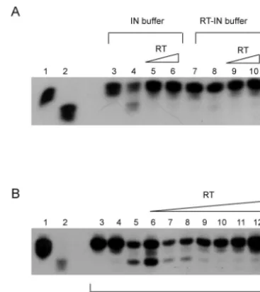
Mutations in HIV-1 IN have been shown to block viral repli-cation at the level of reverse transcription (38). This influence of IN on reverse transcription may be mediated by RT-IN interaction. To evaluate the functional significance of the in-teraction, we first tested the effect of IN on RT function. In standard RT reactions to measure the level of incorporation of dNTP into DNA by RT, the addition of the IN protein at various levels had no effect (data not shown). In order to detect qualitative differences, we measured processive synthesis by RT in the presence or absence of the IN protein in the reac-tions. Using a constant input of RT, the amount of IN was varied to get ratios of RT heterodimer to IN monomer ranging from 1:0.25 to 1:32. After the reaction, the products were separated on a denaturing gel and exposed to autoradiography. Our results show that IN does not influence RT activity either on homopolymeric (data not shown) or on heteropolymeric RNA templates and does not influence its processivity (Fig. 8). To determine the effect of RT on IN activity, we first carried out 3⬘processing reactions in the presence of RT. As before, to ensure that the reaction buffer was optimally suited for RT-IN interaction, we compared 3⬘processing in both the standard IN
buffer and a buffer optimized for RT-IN interaction. Since the 3⬘processing activity of IN was undetectable in RT-IN buffer (Fig. 9A, lane 8), we used standard IN buffer conditions, under which significant levels of processing activity could be detected (Fig. 9A, lane 4). Under these reaction conditions, we found that the addition of RT (either 1:1 or 1:4 ratios of IN to RT) inhibited 3⬘processing by IN. In order to determine the min-imal amount of RT sufficient to inhibit 3⬘processing, we per-formed a dose-dependence assay of this inhibition by varying the proportion of RT to a constant input of 4.68 pmol of IN. The molar ratios of IN monomer to RT heterodimer were 1:0.03125, 1:0625, 1:0.125, 1:0.25, 1:0.5, 1:1, and 1:2. The pres-ence of RT appeared to inhibit the 3⬘processing activity (Fig. 9B) in a dose-dependent manner. The lowest molar ratio of IN monomer to RT heterodimer sufficient to detect inhibition appeared to be 1:0.0625.
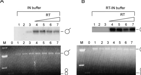
[image:6.603.87.501.73.345.2]We then examined whether RT influenced the strand trans-fer or joining activity of IN. The RT concentration was varied from a 1:0.125 to a 1:1 molar ratio of IN monomer to RT heterodimer, maintaining IN at a constant concentration (Fig. 10A, lanes 4 to 7). The reactions were carried out using a supercoiled plasmid DNA as the recipient and oligonucleotide substrates labeled at the 5⬘end of the joining strand. Insertion of oligonucleotide substrate into the target would result in a tailed plasmid DNA (lariat) that is radioactive and would be apparent on an autoradiogram. While the insertion of a single
FIG. 4. Mapping the RT-binding domain on IN. (A) In an experimental setup for pull-down experiments similar to that in Fig. 3, GST-IN and GST fusions of various truncations of IN were incubated with heterodimeric RT, followed by washing and resolving the bound proteins on SDS-PAGE. As before, one gel was transferred to nitrocellulose and probed with monoclonal␣-RT antibody 5B2B2 (top), and a duplicate gel with the same proteins was stained with Coomassie blue (bottom). Lane 1, empty G beads; lane 2: GST-bound G beads; lanes 3 and 7, GST-IN-bound G beads; lanes 4, 5, 6, and 8, G beads bound to GST-IN Zn finger domain (amino acid residues 1 to 50), GST-IN catalytic core domain (residues 48 to 208), GST-IN C-terminal domain (residues 201 to 288), and GST-IN C-terminal deletion (residues 1 to 220), respectively. (B) Schematic showing a summary of RT-binding abilities of full-length IN and the various truncation mutants of IN tested. The amino acid residues of each mutant are shown at the left of the horizontal bar representing each deletion.⫹, able to bind;⫺, not able to bind.
on November 8, 2019 by guest
http://jvi.asm.org/
oligonucleotide substrate (nonconcerted integration) into the target DNA results in the lariat structures, the concerted in-tegration of two oligonucleotide substrates into the same target DNA results in a linearized plasmid DNA molecule. We found that the addition of low inputs of RT (as low as fourfold-lower molar proportion compared to IN) significantly stimulated the joining activity of IN (Fig. 10A and B, compare lanes 3 and 4). The above-mentioned reactions were carried out using the optimized buffer conditions for IN reactions. To determine if reaction buffers conducive to RT-IN binding would enhance the stimulation observed in IN buffer, we carried out the IN strand transfer reactions in buffers that facilitated maximal interaction of the two proteins. Although IN exhibited barely detectable activity in the absence of RT under these condi-tions, the presence of RT resulted in a dramatic increase (155-fold) in its activity (Fig. 10B, lanes 3 and 4). However, we did not see any linear products, suggesting that RT did not stim-ulate concerted integration. Based on these results, RT
ap-pears to stimulate IN joining activity while inhibiting 3⬘ pro-cessing activity.
DISCUSSION
[image:7.603.44.282.66.225.2]Previous reports have demonstrated protein-protein inter-actions of retroviral RT and IN proteins, which are derived from a single polypeptide in the virus (14, 15, 32, 38) (39). Here, we describe the domains necessary for HIV-1 RT and IN interactions and the influence of each on the other’s function in vitro. Our data demonstrate that in addition to het-erodimeric RT, the p66 homodimers and p51 monomers can also interact with IN. This implies that IN recognizes a feature that is common to all three preparations of HIV-1 RT. The p66 preparations of RT are known to be predominantly in a ho-modimeric form and display up to 70% activity of hetero-dimeric RT (4, 23). Structurally, it is thought that one of the p66 molecules in the homodimer assumes the shape of p51 (36). However, the p51 molecule is known to exist mostly as a monomer in the absence of p66 and homodimerizes only under certain unusual conditions, including a high protein concentra-tion (8). The conformaconcentra-tion of monomeric p51 is unknown, but
FIG. 5. Interaction between the binding domains occurs without nucleic acid bridging. G beads, GST, or GST-IN prepared with or without micrococcal-nuclease treatment were incubated with p66, FP, Conn-R, or C*R similarly prepared with or without micrococcal-nu-clease treatment. (A) Pull-down reactions using micrococcal-numicrococcal-nu-clease- micrococcal-nuclease-treated interaction partners. Plain G beads (lane 1) or G beads bound to 1g of GST (lane 2) or GST-IN (lane 3) were incubated with p66 protein. G beads bound to GST-IN alone were incubated with the FP domain (lane 4). Subsequent to the pull-down and SDS-PAGE (15% acrylamide) analysis of the bound proteins, an immunoblot was pre-pared and probed with the monoclonal antibody 8C4D7, which recog-nizes the epitope located between RT residues 193 and 284, which overlaps with the FP domains. (B) Pull-down reactions using micro-coccal-nuclease-treated interaction partners. Similar to panel A, plain G beads (lane 5) or G beads bound to GST (lane 6) or GST-IN (lane 7) were incubated with p66. The G beads bound to GST-IN were also separately incubated with Conn-R (lane 8) or C*R (lane 9). Although both panels A and B are from the same SDS-PAGE gel, the immu-noblot corresponding to panel B was probed with a different mono-clonal antibody directed to the R domain (residues 440 to 560 of RT p66) to facilitate detection of the C-terminal fragments of RT. (C and D) The lanes are identical to those in panels A and B, except that none of the proteins were treated with micrococcal nuclease. (E and F) The lanes are similar to those in panels A and B, except that GST–C-terminal IN was used instead of GST-IN. All proteins were also treated with micrococcal nuclease. (G and H) The lanes are similar to those in panels E and F in that GST–C-terminal IN was used for pull downs, but no proteins were treated with micrococcal nuclease. The numbers on the left of the panels are molecular weight markers (prestained Invitrogen Benchmark).
FIG. 6. W235 substitutions do not disrupt RT-IN interaction. MBP-IN fusion proteins containing W235A or W235E mutations were concentrated from induced lysates by using amylose resin beads. Amy-lose resins alone (lanes 1) and amyAmy-lose bound to MBP (lanes 2), MBP-INW235A(lanes 3), or MBP-INW235E(lanes 4) protein were
in-cubated with lysates containing HIV-1 RT heterodimer followed by washing the resin and analysis of bound proteins on SDS-PAGE. (A) Immunoblot analysis using␣-RT antibodies. The positions of 66-and 51-kDa subunits of RT are indicated. (B) Gel identical to that in panel A stained with Coomassie brilliant blue. The migration positions of the MBP-IN fusion and MBP are indicated.
on November 8, 2019 by guest
http://jvi.asm.org/
[image:7.603.313.527.333.623.2]if one assumes that the monomeric p51 has a structure similar to that of the p51 present in a heterodimer, p51 would be the common element in all three preparations, and thus it is pos-sibly the subunit with which IN interacts. Our data, however, do not rule out the involvement of the catalytic p66 subunit in the interaction.
Our results indicate that IN binds to two discontinuous re-gions on RT, the FP region (amino acid residues 1 to 242) and the carboxy-terminal half of the connection subdomain (amino acid residues 387 to 422). We have attempted to further de-lineate the binding site within the FP region by expressing smaller segments within the region. In the primary sequence of the RT protein, the FP region is not continuous but rather intermixed and arranged as Fingers1-Palm1-Fingers2-Palm2 (F1-P1-F2-P2) sequences. Unfortunately, all of the smaller segments of FP subdomains (F1-P1, F2-P2, etc.), when ex-pressed alone, led to unstable proteins that could barely be detected or that were expressed at levels that were too low to facilitate binding studies.
Both of the two binding regions mentioned above (FP and half connection), when independently expressed as the entire FP or as half connection-RNase H domains, were able to bind to IN. This result was confirmed by testing nested deletions from both the amino and carboxy termini of RT. Neither the T
[image:8.603.50.272.71.328.2]subdomain nor the R domains, when expressed alone, were able to bind IN. Furthermore, the minimal RT domains were able to bind to the GST-IN protein even when pretreated with micrococcal nuclease, demonstrating the absence of nucleic acid bridging in the interaction. More interestingly, when GST–C-terminal IN was used instead of the GST-IN protein in pull-down reactions, while both the FP and Conn-R fragments of RT retained interaction, C*R, which binds to GST-IN, showed no interaction with the GST–C-terminal IN protein. It is possible that regions outside the carboxy-terminal domain of IN used here (residues 201 to 288) may be required for inter-action with the C*R domain. Whether the role of any such residues is in the direct interaction with RT or indirectly in modulating the overall conformation of the C-terminal domain of IN used here is unclear.
FIG. 7. Effects of mutations at conserved IN residues on RT-IN interaction. GST pull-down experiments were carried out as before by incubating wild-type GST-IN or the substitution mutants H12A, H16A, D116A, and F185A with lysates containing heterodimeric RT. (A) Bound proteins were resolved on SDS-PAGE, transferred to ni-trocellulose, and probed with the␣-RT antibody 5B2B2. Lanes 1 to 3, empty, bound, and IN-bound G beads; lanes 4 to 7, GST-IN–H12A, GST-IN–H16A, GST-IN–D116A, and GST-IN–F185A, re-spectively. The positions of p66 and p51 polypeptides are indicated. (B) A parallel SDS-PAGE gel was Coomassie blue stained to ensure the presence of equivalent inputs of proteins. The positions of GST and GST-IN are indicated.
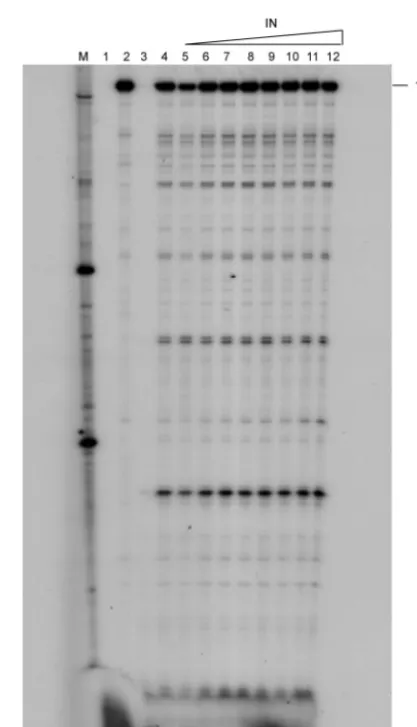
FIG. 8. Effect of IN on the processivity of HIV-1 RT. Using a primer that binds to the primer binding site, processivity reactions were done in the presence or absence of IN. Lane 1, primer alone; lane 2, untrapped reaction in the absence of IN; lane 3, pretrapped reaction where RT was mixed with poly(rA)䡠oligo(dT) prior to the addition of template-primer to ensure the effectiveness of the trap; lane 4, reaction in which dNTPs and trap were added simultaneously; lanes 5 to 12, reactions in which dNTPs and trap were added simultaneously in the presence of increasing amounts of IN ranging from a 1:0.03125 to a 1:4 molar ratio of RT to IN (with a constant input of RT). nt, nucleotides.
on November 8, 2019 by guest
http://jvi.asm.org/
[image:8.603.316.525.265.629.2]The inability of some of the RT fragments to bind IN may result from improper folding. We believe this to be unlikely, as several of the RT fragments utilized in our studies (FP, FPT, T, TCR, Conn-R, C*R, and R) have led to the reconstitution of a functional enzyme when used in mixing experiments (29). For example, when the Conn-R domain alone or the TC frag-ment was added to the enzymatically inactive C*R fragfrag-ment, it resulted in reactivation of R. Furthermore, Unge et al. crys-tallized the FP domain from amino acid residues 1 to 216 and found it to be very close to the structure of the FP domain observed in p66 (34). Similarly, the crystal structure of a frag-ment of HIV-1 RT corresponding to the R domain is largely comparable to the structure of this domain when the entire heterodimer was crystallized (7, 16). Therefore, we believe that the fragments of RT used in our mapping experiments are likely to have been properly folded.
It is interesting that the IN-binding site maps to more than one subdomain of RT. If, as described above, one assumes that the p51 subunit mediates this interaction as discussed above, the structure of the p51 subunit shows the FP and Conn-R subdomains to be physically very near each other (36). In contrast, these subdomains are distal to each other in p66.
We mapped the RT-binding region on IN to the
carboxy-terminal domain between residues 201 and 288. These results have been corroborated by the studies of Zhu et al. (39), which showed that the carboxy-terminal domain is sufficient to inter-act with RT and that a C130S mutation blocks this interinter-action. The C-terminal domain has a nonspecific DNA-binding activ-ity and is also known to be involved in multimeric interactions (1, 3, 13, 20, 25). It is known that multimerization of IN is necessary for its integration activity (9, 10, 12, 18, 35). We have found that a mutant of IN (V260E) defective for IN-IN inter-actions (20) is not defective for RT-IN interinter-actions. These results suggest that (i) the oligomerization of IN may not be necessary for RT-IN interaction and (ii) the surface of the C-terminal domain necessary for IN-IN interactions may be distinct from the region necessary for RT-IN interactions.
Studies of HIV RT and IN demonstrate that they behave differently in vitro and in vivo. For example, in vivo, RT can efficiently synthesize⬃10-kb DNA, whereas in vitro it can only synthesize a few hundred nucleotides. Similarly, in vitro reac-tions of IN are less efficient and require a high input of purified IN protein. These differences between in vivo and in vitro activities are most likely due to the milieu of the nucleoprotein complexes within the virions and possibly to the presence of other viral and/or cellular proteins. Thus, it is likely that these two Pol-derived interacting proteins have an influence on each other’s activities. This conclusion is supported by earlier results showing that several mutations in HIV-1 IN protein blocked viral replication at the level of reverse transcription (38) and by the recent observations that the C130S mutation in IN results in a block to reverse transcription (39).
Our results with testing the effect of IN on RT activity demonstrated that the RNA-dependent DNA polymerase (RDDP) activity of HIV-1 RT, or its processivity, was unaf-fected by the presence of the IN protein. Other observations indicate inhibition of RT activity by the IN protein. For exam-ple, Tasara et al. showed that the presence of IN inhibits DNA-dependent DNA polymerization by HIV-1 RT but not RDDP activity (32). The latter results are in agreement with our results showing that IN protein does not influence RNA-dependent DNA polymerization by RT. Another report by Oz et al. also indicated that IN had no influence on RT activity or processivity (26). The differential effect on RNA- versus DNA-dependent DNA polymerization activities may be due to the ability of IN to bind DNA but not RNA, resulting in compe-tition for the same substrate by both the RT and IN proteins. Thus, it is formally possible that the influence of IN mutations on reverse transcription in infected cells, as implied by Wu et al. (38) and Zhu et al. (39), is indirect, involving the recruit-ment of other viral or host proteins (see below).
[image:9.603.69.253.68.274.2]Our results indicate that RT has no effect on the 3⬘ process-ing activity of IN at lower concentrations but that at higher concentrations it inhibits this activity. This is most likely the result of RT competing with IN for the oligonucleotide sub-strates used for binding. The molar ratios used ranged from 1:0.03125 to 1:2 (IN monomer to RT heterodimer). If we assume IN to be a tetramer, then the ratio would be 4:1 to 1:16 (IN tetramer to RT heterodimer). Thus, 3⬘processing is com-pletely inhibited by RT when the RT-to-IN ratio exceeds 1 to 1 (Fig. 9B, lanes 9 to 12). The inhibition of 3⬘processing by IN in the presence of RT is in agreement with the results of Tasara et al. (32) and Oz et al. (26).
FIG. 9. Effect of RT on 3⬘ processing by IN protein. (A) Using radiolabeled U5.3/U5.4 substrate, a comparison was made between normal IN buffer and RT-IN buffer. Lane 1, unclipped U5.3/U5.4; lane 2, U5.5/U5.4, the preclipped version of U5.3/U5.4; lane 3, 3⬘ process-ing reaction usprocess-ing IN buffer in the absence of IN; lane 4, processprocess-ing reaction using IN buffer in the presence of IN; lanes 5 and 6, process-ing reactions usprocess-ing IN buffer in the presence of IN and RT in a 1:1 to 1:4 molar ratio of IN monomer to RT, respectively; lanes 7 to 10, exactly as for lanes 3 to 6 except using RT-IN buffer. The reactions in lanes 4 to 6 and 8 to 10 were performed with a constant input of IN. (B) All of the 3⬘processing reactions were done using normal IN buffer. Lanes 1 and 2, same as for panel A; lane 3, 3⬘ processing reaction in the absence of IN; lane 4, 3⬘processing reaction in the absence of IN and the presence of 9.36 pmol of RT; lane 5, 3⬘ pro-cessing reaction in the presence of 4.68 pmol of IN monomer; lanes 6 to 12, 3⬘ processing reactions in the presence of 4.68 pmol of IN monomer and the following concentrations of RT: 1:0.03125, 1:0.0625, 1:0.125, 1:0.25, 1:0.5, 1:1, and 1:2 (IN monomer to RT heterodimer), respectively.
on November 8, 2019 by guest
http://jvi.asm.org/
Perhaps the most dramatic effect we observed was the stim-ulation of the strand transfer activity of IN by RT. Under standard IN assay conditions, RT stimulated joining 5-fold, but in buffer optimized for RT-IN interaction, the effect was dra-matic (up to 155-fold). The large stimulation of joining by RT observed here was not previously reported. This was most likely the result of differences in the assays used and the use of buffer conditions optimized for RT-IN interaction. Both Tasara et al. (32) and Oz et al. (26) used radiolabeled oligo-nucleotide substrates to measure integration as a measure of joining activity and observed inhibition by RT. We, too, used radiolabeled IN oligonucleotide substrates, but we measured their integration into supercoiled plasmid DNA. Using the same assay, Carteau et al. also observed stimulation of joining activity by RT (although at a lower level) (6). These authors reported that the addition of p51 stimulated joining by IN, which is in agreement with our finding that p51 prepara-tions bind to the IN protein as efficiently as p66 and p66/p51 heterodimers. We believe that the presence of a bivalent IN-binding protein, such as RT, in the reaction mixture may pro-mote multimerization of IN, increasing the effective concen-tration of the active form, thus leading to stimulation of joining by IN. A similar effect on IN activity had been observed before, when another IN-binding protein, INI1, was present in the reaction mixture (19). At lower concentrations of IN, INI1 stimulated the activity, and at higher concentrations, it had no
effect or had inhibitory activity (19). Thus, the effect of IN-interacting proteins on its activity again suggests that high efficiency of IN activity in vivo can be attributed to the milieu of IN-interacting proteins, including RT, binding to it and modulating its activity.
[image:10.603.46.539.76.340.2]Wu et al. reported a significant block to viral reverse tran-scription caused by the mutations at conserved residues of IN, suggesting that IN interactions with other viral proteins, such as RT, may be important for reverse transcription in addition to integration. This belief is in agreement with the report that reverse transcription in vivo is more efficient than in vitro (17). It is possible that IN is required for recruiting cellular proteins that may be essential to initiate reverse transcription in the intracellular reverse-transcription complexes. It is important to reconcile two important differences between the biochemical findings in this report and the virological findings of Wu et al., who studied the functional effects of RT-IN interaction (38). First, in our GST pull-down assays, the presence of the muta-tions H12A, H16A, D116A, and F185A in the IN protein did not affect the association between IN and RT. Furthermore, the RT-binding domain of IN resides in the carboxy-terminal domain, which was also corroborated by Zhu et al. (39). It is possible that mutations in the N-terminal Zn-binding domain of IN influence the RT-IN interaction in vivo within the archi-tecture of PICs but are unable to affect the interaction in solution. It is also possible that mutations in the Zn-binding
FIG. 10. Effect of RT on the joining reaction by IN. (A) Effect of RT on joining reactions by IN using standard IN buffer. (Top) Autoradiogram displaying products of joining reactions carried out in the presence of increasing concentrations of RT. Lane 1, no IN; lane 2, RT alone (4.68 pmol); lane 3, IN alone (4.68 pmol); lanes 4 to 7, 4.68 pmol of IN with various proportions of RT at molar ratios (IN monomer to RT heterodimer) of 1:0.125, 1:0.25, 1:0.5, and 1:1, respectively. (Bottom) Agarose gel of the same joining reactions. Lane M, markers showing 4-, 3-, 2-, and 1.5-kb bands; lane 0, 0.2g of uncut pBluescript. Lanes 1 to 7 are identical to lanes 1 to 7 above. The starting supercoiled DNA substrate (bottom only; indicated with a twisted circle symbol) and the nonconcerted joining reaction products (top and bottom; indicated with a panhandle symbol) are shown. (B) (Top and bottom) Exactly as described for panel A, except that the reactions were carried out in the presence of RT-IN buffer and the marker lane (M) shows only 3-, 2-, and 1.5-kb bands.
on November 8, 2019 by guest
http://jvi.asm.org/
domain affect the association of IN with another viral or cel-lular protein, which may mediate the IN effect on viral reverse transcription. Second, in spite of the block to reverse transcrip-tion caused by IN mutatranscrip-tions during viral replicatranscrip-tion, the in vitro association of IN with RT did not lead to any stimulation of RDDP activity in our hands. In fact, two groups have shown inhibition of DNA-dependent DNA polymerase activity of RT by IN. It is possible that this inhibition merely represents com-petition for nonspecific binding to the same DNA by two DNA-binding proteins. This inhibition may not be observed in vivo, since the viral DNA is likely coated by the nucleocapsid protein, minimizing nonspecific binding.
In order to highlight the significance of the interaction for virus replication, it would be necessary to study mutants that disrupt RT-IN interaction without affecting their enzymatic activities. Unfortunately, the two potential RT interaction-neg-ative IN mutants (W235A and W235E) reported by Ishikawa et al. (15) displayed wild-type levels of interaction with RT in our repeated attempts using two different fusion partners. The C130S mutation, which abolished interaction with RT and led to virions defective for reverse transcription (39), offers a use-ful avenue for further studies. These results are reminiscent of the reports that several mutations in the HIV-1 IN protein did not affect in vitro IN or RT activity but displayed an in vivo DNA synthesis defect (22). Our results and those of Zhu et al. together lay a foundation for further studies aimed at under-standing the dynamics of RT-IN interactions during viral re-verse transcription in vivo.
ACKNOWLEDGMENTS
We thank Ron Swanstrom, Duane Grandgenett, and M. A. Wain-berg for generously sharing the IN expression plasmids pT7IN and pT7⌬IN,␣-IN antibodies, and the pHIV-PBS plasmid, respectively; Samson Chow (UCLA) for sharing unpublished results, as well as critically reading the manuscript; and William C. Drosopoulos for helpful advice.
E.A.H. is grateful for support from NIH Institutional Training Grant T32-GM07491. This work was supported by Public Service Grants RO1-AI30861 to V.R.P. and AI/GM 39951 to G.V.K.
REFERENCES
1. Andrake, M. D., and A. M. Skalka.1995. Multimerization determinants reside in both catalytic core and C terminus of avian sarcoma virus integrase.
J. Biol. Chem.270:29299–29306.
2. Arts, E. J., X. Li, Z. Gu, L. Kleiman, M. A. Parniak, and M. A. Wainberg.
1994. Comparison of deoxyoligonucleotide and tRNA(Lys-3) as primers in an endogenous human immunodeficiency virus-1 in vitro reverse
transcrip-tion/template-switching reaction. J. Biol. Chem.269:14672–14680.
3. Asante-Appiah, E., and A. M. Skalka.1999. HIV-1 integrase: structural
organization, conformational changes, and catalysis. Adv. Virus Res.52:351–
369.
4. Beard, W. A., and S. H. Wilson.1993. Kinetic analysis of template-primer interactions with recombinant forms of HIV-1 reverse transcriptase.
Bio-chemistry32:9745–9753.
5. Bukrinsky, M. I., N. Sharova, T. L. McDonald, T. Pushkarskaya, W. G. Tarpley, and M. Stevenson.1993. Association of integrase, matrix, and reverse transcriptase antigens of human immunodeficiency virus type 1 with viral nucleic acids following acute infection. Proc. Natl. Acad. Sci. USA
90:6125–6129.
6. Carteau, S., R. J. Gorelick, and F. D. Bushman.1999. Coupled integration of human immunodeficiency virus type 1 cDNA ends by purified integrase in
vitro: stimulation by the viral nucleocapsid protein. J. Virol.73:6670–6679.
7. Davies, J. F., Z. Hostomska, Z. Hostomsky, S. R. Jordan, and D. A. Mat-thews.1991. Crystal structure of the ribonuclease H domain of HIV-1
re-verse transcriptase. Science252:88–95.
8. Dirani-Diab, R. E., M.-L. Andreola, G. Nevinsky, D. Tharaud, P. J. Barr, S. Litvak, and L. Tarrago-Litvak.1992. Biochemical characterization of the p51 subunit of human immunodeficiency virus reverse transcriptase in
homo-and heterodimeric recombinant forms of the enzyme. FEBS Lett.301:23–28.
9. Ellison, V., J. Gerton, K. A. Vincent, and P. O. Brown.1995. An essential interaction between distinct domains of HIV-1 integrase mediates assembly
of the active multimer. J. Biol. Chem.270:3320–3326.
10. Engelman, A., F. D. Bushman, and R. Craigie.1993. Identification of dis-crete functional domains of HIV-1 integrase and their organization within an
active multimeric complex. EMBO J.12:3269–3275.
11. Fassati, A., and S. P. Goff.2001. Characterization of intracellular reverse transcription complexes of human immunodeficiency virus type 1. J. Virol.
75:3626–3635.
12. Grandgenett, D. P., and G. Goodarzi.1994. Folding of the multidomain
human immunodeficiency virus type-I integrase. Protein Sci.3:888–897.
13. Heuer, T. S., and P. O. Brown.1998. Photo-cross-linking studies suggest a model for the architecture of an active human immunodeficiency virus type
1 integrase-DNA complex. Biochemistry37:6667–6678.
14. Hu, S. C., D. L. Court, M. Zweig, and J. G. Levin.1986. Murine leukemia
viruspolgene products: analysis with antisera generated against reverse
transcriptase and endonuclease fusion proteins expressed inEscherichia coli.
J. Virol.60:267–274.
15. Ishikawa, T., N. Okui, N. Kobayashi, R. Sakuma, T. Kitamura, and Y. Kitamura.1999. Monoclonal antibodies against the minimal DNA-binding domain in carboxy-terminal region of human immunodeficiency virus type 1
integrase. J. Virol.73:4475–4480.
16. Jacobo-Molina, A., J. Ding, R. G. Nanni, A. D. Clark, Jr., X. Lu, C. Tantillo, R. L. Williams, G. Kamer, A. L. Ferris, P. Clark, et al.1993. Crystal structure of human immunodeficiency virus type 1 reverse transcriptase complexed
with double-stranded DNA at 3.0 A˚ resolution shows bent DNA. Proc. Natl.
Acad. Sci. USA90:6320–6324.
17. Ji, X., G. J. Klarmann, and B. D. Preston.1996. Effect of human immuno-deficiency virus type 1 (HIV-1) nucleocapsid protein on HIV-1 reverse
transcriptase activity in vitro. Biochemistry35:132–143.
18. Kalpana, G. V., and S. P. Goff.1993. Genetic analysis of homodimeric interactions of human immunodeficiency virus type 1 integrase using the
yeast two-hybrid system. Proc. Natl. Acad. Sci. USA90:10593–10597.
19. Kalpana, G. V., S. Marmon, W. Wang, G. R. Crabtree, and S. P. Goff.1994. Binding and stimulation of HIV-1 integrase by a human homologue of yeast
transcription factor SNF5. Science266:2002–2006.
20. Kalpana, G. V., A. Reicin, G. S. Cheng, M. Sorin, S. Paik, and S. P. Goff.
1999. Isolation and characterization of an oligomerization-negative mutant
of HIV-1 integrase. Virology259:274–285.
21. Kew, Y., Q. Song, and V. Prasad.1994. Subunit selective mutagenesis of Glu89 residue in human immunodeficiency virus reverse transcriptase.
J. Biol. Chem.269:15331–15336.
22. Leavitt, A. D., G. Robles, N. Alesandro, and H. E. Varmus.1996. Human immunodeficiency virus type 1 integrase mutants retain in vitro integrase activity yet fail to integrate viral DNA efficiently during infection. J. Virol.
70:721–728.
23. Le Grice, S. F., T. Naas, B. Wohlgensinger, and O. Schatz.1991. Subunit-selective mutagenesis indicates minimal polymerase activity in
heterodimer-associated p51 HIV-1 reverse transcriptase. EMBO J.10:3905–3911.
24. Luan, D. D., M. H. Korman, J. L. Jakubczak, and T. H. Eickbush.1993. Reverse transcription of R2Bm RNA is primed by a nick at the chromosomal
target site: a mechanism for non-LTR retrotransposition. Cell72:595–605.
25. Lutzke, R. A., and R. H. Plasterk.1998. Structure-based mutational analysis of the C-terminal DNA-binding domain of human immunodeficiency virus type 1 integrase: critical residues for protein oligomerization and DNA
binding. J. Virol.72:4841–4848.
26. Oz, I., O. Avidan, and A. Hizi.2002. Inhibition of the integrases of human immunodeficiency viruses type 1 and type 2 by reverse transcriptases.
Bio-chem. J.361:557–566.
27. Prasad, V. R.1993. Genetic analysis of retroviral reverse transcriptase
struc-ture, p. 135–161.InS. P. Goff and A. M. Skalka (ed.), Reverse transcriptase.
Cold Spring Harbor Laboratory Press, Plainview, N.Y.
28. Reicin, A. S., G. Kalpana, S. Paik, S. Marmon, and S. Goff.1995. Sequences in the human immunodeficiency virus type 1 U3 region required for in vivo
and in vitro integration. J. Virol.69:5904–5907.
29. Smith, J. S., K. Gritsman, and M. J. Roth.1994. Contributions of DNA polymerase subdomains to the RNase H activity of human
immunodefi-ciency virus type 1 reverse transcriptase. J. Virol.68:5721–5729.
30. Soltis, D., and A. M. Skalka.1988. The a and b chains of avian retrovirus
reverse transcriptase independently expressed inE. coli: characterization of
enzymatic activities. Proc. Natl. Acad. Sci. USA85:3372–3376.
31. Szilvay, A. M., S. Nornes, I. R. Haugan, L. Olsen, V. R. Prasad, S. P. Goff, C. Endresan, and D. Helland.1992. Epitope mapping of HIV-1 reverse transcriptase with monoclonal antibodies which inhibit the polymerase and
the RNAse H activities. J. Acquired Immune Defic. Syndr.5:647–657.
32. Tasara, T., G. Maga, M. O. Hottiger, and U. Hubscher.2000. HIV-1 reverse transcriptase and integrase enzymes physically interact and inhibit each
other. FEBS Lett.507:39–44.
33. Trentin, B., N. Rebeyrotte, and R. Z. Mamoun.1998. Human T-cell leukemia virus type 1 reverse transcriptase (RT) originates from the pro and pol open reading frames and requires the presence of RT-RNa H (RH) and
RT-RH-integrase proteins for its activity. J. Virol.72:6504–6510.
on November 8, 2019 by guest
http://jvi.asm.org/
34. Unge, T., S. Knight, R. Bhikhabhai, S. Lovgren, Z. Dauter, K. Wilson, and B. Strandberg.1994. 2.2 A˚ resolution structure of the amino-terminal half of
HIV-1 reverse transcriptase (fingers and palm subdomains). Structure2:953–
961.
35. van Gent, D. C., C. Vink, A. A. Groeneger, and R. H. Plasterk.1993. Comple-mentation between HIV integrase proteins mutated in different domains.
EMBO J.12:3261–3267.
36. Wang, J., S. J. Smerdon, J. Jager, L. A. Kohlstaedt, P. A. Rice, J. M. Friedman, and T. A. Steitz.1994. Structural basis of asymmetry in the human immunodeficiency virus type 1 reverse transcriptase heterodimer. Proc. Natl.
Acad. Sci. USA91:7242–7246.
37. Werner, S., P. Hindmarsh, M. Napirei, K. Vogel-Bachmayr, and B. M. Wohrl.2002. Subcellular localization and integration activities of Rous
sar-coma virus reverse transcriptase. J. Virol.76:6205–6212.
38. Wu, X., H. Liu, H. Xiao, J. A. Conway, E. Hehl, G. V. Kalpana, V. R. Prasad, and J. C. Kappes.1999. Human immunodeficiency virus type 1 integrase protein promotes reverse transcription through specific interactions with the
nucleoprotein reverse transcription complex. J. Virol.73:2126–2135.
39. Zhu, K., C. Dobard, and S. A. Chow.2004. Requirement for integrase during reverse transcription of human immunodeficiency virus type 1 and the effect of cysteine mutations of integrase on its interactions with reverse
transcrip-tase. J. Virol.78:5045–5055.