Copyright © 2002, American Society for Microbiology. All Rights Reserved.
Differential Effect of Murine Alpha/Beta Interferon Transgenes on
Antagonization of Herpes Simplex Virus Type 1 Replication
Peter Härle,
1Vanessa Cull,
2Martin-Paul Agbaga,
1Robert Silverman,
3Bryan R. G. Williams,
3Cassandra James,
2and Daniel J. J. Carr
1,4*
Departments of Ophthalmology
1and Microbiology and Immunology,
4The University of Oklahoma Health Sciences Center,
Oklahoma City, Oklahoma 73104; Division of Veterinary & Biomedical Science, Murdoch University, Perth, Australia 6150
2;
and Department of Cancer Biology, Lerner Research Institute, Cleveland Clinic Foundation, Cleveland, Ohio 44195
3Received 21 December 2001/Accepted 5 April 2002
Alpha/beta interferons (IFN-
␣
/

) are potent, endogenous antiviral cytokines that suppress the replication of
RNA and DNA viruses, including herpes simplex virus type 1 (HSV-1). The present study compared the
efficacies of IFN-
␣
/

transgenes, including IFN-
␣
1, -
␣
4, -
␣
5, -
␣
6, -
␣
9, and -

, against HSV-1 infection. L929
cells transfected with the IFN-
␣
/

transgenes produced similar levels of IFN, as measured by bioassay and
enzyme-linked immunosorbent assay. In addition, transfected cells were less susceptible to HSV-1 infection
than were cells transfected with a plasmid vector control. The murine IFN-

plasmid construct exhibited the
greatest reduction, while the murine IFN-
␣
5 transgene showed a modest inhibitory effect in viral titers
recovered from the supernatants of transfected, infected L929 cultures. Consistent with this observation, the
IFN-

transgene antagonized viral transcript levels, including infected cell protein 27, thymidine kinase, and
glycoprotein B, to a greater extent than did the IFN-
␣
transgenes at 6 to 10 h postinfection as determined by
real-time PCR. Cells transfected with the IFN-
␣
4, IFN-
␣
9, or IFN-

transgenes showed the greatest reduction
in viral protein expression relative to the other transfected cells, which was associated with increased STAT1
expression. The absence of the IFN-responsive protein kinase R (PKR) gene completely abrogated the antiviral
induction by all IFN-
␣
/

against HSV-1. In the absence of RNase L, viral yields were increased 10-fold, but the
antiviral effect of IFN was either unaffected or enhanced. These results suggest that the predominant
IFN-mediated, antiviral pathway during HSV-1 infection taken by IFN-
␣
/

in L929 cells utilizes PKR.
Herpes simplex virus type 1 (HSV-1) is an important
neu-rotropic virus with a worldwide distribution that seems to have
coevolved with the vertebrate immune system (44). Upon
in-fection, HSV-1 genes are expressed in a sequential cascade,
resulting in progeny viruses that are eventually cleared from
the host or alternatively establish a latent infection in the
neurons of the sensory ganglia. During the acute infection, the
host immune response initially consists of neutrophils,
macro-phages, and natural killer cells as well as the proinflammatory
cytokines (tumor necrosis factor alpha, interleukins 1 and 6)
and the alpha/beta interferons (IFN-
␣
/

), including IFN-
␣
and
IFN-

(1, 6, 15, 27, 36, 62, 71). Typically, following this initial
onslaught, CD4
⫹and CD8
⫹T cells infiltrate the infected area
(41, 61) and appear to play a dominant role in clearing the
acute infection through direct or indirect means (7, 42, 64). To
circumvent the hostile environment, HSV-1 has evolved
mech-anisms to evade immune surveillance, including antagonizing
antigen processing and presentation (2, 28, 32, 70), ultimately
hindering virus-infected cell recognition by CD8
⫹cytotoxic T
lymphocytes (19), blocking complement activation (17, 72),
and encoding a glycoprotein that works as a decoy receptor for
immunoglobulin G (51). Furthermore, HSV-1 reportedly
in-duces apoptosis of T cells (31, 56) and renders infected target
cells resistant to the lethal hit of cytotoxic T lymphocytes (33).
In addition, HSV-1-infected peripheral blood mononuclear
cells produce transforming growth factor

1 (45), which is
known to suppress major histocompatibility complex class II
expression (37). Collectively, there is strong evidence
indicat-ing that the virus has adopted several strategies to elude the
adaptive immune response and to promote the establishment
of a lifelong infection in the host.
IFN-
␣
/

are endogenous, antiviral cytokines that have
pre-viously been found to antagonize HSV-1 replication in vitro at
the transcriptional and translational levels (40, 47, 50, 65).
Likewise, in vivo studies have determined that IFN-
␣
/

play a
dominant role in controlling acute HSV-1 infection (22, 26, 66,
67) and facilitate the adaptive immune response to infection
(16). Although it was originally thought that the target of
IFN-
␣
/

might include viral protein 16 (13), it has
subse-quently been suggested that the level of antagonism might be
more generalized (52, 60). Similar to other antiviral pathways,
HSV-1 has developed mechanisms to obstruct the action of
IFN. For example, HSV-1-infected cell protein 0 (ICP0) and
ICP34.5 have been demonstrated to confer resistance to the
effects of IFN-
␣
/

(24, 25, 38, 49). Resistance by ICP34.5
seems to reside in antagonizing the anitviral effects of
IFN-inducible, double-stranded-RNA-dependent protein kinase R
(PKR) (9), whereas the level by which ICP0 antagonizes IFN is
presently unknown.
Most investigations evaluating the antiviral effects of
IFN-␣
/

use hybrid or recombinant proteins at seemingly high
con-centrations (
⬎
100 U). Little attention has focused on relative
differences between or within the endogenous IFN subtypes as
they relate to antiviral activity. In fact, murine IFN-
␣
subtypes
as well as IFN-

act through the same receptor (59) yet evoke
* Corresponding author. Mailing address: Department of
Ophthal-mology, DMEI no. 415, OUHSC, 608 Stanton L. Young Blvd.,
Okla-homa City, OK 73104. Phone: (405) 271-1084. Fax: (405) 271-8781.
E-mail: [email protected].
6558
on November 8, 2019 by guest
http://jvi.asm.org/
different antiviral or antiproliferative efficacies (5, 10, 68, 73).
Within the human system, these functional differences reside
at the receptor level (18, 55) and the activation of downstream
signaling pathways (3, 29) and impact on specific gene
induc-tion (12). The present study was undertaken to evaluate the
antiviral potency of five different murine IFN-
␣
subtypes and
IFN-

in transfected L929 cells as well as in wild-type and
double-stranded RNA-activated kinase (PKR) or 2
⬘
,5
⬘
-oli-goadenylate synthetase (OAS)/RNase L-deficient cell lines
in-fected with HSV-1 (McKrae strain). The results of this study
show that IFN-

is the dominant antiviral IFN-
␣
/

against
HSV-1 infection, antagonizing viral replication at the
tran-scriptional and protein level to a greater degree than do the
IFN-
␣
subtypes. They also show that PKR is essential for the
anti-HSV-1 activity of IFN.
MATERIALS AND METHODS
Cells and virus.The murine fibroblast cell line L929 and the African green monkey kidney cell line, Vero, were obtained from the American Type Culture Collection (Manassas, Va.). Propagation and experiments were carried out in Dulbecco’s modified Eagle medium (DMEM) (American Type Culture Collec-tion) for L929 cells or RPMI medium (Gibco-BRL, Gaithersburg, Md.) supple-mented with 10% fetal bovine serum and an antibiotic-antimycotic mixture (Gibco), which is referred to as complete medium. Wild-type (mouse embryo fibroblasts, MEF-6) and PKR-gene-disrupted cells (PKR KO) (35) were
propa-gated in complete DMEM. Likewise, wild-type (RL⫹/⫹) and RNase
L-gene-disrupted cells (RL⫺/⫺) (75) were propagated in complete DMEM. HSV-1
(McKrae strain) and vesicular stomatitis virus (a gift from Robert Fleischmann, University of Texas Medical Branch, Galveston, Tex.) were propagated in Vero
cells as previously described (21). The cells were incubated at 37°C, 5% CO2, and
95% humidity at all times.
Plasmid DNA constructs.All murine IFN transgenes were cloned into the eukaryotic expression vector PKCMVintPolylinker (5,087 bp, Vical Inc., San Diego, Calif.) containing a simian virus 40 polyadenylation signal and a kana-mycin resistance gene. The IFN genes (575 to 626 bp) are expressed under the control of a human cytomegalovirus immediate-early enhancer/promoter.
Clon-ing sites within the vector are as follows: IFN-␣1-BamHI/BglII, IFN-␣4-PstI/SalI,
IFN-␣5-SalI/BglII, IFN-␣6-PstI/BglII, IFN-␣9-XbaI/BglII, and IFN--SalI/XbaI
(10). The plasmid constructs were transformed into theEscherichia colistrain
INV␣F’ (Invitrogen, Carlsbad, Calif.) and grown in Terrific broth, containing 50
g of kanamycin/ml, followed by purification using Qiagen Maxi kits (Qiagen,
Inc., Valencia, Calif.). After each plasmid isolation, restriction enzyme digestion assays were conducted and the products were analyzed by agarose gel electro-phoresis.
Transfection and infection of L929 cells.Prior to the transfection, 3⫻105
L929 cells were plated in duplicate in six-well tissue cultures. Following overnight
incubation, the cells were transfected with 3g of plasmid DNA and 45l of
Superfect (Qiagen) in 0.5 ml of complete DMEM for 6 h followed by a change of fresh, prewarmed complete DMEM. Twenty-four hours posttransfection, su-pernatants were collected and cells were infected with HSV-1 at a multiplicity of infection (MOI) of 0.5. The viral inoculum was removed 1 h postinfection (p.i.) and replaced with fresh complete DMEM. After 24 h, the supernatants were collected and the HSV-1 titer was determined in microtiter plate plaque assays using Vero cells.
IFN bioassay.To quantitate biologically active amounts of IFN secreted at the end of the 24-h transfection period, pooled supernatants from two samples were assayed as previously described (22).
Incubation of cells with biologically active IFN-␣/.To determine the 50%
inhibitory concentration (IC50) for each IFN-␣/subtype, supernatants from the
transfected L929 cells containing equivalent concentrations of biologically active IFN (1,000 U/ml) were diluted (half-log dilutions) and added to fresh L929 cells (500,000 cells/well). Following an overnight incubation (18 h), the supernatant
was removed and cells were infected with HSV-1 (McKrae strain, MOI⫽0.5).
After 60 min, the virus-containing supernatant was removed and fresh complete DMEM was added. Following an additional 28- to 36-h incubation period, the
cells were frozen and thawed and the clarified supernatant (10,000⫻g, 1 min)
was assayed for infectious virus by plaque assay using Vero cells. The IC50was
determined by linear regression analysis for each sample. The correlation
coef-ficient for each sample analyzed over 6 half-log dilutions ranged from 0.90 to 0.99.
To determine the susceptibility of wild-type or gene-disrupted cells to HSV-1
infection following exposure to IFN-␣/, supernatants from transfected L929
cells containing equivalent amounts of IFN-␣/subtypes (50 to 100 U/ml) were
added to the MEF/6, PKR KO, RL⫹/⫹, or RL⫺/⫺cells (250,000 cells/well).
Following an overnight incubation, the supernatant was removed and the cells
were infected with HSV-1 (McKrae strain, MOI⫽1.0). After 60 min, the
virus-containing supernatant was removed and fresh complete DMEM was added. Following an additional 28- to 36-h incubation period, the cells were
frozen and thawed and the clarified supernatant (10,000⫻g, 1 min) was assayed
for infectious virus by plaque assay using Vero cells.
IFN-ELISA.To quantitate the amounts of IFN protein produced at the end of the 24-h transfection period, pooled supernatants from two samples were assayed in triplicate using an enzyme-linked immunosorbent assay (ELISA) specific for
mouse IFN-␣(PBL Biomedical Laboratories). The ELISA was carried out
ac-cording to the manufacturer’s protocol and analyzed at an absorbance of 450 nm using a FL600 microplate fluorescence reader (Bio-Tek Instruments, Inc.,
Wi-nooski, Vt.). The lower end of the sensitivity range of the IFN-␣ELISA was 12.5
pg/ml.
Semiquantitative real-time PCR for viral genes and IFN-inducible genes.
Total cell RNA was isolated at 6 and 10 h p.i. in Ultraspect RNA isolation reagent (Biotecx Inc., Houston, Tex.) according to the manufacturer’s protocol. Before the reverse transcription step, DNA contamination was removed using DNase I according to the manufacturer’s protocol (Gibco-BRL). First-strand cDNA was synthesized using avian myeloblastosis virus reverse transcriptase (Promega, Madison, Wis.) and an oligo(dT) primer (Promega). Real-time PCR was carried out in 96-well PCR plates (Rad, Hercules, Calif.) using a Bio-Rad iCycler. Real-time PCR conditions for all primers included an initial dena-turing step for 3 min at 95°C, followed by 30 cycles at 95°C for 10 s and
annealing/elongation at 61°C for 35 s. Each reaction contained 45l of PCR
Platinum SuperMix (Gibco-BRL) and CYBRgreen I (Molecular Probes,
Eu-gene, Oreg.) at a final dilution of 1:100,000. MgCl2was supplemented as
indi-cated with the primer sequences. During the optimization procedures of the primers, 1% agarose gel analysis verified the amplification of one product of the predicted size with no primer-dimer bands. The absence of primer-dimer for-mation for each oligonucleotide set was also validated by establishing the melting curve profile. The PCR results were analyzed on the iCycler Software (version 2.3), and threshold cycles were determined as follows: after subtraction of the background fluorescence for each sample, the threshold fluorescence for each gene was determined at that point where the relative light units reached a level of more than 10 standard deviations above the baseline relative light units. At 40 cycles, the primers-only control did not give a signal above the threshold. At 35 cycles the primers with uninfected cells did not give a signal above the threshold. The semiquantitative comparison between samples was calculated as follows: the data were normalized by subtracting the difference of the threshold cycles
(CT) between the gene of interest’sCTand the “housekeeping” gene GAPDH’s
CT(gene of interestCT⫺GAPDHCT⫽ ⌬CT) for each sample. The⌬CT
was then compared to the expression levels of the vector control sample
(sample⌬CT ⫺vector⌬CT). To determine the relative enhanced
expres-sion of the gene of interest, the following calculation was made: fold change⫽
2(⫺sample⌬CT ⫺vector⌬CT). If the difference (sample⌬C
T⫺ vector⌬CT)
was a positive value, then it was calculated as follows: fold change ⫽
⫺1/2(⫺sample⌬CT⫺vector⌬CT)in order to get a negative value expressing the
relative suppression of the gene of interest. Oligonucleotide sequences for
the targeted genes include the following:GAPDH, 5⬘-GAATCTACTGGC
GTCTTCACC-3⬘ and 5⬘-GTCATGAGCCCTTCCACGATGC-3⬘ (2 mM
MgCl2);icp27, 5⬘-TGACGCCGAGACCAGAC-3⬘and 5⬘-GGCAAAAGTGC
GATAGAGG-3⬘(3 mM MgCl2); thymidine kinase (tk), 5⬘-AAACCACCAC
CACGCAAC-3⬘and 5⬘-ACACCCGCCAGTAAGTCATC-3⬘(3 mM MgCl2);
andgB, 5⬘-CGTTTCGCAGGTGTGGTTC-3⬘and 5⬘-ATGTCGGTCTCGTG
GTCG-3⬘(3 mM MgCl2).
Western blot analysis.Twenty-four hours posttransfection or at various times p.i., total cell protein was harvested following disruption of cells in sodium dodecyl sulfate (SDS) containing SDS lysis buffer supplemented with a cocktail of EDTA-free protease inhibitors (Boehringer Mannheim, Mannheim, Ger-many) and 1 mM sodium orthovanadate (Sigma, St. Louis, Mo.). In experiments assessing viral protein expression, cells that had been transfected for 24 h were
subsequently infected with HSV-1 (MOI⫽0.5) and total protein was obtained
from lysed cells at 12 or 24 h p.i. Protein was quantitated with the BCA Protein Assay Reagent (Pierce, Rockford, Ill.) using the FL600 fluorescence plate reader
(Bio-Tek). Total cell protein (10 to 30g) was electrophoresed on SDS–7.5%
polyacrylamide gels (criterion precast gels; Bio-Rad) and transferred by semidry
V
OL. 76, 2002
HSV-1 AND IFN-
␣
/

6559
on November 8, 2019 by guest
http://jvi.asm.org/
blotting onto polyvinylidene difluoride (PVDF) membranes (Bio-Rad). The membranes were blocked with 5% dry milk–2.5% bovine serum albumin in TTBS (0.1% Tween, 0.1 M Tris, 0.4 M NaCl [pH 7.4]) overnight at 4°C and incubated with a polyclonal rabbit anti-HSV antibody (Dako, Carpinteria, Cal-if.), a polyclonal anti-JAK1 p130 antibody (Biosource, Camarillo, CalCal-if.), a poly-clonal anti-STAT1 p91/p84 antibody (Santa Cruz Biotechnology, Inc., Santa
Cruz, Calif.), a polyclonal phospho-JAK1 (Y1022/1023) antibody (Biosource), or a
polyclonal phospho-STAT1 (Y701) antibody (New England Biolabs, Beverly, Mass.) for 2 h at room temperature on a shaking platform. The primary antibody was detected via an anti-rabbit-antibody coupled with horseradish peroxidase (Santa Cruz) followed by detection using chemiluminescence detection reagents (Pierce). The resultant blots were digitalized using the Bio-Rad 1000 image documentation system and software (Quantity One 4.0.3; Bio-Rad).
Statistics.Analysis of data was carried out by one-way analysis of variance (ANOVA) and Scheffe multiple-comparison test or the nonparametric Wilcoxon
signed-rank test to determine statistically significant differences (P⬍0.05)
be-tween the vector and IFN-␣/transgene groups.
RESULTS
Production of biologically active IFN by transfected L929
cells.
In order to compare the potency of IFN-
␣
/

against
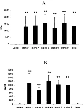
[image:3.587.129.459.71.494.2]HSV-1 infection, transfected L929 cells were initially assayed
for the production of biologically active IFN. Twenty-four
hours posttransfection, culture supernatants were collected
and assayed for IFN content. The results show that, with the
exception of cells transfected with the plasmid vector alone, all
transfected cells secreted equivalent amounts of biologically
active IFN-
␣
/

(Fig. 1A). For those cells transfected with the
IFN transgenes, quantitative comparisons of IFN-
␣
protein
expression were conducted by ELISA (Fig. 1B). Cells
trans-fected with the IFN-
␣
subtype transgenes secreted similar
FIG. 1. Transfected L cells secrete biologically active IFN. L929 cells (500,000 cells/well) were transfected with 3
g of plasmid DNA alone
(Vector) or plasmid containing the indicated IFN-
␣
or -

transgene. Following a 24-h incubation, the supernatants were harvested and assayed
for biologically active IFN (A) or quantitated for IFN-
␣
by ELISA (B). The figure is a summary of three experiments for each transgene or vector.
Bars represent the mean
⫾
standard deviation.
ⴱⴱ
,
P
⬍
0.01 in comparison of cells transfected with the IFN-
␣
/

transgene to the vector control
(A) or in comparison of cells transfected with the IFN-
␣
transgenes to the IFN-

transgene or plasmid vector groups (B) as determined by ANOVA
and Scheffe multiple-comparison test.
on November 8, 2019 by guest
http://jvi.asm.org/
quantities of IFN-
␣
, whereas cells transfected with the plasmid
vector alone or the plasmid containing the IFN-

transgene
secreted nominal amounts of IFN-
␣
(Fig. 1B). Although
trans-fection efficiencies were not determined using the plasmid
construct, a previous study employing a similar construct
(pCMV-

) found that transduction efficiencies ranged from 10
to 25% of cells between assays (data not shown).
Comparison of the efficacy of IFN-
␣
/

transgenes against
HSV-1 infection.
Since the transfection of target cells with the
various murine IFN-
␣
/

transgenes generated similar
quanti-ties of biologically active IFN, the cells were assayed for their
sensitivity to HSV-1 infection. Viral titers were determined in
transfected cells infected with a highly neurovirulent strain
(McKrae) of HSV-1. The results show a reduction in infectious
virions recovered in the supernatants from cells transfected
with the IFN-
␣
or IFN-

transgenes in comparison to the
vector control (Fig. 2). However, cells transfected with the
murine IFN-

transgene consistently showed significantly
lower viral titers than did cells transfected with the IFN-
␣
transgene subtypes (Fig. 2). The viability of cells did not differ
between transgene-transfected groups (97.0%
⫾
0.8%) and
nontransfected cells (97.8%
⫾
0.5%). To further investigate
this difference, the IC
50for each IFN-
␣
/

subtype was
deter-mined. Consistent with the above observation, IFN-

was
found to have the lowest IC
50in comparisons with the IFN-
␣
subtypes, having statistically significant differences with
IFN-␣
1, IFN-
␣
5, and IFN-
␣
6 (Table 1).
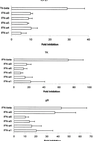
IFN-
␣
/

are known to suppress viral replication at the
tran-scriptional and translational levels (50, 65). To address the
degree and level of viral suppression, HSV-1 gene and protein
expression was measured at times p.i. Regarding gene
expres-sion, HSV-1 immediate-early (
icp27
), early (
tk
), and late (
gB
)
transcript levels were evaluated by real-time PCR. The results
show that, similar to what is found in viral titers, cells
trans-fected with the murine IFN-

transgene showed the greatest
reduction in viral gene expression (immediate early, early, and
late HSV-1 genes) in comparison to cells transfected with the
IFN-
␣
transgenes, determined at 6 to 10 h p.i. (Fig. 3).
How-ever, IFN-
␣
9 and IFN-

suppressed HSV-1
gB
gene expression
to a similar degree. Consistent with the above findings, HSV-1
antigen expression was reduced in cells transfected with some
IFN-
␣
/

transgenes, compared to expression in plasmid
vec-tor-transfected cells (Fig. 4). Specifically, while the IFN-

,
IFN-
␣
4, and IFN-
␣
9 transgene-transfected cells showed the
greatest reduction in viral antigen expression (evident at
pro-teins migrating at an apparent molecular mass of 120 to 125
kDa), there was no significant reduction in those cells
trans-fected with the IFN-
␣
1, IFN-
␣
5, or IFN-
␣
6 transgenes (Fig. 4;
Table 2).
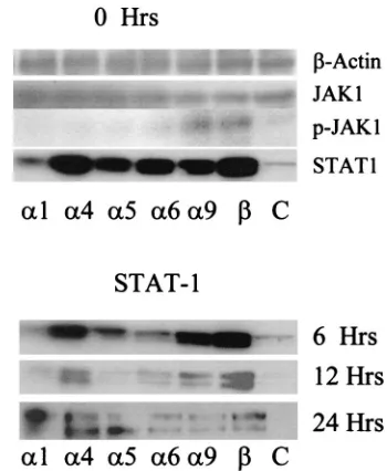
IFN-
␣
4 and IFN-

transgenes augment STAT1 expression
in transfected L929 cells.
IFN-
␣
/

utilize the JAK/STAT
[image:4.587.129.443.74.235.2]trans-duction pathway in inducing IFN-responsive gene expression
(11). Therefore, it was of interest to determine if changes in
[image:4.587.302.542.563.638.2]FIG. 2. Cells transfected with IFN-
␣
/

transgenes are less sensitive to HSV-1 infection. L929 cells (500,000 cells/well) were transfected with 3
g of plasmid DNA alone (Vector) or plasmid containing the indicated IFN-
␣
or -

transgene. Following an overnight incubation, the supernatant
was removed and the cells were infected with HSV-1 (McKrae strain) at an MOI of 0.5. Twenty-four hours p.i., the cultures were frozen and
thawed. Clarified supernatants were assayed for viral titer by plaque assay using Vero cells. This figure is a representative of two experiments, each
conducted in duplicate. Bars represent the mean
⫾
standard deviation.
ⴱⴱ
,
P
⬍
0.01 in comparisons of the IFN-
␣
/

transgene-transfected cells to
cells transfected with the plasmid vector alone, as determined by ANOVA and Scheffe multiple-comparison test.
⌬
,
P
⬍
0.05 when IFN-

transgene-transfected cells are compared to IFN-
␣
transgene-transfected cells.
TABLE 1. IC
50of IFN-
␣
/

against HSV-1
aGroup IC50
IFN-
␣
1 ...69
⫾
32
bIFN-
␣
4 ...26
⫾
18
IFN-
␣
5 ...84
⫾
34
IFN-
␣
6 ...70
⫾
40
IFN-
␣
9 ...28
⫾
12
IFN-

...13
⫾
6*
aSupernatants from transfected cells were diluted (half-log) and added to
L929 cells (500,000/well) for overnight incubation. The following day, the super-natant was removed and HSV-1 (McKrae strain) was added to the cells at an MOI of 0.5. Following a 28- to 36-h period, the cultures were frozen and thawed, and the clarified supernatants were assayed for infectious virus by plaque assay
using Vero cells. The IC50was determined by linear regression analysis for each
sample. The correlation coefficient for each sample analyzed over 6 half-log dilutions ranged from 0.9 to 0.99.
bNumbers represents means⫾standard deviations. This table is a summary of
three experiments.ⴱ,P⬍0.05 in comparisons of the IC50of IFN-with IC50s of
IFN-␣1, IFN-␣5, and IFN-␣6 as determined by the Wilcoxon signed-rank test.
V
OL. 76, 2002
HSV-1 AND IFN-
␣
/

6561
on November 8, 2019 by guest
http://jvi.asm.org/
JAK1 and STAT1 expression following transfection of cells
with the IFN-
␣
/

plasmid constructs correlated with resistance
to HSV-1 infection. To this end, L929 cells transfected with
plasmid vector control or plasmids containing the IFN-
␣
/

transgenes were assessed for JAK1 and STAT1 expression
prior to and/or after infection with HSV-1. Similar levels of the
housekeeping gene

-actin and JAK1 were expressed by all
transfected cells (Fig. 5). In addition, all IFN-
␣
/

transgene-transfected cells showed increased levels of STAT1 expression
prior to infection, compared to the nontransfected controls
(Fig. 5). However, cells transfected with the IFN-
␣
4 or IFN-

[image:5.587.136.454.83.563.2]transgenes consistently expressed the greatest amount of
FIG. 3. Expression of HSV-1
␣
,

, and
␥
genes in infected cells transfected with IFN-
␣
/

plasmid constructs. L929 cells (500,000 cells/well) were
transfected with 3
g of plasmid DNA alone (Vector) or plasmid containing the indicated IFN-
␣
or -

transgene. Following an overnight
incubation, the supernatant was removed and the cells were infected with HSV-1 (McKrae strain) at an MOI of 0.5. At 6 to 10 h p.i., the cells were
collected. Then RNA was harvested and used to generate cDNA template with reverse transcriptase. Using oligonucleotide primers specific for
icp27
,
tk
, and
gB
, the relative amount of each HSV-1 gene was determined by real-time PCR. The change (fold decrease) in viral gene expression
was calculated from the relative level of expression for each IFN-
␣
/

transgene group in comparison to that for the plasmid vector control. Bars
represent mean
⫾
standard error of the mean. This figure is a summary of four experiments.
on November 8, 2019 by guest
http://jvi.asm.org/
STAT1, compared to the other IFN-
␣
/

transgene-transfected
cells (Fig. 5). By 24 h p.i., STAT1 expression was similar in all
transfected cells but had declined from noninfected (0 h) cells.
Since it is phosphorylated STAT1 (p-STAT1) that ultimately
complexes with p-STAT2 and p48 to form ISGF3, p-STAT1
levels were measured in the transfected cells. Only in those
cells transfected with the IFN-

transgene were the p-STAT1
levels consistently detected at very low levels (data not shown).
Occasionally, cells transfected with the IFN-
␣
4 and IFN-
␣
9
transgenes expressed detectable p-STAT1. On the other hand,
cells transfected with the IFN-
␣
9 or IFN-

transgene showed
an increase in the expression of p-JAK1 (Fig. 5).
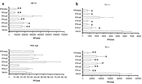
The antiviral effects elicited by IFN-
␣
/

require PKR.
IFN-␣
/

have been shown to activate a number of genes, including
the IFN-stimulated genes OAS/RNase L and PKR (12). By
infecting cells in which either the PKR or OAS/RNase L gene
is disrupted, it might be possible to distinguish the antiviral
efficacies of the different murine IFN-
␣
and IFN-

subtypes.
Consistent with previous observations (35), cells deficient in
either PKR or OAS/RNase L were highly susceptible to HSV-1
infection, with a significant increase in virus yield compared to
that of the wild-type counterparts, suggesting that PKR and
OAS/RNase L are central in constitutive resistance to HSV-1.
Specifically, in the absence of PKR, viral titers rose fivefold, as
opposed to a 14-fold rise in viral titers in the absence of the
OAS/RNase L gene compared to results for wild-type cells
(data not shown). Pretreating cells deficient in PKR with
IFN-␣
/

conferred no resistance to HSV-1 infection, whereas
pre-treatment of the corresponding wild-type cells with most
IFN-
␣
subtypes or IFN-

(50 to 100 U/ml) resulted in a two- to
sixfold reduction in viral titers (Fig. 6a). Wild-type cells treated
with IFN-
␣
6 did not show a significant reduction in viral titers.
By comparison, the absence of OAS/RNase L had no
signifi-cant impact on the antiviral effect of IFN-
␣
/

s with the
excep-tion of IFN-
␣
6 (Fig. 6b). Specifically, the treatment of
wild-type cells with IFN-
␣
6 did not reduce viral titers, whereas
exposure of RL
⫺
/
⫺
cells to IFN-
␣
6 resulted in a significant
reduction (Fig. 6b). We interpret these results to suggest that
the absence of OAS/RNase L does not impact on resistance to
HSV-1 conferred by most IFN-
␣
/

(with the exception being
IFN-
␣
6) under these experimental conditions.
DISCUSSION
In the present study, the efficacies of different types of
[image:6.587.331.506.71.284.2]IFN-␣
/

against HSV-1 were compared using cells transfected with
plasmid constructs containing IFN-
␣
/

transgenes. We have
FIG. 4. IFN-
␣
/

transgenes antagonize HSV-1 protein synthesis in
transfected cells. Total cell lysates (10
g/lane) from transfected L929
cells previously infected with HSV-1 (24 h earlier) were
electropho-resed on polyacrylamide gels, transferred onto PVDF membranes, and
used for Western blot analysis with a polyclonal anti-HSV antibody.
This figure is a representative of four experiments with identical
out-comes. Western blot for HSV-1 antigen. Note the diminished
expres-sion of proteins migrating at approximately 50, 75, and 120 to 125 kDa
(arrows) in comparisons of the plasmid vector (V) to the IFN-
␣
or -

transgene groups. A corresponding Coomassie blue-stained
polyacryl-amide gel showed an equivalent level of protein loaded in each lane
(data not shown). C, control.
FIG. 5. STAT1 and p-JAK1 are elevated in cells transfected with
the IFN-

transgene. Shown are total cell lysates (10 to 30
g/lane)
from transfected or nontransfected (C) L929 cells that were
nonin-fected (0 h) or innonin-fected with HSV-1 (MOI
⫽
0.5) and were
electro-phoresed on polyacrylamide gels, transferred to PVDF membranes,
and used for Western blot analysis with polyclonal antibody specific for
the designated protein. This figure is a representative of two to eight
experiments/time point with similar outcomes.
TABLE 2. IFN-
␣
4- and IFN-

-transgene-transfected cells express
less HSV-1 antigen
aPlasmid construct
or control HSV-1 expression in 120-to 125-kDa protein
PKCMV alone ... 9.8
⫾
1.9
bPKCMV-IFN-
␣
1... 6.9
⫾
2.2
PKCMV-IFN-
␣
4... 1.8
⫾
0.4*
PKCMV-IFN-
␣
5... 8.4
⫾
2.5
PKCMV-IFN-
␣
6... 7.9
⫾
1.3
PKCMV-IFN-
␣
9... 4.3
⫾
0.9*
PKCMV-IFN-

... 1.7
⫾
0.7*
Nontransfected control ...14.3
⫾
3.8
aProtein from cell lysates (10g) of transfected L929 cells infected with
HSV-1 (MOI⫽0.5) obtained 24 h p.i. were resolved on SDS–7.5%
polyacryl-amide gels, transferred onto PVDF membranes, and probed for HSV-1 antigen using a polyclonal rabbit anti-HSV antibody. This table is a summary of four experiments using densitometric scanning software (ONE-Dscan software; Scanalytics, Billerica, Mass.) for image analysis of the protein migrating at 120 to 125 kDa in each of the transfected and nontransfected cells. A representative example is shown in Fig. 4.
bNumbers represent the mean pixel equivalent⫾standard error of the mean.
ⴱ,P⬍0.05 in comparisons of IFN-␣/transgene-transfected cells to the plasmid
vector-transfected control as determined by Wilcoxon signed-rank test.
V
OL. 76, 2002
HSV-1 AND IFN-
␣
/

6563
on November 8, 2019 by guest
http://jvi.asm.org/
[image:6.587.52.275.72.189.2]previously found that, following transfection, the IFN
trans-gene product elicits an antiviral state following secretion into
the culture milieu. Specifically, neutralizing antibody to
IFN-␣
/

applied to the culture supernatant of the transfected cells
blocks the antiviral effect (D. J. J. Carr, unpublished
observa-tion). These results suggest that the transgene product must
bind extracellularly to the IFN-
␣
/

receptor in order to confer
resistance to viral infection through the induction of numerous
IFN-responsive genes. Such results are consistent with a
pre-vious finding indicating that a functional IFN-
␣
/

receptor is
required to elicit antiviral effects through autocrine IFN-
␣
/

production (58). IFN-

was found to suppress HSV-1
replica-tion to the greatest extent, in comparison to IFN-
␣
subtypes
when viral titers from the cultures of infected, transfected L929
cells are measured. Viral titers in L929 cells transfected with
the IFN-

transgene were reduced by 30-fold compared to
results for the IFN-
␣
5 transgene, which elicited the least
fa-vorable repression of viral replication with only a threefold
reduction in viral titer. Since the L929 cells transfected with the
IFN-
␣
/

transgenes produced similar amounts of biologically
active IFN-
␣
/

, the disparity in efficacy between the IFN-

transgene and the IFN-
␣
plasmid constructs cannot be based
on the quantity of the transgene product secreted.
Unfortu-nately, the IFN-

product could only be measured by bioassay,
so, therefore, a true quantitative measurement could not be
determined. Nevertheless, similar amounts of IFN-
␣
were
gen-erated as determined by ELISA and bioassay and yet
differ-ences in antiviral activity were observed between IFN-
␣
sub-types, suggesting that, even within the same family of IFN-
␣
/

,
the efficacy against a viral pathogen can differ.
Cells transfected with the IFN-

transgene strongly
sup-pressed HSV-1 immediate early, early, and late gene
expres-sion as represented by
icp27
,
tk
, and
gB
, respectively. In
com-parison, cells transfected with the IFN-
␣
transgenes antagonized
HSV-1 gene expression to similar degrees, with the notable
exception of the IFN-
␣
9 transgene, which suppressed HSV-1
gB
gene expression to a level nearly equivalent to that of the
IFN-

transgene. However, at the protein level, the major
HSV-1 protein(s) identified (migrating at an apparent
molec-ular mass of 120 to 125 kDa) was equally suppressed in cells
transfected with either the IFN-
␣
4 or IFN-

transgene.
Like-wise, cells transfected with the IFN-
␣
9 transgene showed a
significant reduction in the expression of the 120- to 125-kDa
HSV-1 protein. These findings are in agreement with the
out-FIG. 6. IFN-
␣
/

antagonizes HSV-1 replication through PKR. (a) Wild-type (MEF/6) or PKR-null cells (2.5
⫻
10
5cells/well) were incubated
with equivalent amounts of the indicated IFN-containing supernatants from transfected L929 cells (50 to 100 U/ml based on bioassay). Following
an overnight incubation, the cells were infected with HSV-1 (MOI
⫽
1.0) and the cultures were assayed for viral titers 28 h p.i. This figure is a
summary of three experiments. Bars represent mean
⫾
standard error of the mean.
ⴱ
,
P
⬍
0.05;
ⴱⴱ
,
P
⬍
0.01 in comparisons of the cells incubated
with IFN-
␣
/

-containing supernatants with the corresponding wild-type cells incubated with vector-transfected culture supernatants, as determined
by ANOVA and Scheffe multiple-comparison test. (b) Wild-type (RL
⫹
/
⫹
) or OAS/RNase L-null cells (RL
⫺
/
⫺
) cells (2.5
⫻
10
5cells/well) were
incubated with equivalent amounts of the indicated IFN-containing supernatants from transfected L929 cells (50 to 100 U/ml based on bioassay).
Following an overnight incubation, the cells were infected with HSV-1 (MOI
⫽
1.0) and the cultures were assayed for viral titers 28 h p.i. This
figure is a summary of four experiments. Bars represent mean
⫾
standard error of the mean.
ⴱ
,
P
⬍
0.05;
ⴱⴱ
,
P
⬍
0.01 when the corresponding
wild-type or knockout cells incubated with IFN-
␣
/

-containing supernatants are compared to the cells incubated with vector-transfected culture
supernatants as determined by ANOVA and Scheffe multiple-comparison test.
on November 8, 2019 by guest
http://jvi.asm.org/
[image:7.587.46.540.68.362.2]come of viral loads recovered from transfected L929 cells, in
which fold reduction in viral titers by the IFN-

, IFN-
␣
9, and
IFN-
␣
4 transgenes was 30, 9.0, and 8.6, respectively, compared
to 6.7-, 5.0-, and 3.0-fold reduction by the IFN-
␣
6, IFN-
␣
1, and
IFN-
␣
5 transgenes, respectively. Taken together, the data
clearly indicate that the IFN-

transgene shows the greatest
degree of efficacy against HSV-1 replication in vitro, followed
by IFN-
␣
4 and IFN-
␣
9, a finding supported by data
establish-ing the IC
50for each of the IFN-
␣
/

subtypes. Of note, a
previous study suggests that, following viral infection, either
IFN-

or IFN-
␣
4 is produced, which can subsequently
stimu-late the expression of other IFN-
␣
species (including IFN-
␣
2,
-
␣
5, -
␣
6, and -
␣
8) through the IFN-
␣
/

receptor, ultimately
conferring resistance to viral infection (43). In the present
study, it is tempting to speculate that the transfection of cells
with the IFN-
␣
4 or IFN-

transgene prior to infection allows
cells sufficient time to express other delayed IFN-
␣
species to
counter HSV-1 replication. Although other IFN-
␣
transgenes
(IFN-
␣
9 notwithstanding) are capable of antagonizing HSV-1
replication, it is interesting that the degree of suppression is
not as strong as that for IFN-
␣
4, IFN-
␣
9, or IFN-

, implying
that either (i) additional IFN-
␣
/

subtypes are not expressed in
L929 cells following transfection with the IFN-
␣
1, IFN-
␣
5, or
IFN-
␣
6 transgene or that (ii) the time needed to express
ad-ditional IFN-
␣
/

subtypes is greater than the incubation period
between transfection and infection (18 h).
IFN-
␣
/

act through the same receptor (48), facilitating the
transphosphorylation and activation of Tyk2 and JAK1 (54),
ultimately leading to the phosphorylation of the STAT1
mol-ecule (63). Consequently, the antiviral resistance induced by
the IFN-
␣
/

may, in part, reside with the level of JAK1 or
STAT1 expression and/or phosphorylation (14). To this end,
JAK1 and STAT1 expression was assessed in transfected L929
cells prior to and at various times p.i. Although JAK1
expres-sion was equivalent in all cells analyzed, the induction of
STAT1 expression prior to infection was greatest in cells
trans-fected with the IFN-
␣
4 or IFN-

transgene. Correlating with
the elevated STAT1 expression in the IFN-

transgene-trans-fected cells, there was also the detection of p-STAT1 and
p-JAK1 expression, indicating that the IFN-

transgene
acti-vates STAT1 and JAK1 within 18 h following transfection. At
6 to 12 h p.i., STAT1 expression was still elevated in those cells
transfected with the IFN-
␣
4 or IFN-

transgenes, compared to
expression in the other transfected groups. It is interesting that
these cells showed the greatest degree of resistance to HSV-1
infection based on viral titers and viral protein expression.
Although the relationship between STAT1 expression and
HSV-1 infection has not been clearly defined, a recent article
suggests that HSV-1 antagonizes the phosphorylation of
STAT1 through the failure to activate JAK1 (74). Presumably
then, the inhibition of p-STAT1 accumulation would prevent
the formation of the transcription factor ISGF3 and reduce the
activation of IFN-stimulatory genes driven by the
IFN-stimu-lated response element. Although the present study did not
assess p-STAT1 levels at times p.i., our lab has found that Vero
cells transfected with a plasmid containing the human IFN-

transgene express pSTAT1 levels 24 h p.i., whereas expression
of pSTAT1 in cells transfected with the human IFN-
␣
2
trans-gene is absent, similar to expression in nontransfected, infected
cells, suggesting that IFN-

may counter the action of HSV-1
in preventing the activation of JAK1 (23). This notion is
sup-ported with the increased expression of p-JAK1 expression in
cells transfected with the IFN-

transgene. While the
respon-sible agent antagonizing the activation of the IFN signaling
cascade during HSV-1 infection is presently unknown, it may
involve the immediate early gene product ICP0 (74).
Two prominent antiviral pathways activated following
IFN-␣
/

receptor ligation are OAS and PKR (20). OAS leads to the
activation of endo-RNase L, which degrades viral transcripts
(53) and inactivates translational machinery (30). PKR
phos-phorylates the
␣
subunit of eukaryotic initiation factor 2
␣
,
resulting in the inhibition of protein synthesis (46, 57). HSV-1
has been found to prevent UV- or anti-Fas antibody-induced
apoptosis associated with a marked reduction in caspase-3 and
caspase-8 activity (34). By preventing apoptosis of infected
cells, cellular machinery is preserved for efficient viral
replica-tion and packaging of progeny virus. However, IFN-
␣
/

pro-mote apoptosis (4) through a PKR-mediated pathway (69) and
therefore would reduce the potential for viral replication.
Al-though there are multiple IFN-responsive genes activated
through the IFN-
␣
/

receptor, it might be possible to segregate
the antiviral efficacy for each IFN-
␣
/

species by initially
fo-cusing on the dominant PKR and OAS/RNase L antiviral
path-ways known to impact on HSV-1 infection (35). In the absence
of RNase L, the antiviral efficacy of IFN-
␣
6 was significantly
increased. In addition, the strength of the inhibition as
mea-sured by recoverable virus was increased in the RNase L
knockout cells treated with the other IFN-
␣
/

. A similar
ob-servation was found when these cells were transduced with an
adenoviral construct expressing the murine IFN-

gene
(un-published observation). We interpret these results to indicate
that the activation of the downstream effector molecule of
OAS, RNase L, targets not only viral RNA but also
IFN-inducible transcripts as well. In the absence of this pathway, the
longevity of IFN-stimulatory cascades would be prolonged,
increasing the antiviral environment induced by the IFN-
␣
/

.
In contrast to the results with the RL
⫺
/
⫺
cells, in the absence
of PKR, the efficacy of IFN-
␣
/

against HSV-1 is completely
lost. These results substantiate the contributions of other
in-vestigators, illustrating the countering effect of the HSV-1
␥
134.5 protein on PKR action (8, 39). Since, under normal
conditions, the virus devotes a considerable amount of energy
to encoding the
␥
134.5 protein, in order to circumvent the
antiviral action of PKR by dephosphorylating eukaryotic
initi-ation factor 2
␣
following PKR-induced phosphorylation (25),
the PKR pathway is undoubtedly an important endogenous
contributor to antagonizing HSV-1 replication. Future studies
are required to evaluate the levels of other components of the
ISGF3 complex in the transfected cells, in addition to the
downstream effector molecules generated from the activation
of the IFN-inducible genes, as a means to address differences
in antiviral efficacy between different IFN-
␣
/

(i.e., IFN-
␣
and
-

).
In summary, the present study has shown that cells
trans-fected with an IFN-

plasmid construct are less sensitive to
HSV-1 infection than are cells transfected with plasmid
con-structs containing five different IFN-
␣
transgenes. Each of
these transgenes, nearly equal in size, was cloned into the same
plasmid and produced a similar amount of biologically active
IFN. To our knowledge, this is the first report that has
com-V
OL. 76, 2002
HSV-1 AND IFN-
␣
/

6565
on November 8, 2019 by guest
http://jvi.asm.org/
pared murine IFN-
␣
/

against HSV-1 infection and found
murine IFN-

to provide superior protection against the
HSV-1 infection, as measured by viral titers and viral gene and
protein expression.
ACKNOWLEDGMENTS
We thank Emmalene Bartlett for her help with plasmid construct
development. In addition, we are grateful to Blake Shockley for his
assistance in the plaque assays and Benitta John-Philip for her help
with the detection of

-actin, STAT1, JAK1, p-JAK1, p-STAT1, and
HSV-1 antigen.
This work was supported by Public Health Service grants EY12409
(D.J.J.C.), AI34039 (B.R.G.W.), and CA44059 (R.S.); NEI core grant
P30 EY12190; an unrestricted grant from Research to Prevent
Blind-ness (RPB), Inc.; and an RPB Stein Research Professorship (D.J.J.C.).
Peter Härle is a recipient of a research fellowship from the Deutsche
Forschungsgemeinschaft (HA2993/1-1).
REFERENCES
1.Adler, H., J. L. Beland, N. C. Del-Pan, L. Kobzik, R. A. Sobel, and I. J. Rimm. 1999. In the absence of T cells, natural killer cells protect from
mortality due to HSV-1 encephalitis. J. Neuroimmunol.93:208–213.
2.Ahn, K., T. H. Meyer, S. Uebel, P. Sempe, H. Djaballay, Y. Yang, P. A. Peterson, K. Fruh, and R. Tampe.1996. Molecular mechanism and species specificity of TAP inhibition by herpes simplex virus protein ICP47. EMBO
J.15:3247–3255.
3.Arora, T., G. Floyd-Smith, M. J. Epsy, and D. F. Jelinek.1999. Dissociation
between IFN-␣-induced anti-viral and growth signaling pathways. J.
Immu-nol.162:3289–3297.
4.Balachandran, S., P. C. Roberts, T. Kipperman, K. N. Bhalla, R. W. Com-pans, D. R. Archer, and G. N. Barber.2000. Alpha/beta interferons poten-tiate virus-induced apoptosis through activation of the FADD/caspase-8
death signaling pathway. J. Virol.74:1513–1523.
5.Beilharz, M. W., N. Swaminathan, C. M. Lai, P. M. Pitha, and S. J. Boyer.
1991. Relative antiviral activity ofin vitro-synthesized murine interferon-␣4
and -␣1. J. Interferon Res.11:9–15.
6.Bukowski, J. F., and R. M. Welsh.1986. The role of natural killer cells and interferon in resistance to acute infection of mice with herpes simplex virus
type 1. J. Immunol.136:3481–3485.
7.Cantin, E. M., D. R. Hinton, J. Chen, and H. Openshaw.1995. Gamma interferon expression during acute and latent nervous system infection by
herpes simplex virus type 1. J. Virol.69:4898–4905.
8.Cheng, G., M.-E. Brett, and B. He.2001. Val193and Phe195of the␥ 134.5
protein of herpes simplex virus 1 are required for viral resistance to
inter-feron-␣/. Virology290:115–120.
9.Chou, J., J.-J. Chen, M. Gross, and B. Roizman.1995. Association of a Mr
90,000 phosphoprotein with protein kinase PKR in cells exhibiting enhanced
phosphorylation of translation initiation factor eIF-2␣and premature shutoff
of protein synthesis after infection with␥134.5⫺mutants of herpes simplex
virus 1. Proc. Natl. Acad. Sci. USA92:10516–10520.
10.Cull, V. S., E. Bartlett, and C. M. James.Type I interferon gene therapy protects against cytomegalovirus-induced myocarditis. Immunology, in press. 11.Darnell, J. E., Jr., I. M. Kerr, and G. R. Stark.1994. Jak-STAT pathways and transcriptional activation in response to IFNs and other extracellular
signal-ing proteins. Science264:1415–1421.
12.Der, S. D., A. Zhou, B. R. G. Williams, and R. H. Silverman.1998.
Identi-fication of genes differentially regulated by interferon␣,, and␥using
oligonucleotide arrays. Proc. Natl. Acad. Sci. USA95:15623–15628.
13.De Stasio, R. P., and M. W. Taylor.1990. Specific effect of interferon on the
herpes simplex virus type 1 transactivation event. J. Virol.64:2588–2593.
14.Durbin, J. E., R. Hackenmiller, M. C. Simon, and D. E. Levy.1996. Targeted disruption of the mouse STAT1 gene results in compromised innate
immu-nity to viral disease. Cell84:223–226.
15.Engler, H., R. Zawatzky, A. Goldbach, C. H. Schroder, C. Weyand, G. J. Hammerling, and H. Kirchner.1981. Experimental infection of inbred mice with herpes simplex virus. II. Interferon production and activation of natural
killer cells in the peritoneal exudate. J. Gen. Virol.55:25–30.
16.Franchini, M., C. Abril, C. Schwerdel, C. Ruedl, M. Ackermann, and M. Suter.2001. Protective T-cell-based immunity induced in neonatal mice by a
single replicative cycle of herpes simplex virus. J. Virol.75:83–89.
17.Friedman, H. M., G. H. Cohen, R. J. Eisenberg, C. A. Seidel, and D. B. Cines.
1984. Glycoprotein C of herpes simplex virus type 1 acts as a receptor for the
C3b complement component on infected cells. Nature309:633–635.
18.Ghislain, J., G. Sussman, S. Goelz, L. E. Ling, and E. N. Fish.1995.
Con-figuration of the interferon-␣/receptor complex determines the context of
the biological response. J. Biol. Chem.270:21785–21792.
19.Goldsmith, K., W. Chen, D. C. Johnson, and R. L. Hendricks.1998. Infected
cell protein (ICP) 47 enhances herpes simplex virus neurovirulence by
block-ing the CD8⫹T cell response. J. Exp. Med.187:341–348.
20.Guidotti, L. G., and F. V. Chisari.2001. Noncytolytic control of viral infec-tions by the innate and adaptive immune repsonse. Annu. Rev. Immunol.
19:65–91.
21.Halford, W. P., B. M. Gebhardt, and D. J. J. Carr.1996. Persistent cytokine expression in trigeminal ganglion latently infected with herpes simplex virus
type 1. J. Immunol.157:3542–3549.
22.Halford, W. P., L. A. Veress, B. M. Gebhardt, and D. J. J. Carr.1997. Innate
and acquired immunity to herpes simplex virus type 1. Virology236:328–337.
23.Härle, P., E. Lauret, P. M. Pitha, E. De Maeyer, and D. J. J. Carr.2001. Expression of human and macaque type I IFN transgenes interfere with
HSV-1 replication at the transcriptional and translational level: IFN-is
more potent than IFN-␣2. Virology290:237–248.
24.Härle, P., B. Sainz, Jr., D. J. J. Carr, and W. P. Halford.2002. The imme-diate-early protein, ICP0, is essential for the resistance of herpes simplex
virus to interferon-␣/. Virology293:295–304..
25.He, B., M. Gross, and B. Roizman.1997. The␥134.5 protein of herpes
simplex virus 1 complexes with protein phosphatase 1 alpha to dephosphor-ylate the alpha subunit of the eukaryotic translation initiation factor 2 and preclude the shutoff of protein synthesis by double-stranded RNA-activated
protein kinase. Proc. Natl. Acad. Sci. USA90:843–848.
26.Hendricks, R. L., P. C. Weber, J. L. Taylor, A. Koumbis, T. M. Tumpey, and J. C. Glorioso.1991. Endogenously produced interferon␣ protects mice
from herpes simplex virus type 1 corneal disease. J. Gen. Virol.72:1601–
1610.
27.Hendrzak, J. A., and P. S. Morahan.1994. The role of macrophages and macrophage cytokines in the host resistance to herpes simplex virus.
Immu-nol. Ser.60:601–617.
28.Hill, A., P. Jugovic, I. York, G. Russ, J. Bennink, J. Yewdell, H. Ploegh, and D. Johnson.1995. Herpes simplex virus turns off the TAP to evade host
immunity. Nature375:411–415.
29.Hu, R., J. Bekisz, M. Hayes, S. Audet, J. Beeler, E. Petricoin, and K. Zoon.
1999. Divergence of binding, signaling, and biological responses to
recom-binant human hybrid IFN. J. Immunol.163:854–860.
30.Iordanov, M. S., J. M. Paranjape, A. Zhou, J. Wong, B. R. Williams, E. F. Meurs, R. H. Silverman, and B. E. Magun.2000. Activation of p38
mitogen-activated protein kinase and c-Jun NH2-terminal kinase by double-stranded
RNA and encephalomyocarditis virus: involvement of RNase L, protein
kinase R, and alternative pathways. Mol. Cell. Biol.20:617–627.
31.Ito, M., M. Watanabe, H. Kamiya, and M. Sakurai.1997. Herpes simplex virus type 1 induces apoptosis in peripheral blood T lymphocytes. J. Infect.
Dis.175:1220–1224.
32.Jennings, S. R., P. L. Rice, E. D. Kloszewski, R. W. Anderson, D. L. Thomp-son, and S. S. Tevethia.1985. Effect of herpes simplex virus types 1 and 2 on surface expression of class I major histocompatibility complex antigens on
infected cells. J. Virol.56:757–766.
33.Jerome, K. R., J. F. Tait, D. M. Koelle, and L. Corey.1998. Herpes simplex virus renders infected cells resistant to cytotoxic T-lymphocyte-induced
ap-optosis. J. Virol.72:436–441.
34.Jerome, K. R., R. Fox, Z. Chen, A. E. Sears, H.-Y. Lee, and L. Corey.1999. Herpes simplex virus inhibits apoptosis through the action of two genes, Us5
and Us3. J. Virol.73:8950–8957.
35.Khabar, K. S. A., M. Dhalla, Y. Siddiqui, A. Zhou, M. N. Al-Ahdal, S. D. Der, R. H. Silverman, and B. R. G. Williams.2000. Effect of deficiency of the double-stranded RNA-dependent protein kinase, PKR, on antiviral resis-tance in the presence or absence of ribonuclease L: HSV-1 replication is particularly sensitive to deficiency of the major IFN-mediated enzymes.
J. Interferon Cytokine Res.20:653–659.
36.Kodukula, P., T. Liu, N. Van Rooijen, M. J. Jager, and R. L. Hendricks.
1999. Macrophage control of herpes simplex virus type 1 replication in the
peripheral nervous system. J. Immunol.162:2895–2905.
37.Lee, Y. J., Y. Han, H. T. Lu, V. Nguyen, H. Qin, P. H. Howe, B. A. Hocevar, J. M. Boss, R. M. Ransohoff, and E. N. Benveniste.1997. TGF-suppresses
IFN-␥induction of class II MHC gene expression by inhibiting class II
transactivator messenger RNA expression. J. Immunol.158:2065–2075.
38.Leib, D. A., T. E. Harrison, K. M. Laslo, M. A. Machalek, N. J. Moorman, and H. W. Virgin.1999. Interferons regulate the phenotype of wild-type and
mutant herpes simplex viruses in vivo. J. Exp. Med.189:663–672.
39.Leib, D. A., M. A. Machalek, B. R. G. Williams, R. H. Silverman, and H. W. Virgin.2000. Specific phenotypic restoration of an attenuated virus by
knock-out of a host resistance gene. Proc. Natl. Acad. Sci. USA97:6097–6101.
40.Linnavuori, K., and T. Hovi.1983. Restricted replication of herpes simplex
virus in human monocyte cultures: role of interferon. Virology130:1–9.
41.Liu, T., Q. Tang, and R. L. Hendricks.1996. Inflammatory infiltration of the trigeminal ganglion after herpes simplex virus type 1 corneal infection. J.
Vi-rol.70:264–271.
42.Manickan, E., and B. T. Rouse.1995. Roles of different T-cell subsets in control of herpes simplex virus infection determined by using T-cell-deficient
mouse models. J. Virol.69:8178–8179.
43.Marie, I., J. E. Durbin, and D. E. Levy.1998. Differential viral induction of
on November 8, 2019 by guest
http://jvi.asm.org/
distinct interferon-␣genes by positive feedback through interferon
regula-tory factor-7. EMBO J.17:6660–6669.
44.McGeoch, D. J., S. Cook, A. Dolan, F. E. Jamieson, and E. A. Telford.1995. Molecular phylogeny and evolutionary timescale for the family of
mamma-lian herpesviruses. J. Mol. Biol.247:443–458.
45.Mendez-Samperio, P., M. Hernandez, and H. E. Ayala.2000. Induction of
transforming growth factor-1production in human cells by herpes simplex
virus. J. Interferon Cytokine Res.20:273–280.
46.Meurs, E., K. Chong, J. Galabru, N. S. Thomas, I. M. Kerr, B. R. G. Williams, and A. G. Hovanessian.1990. Molecular cloning and character-ization of the human double-stranded RNA-activated protein kinase induced
by interferon. Cell62:379–390.
47.Mittnacht, S., P. Straub, H. Kirchner, and H. Jacobsen.1988. Interferon treatment inhibits onset of herpes simplex virus immediate-early
transcrip-tion. Virology164:201–210.
48.Mogensen, K. E., M. Lewerenz, J. Reboul, G. Lutfalla, and G. Uze.1999. The type I interferon receptor: structure, function, and evolution of a family
business. J. Interferon Cytokine Res.19:1069–1098.
49.Mossman, K. L., H. A. Saffran, and J. R. Smiley.2000. Herpes simplex virus
ICP0 mutants are hypersensitive to interferon. J. Virol.74:2052–2056.
50.Munoz, A., and L. Carrasco.1984. Formation of non-infective herpesvirus particles in cultured cells treated with human interferon. J. Gen. Virol.
65:1069–1078.
51.Nagashunmugam, T., J. Lubinski, L. Wang, L. T. Goldstein, B. S. Weeks, P. Sundaresan, E. H. Kang, G. Dubin, and H. M. Friedman.1998. In vivo immune evasion mediated by the herpes simplex virus type 1
immunoglob-ulin G Fc receptor. J. Virol.72:5351–5359.
52.Nicholl, M. J., and C. M. Preston.1996. Inhibition of herpes simplex virus type 1 immediate-early gene expression by alpha interferon is not VP16
specific. J. Virol.70:6336–6339.
53.Nilsen, T. W., and C. Baglioni.1979. Mechanism for discrimination between viral and host mRNA in interferon-treated cells. Proc. Natl. Acad. Sci. USA
76:2600–2604.
54.Novick, D., B. Cohen, and M. Rubinstein. 1994. The human interferon
alpha/beta receptor: characterization and molecular cloning. Cell77:391–
400.
55.Pellegrini, S., J. John, M. Shearer, I. Kerr, and G. Stark.1989. Use of a selectable marker regulated by alpha interferon to obtain mutations in the
signaling pathway. Mol. Cell. Biol.9:4605–4612.
56.Raftery, M. J., C. K. Behrens, A. Muller, P. H. Krammer, H. Walczak, and G. Schonrich.1999. Herpes simplex virus type 1 infection of activated cyto-toxic T cells: induction of fratricide as a mechanism of viral immune evasion.
J. Exp. Med.190:1103–1113.
57.Roberts, W. K., A. Hovanessian, R. E. Brown, M. J. Clemens, and I. M. Kerr.
1976. Interferon-mediated protein kinase and low-molecular-weight
inhibi-tor of protein synthesis. Nature264:477–480.
58.Rousseau, V., I. Cremer, E. Lauret, I. Riviere, M. Aguet, and E. De Maeyer.
1995. Antiviral activity of autocrine interferon-beta requires the presence of
a functional interferon type I receptor. J. Interferon Cytokine Res.15:785–
789.
59.Samuel, C. E.1991. Antiviral action of interferon. Interferon-regulated cel-lular proteins and their surprisingly selective antiviral activities. Virology
183:1–11.
60.Shibaki, T., T. Suzutani, I. Yoshida, M. Ogasawara, and M. Azuma.2001.
Participation of type I interferon in the decreased virulence of the UL13 gene-deleted mutant of herpes simplex virus type 1. J. Interferon Cytokine
Res.21:279–285.
61.Shimeld, C., J. L. Whiteland, S. M. Nicholls, E. Grinfeld, D. L. Easty, H. Gao, and T. J. Hill.1995. Immune cell infiltration and persistence in the mouse trigeminal ganglion after infection of the cornea with herpes simplex
virus type 1. J. Neuroimmunol.61:7–16.
62.Shimeld, C., J. L. Whiteland, N. A. Williams, D. L. Easty, and T. J. Hill.
1997. Cytokine production in the nervous system of mice during acute and
latent infection with herpes simplex virus type 1. J. Gen. Virol.78:3317–3325.
63.Shuai, K., C. M. Horvath, L. H. Huang, S. A. Qureshi, D. Cowburn, and J. E. Darnell, Jr.1994. Interferon activation of the transcription factor Stat91 involves dimerization through SH2-phosphotyrosyl peptide interactions. Cell
76:821–828.
64.Simmons, A., and D. C. Tscharke.1992. Anti-CD8 impairs clearance of herpes simplex virus from the nervous system: implications for the fate of
virally infected neurons. J. Exp. Med.175:1337–1344.
65.Straub, P., D. H. Kirchner, H. Jacobsen, and A. Panet.1986. Synthesis of herpes simplex virus proteins and nucleic acids in interferon-treated
macro-phages. Virology150:411–418.
66.Su, Y.-H., J. E. Oakes, and R. N. Lausch.1990. Ocular avirulence of a herpes simplex virus type 1 strain is associated with heightened sensitivity to alpha/
beta interferon. J. Virol.64:2187–2192.
67.Su, Y.-H., J. E. Oakes, and R. N. Lausch.1993. Mapping the genetic region
coding for herpes simplex virus resistance to mouse interferon-␣/. J. Gen.
Virol.74:2325–2332.
68.Swaminathan, N., C. M. Lai, S. J. Boyer, M. W. Beilharz, and S. P. Klinken.
1992. Biological activities of recombinant murine interferons alpha 1 and
alpha 4: large difference in antiproliferative effect. Antivir. Res.19:149–159.
69.Tan, S.-L., and M. G. Katze.1999. The emerging role of the interferon-induced PKR protein kinase as an apoptotic effector: a new face of death?
J. Interferon Cytokine Res.19:543–554.
70.Tomazin, R., A. B. Hill, P. Jugovic, I. York, P. V. Endert, H. L. Ploegh, D. W. Andrews, and D. C. Johnson.1996. Stable binding of the herpes simplex virus
ICP47 protein to the peptide binding site of TAP. EMBO J.15:3256–3266.
71.Tumpey, T. M., S. Chen, J. E. Oakes, and R. N. Lausch.1996. Neutrophil-mediated suppression of virus replication after herpes simplex virus type 1
infection of the murine cornea. J. Virol.70:898–904.
72.Van Strijp, J. A., K. P. van Kessel, L. A. Miltenburg, A. C. Fluit, and J. Verhoef.1988. Attachment of human polymorphonuclear leukocytes to her-pes simplex virus-infected fibroblasts mediated by antibody-independent
complement activation. J. Virol.62:847–850.
73.Yeow, W.-S., C. M. Lawson, and M. W. Beilharz.1998. Antiviral activities of individual murine interferon alpha subtypes in vivo: intramuscular injection of interferon expression constructs reduces cytomegalovirus replication.
J. Immunol.160:2932–2939.
74.Yokota, S.-I., N. Yokosawa, T. Kubota, T. Suzutani, I. Yoshida, S. Miura, K. Jimbow, and N. Fujii.2001. Herpes simplex virus type 1 suppresses the interferon signaling pathway by inhibiting phosphorylation of STATs and
Janus kinases during an early infection stage. Virology286:119–124.
75.Zhou, A., J. Paranjape, T. L. Brown, H. Nie, S. Naik, B. Dong, A. Chang, B. Trapp, R. Fairchild, C. Colmenares, and R. H. Silverman.1997. Interferon
action and apoptosis are defective in mice devoid of 2⬘,5⬘
-oligoadenylate-dependent RNase L. EMBO J.16:6355–6363.
V
OL. 76, 2002
HSV-1 AND IFN-
␣
/

6567
on November 8, 2019 by guest
http://jvi.asm.org/