Dissertation on
A STUDY ON THE BRANCHING PATTERN AND VARIATIONS OF THE COELIAC ARTERY AND ITS BRANCHES
Submitted in partial fulfillment for
M.D. DEGREE EXAMINATION BRANCH-XXIII, ANATOMY
Upgraded Institute of Anatomy
Madras Medical College & Rajiv Gandhi Government General Hospital Chennai-600 003
THE TAMILNADU Dr. M.G.R. MEDICAL UNIVERSITY CHENNAI-600 032
TAMILNADU
CERTIFICATE
This is to certify that this dissertation entitled
“A STUDY ON THE BRANCHING PATTERN AND VARIATIONS OF
THE COELIAC ARTERY AND ITS BRANCHES”
is a bonafide record of the research work done by Dr.E.SRIVIDHYA, Post
graduate in the Institute of Anatomy, Madras Medical College and Rajiv
Gandhi Government General Hospital, Chennai-03, in partial fulfillment of the
regulations laid down by The Tamil Nadu Dr. M.G.R. Medical University for
the award of M.D. Degree Branch XXIII- Anatomy, under my guidance and
supervision during the academic year 2012-2015.
Dean
Madras Medical College and
Rajiv Gandhi Government General
Hospital
Chennai-600 003
Dr.Sudha Seshayyan, M.B.B.S.,M.S., Director and Professor,
Institute of Anatomy,
Madras Medical College,
Chennai 600003.
ACKNOWLEDGEMENT
I wish to express exquisite thankfulness and gratitude to my most
respected teacher and guide Dr. Mrs. Sudha Seshayyan, M.S., Director and
Professor, Institute of Anatomy, Madras Medical College, Chennai – 3, for
her valuable guidance, persistent support and quest for perfection which has
made this dissertation take its present shape.
I am thankful to Dr.R.Vimala M.D., Dean, Madras Medical College,
Chennai – 3 for permitting me to avail the facilities in this college for
performing this study.
My heartfelt thanks to Dr.B.Chezhian, Dr.V.Lokanayaki,
Dr.B.Santhi, Associate Professors, Dr.V.Lakshmi, Dr.T.Anitha,
Dr.P.Kanagavalli, Dr.J.Sreevidya, Dr.Elamathi Bose, Dr.S.Arrchana,
Assistant Professors, Institute of Anatomy, Madras Medical College, Chennai
for their valuable suggestions and encouragement throughout the study.
My gratefulness to Dr.K.Vanitha, Director, Barnard Institute of
Radiology, Govt. General Hospital, Chennai – 3 , for the help in radiological
I earnestly thank my seniors Dr.K.Arumugam, Dr.P.Radhakrishnan
and my helpful juniors Dr.S.Keerti, Dr.P.R.Prefulla, Dr.N.V.Ganga, and
other members of faculty who have been supportive and encouraging
throughout the study.
I extend my heartfelt thanks to my colleagues Dr.M.Anuradha,
Dr.B.J.Bhuvaneswari, and Dr.S.Elizabeth Priyadarisini for their constant
encouragement and unstinted co-operation.
I’m especially thankful to Mr.Mathews and Mr.Senthilkumar,
technicians, who extended great support for this study and all other staff
members including Mr.Jagadeesan, Mr.Maneesh, and Mr.Devaraj for
helping me to carry out the study.
I’m grateful to my family members for their encouragement.
I’m especially grateful to my Mother who has helped making this study
a reality.
Above all, I thank the ALMIGHTY GOD who has showered His
LEGEND
AA
-
Abdominal Aorta
CA
-
Coeliac artery
CHA
-
Common Hepatic Artery
DPA
-
Dorsal Pancreatic Artery
GDA
-
Gastro Duodenal Artery
LGA
-
Left Gastric Artery
LHA
-
Left Hepatic Artery
LIPA -
Left Inferior Phrenic Artery
MAL -
Median Arcuate Ligament
PHA
-
Proper Hepatic Artery
RGA
-
Right Gastric Artery
RHA
-
Right Hepatic Artery
RIPA -
Right Inferior Phrenic Artery
SA
-
Splenic Artery
SMA
-
Superior
Mesenteric Artery
CONTENTS
SL.
NO. TITLE
PAGE NO.
1. INTRODUCTION 1
2. AIM OF THE STUDY 11
3. REVIEW OF LITERATURE 16
4. EMBRYOLOGY 44
5. MATERIALS AND METHODS 49
6. OBSERVATION 52
7. DISCUSSION 66
8. CONCLUSION 104
ABSTRACT
A STUDY ON THE BRANCHING PATTERN AND VARIATIONS OF THE COELIAC ARTERY AND ITS BRANCHES
Coeliac artery is the major source of blood supply to the supracolic
abdominal compartment. It commonly divides into left gastric, splenic and
common hepatic arteries. Variations in its origin and branching pattern are
frequently observed during cadaveric dissection and radiological imaging.
These variations although asymptomatic become significant in patients
undergoing diagnostic angiography and invasive procedures of the abdomen.
In the present study the coeliac artery was complete with all three
branches emerging from it in 78% and had aberrant branches in 22%. The
aberrant branches encountered were the right and left inferior phrenic arteries
and the dorsal pancreatic artery. Replaced left hepatic artery arising from left
gastric artery was observed in 4%. In 2% of the specimens the common hepatic
artery quadrifurcated into left hepatic artery, right hepatic artery, gastroduodenal
artery and cystic artery.
The observations made in this study could help surgeons minimize
iotrogenic injury during abdominal surgeries. It may also facilitate accurate
radiological interpretation
1
INTRODUCTION
‘Variability is the law of life’ – Sir William Osler
Variations in the vasculature of the abdomen especially the coeliac
artery are very common and have an embryological basis. These
variations not only interest the anatomist but a thorough knowledge of
their anatomy is very essential for surgeons and radiologists because of
their impact in visceral surgery and medicolegal implications.
Surgeons must have expertise in anatomy which helps them to
recognize the variations and plan the surgery accordingly. This improves
the safety of the surgery and lowers morbidity.
The value of knowledge of arterial anatomy of the abdomen has
increased immensely with the introduction of laparoscopic procedures of
the abdomen where the surgical field of vision is limited and mistakes are
common. Ligation of a variant artery with subsequent ischemia of the part
2
The importance of anatomy of visceral vasculature has increased
furthermore with the introduction of organ transplantation. In the era of
computerized surgery, anatomy becomes indispensable.
Coeliac artery is the artery supplying the infradiaphragmatic part of
the foregut namely the stomach, the part of duodenum till the opening of
common bile duct, liver, pancreas, spleen.
The coeliac artery is a wide branch, approximately 1.25 cm long,
from the ventral aspect of abdominal aorta; just below the aortic opening
of the diaphragm. It passes horizontally forward and to the right, superior
to the splenic vein and pancreas and divides into three branches
1) Left gastric artery
2) Common hepatic artery
3) Splenic artery
One or both the right and left inferior phrenic arteries may
originate from coeliac artery. The coeliac artery and superior mesenteric
artery may originate from a common trunk from the aorta or the classic
3
THE LEFT GASTRIC ARTERY
Left gastric artery is the smallest of the branches of coeliac artery.
It ascends towards the cardiac end of the stomach. On reaching it, the
artery gives off two or three oesophageal branches. It then arches forward
to reach the lesser curvature of the stomach dragging a fold of
peritoneum with it – the left gastropancreatic fold. Situated between the
layers of the lesser omentum, it descends along the lesser curvature of the
stomach and anastomoses with the right gastric artery. Gastric,
oesophageal, aberrant left hepatic artery (accessory or replaced) arise
from it.
THE SPLENIC ARTERY
Splenic artery is the largest branch. It takes a tortuous course to left
along the upper border of the pancreas, crossing the left crus of the
diaphragm, the left suprarenal gland and half the breadth of left kidney.
It then reaches the hilum of spleen by passing between the layers of
lieno-renal ligament and divides into five or more segmental branches.
Pancreatic branches, short gastric and left gastroepiploic artery emerge
4
THE COMMON HEPATIC ARTERY
It takes a course to the right along the upper border of the pancreas
and reaches the front of portal vein. It then divides into two limbs, an
ascending limb- the proper hepatic artery and a descending limb- the
gastroduodenal artery. Situated between the layers of lesser omentum the
proper hepatic artery reaches the porta hepatis. There it divides into the
right and the left hepatic artery which supplies the corresponding lobes of
the liver. The right gastric artery arises from proper hepatic artery. The
common hepatic artery may originate from superior mesenteric artery or
directly from aorta. The right hepatic artery may arise from superior
mesenteric artery. The left hepatic artery may arise from left gastric
artery.
These arteries when present along with the usual branches are
called accessory hepatic arteries. When they replace the usual branches,
they are called replaced hepatic arteries.
THE INFERIOR PHRENIC ARTERIES
They are the two small arteries, the right inferior phrenic artery and
left inferior phrenic artery. They supply the diaphragm. They may take
5
the aorta or the coeliac artery. Sometimes one artery arises from the aorta
and the other from right or left renal artery.
THE GASTRODUODENAL ARTERY
It arises from the common hepatic artery, behind or above the first
part of duodenum. It descends between the first part of the duodenum and
the neck of the pancreas. It lies usually to the left of bile duct or in front
of it. It divides into superior pancreatico duodenal and right gastro
epiploic arteries at the lower border of first part of duodenum.
THE DORSAL PANCREATIC ARTERY
It arises from splenic artery as it runs along the upper border of the
pancreas. It supplies the neck of the pancreas. It may arise from superior
6
7
Coeliac artery and its branches - Stomach reflected
Coel
liac arteery and i
Dorsa
8
its branc
al pancr
ches. Dia
reatic art
agram to
tery
9
MEDIAN ARCUATE LIGAMENT
The medial tendinous margins of the right and left diaphragmatic
crura meet in the median plane at the level of twelfth thoracic vertebra to
form the median arcuate ligament. It forms a fibrous arch across the front
of the aorta. Usually it is poorly defined. It is highly variable in position,
shape, size and connective tissue content. It may be a definitive cord of 1
to 3 mm diameter or may be amorphous and hard to identify. Median
arcuate ligament usually crosses the aorta above the level of origin of
coeliac artery.
In some individuals (33% according to Harold H. Lindner et al10 ),
it crosses at or below the level of origin of coeliac artery. This causes
pathologic compression of coeliac artery and results in coeliac artery
compression syndrome. The cause for this compression is either a high
origin of coeliac artery or lower position of median arcuate ligament. In
adults the position of coeliac artery is not consistent. This is because of
high cervical origin and variable caudal migration during embryonic
development. A high origin of the coeliac artery with the inferior phrenic
arteries taking origin from it, frequently contributes to the entrapment
syndrome. In women the coeliac artery takes origin at a more cephalic
Median
n arcuat
10
11
AIM OF THE STUDY
Ligation of an abnormal artery causes ischemia of the part it
supplies. This can cause unnecessary post-operative complication and
sometimes even death of the patient on table. Knowing the anatomy and
the possible variations pre-operatively reduces iotrogenic injury.
Knowledge of branching pattern of coeliac artery is essential as it
decides the plane of resection in liver transplantation surgeries. It is also
essential for appropriate placement of chemo therapy pump to avoid
misperfusion, chemo toxicity and bleeding.
Post-operative complications are increased in transplant recipients
with variant anatomy. Such patients require extra care during
pre-operative evaluation and planning of surgery. Anatomy of coeliac artery
and its branches is of supreme importance in oesophageal, gastric,
pancreatic, hepatobiliary malignancies where the surgery involves
resection of the affected part and preservation of the unaffected part.
Therefore a thorough knowledge about the normal and variant
anatomy of coeliac artery and its branches becomes indispensable in any
invasive procedure of the abdomen. The present study aims at observing
the coeliac artery and its branches and the variations in its branching
12
PARAMETERS:
LEVEL OF ORIGIN OF COELIAC ARTERY
Upper border of T12
Lower border of T12
Intervertebral disc between T12 and L1
Upper border of L1
ORIGIN OF COELIAC ARTERY IN RELATION TO THE
MEDIAN ARCUATE LIGAMENT OF THE DIAPHRAGM
Below the level of median arcuate ligament
At the level of median arcuate ligament
Above the level of median arcuate ligament
LENGTH OF COELIAC ARTERY FROM ITS ORIGIN TO
EMERGENCE OF FIRST BRANCH
TYPE OF COELIAC ARTERY
Complete:
Left Gastric, splenic, common hepatic
13
Hepatosplenic with left gastric artery from aorta
Gastrosplenic with common hepatic artery from superior
mesenteric artery or aorta
Hepatogastric with splenic artery from aorta
Coeliacomesenteric artery
Absent coeliac artery
Coeliac artery with aberrant branches.
BRANCHES OF COELIAC ARTERY
SOURCE OF LEFT GASTRIC ARTERY
From coeliac artery
From aorta
SOURCE OF SPLENIC ARTERY
From coeliac artery
From aorta
SOURCE OF COMMON HEPATIC ARTERY
From coeliac artery
From aorta
14
BRANCHES OF COMMON HEPATIC ARTERY
SOURCE OF LEFT HEPATIC ARTERY
From proper hepatic artery
From left gastric artery
From common hepatic artery
SOURCE OF RIGHT HEPATIC ARTERY
From proper hepatic artery
From superior mesenteric artery
From common hepatic artery
OTHER/ABERRANT BRANCHES OF COELIAC ARTERY
RIGHT INFERIOR PHRENIC ARTERY
Present
Absent
LEFT INFERIOR PHRENIC ARTERY
Present
15
GASTRODUODENAL ARTERY
Present
Absent
DORSAL PANCREATIC ARTERY
Present
16
REVIEW OF LITERATURE
LEVEL OF ORIGIN OF COELIAC ARTERY
J.C. Boileau Grant5 [1958] stated that the CA takes origin from AA at
the level of twelfth thoracic vertebra.
Bulent Yalcin et al6 [2004] reported a case of 25 year old woman where
the CA originated at the level of intervertebral disc between T12 and L1.
Pushpalatha25 [2006] reported that the CA took origin at the level of
upper border of T12 in 6% of cases; lower border of T12 in 66% of cases;
between T 12 and L 1 in 4% of cases; upper border of L 1 in 24% of cases.
Susan Standring39 [2008] stated that the origin of CA from AA is at the
level of T12 / L1 vertebra.
S.R. Nayak et al22 [2008] reported a case where the CA originated from
AA at the level of L1 vertebra.
Kalthur S.G. et al16 [2011] observed that CA originated from AA
17
Ambica Wadhwa et al2 [2011] reported that the CA originated from the
AA at the level of intervertebral disc between T12 and L1 in 73.3% and
upper one-third of L1 vertebra in 26.6%.
Chummy S. Sinnatamby7 [2011] stated that the CA arises from AA at
the level of the body of the twelfth thoracic vertebra.
Richard S. Snell28 [2012] stated that the origin of CA is at the level of
T12 vertebra.
ORIGIN OF COELIAC ARTERY IN RELATION TO MEDIAN
ARCUATE LIGAMENT OF THE DIAPHRAGM.
J.David Dunbar9 et al [1965] reported 27 cases with abdominal angina
among which 15 patients presented with compression of CA by MAL of
the diaphragm. 13 of them were relieved of the symptoms after surgical
decompression. He also stated that in each case both LIPA and RIPA
arose from the CA.
James H. Curl et al12 [1970] reported abdominal angina in 39 year old
female due to compression of CA and proximal SMA against the aorta by
18
Harold H. Lindner et al10 [1971] stated that the origin of CA was at or
above the median arcuate ligament in 33%. This was because the
ligament had moved down in position and not because of high arterial
origin; the coeliac ganglionic cuirass contributes to the compression of
CA; there is higher incidence of compression in females.
Selma Petrella et al34 [2006] reported that the MAL was distant from the
origin of CA in 14.46%; the MAL was touching the origin of CA in
42.17% ; and MAL was overlapping the origin of CA in 43.37%
Susan Standring39 [2008] stated that the origin of CA may be
compressed by the right crus of the diaphragm giving the appearance of a
stricture.
Chummy S. Sinnatamby7 [2011] stated that the CA takes origin from
19
LENGTH OF COELIAC ARTERY FROM ITS ORIGIN TO
EMERGENCE OF FIRST BRANCH
Benjamin Lipshutz3 [1917] stated the range for the length of the CA to
be 1 to 3 cm.
Pushpalatha25 [2006] stated that the length of the CA was between 0.4
and 2.9 cm
Selma Petrella et al32 [2007] found the mean length of the CA as 1.23cm
in male and 1.11cm in female.
Susan Standring39 [2008] stated that the length of CA is between 1.5 cm
and 2 cm.
Ambica Wadhwa et al2 [2011] reported that the length of CA was
between 0.8 cm and 2.1 cm with maximum number of cases falling
between 1cm and 1.3 cm.
Prakash et al24 [2012] observed the length of CA to be between 1.2 cm
and 1.4 cm.
Satheesha Nayak B et al31 [2012] reported a case of 65 year old with CA
20
TYPE OF COELIAC ARTERY
Benjamin Lipshutz3 [1917] reported the following findings regarding
the type of CA.
Complete CA which gave rise to left gastric, splenic, common hepatic
branches were observed in 72.2% (Type 1)
Incomplete CA with any one of its branches arising from AA were
observed in 25%. This included:
Hepato splenic with LGA from aorta in 15% (Type 2)
Gastro hepatic with SA from aorta in 6% (Type 3)
Gastro splenic with CHA from aorta in 4% (Type 4)
Coeliaco mesenteric trunk was observed in 2.4%
J.C. Boileau Grant5 [1958] stated that CA divides into three branches,
the LGA, SA and CHA.
G.J. Romanes29 [1972] stated that the CA divides into three branches the
LGA, SA and CHA. He also stated that the CA maybe absent with its
21
W.Henry Hollinshead11 [1975] stated that CA gives rise to three
branches of which the LGA originates first followed by bifurcation into
SA and CHA.
Vandamme JP et al42 [1985] reported that the main branches of the CA
are the LGA, CHA and SA. In most of the cases, the CA bifurcates into
CHA and SA. The LGA has a variable origin sliding between the aorta,
all over the CA up to trifurcation.
Shoumura S et al35 [1991] reported the following findings regarding the
type of CA based on Adachi’s classification.
Complete CA (Adachi Type 1) was observed in 90.2%.
Hepatosplenic trunk (Adachi Type 2) was observed in 3.8%.
Hepatospleno mesenteric trunk (Adachi Type 3) was observed in
1%.
Coeliacomesenteric trunk (Type 4) was observed in 0.5%
Gastrosplenic and Hepatomesenteric trunks(Type 5) were observed
22
Gastrosplenic trunk and accessory RHA from SMA (Type 6) were
observed in 0.5%.
Gastrosplenic trunk and accessory RHA from Gastrosplenic trunk
(Type 6) was observed in 0.5%.
1.5% of cases were not classified based on Adachi.
Gastrosplenic trunk with CHA from aorta was observed in 1%
Splenomesenteric trunk and Gastro hepatic trunk was observed in
0.5%
R.M. Jones et al14 [2001] reported that the CA was complete in 92%. It
was incomplete in 1.7%. Hepato splenic trunk with LGA from SA was
observed in 1.1%. Hepato splenic trunk with LGA from AA was
observed 0.6%.
Coeliaco mesenteric trunk was observed in 1.6%.
Coeliac artery was absent in 1.1%.Aberrant branches were present in
0.6%.
Muhammad Saeed et al19 [2003] stated that the CA was complete with
23
from it in 88.3%. It was incomplete with LGA and SA arising from it and
CHA from SMA in 1.9%. CA with aberrant branches was found in 9.6%.
Nakamura Y et al21 [2003] reported three cases of gastrosplenic and
hepatomesenteric trunks. The gastrosplenic trunk gave origin to LIPA.
The LGA gave origin to LHA. Hepatomesenteric trunk gave origin to
RHA.
Bulent Yalcin et al6 [2004] reported a case of a 25 year old in which CA
gave origin to four branches namely, CHA, SA, left middle suprarenal
artery and a common stem consisting of LIPA and LGA.
Pushpalatha25 [2006] reported that CA was complete in 72%;
incomplete in 4%; absent in 4%; aberrant branches were present in 20%.
Selma Petrella et al32 [2007] stated that CA was complete in 82.02%
with classic trifurcation in 20.22%.
It was incomplete in 6.6%. Gastrosplenic trunk with CHA from SMA was
found in 3.37%; Hepatosplenic trunk with LGA from AA was found in
2.25%; CHA from CA with LGA, SA from aorta was found in 1.12%.
CA was absent with LGA, SA, CHA directly from aorta in 1.12%.
Aberrant branches were present in 7.86%. Acc. LHA from CA was found
24
S.R. Nayak et al22 [2008] reported a case of CA with five branches
namely, LIPA, LGA, SA, CHA, and GDA.
Susan Standring39 [2008] stated the following regarding the CA and its
branches. The CA divides into LGA, SA and CHA. One or both the IPAs
may arise from CA. The CA may originate from AA along with SMA as
a common trunk; the CA may give rise to one or more of the branches of
SMA.
M S Ugurel et al40 [2010] reported the following findings regarding the
types of CA based on Uflacker’s classification.
CA was complete in 89% with all the three classic branches emerging
from it. CA was incomplete in 8% (hepatosplenic 3%, hepatogastric 1%,
gastrosplenic 4%). CA was absent in 1%.
They also reported hepatosplenomesenteric trunk in 1% and spleno
mesenteric trunk (not described in literature) in 1% of the cases.
Soon-Young Song et al37 [2010] reported the following findings:
Complete CA was found in 89.1%
12 specific types of CA were observed based on variations in the
25
Incomplete CA was observed in 4.66%. This included
Hepatosplenic trunk with LGA from aorta in 4.42%
Hepato gastric trunk with SA from aorta in 0.02 %
Gastrosplenic trunk with CHA from aorta in 0.22%
Coeliacomesenteric trunk was observed in 1.06%
Absent CA was observed in 0.1%
Hepatomesenteric trunk with gastrosplenic trunk was observed in 2.64%.
Hepatosplenomesenteric trunk with LGA from aorta was observed in
0.68%.
Hepatomesenteric trunk with LGA and SA from aorta was observed in
0.24%.
Hepatogastric trunk with splenomesenteric trunk was observed in 0.16%
Gastrosplenomesenteric trunk with CHA from aorta was observed in
0.06%.
Splenomesenteric trunk with CHA and LGA from aorta was observed in
26
Hepatosplenic trunk with Gastromesenteric trunk was observed in
0.02%.
Mburu K S et al18 [2010] reported the following findings regarding the
type of CA.
Complete CA with LGA,SA and CHA emerging from it was observed in
61.7%; among them classical trifurcation was observed in 32.5%. Non
classical trifurcation with a separate left gastric and hepatosplenic trunk
was observed in 29.3%.
Incomplete CA was observed in 17.9%; among them gastro splenic with
CHA from SMA was observed in 4.9%; hepato splenic with LGA from
AA was observed in 13.1%
27
Ambica Wadhwa et al2 [2011] reported that CA was complete in 93.3%.
It was incomplete with CHA and SA from CA, and LGA from AA in
6.6%.
Rajesh B. Astik et al26 [2011] reported a case of CA which gave origin
to left superior suprarenal artery, left middle suprarenal artery, GDA and
RIPA in addition to LGA, SA and CHA
Kalthur S G et al16 [2011] reported a case of CA with four branches
namely LGA, CHA, SA and DPA.
Salve V M et al30 [2011] reported a case of CA which trifurcated into
three branches. The first branch was the LIPA. The second branch
consisted of LGA and replaced LHA. The third branch divided into SA
and CHA.
Chummy S. Sinnatamby7 [2011] stated that the CA gives of three
branches namely the LGA, CHA and SA.
Satheesha Nayak B et al31 [2012] reported a case of 65 year old with CA
quadrifurcating into CHA, SA, LGA and LHA. The CHA gave origin to
28
Jyothi Krishnarajanagar Chandrachar et al15 [2012] reported a case
of CA from which LIPA and RIPA and accessory RHA originated in
addition to LGA, SA and CHA.
Prakash et al24 [2012] stated the following findings:
CA was complete in 86%, with classic trifurcation in 10%.
CA was incomplete in 10% which included hepatosplenic trunk
with LGA from AA in 8% and gastrohepatic trunk with SA from
AA in 2%
CA was absent in 4%
Binit Sureka et al4 [2013] reported the following findings:
Complete CA with the LGA, SA and CHA originating from it was
observed in 91%
Incomplete CA was observed in 4.2% which included
hepatosplenic trunk with LGA from aorta (2.83%) and
gastrosplenic trunk with CHA from aorta (0.83%). Gastrosplenic
29
Coeliacomesenteric trunk was observed in 0.66%.
Hepatomesenteric trunk with LGA and SA from aorta was
observed in 0.33%.
Hepatosplenomesenteric trunk with LGA from aorta was observed
in 0.16%.
Ambiguous anatomy was observed in 3.5%.
BRANCHES OF COELIAC ARTERY
LEFT GASTRIC ARTERY
Benjamin Lipshutz3 [1917] stated that LGA arose directly from the AA
in 15% and from CA in 85%.
J.C. Boileau Grant5 [1958] stated that LGA takes origin from CA.
G.J Romanes29 [1972] stated that CA gives rise to LGA.
Shoumura S et al35 [1991] reported that LGA originated from AA in
4.8% and from CA in 95.2%.
R. M. Jones et al14 [2001] stated that LGA arose from CA in 96.7%;
30
Pushpalatha25 [2006] reported that LGA originated directly from the AA in 4% and from CA in 96%
Selma Petrella et al32 [2007] reported that LGA arose directly from the
AA in 4.48% and from CA in 95.52%.
Randjelovic D T et al27 [2007] reported that LGA emerged from AA in
1.8% and from CA in 98.2%.
Soon-Young Song et al37 [2010] reported that LGA originated directly
from AA in 5.4% and from CA in 93.3% and ambiguous anatomy was
present in 1.26%.
M S Ugurel et al40 [2010] reported that LGA arose from AA in 5% and
from CA in 95%.
Mburu K S et al18 [2010] reported that LGA originated from AA in
13.1% and from CA in 86.9%.
Ambica Wadhwa et al2 [2011] reported that LGA emerged from AA in
6.6% and from CA in 93.3%.
Chummy S. Sinnatamby7 [2011] stated that LGA arises from CA.
Prakash et al24 [2012] observed the LGA to take origin from AA in 12%
31
Sunita U. Sawant et al38 [2013] stated that LGA arose from CA in 98%
of the cases and LGA arose from AA in 2%.
Binit sureka et al4 [2013] reported that LGA originated directly from
AA in 3.32% and from CA in 96.68%.
SPLENIC ARTERY
Benjamin Lipshutz3 [1917] reported that SA arose directly from AA in
6% and from CA in 94%.
J.C. Boileau Grant5 [1958] stated that SA takes origin from CA.
G.J Romanes29 [1972] stated that CA gives rise to SA.
Vandamme J P et al41 [1986] reported that variations in the origin of
splenic artery are very rare and that it emerges from CA.
R. M. Jones et al14 [2001] reported that SA arose from AA in 1.6% and
from CA in 98.4%.
S. K. Pandey et al23 [2004] reported that SA originated from AA in 8.1%
and from CA in 91.9%.
Pushpalatha25 [2006] reported that SA originated from AA in 4% and
32
Selma Petrella et al32 [2007] noted that SA took origin from AA in
2.24% and from CA in 97.76%.
Soon-Young Song et al37 [2010] reported that SA arose from AA in
0.36% and from CA in 98.4% and ambiguous anatomy was present in
1.26%.
M S Ugurel et al40 [2010] reported that SA arose from AA in 2% and
from CA in 97% and as splenomesenteric trunk in 1%.
Chummy S. Sinnatamby7 [2011] stated that SA arises from CA.
Prakash et al24 [2012] observed that SA arose directly from AA in 6%
and from CA in 94%
COMMON HEPATIC ARTERY
Benjamin Lipshutz3 [1917] stated that CHA is given by CA in 96% and
by AA in 4%.
J.C. Boileau Grant5 [1958] stated that CHA takes origin from CA.
G.J Romanes29 [1972] stated that CA gives rise to CHA. He also stated
33
W. Henry Hollinshead11 [1975] stated that in 4% CHA/PHA arises from
SMA/AA/LGA and in 96% it arises from CA.
Jonathan R. Hiatt et al13 [1994] reported that CHA arose from CA in
96%; from SMA in 1.5%; and from AA in 0.2%. There was a
combination of replaced or accessory LHA and replaced or accessory
RHA in 2.3%.
R.M. Jones et al14 [2001] stated that CHA arose from CA in 98.3%; from
SMA in 0.6%; and from AA in 1.1%.
Muhammad Saeed et al19 [2003] reported that CHA arose from CA in
96.2%; from SMA in 1.9%; and as double CHA both from CA and SMA
in 1.9%.
Koops et al17 [2004] reported that CHA arose from CA in 94%; from
SMA in 2.8%; and from AA in 0.2%. Combined anomalous origin of
LHA and RHA were present in 3%.
Pushpalatha25 [2006] reported that CHA arose from CA in 92% and
from AA 8%.
S S Abdullah et al36 [2006] stated that CHA emerged from CA in 89.6%;
from SMA in 1.6%; and from AA in 0.3%. Combined anomalous origin
34
Yang S H et al43 [2007] stated that CHA arose from CA in 97.7%; and
from SMA in 2.34%.
Corinne B. Winston et al8 [2007] reported that CHA arose from CA in
97%; from SMA in 1.5%; and from AA in 1.5%.
Selma Petrella et al32 [2007] stated that CHA arose from CA in 95.5%;
from SMA in 3.37%; and from AA in 1.12%.
M S Ugurelet al40 [2010] stated that CHA originated from CA in 96%;
from SMA in 2%; from AA in 1% and had ambiguous origin in 1%.
Mburu K S et al18 [2010] stated that CHA originated from CA in 95.1%
and from SMA in 4.9%.
Yoshitaka Okada et al44 [2010] reported three cases in which the CHA
originated from LGA.
Soon-Young Song et al37 [2010] stated that CHA arose from CA in
96.44%, from SMA in 3%; from AA in 0.4%; and from LGA in 0.16%.
Chummy S. Sinnatamby7 [2011] stated that CHA arises from CA and
35
Prakash et al24 [2012] stated that CHA emerged from CA in 96%; and
from AA in 4%.
Binit Sureka et al4 [2013] stated that CHA originated from CA in
95.83%; from SMA in 1%; from AA in 0.33 %; from ambiguous dual
pathway: HM trunk in 0.66%.Its origin was not determined in 2.16%.
LEFT HEPATIC ARTERY
J.C. Boileau Grant5 [1958] stated that LHA arises from LGA in 11.5%
and from PHA in 88.5%.
G.J. Romanes29[1972] stated that LHA or accessory HA may arise from
LGA.
W. Henry Hollinshead11 [1975] stated that accessory or replaced LHA
may arise from LGA.
Jonathan R. Hiatt et al13 [1994] stated that LHA originated from PHA
in 88%; replaced or accessory LHA arose from LGA in 12%.
Muhammad Saeed et al19 [2003] stated that LHA arose from PHA in
36
Koops et al17 [2004] stated that LHA arose from PHA in 94%; from
LGA in 4.3% and from CA in 1.7%.
Pushpalatha25 [2006] stated LHA arose from PHA in 76%; from CHA in
12%; and from LGA in12%.
S S Abdullah et al36 [2006] stated that LHA emerged from PHA in
81.1%; from CHA 2.3%; from LGA in 14.5% and from CA in 2.1%.
Randjelovic D T et al27 [2007] stated that LHA arose from PHA in 98%
and from LGA in 1.6%. Combined anomalies of LHA and RHA were
present in 0.4%. They also stated that accessory LHA arose from LGA in
4.4%.
Corinne B. Winston et al8 [2007] stated that LHA originated from PHA
in 87.9%; from LGA in 7.6%; from CHA in 4.3%; and from CA in 0.2%.
They also reported accessory LHA from LGA in 4%
Susan Standring39[2008] stated that replaced or accessory LHA arises
from LGA and in some cases the CHA trifurcates into LHA, RHA and
GDA.
MS Ugurel et al40 [2010] stated that LHA arose from PHA in 87%; from
LGA in 12%; and from CHA in 1%. They also reported accessory LHA
37
Chummy S. Sinnatamby7 [2011] stated that LHA arises from PHA in
80% and from LGA in 20%.
Binit Sureka et al4 [2013] stated that LHA originated from PHA in
89.1%; from LGA in 10.5%; and from AA in 0.3%. Accessory LHA
originated from LGA in 7.6%.
RIGHT HEPATIC ARTERY
J.C. Boileau Grant5 [1958] stated that RHA arose from PHA in 88.5%
and from SMA in 11.5%.
W. Henry Hollinshead11 [1975] stated that accessory or replaced RHA
arises from SMA or AA/GDA/LHA.
Jonathan R Hiatt et al13 [1994] stated that RHA emerged from PHA in
87.1% and accessory or replaced RHA arose from SMA in 12.9%.
Muhammad Saeed et al19 [2003] stated that the RHA arose from PHA in
96.2%; and from SMA in 3.8%.
Koops et al17 [2004] stated that the RHA emerged from PHA in 85.1%;
accessory or replaced RHA originated from SMA in 13.2%. Combined
38
Pushplalatha25 [2006] stated that RHA arose from PHA in 76%; from
CHA in 20%;and from GDA in 4%.
S S Abdullah et al36[2006] stated that RHA emerged from PHA in 79%;
accessory or replaced RHA originated from SMA in 16.6%; and from
CHA in 2.3%. Combined anomalous origin of LHA and RHA were
present in 2.1%.
Selma Petrella et al32 [2007] stated that accessory RHA arose from CA
in 2.25%.
Randjelovic D T et al27 [2007] stated that RHA arose from PHA in
98.5%; and from SMA in 1.1%. Combined anomalies of LHA and RHA
were present in 0.4%.
Yang S.H et al43 [2007] stated that RHA originated from PHA in 90.2%;
and from SMA in 9.82%.
Corinne B. Winston et al8 [2007] stated that RHA originated from PHA
in 77.7%; from SMA in 13.7%; and from CHA in 3.8%. RHA arose from
GDA and CA in 4.8%.
Susan Standring39 [2008] stated that replaced or accessory RHA arises
39
M S Ugurel et al40 [2010] stated that RHA originated from PHA in 79%;
from SMA in 19%; from middle colic artery in 1% and from AA in 1%.
Accessory RHA was observed in 2%.
Chummy S. Sinnatamby7 [2011] stated that the RHA may take origin
from SMA in 15% and from PHA in 85%.
Binit Sureka et al4 [2013] stated that the RHA arose from PHA in
84.9%; from SMA in 13.5%; from CA in 1.3% and from aorta in 0.3%.
Accessory RHA emerged from SMA in 3.5%.
Sunita U. Sawant et al38[2013] stated that the accessory RHA arose from
SMA in 2%.
ABERRANT BRANCHES
THE LEFT INFERIOR PHRENIC ARTERY
Benjamin Lipshutz3 [1917] stated that the LIPA arose from CA in
15.6% of cases.
Nakamura Y. et al21 [2003] reported 3 cases of Gastrosplenic and
Hepatomesenteric trunks. LIPA originated from the Gastrosplenic trunk.
Bulent Yalcin et al6 [2004] reported a case of a 25 year old in which the
40
Selma Petrella et al33 [2006] reported the following findings. IPA arose
from CA in 34.83% of cases, among which LIPA arose in 21.35% of
cases.
Pushpalatha25 [2006] stated that LIPA arose from CA in 18% of cases.
S. R. Nayak et al22 [2008] reported a case of CA in which it gave origin
to LIPA in addition to its classic branches.
Susan Standring39 [2008] stated that one or both the RIPA and LIPA
may orginate from CA either independently or as a common stem.
Salve V.M. et al30 [2011] reported a case of CA in which the LIPA took
origin as the first branch.
Sunita U. Sawant et al38 [2013] reported that LIPA took origin from CA
in 2% of cases.
THE RIGHT INFERIOR PHRENIC ARTERY
Selma Petrella et al33 [2006] reported the following findings. IPA arose
from CA in 34.83% of cases. RIPA alone arose in 5.62% of cases.
41
Susan Standring39 [2008] stated that one or both the RIPA and LIPA
may orginate from CA either independently or as a common stem.
Mburu K S et al18 [2010] stated the presence of aberrant branches in
20.3% of the cases studied among which RIPA were present in 4.9% of
the cases.
Rajesh B.Astik et al26 [2011] reported a case where RIPA originated
from CA.
THE RIGHT AND THE LEFT INFERIOR PHRENIC ARTERIES
Muhammad Saeed et al19 [2003] stated that both the IPA arose directly
from CA either separately or as a common stem in 9.6% of cases.
Selma Petrella et al33 [2006] reported the following findings. IPA arose
from CA in 34.83% of cases. Both arose from CA in 5.62% of cases.
Both arteries arose as a single stem in 2.25% of cases.
Ahmet Songur et al1 [2010] stated that the IPA originated from CA as a
single stem in 4.2% of cases.
GASTRODUODENAL ARTERY
Benjamin Lipshutz3 [1917] stated that the GDA arose from CA in 3.7%
42
R. M. Jones et al14 [2001] stated that GDA arose from CA in 0.5% of
cases.
Selma Petrella et al32 [2007] stated that the GDA arose from CA in
6.74% of cases, gastro duodenocolic artery arose from CA in 1.12% of
cases.
S. R. Nayak et al22 [2008] reported a case of CA in which it gave origin
to GDA in addition to its classic branches.
Mburu K S et al18 [2010] stated the presence of aberrant branches in
20.3% of the cases studied among which GDA originated from CA in
3.3% of the cases.
Rajesh B.Astik et al26 [2011] reported a case where GDA originated
from CA.
DORSAL PANCREATIC ARTERY
W. Henry Hollinshead11 [1975] stated that DPA arises from SA close to
its origin from CA or may arise from HA or directly from CA.
Pushpalatha25 [2006] reported the presence of DPA in 2% of the cases
43
Mustafa Karakose et al20 [2006] reported a case of 62 year old where
the CA trifurcated into DPA, CHA, SA and the LGA originated proximal
to the trifurcation.
Mburu K S et al18 [2010] stated the presence of aberrant branches in
20.3% of the cases studied, among which DPA were present in 14.8% of
the cases.
Kalthur S. G. et al16 [2011] reported a case of CA where DPA originated
from CA in addition to its classic branches.
Sunita U. Sawant et al38 [2013] reported that the DPA took origin from
44
EMBRYOLOGY
The coeliac artery develops from the ventral splanchnic arteries.
The ventral splanchnic arteries are segmental branches originating from
the dorsal aorta on each side and supply the primitive gut. Initially they
are paired. During the fourth week of intra-uterine life, they become
unpaired after the fusion of dorsal aortae.
They are connected by longitudinal anastomotic channels along the
dorsal and ventral aspects of the digestive tube. They form dorsal and
ventral splanchnic anastomoses. After the formation of longitudinal
anastomoses, only a few sub diaphragmatic ventral splanchnic arteries are
needed. So their number is reduced to three- The coeliac artery, the
superior mesenteric artery and inferior mesenteric artery.
There are initially four roots of ventral splanchnic arteries
connected by longitudinal anastomoses. The first three roots form the left
gastric, splenic and common hepatic arteries and the fourth root forms the
superior mesenteric artery. Usually the part of second and third root
proximal to anastomosis disappears. The anastomosis between the third
45
This leads to origin of the left gastric artery, splenic artery,
common hepatic artery from coeliac artery, which is formed from the first
root. If there occurs any variation in this process like, the part that
normally disappears does not do so, or the part that usually persists
disappears, this leads to variation in the branching pattern of coeliac
artery. This explains the reason for displacement of any of the classic
branches of coeliac artery to superior mesenteric artery (usually common
hepatic artery) or its branches directly arising from the aorta.
As the organs descend into the abdomen, origin of the arteries
supplying them also descend by differential growth. This descent
transfers the origin of coeliac artery from seventh cervical segment to
twelfth thoracic segment. This explains the inconsistent position of
coeliac artery in relation to median arcuate ligament and its compression
Diagra
abd
am showi
dominal a
ana
ng the seg
aorta. Not
astomoses
46
gmental a
te the dor
s in relati
and inters
rsal and v
ion to the
segmenta
ventral lon
gut tube.
l branche
ngitudina
.
es of
Norm Co
A
mal Branch omplete coe
Absent coeli
hing Patter eliac artery
iac artery
48 rn
y Inco
Coel
omplete coe
iaco mesen
eliac artery
nteric trunk y
49
MATERIALS AND METHODS
Study material
1. 50 embalmed adult human cadavers
2. 25 adult CT coeliac angiograms
Method of study
1. Conventional dissection method
2. Radiological study
Specimen collection
1. Embalmed adult human cadavers allotted for the routine academic
dissection to the first year MBBS and BDS students at the Institute
of Anatomy, Madras Medical College, Chennai.
2. 25 adult CT coeliac angiograms were collected irrespective of the
patient particulars from the Barnard Institute of Radiology, Rajiv
Gandhi Government General Hospital, Chennai.
Conventional Dissection Method:
The skin incision and the dissection of the layers of the anterior
abdominal wall was done as per Cunningham’s manual and the
50
up. Its continuity with the stomach and transverse colon were noted. The
arterial arcade formed by the gastro-epiploic arteries in the greater
omentum were identified. Anterior layer of the greater omentum was cut
2-3cm inferior to the arteries and the lower part of the lesser sac was
opened. The liver was pulled superiorly. Its inferior margin was tilted
anteriorly and the lesser omentum was exposed. If the lesser omentum
was not sufficiently exposed, the left lobe of the liver was cut to the left
of, falciform ligament and fissure for ligamentum teres.
The attachment of the left lobe to the lesser omentum was also cut.
The anterior layer of the lesser omentum was removed close to the lesser
curvature of the stomach. The LGA present in the lesser omentum was
traced. The RGA was traced to its origin from the PHA. The PHA and its
branches, the RHA and LHA were exposed till they reach the porta
hepatis. The remaining lesser omentum was removed leaving the vessels
in place. The abdominal wall posterior to the lesser omentum and the
lesser sac were examined. The lienorenal ligament posterior to the spleen
was identified and the SA present in it was exposed. The stomach along
with the RGA and epiploic vessels were cut to the left of the pylorus and
the lesser sac was exposed. The peritoneum present in the posterior wall
of lesser sac was removed. The CA was identified and the dense
51
All the branches derived from the CA namely the LHA, SA and CHA
were carefully traced. Other additional significant branches were noted
when encountered. Then the abdominal viscera were removed
systematically according to Cunningham’s manual. The level of origin of
CA and its relation to MAL were noted. Photographs were taken and
recorded. The details about the CA of every specimen were recorded and
analysed.
Radiological study:
25 adult CT coeliac angiograms were obtained from the archives of
52
OBSERVATION
LEVEL OF ORIGIN OF COELIAC ARTERY
Among 50 adult specimens the origin of CA was at the level of
upper border of T12 in 5 specimens, at the lower border of T12 in 32
specimens, at the intervertebral disc between T12 and L1 in 3 specimens
[image:66.595.106.522.398.721.2]and at the upper border of L1 in 10 specimens
TABLE 1 : LEVEL OF ORIGIN OF CA
Level of origin of CA Frequency Percentage
Upper border of T12 5 10%
Lower border of T12 32 64%
Between T12 and L1 3 6%
the upper border of L1 10 20%
CHART 1 : LEVEL OF ORIGIN OF CA
10% 64% 6% 20% 0% 10% 20% 30% 40% 50% 60% 70%
Fig. 2A. Origin of coeliac artery at the level of median arcuate ligament Fig.1 : Origin of coeliac artery above the level of median arcuate ligament
Right crus
MAL
CA
L
G
A
SA
CH A
SMA
CA
SMA Left
crus
Fig. 2B. Origin of coeliac artery at the level of median arcuate ligament
CA
SMA
Fig. 2C. Origin of coeliac artery at the level of median arcuate ligament
CHA
SMA SA
LGA
CA
Arrow indicates MAL
Fig. 3 Origin of coeliac artery below the level of median arcuate ligament
CA
SMA
AA MAL Right
crus
53
ORIGIN OF COELIAC ARTERY IN RELATION TO MEDIAN
ARCUATE LIGAMENT OF DIAPHRAGM
Among the 50 adult specimens the origin of CA was located above
the MAL in 9 specimens (Fig1), at the level of MAL in 26 specimens
[image:70.595.109.520.472.750.2](Fig2A,B&C) and below the level of MAL in 15 specimens (Fig3).
TABLE 2 : ORIGIN OF CA IN RELATION TO MAL
Origin of CA in relation
to MAL Frequency Percentage
Above the level of MAL 9 18%
At the level of MAL 26 52%
Below the level of MAL 15 30%
CHART 2 : ORIGIN OF CA IN RELATION TO MAL
18%
52%
30%
0% 10% 20% 30% 40% 50% 60%
54
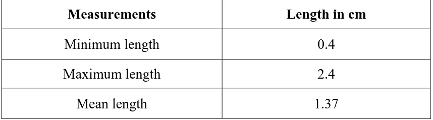
LENGTH OF CA FROM ITS ORIGIN TO EMERGENCE OF
FIRST BRANCH
Length of CA was measured from the point of origin to the point of
[image:71.595.101.530.283.402.2]emergence of first branch. The following were observed.
TABLE 3 : LENGTH OF CA FROM ITS ORIGIN TO EMEREGENCE OF FIRST BRANCH
Measurements Length in cm
Minimum length 0.4
Maximum length 2.4
Mean length 1.37
TABLE 4 : LENGTH OF CA
Length of CA Frequency Percentage
<= 0.5cm 2 4%
0.6 - 1 cm 10 20%
1.1 – 1.5 cm 19 38%
1.6 – 2 cm 15 30%
[image:71.595.100.529.384.705.2]55
CHART 3 : LENGTH OF CA
TYPEOF CA
Among the 50 specimens CA was complete with all the 3 classic
branches (LGA, SA, CHA) arising from it in 39 specimens (Fig4&5) and
had aberrant branches in addition to classic branches in 11 specimens
(Fig7&8).
In dissection done on a three month old infant in the Institute Of
Anatomy, Madras Medical College, the CA was complete. The LGA
originated first followed by bifurcation into CHA and SA (Fig6). 4%
20%
38%
30%
8%
0% 5% 10% 15% 20% 25% 30% 35% 40%
Fig. 5. Coeliac Artery- complete type, classical trifurcation Fig. 4. Coeliac artery- complete type, non classical trifurcation
LIVER
PV C
H A
LG A
A C
A S
LIVER
CHA
LGA
CA
Fig. 6. Infant dissection - Coeliac artery, complete type, non classical trifurcation
Fig. 7. Coeliac artery with aberrant branches- the RIPA and LIPA
LGA
CHA
SA
PV
PANCREAS
CA
LIVER STOMACH
LGA
RIPA
LIPA
CHA
56
TABLE 5 : TYPE OF CA
Type of CA Frequency Percentage
Complete 39 78%
Incomplete - 0%
Absent - 0%
Coeliacomesentric trunk - 0%
Aberrant branches 11 22%
CHART 4 : TYPE OF CA
78%
0% 0% 0%
22%
0% 10% 20% 30% 40% 50% 60% 70% 80% 90%
Complete Incomplete Absent Coeliacomesentric
Fig. 8. Coeliac artery with aberrant branch- dorsal pancreatic artery
Fig. 9. Gastrophrenic trunk from coeliac artery - RIPA and LGA.
LVI ER
H C
A
LG A
SA
CA DPA
PANCREAS
S OM
CH
T A
AA
LIPA
CA RIPA
LGA
57
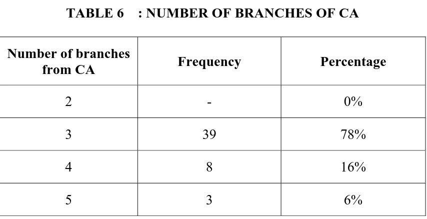
Branching pattern of CA
In 34 specimens the CA bifurcated into hepato splenic trunk with
the LGA arising separately proximal to the bifurcation(Fig4). In 5
specimens the CA trifurcated classically( all the branches arising from
CA at the same point) (Fig5). In 2 specimens CA quadrifurcated into
LGA, SA, CHA and DPA(Fig8). In 1 specimen CA gave rise to a
gastrophrenic trunk and hepatosplenic trunk(Fig9).
In 3 specimens both the LIPA & RIPA arose from CA separately
proximal to origin of LGA(Fig7).
In 5 specimens the RIPA arose from CA proximal to the
[image:77.595.103.530.502.721.2]emergence of its three branches namely LGA, SA, CHA
TABLE 6 : NUMBER OF BRANCHES OF CA
Number of branches
from CA Frequency Percentage
2 - 0%
3 39 78%
4 8 16%
Fig. 10.Left gastric artery arising from coeliac artery
Fig. 11. Splenic artery arising from coeliac artery CH
A SM
A
SA
LG A
CA
IVE
L R
S MA TO
CH LGA
CHA
CA
A S
Fig. 12. Common hepatic artery arising from coeliac artery
Fig. 13. Left hepatic artery arising from proper hepatic artery
LIVER RHA GDA PV HA C LGA SA CA SMA STOMACH
LIVER Le t L
e f ob LHA PH A RHA GDA
CHA LGA
SA
CA
58
CHART 5 : NUMBER OF BRANCHES OF CA
BRANCHES OF CA
LGA
o In all the 50 specimens the LGA arose from CA (Fig10).
SA
o In all the 50 specimens the SA arose from the CA (Fig11).
CHA
o In all the 50 specimens the CHA arose from the CA (Fig12). 0%
78%
16%
6%
0% 10% 20% 30% 40% 50% 60% 70% 80% 90%
Fig. 14.Replaced left hepatic artery arising from left gastric artery
Fig. 15. Accessory left hepatic artery arising from left gastric artery
LIVER
LHA
RHA
CHA
PV GDA
STOMACH
GA L
SA
CA
RHA
LHA
PHA
GDA
A
CH ACCESSORY LHA
CA
SA
Fig. 16. Left hepatic artery arising from common hepatic artery
Fig. 17. Right hepatic artery arising from proper hepatic artery
LIVER LEFT LOBE H L A RHA CYSTIC ARTERY G D A CHA LGA SA STOM ACH LHA PH A RHA GDA
CHA LGA
SA
CA
S OT MACH
59
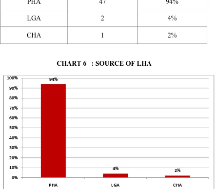
BRANCHES OF CHA
LHA
Among 50 specimens the LHA originated from PHA in 47
specimens (Fig13), from LGA as replaced LHA in 2 specimens (Fig14)
and from CHA in 1 specimen (Fig16). Accessory LHA was observed in 2
[image:83.595.102.545.347.735.2]specimens(Fig15).
TABLE 7 : SOURCE OF LHA
Source of LHA Frequency Percentage
PHA 47 94%
LGA 2 4%
CHA 1 2%
CHART 6 : SOURCE OF LHA
94% 4% 2% 0% 10% 20% 30% 40% 50% 60% 70% 80% 90% 100%
60
RHA
In 49 specimens RHA arose from PHA (Fig17). In the remaining 1
[image:84.595.110.531.255.681.2]specimen it originated from CHA (Fig18).
TABLE 8 : SOURCE OF RHA
Source of RHA Frequency Percentage
PHA 49 98%
CHA 1 2%
CHART 7 : SOURCE OF RHA
98%
2% 0%
20% 40% 60% 80% 100% 120%
PHA CHA
Fig.18.Right hepatic artery arising from common hepatic artery
Fig.19.RIPA and LIPA from coeliac artery as independent branches
A CH
LIVER
RIPA
LI AP
61
ABERRANT BRANCHES
LIPA
In 47 specimens the LIPA arose from AA. In 3 specimens it arose
[image:86.595.107.544.331.687.2]from CA (Fig19).
TABLE 9 : SOURCE OF LIPA
Source of LIPA Frequency Percentage
CA 3 6%
Aorta 47 94%
CHART 8 : SOURCE OF LIPA
6%
94%
0% 10% 20% 30% 40% 50% 60% 70% 80% 90% 100%
Fig. 20.RIPA from coeliac artery and LIPA from aorta
Fig. 21A. Dorsal pancreatic artery arising from coeliac artery
AA
LI AP
CA RIPA
I ER L V
A C
H
LGA
SA
62
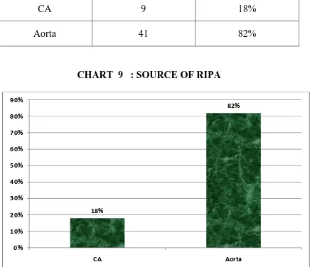
RIPA
Among 50 specimens the RIPA arose from CA in 9 specimens
[image:88.595.106.542.258.634.2](Fig20). It arose from AA in 41 specimens.
TABLE 10 : SOURCE OF RIPA
Source of RIPA Frequency Percentage
CA 9 18%
Aorta 41 82%
CHART 9 : SOURCE OF RIPA
Of the 50 specimens in 3 specimens both the RIPA and LIPA arose
from CA (Fig19), in 6 specimens the RIPA arose from the CA and the
LIPA from the AA (Fig20). 18%
82%
0% 10% 20% 30% 40% 50% 60% 70% 80% 90%
Fig. 21B. Dorsal pancreatic artery arising from coeliac artery
I L VER
CHA
SA
LG A
D PA
LHA
63
DPA
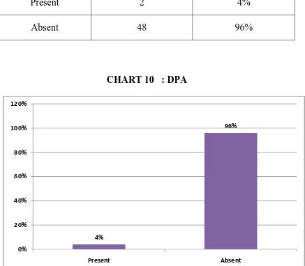
[image:90.595.105.542.235.617.2]In 2 specimens the DPA arose from the CA(Fig21 A&B).
TABLE 11 : DPA
DPA Frequency Percentage
Present 2 4%
Absent 48 96%
CHART 10 : DPA
4%
96%
0% 20% 40% 60% 80% 100% 120%
64
ABERRANT BRANCHES
Among the 11 specimens in which aberrant branches were present,
in 9 specimens inferior phrenic arteries were the aberrant branches and in
[image:91.595.109.533.402.695.2]2 specimens DPA was the aberrant branch.
TABLE 12 : ABERRANT BRANCHES FROM CA
Aberrant branch Frequency Percentage
IPA 9 18%
DPA 2 4%
CHART 11 : ABERRANT BRANCHES FROM CA
18%
4%
0% 2% 4% 6% 8% 10% 12% 14% 16% 18% 20%
Fig. 22.Normal adult CT coeliac angiogram
Fig.23.CT angiogram showing classical trifurcation of coeliac artery A
A
CA LGA
A S CHA
AA
CA LGA
SA CHA
GDA A LH
Fig. 24.Coeliac artery is absent with all its branches arising directly from aorta
Fig. 25.Coeliac artery with aberrant branches - LIPA and RIPA
AA LGA
SA
CHA
AA
CA RIPA
LIPA
LGA
SA
Fig. 26.Coeliac artery with aberrant branch - LIPA
Fig. 27.Coeliac artery with aberrant branch - DPA AA
CA LGA
SA CHA
LIPA
AA
CA
LGA
SA
CHA
Fig. 28.LHA arising from LGA and RHA arising from SMA with absent CHA
Fig. 29.Left hepatic artery arising from common hepatic artery.
AA
CA LGA
SMA
AA
CA LGA
SA
CHA
LHA
GDA RHA
LHA
Fig. 30. LIPA arising from coeliac artery
AA
CA
SA
G L
A
65
RADIOLOGICAL STUDY
Among the 25 adult CT coeliac angiograms CA was complete in
19 cases (76%) (Fig22) with classical trifurcation in 1 case (4%)
(Fig23).
It was absent in 1 case (4%) (Fig24).
It had aberrant branches in 5 cases (20%) (Fig25,26& 27).
LGA originated from AA in 1 case (4%) (Fig24).
SA originated from AA in 1 case (4%) (Fig24).
CHA originated from AA in 1 case (4%) (Fig24).
LHA arising from LGA and RHA arising from SMA with absent
CHA was found in 2 cases (8%) (Fig28).
LHA arising from CHA was found in 1 case (4%) (Fig29).
RIPA and LIPA from CA were noticed in 1 case (4%) (Fig25).
LIPA alone from CA was noticed in 3 cases (12%) (Fig26&30).
66
DISCUSSION
LEVEL OF ORIGIN OF COELIAC ARTERY
J.C. Boileau Grant5 [1958] stated that the AA gives off CA at the level
of T12 vertebra.
Pushpalatha25 [2006] reported that in 6% of the specimens the CA
originated at the level of upper border of T12; in 66% it originated at the
level of lower border of T12 ; in 4% CA originated between T12 and L1; in
24% CA originated at the level of upper border of L1.
Ambica Wadhwa et al2 [2011] reported that the CA originated from AA
at the level of inter vertebral disc between T12 and L1 in 73.3% and upper
one third of L1 vertebra in 26.6%.
Chummy S. Sinnatamby7 [2011] stated that the CA arises from AA at
the level of the body of the twelfth thoracic vertebra.
In the present study in 10% the CA originated at the level of upper
border of T12 . In 64% it originated at the level of lower border of T12; in
6% CA originated between T12 and L1; and in 20% CA originated at the
level of upper border of L1. The observations of the present study were
67
Variation in the level of origin of CA implies that the planning of
surgery must be individualized for carcinoma pancreas, stomach, liver
[image:100.595.109.532.467.747.2]and extrahepatic biliary tree as nodes at risk lie near the artery.
TABLE 13 : LEVEL OF ORIGIN OF COELIAC ARTERY
Level of origin of CA Pushpalatha
[2006] Present study
Upper border of T12 6% 10%
Lower border of T12 66% 64%
Between T12 and L1 4% 6%
Upper border of L1 24% 20%
CHART 12 : LEVEL OF ORIGIN OF COELIAC ARTERY
6% 66% 4% 24% 10% 64% 6% 20% 0% 10% 20% 30% 40% 50% 60% 70% Upper border of T12 Lower border of T12 Between T12 and L1
Upper border of
L1
68
ORIGIN OF COELIAC ARTERY IN RELATION TO MEDIAN
ARCUATE LIGAMENT OF THE DIAPHRAGM
Harold H. Lindner et al10 [1971] reported that in 33% the origin of
coeliac artery was at or above the level of MAL.
Selma Petrella et al34 [2006] reported that in 14.46% the MAL was
distant from the origin of coeliac artery; in 42.17% , the MAL was
touching the origin of CA; and in 43.37% MAL was overlapping the
origin of CA.
Susan Standring39[2008] stated that the origin of CA may be
compressed by the right crux of the diaphragm giving the appearance of a
stricture.
Chummy S. Sinnatamby7 [2011] stated that the CA takes origin from
AA a little below the MAL.
In the present study in 18% the origin of coeliac artery was above
the level of MAL; and in 52% the origin of coeliac artery was at the level
of MAL and in 30% the origin of coeliac artery was below the level of
MAL.
In cases where the CA takes origin above the level of MAL it gets
69
loss of weight, the symptoms of MAL syndrome. These patients are
[image:102.595.110.525.477.761.2]relieved of the symptoms by the surgical division of the MAL.