“COMPARATIVE STUDY ON THE EFFICACY OF
MUSCLE ENERGY TECHNIQUE VERSUS
MAITLAND MOBILIZATION IN TREATING SACRO
ILIAC JOINT DYSFUNCTION”
A Dissertation Submitted to
The Tamilnadu Dr.M.G.R.Medical University,
CHENNAI
In partial fulfillment of the requirements
For the award of the
MASTER OF PHYSIOTHERAPY
(Advanced Physiotherapy in Orthopaedics)
DEGREE
Submitted by
Reg.No.271410062
NANDHA COLLEGE OF PHYSIOTHERAPY
“
COMPARATIVE STUDY ON THE EFFICACY OF
MUSCLE ENERGY TECHNIQUE VERSUS
MAITLAND MOBILIZATION IN TREATING SACRO
ILIAC JOINT DYSFUNCTION”
NANDHA COLLEGE OF PHYSIOTHERAPY
ERODE -638 052
The dissertation entitled
Submitted by
Reg.No.271410062
Under the guidance of
Prof. V.MANIVANNAN.,M.P.T(ORTHO)
A Dissertation Submitted to
The Tamilnadu Dr.M.G.R.Medical University,
CHENNAI
Dissertation Evaluated on ………..
CERTIFICATE BY THE HEAD OF THE INSTITUTION
PROF.V.MANIVANNAN, M.P.T. (ORTHO)
Principal/Head of the Institution,
NANDHA COLLEGE OF PHYSIOTHERAPY,
ERODE -636 052.
This is Certify that
(Reg. No. 271410062)
is a Bonafide student of
Nandha College of Physiotherapy, studying Master of Physiotherapy
(Advanced Physiotheraphy in orthopaedics)
degree course from the year
2014-2016. The dissertation entitled,
“COMPARATIVE STUDY ON THE
EFFICACY OF MUSCLE ENERGY TECHNIQUE VERSUS MAITLAND
MOBILIZATION
IN
TREATING
SACRO
ILIAC
JOINT
DYSFUNCTION”
is a record of original and independent work done by her
under the guidance of me.
I wish him a great success in his dissertation work.
Place :
Signature of Principal/HOD
CERTIFICATE BY THE GUIDE
This is to certify that this dissertation entitled, “
COMPARATIVE
STUDY ON THE EFFICACY OF MUSCLE ENERGY TECHNIQUE
VERSUS MAITLAND MOBILIZATION IN TREATING SACRO ILIAC
JOINT DYSFUNCTION”
submitted by (Reg.No.271410062) is a record of
original and independent work done by the candidate during the period of study
under my supervision and guidance. The dissertation represents entirely and
independent work on the part of the candidate but for the general guidance by
me.
Place :
Date:
Signature of Guide
DECLARATION
I here, by declare and present my project work entitled
“COMPARATIVE STUDY ON THE EFFICACY OF MUSCLE
ENERGY TECHNIQUE VERSUS MAITLAND MOBILIZATION
IN TREATING SACRO ILIAC JOINT DYSFUNCTION”
is outcome
of original research work was under taken and carried out by me under
the guidance of Prof.
.V.MANIVANNAN.,M.P.T.,
To the best of my knowledge this dissertation has not been formed
in any other basic for the award of any other degree, diploma,
associateship, fellowship, previously form, any other medical university.
ACKNOWLEDGEMENT
First and foremost thank to the lord almighty for bestowing me with knowledge
and lige that has enabled me to begin and complete this project successfully.
It is impossible to do a project without involvement of various experts and
other concerned people. So, I use this incredible moment to thank everyone who used
to strongly influence and facilitate me towards selection, progression and successful
completion of this project work.
My sincere thanks to our principal and guide,
Prof.V.Manivannan
MPT(Ortho),
for supporting me and who was instrumental in bringing out if this
project. He constantly encouraged me to give my best
My hearty thanks to my research advisor
Prof. K. Saranya Devi MPT
(Ortho),
Assistant professor,
Prof. V. Vijayaraj MPT (Neuro),
Nandha College of
Physiotherapy who moulds me an effective person by his encouragement and
enormous support.
I am grateful to all our
Professors of Nandha College of Physiotherapy,
for
their valuable inputs and advice they gave me regarding the various aspects of
application of this technique.
I also thank
Prof. K. Dhanapal
, M.Sc., who scrutinized and rectified the
study’s blue print and assisted in the analytical aspects of this research work.
I thank all my Staff Members, My Seniors, My Friends who helped in
compiling the data, manuscripts, typing, etc.,
TABLE OF CONTENTS
CHAPTERS CONTENTS PAGE NO
1 INTRODUCTION 1
1.1 Aim of the study 3
1.2 Objectives 4
1.3 Hypothesis 4
2 REVIEW OF LITERATURE 5 3 MATERIALS AND METHODOLOGY 13
3.1 Materials 13
3.2 Study design 13
3.3 Study setting 13
3.4 Study Sampling 13
3.5 Study duration 14
3.6 Inclusion criteria 14
3.7 Exclusion criteria 14
3.8 Parameters 15
3.9 Procedure 15
3.10 Statistical tools 16
4 DATA PRESENTATION 18 5 DATA ANALYSIS AND INTERPRETATION 19
6 DISCUSSION 27
7 SUMMARY AND CONCLUSION 31 8 RECOMMENDATIONS 33
9 BIBLIOGRAPHY 34
REFERENCES 35
APPENDIX 36
Informed Consent 36
Assesment Chart 37
Parameters 38
LIST OF TABLES
Table I : Data Presentation
18
Table II : Oswestry Disability Index for Group A
19
Table III : Oswestry Disability Index for Group B
20
Table IV : Oswestry Disability Index for Group A & Group B
[image:8.595.100.545.494.752.2]21
Table V : Visual analogue Scale For Group A
23
Table VI : Visual analogue Scale For Group B
24
Table VII : Visual analogue Scale For Group A & Group B
25
LIST OF GRAPHS
Graph I : Oswestry Disability Index for Group A
19
Graph II : Oswestry Disability Index for Group B
20
Graph III : Oswestry Disability Index for Group A & Group B
21
Graph IV : Visual analogue Scale For Group A
22
Graph V : Visual analogue Scale For Group B
25
1.
INTRODUCTION
Sacroiliac joint dysfunction is a condition in which the joint is locked,
Partially dislocated (or) subluxated in a non-anatomical position due to hyper
Mobility (or) hypo mobility within the joint.
Dysfunction of sacroiliac joint may causes low back (or) leg pain.
Sacroiliac Joint dysfunction can mimic the pain caused by a number of others
spinal structures. Lumbar disc, nerve root, facet joint. The pain is typically felt
on one side of buttocks, and can radiate down to the leg.
The pain usually remains above the knee but at times pain can extend to
the ankle or foot. It is reported that it 15% to 38% of general population with
women being 3 (or) 4 times more likely to be affected than men.
There are many different causes of sacroiliac joint pain, pregnancy may
be a factor in developing of sacroiliac joint dysfunction. Also it is a lesion has
one leg is shorter than the other leg. The abnormal alignment may end up
The most important role is played by the physiotherapist, in which
Physiotherapy reduces pain and improves the joint range of motion and muscle
Power.
Many physiotherapeutic interventions are done which include exercise
therapy and electro therapy. Electro therapy includes paraffin wax path.
Exercise therapy includes active and passive movements, soft tissue stretch and
isometric stabilizing exercise, general grip strengthening exercise and passive
joint mobilization technique.
Muscle Energy Technique t is a type of osteopathic manipulative
treatment in which patient's use their muscles actively upon request. It is used to
treat somatic dysfunction, decreased ROM, muscular hypertonicity, muscular
spasm, and pain. It’s also engages and regulates sensorimotor impulses and any
musculature that moves a particular body joint. MET is most effective for the
mobilization of joints, correction of postural and movement asymmetries,
stretching of muscles and reduction of pain.
The Maitland technique is a passive skilled manual therapy technique
Applied to joint and related soft tissue at varying speeds and amplitudes, using
Mobilization provides strong inhibitory stimulus through large afferent
myelinated fibers of the dorsal horn to block the small diameter of nociceptive,
Input through a pain gate mechanism and enhance the physiological
movements.
The study aims to compare study between Muscle Energy Technique
versus Maitland’s mobilization in treating patients with sacroiliac joint
dysfunction.
1.1.
AIM OF THE STUDY
The aim of the study was to compare the efficacy of Muscle
Energy Technique versus Maitland’s mobilization in treating patients with
1.2.
OBJECTIVES OF STUDY
1.
To determine the efficacy of the muscle energy technique.
2. To determine the efficacy of Maitland’s mobilization.
3. To determine the difference between the efficacy of Muscle Energy
Technique versus Maitland’s Mobilization in treating patients with
sacroiliac joint Dysfunction.
1.3
HYPOTHESIS
NULL HYPOTHESIS
The null hypothesis states that there was no significant difference
between the efficacy of muscle energy technique versus Maitland’s mobilization
in treating patients with sacroiliac joint dysfunction.
ALTERNATIVE HYPOTHESIS
The alternative hypothesis states that there was significant difference
Between the efficacy of muscle energy technique, versus Maitland’s
2.
REVIEW OF LITERATURE
1)
Rana Kanchan and Bansal Nitesh (2009)
Conducted an experimental study on 45 subjects with low back pain. The aim of
the study was to compare the effects of muscle energy technique and Maitland’s
mobilization along with exercise. The main outcome range of oswestry disability
index and visual analogue scale were measured. The result of the study conclude that
the active exercise muscle energy technique (MET) is moderately significant over the
G.D Maitland’s technique of mobilization in improving functional ability and
increased the medial rotation of hip joint in mechanical chronic low back pain caused
by sacroiliac joint dysfunction.
2)
Dhinkaran Mullai and Sareen Aarti (2011)
Conducted an experimental study on 30 subjects with scaroiliac joint dysfunction.
The aim of the study compare the effects of muscle energy technique with
conventional therapy. The main outcome range of Oswestry disability index (ODI)
and numeric pain rating scale (NPRS) were measured. The result of the Study
conducted. The average of the Oswestry disability index (1%) relief decrease for
Group A is 27.15% and Group B it is 19.67% and the average of numeric pain rating
scale relief for pain Group A is 3.40 and for Group B is 2.60 so the result of the study
showed that along with corrective exercises. MET is moderately significant over
3)
Karen L Lenehan Bsc, Gary Fryer BappSC (2005)
Conducted an experimental study on 59 subjects with sacroiliac joint dysfunction.
The purpose of study was evaluate the effect of the muscle energy technique. The
result of the study concluded that the muscle energy technique is restricts rotation of
thoracic spine and increases the range of active trunk rotation.
4)
Niemisto, Leena MD and Lahtinen-suopanki
Conducted a randomized prospective trial on 204 patients with low back ache.
The aim of the study was to find out the combined effectiveness of combined
manipulation treatments and stabilizing exercise and physician consultation. The main
outcome was reduction of the pain and disability. The study concluded that
manipulation with stabilizing exercise was more effective in reducing pain, Intensity
and disability.
5)
Scrimshaw S.R. et.,al (2001)
Conducted a comparative study between responsiveness of visual analogue Scale
and McGill pain questionnaire. It was measured in 75 patients and conclude. That
VAS was a better tool than the McGill pain questionaires for measuring pain In
clinical practice. “Journal of manipulative physical therapy”, vol 24
6)
George Lewith, et.al.,
Conducted an experimental study on 36 subjects with low back pain and
They were randomly divided into 2 groups. One group received manipulation
therapy, another group received standard physical therapy (heat modalities and
exercise). The visual analogue scale was used for evaluating the pain. The result of
the study showed that there was as significant reduction of pain intensity and
improvement in range of motion in the manipulative therapy group compared to
standard physical therapy group.
7)
O’SULLIVAN, PETTER.B.,et.al., (1997)
Conducted a randomized, controlled study on 44 patients, with chronic low Back
pain and they were divided into two groups randomly. One group received 10 week
specific spinal exercise program, another group received conventional Therapy. The
study showed that there was a statistically significant reduction of Pain intensity and
8)
FOWLER.B., et.al., (1995)
Conducted an experimental study on 23 patients with mechnaical low back pain.
They were treated with spinal manipulative therapy for a period of 7-10 days. The
Study explained that 18 patients showed that definite clinical improvements with
reduction in low back pain. The study concluded that the mechanical low back pain
could be effectively treated by spinal manipulative therapy.
9)
LEON CHAIT N.D (2007)
Conducted a experimental study with pelvic pain and dysfunction. The aim of the
study was to compare the effects of physiotherapy approach and manual therapy. The
main outcome were range of active and pain were measured. The result of the study
conducted that physiotherapy approach and manual therapy techniques increases the
range of motion and decrease the pain level.
10)
Ulrika Holmgren & Kerstin Waling (2007)
Conducted an experimental study on 25 patients with low back pain, purpose of
the study was to evaluate the effects of muscle energy technique application were
11)
Fairbank, Jeremy C. T. MD, FRCS*; Pynsent, Paul B. PhD† 2000
Conducted a systematic review of the Oswestry Disability Index (ODI) . they
found that the ODI remains a valid and vigorous measure and has been a worthwhile
outcome measure after comparing 200 citations with at least 114 studies that contain
usable data. These data provide both validation and standards for other users and indicate
the power of the instrument for detecting change in sample populations.
12)
PE Bijur, W Silver, EJ Gallagher - Academic emergency …, 2001
Conducted a study to assess the reliability of the VAS for measurement of acute
pain. The paired measurements were more reproducible at the extremes of pain intensity
than at moderate levels of pain. The Reliability of the VAS for acute pain measurement
as assessed by the ICC appears to be high. Ninety percent of the pain ratings were
reproducible within 9 mm. These data suggest that the VAS is sufficiently reliable to be
13)
Franke H, Fryer G, Ostelo RW, Kamper SJ. (2015)
Low-back pain (LBP) is responsible for considerable personal suffering due
to pain and reduced function, as well as the societal burden due to costs of health care and
lost work productivity. For the vast majority of people with LBP, no specific anatomical
cause can be reliably identified. For these people with non-specific LBP there are
numerous treatment options, few of which have been shown to be effective in
reducingpain and disability. The muscle energy technique (MET) is a
treatment technique used predominantly by osteopaths, physiotherapists and chiropractors
which involves alternating periods of resisted muscle contractions and assisted stretching.
14)
Day JM, Nitz AJ. (2012)
Low back pain is the most common type of pain reported by adults in the United
States. A variety of manual therapy techniques are used in the management of low back
pain to reduce pain, improve function, and reduce disability. In recent
years, muscle energy techniques have been increasingly used in clinics to treat low back
15)
Richardson CA, Snijders CJ, Hides JA, Damen L, Pas MS, Storm J. (2002)
Contraction of the transversus abdominis significantly decreases the laxity of the
sacroiliac joint. This decrease in laxity is larger than that caused by a bracing action using
all the lateral abdominal muscles. These findings are in line with the authors'
biomechanical model predictions and support the use of independent transversus
abdominis contractions for the treatment of low back pain.
16)
George SZ, Wittmer VT, Fillingim RB, Robinson ME. (2010)
Physical therapy supplemented with graded exercise or graded exposure resulted
in equivalent clinical outcomes for pain intensity and disability. The overall treatment
effects were modest in this setting. Instead of being associated with a specific behavioral
intervention, reductions in pain and disability were associated with reductions in
depressive symptoms and pain catastrophizing, respectively.
17)
Hurley DA, McDonough SM, Baxter GD, Dempster M, Moore AP. (2005)
The use of mobilization techniques within the trial was comparable with their
usage by the general population of physiotherapists in Britain and Ireland for LBP
management. However, the usage of manipulation techniques was considerably higher
than reported in physiotherapy surveys and may reflect the postgraduate training of trial
18)
Meade TW, Dyer S, Browne W, Townsend J, Frank AO. (1991)
In this study, patients with low back pain in whom manipulation is not
contraindicated, chiropractic almost certainly confers worthwhile, long-term benefit in
comparison with hospital outpatient management. The benefit is seen mainly in those
with chronic or severe pain. Introducing chiropractic into NHS practice should be
3.
MATERIALS AND METHODOLOGY
3.1
MATERIALS
Couch
Pillows
Towel
Back rest chair
Visual analogue scale
Oswestry disability index
METHODOLOGY
3.2 STUDY DESIGN
Quasi experimental study with Pre Versus Post test design
3.3 STUDY SETTING
Nandha College of Physiotherapy-Erode
Government headquarters hospital-Erode
LKM Hospital, Erode.
SIMS Hospital, Erode.
3.4 STUDY SAMPLING
A Total number of 30 subjects were selected by convenient sampling Method
after giving due consideration to inclusion and exclusion criteria and they
3.5 STUDY DURATION
The Study was conducted for a period of 4 weeks.
3.6 INCLUSION CRITERIA
Age group 30-45 Years.
Sex – Both male and female
Sacroiliac dysfunction patients
3.7 EXCLUSION CRITERIA
Pregnancy
IVDP
Fracture
Spondylolisthesis
Associated cardiovascular disease
Sacroiliac tumors
Osteoporosis
Spinal stenosis
Myofascial syndrome
Piriformis syndrome
Gout
3.8 PARAMETERS
Visual Analogue Scale (VAS)
Visual Analogue Scale was used to measure the severity of pain response that
the patient experience. It is a 10 cm horizontal line with two ends labeled, no pain (0)
at one end and severe pain (10) at other end. The pain marked on the line which
corresponds to severity of pain patients experienced.
Oswestry Disability Index
Functional ability was measured by using Oswestry Disability Index which
consist of 10 sections with 6 statements contained in each section. The patients were
made to mark on the one statement in each section which described their limitation
most accurately.
3.9 PROCEDURES
A total number of thirty subjects who met inclusion criteria and exclusion
criteria were recruited. After informed consent was obtained, they were divided into
two groups A and B with subjects 15 each.
Prior to the treatment, pre test were conducted for Group A and Group B with
Visual Analogue Scale for pain, Oswestry, Disability Index for functional ability.
After a brief demonstration about muscle energy technique, Group A were
treated with the same for a period of 4 weeks.
The post test were conducted for Group A and Group B by Visual Analogue
Scale for pain, Oswestry Disability Index for functional ability.
Pre and Post results were recorded and analyzed.
3.10 STATISTICAL TOOLS
The collected data was subjected to statistical analysis using paired and
unpaired “t” test to find out the research effectiveness.
PAIRED –“t”-TEST
The paired “t” test was used to compare the pre and post test values of
Visual Analogue Scale for pain, Oswestry Disability Index for functional ability.
FORMULA: Paired “t” test
𝑆 = 𝑑
2−( 𝑑)2 𝑛
𝑛−1
𝑡 = 𝑑 𝑛𝑆
d = Difference between the pre test versus post test
𝑑 = Mean difference
n = Total number of subjects
UNPAIRED “t” – TEST
The unpaired “t” test was used to compare the mean difference between Group A
and Group B subjects treated with Muscle Energy Technique, and Maitland’s
Mobilization.
FORMULA :- Unpaired “t” – Test
𝑆 = 𝑛1− 1 𝑆12+ 𝑛2− 1 𝑆22 𝑛1+ 𝑛2− 2
𝑡 = 𝑋 − 𝑋1 2 𝑆 𝑛1
1 + 1 𝑛2
𝑛1 = Total number of subject in Group - A
𝑛2 = Total number of subject in Group – B
𝑋1 = Difference between pre test versus post test of Group – A
𝑋1
= Mean difference between pre test versus post text of Group – A
𝑋2 = Difference between pre test versus post text of Group – B
𝑋2
= Mean difference between pre test versus post text of Group – B
4.
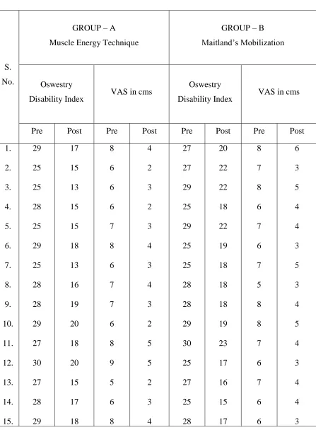
DATA PRESENTATION
TABLE – I
S.
No.
GROUP – A
Muscle Energy Technique
GROUP – B Maitland’s Mobilization
Oswestry
Disability Index VAS in cms
Oswestry
Disability Index VAS in cms
Pre Post Pre Post Pre Post Pre Post
5. DATA ANALYSIS AND INTERPRETATION
This section deals with analysis and interpretation of data’s from pre and Post
test result of Group A and Group B
TABLE – II Group – A
The comparative main value, mean difference, standard deviation and Paired
t – value between pre Vs post test of Oswestry Disability Index in Group A.
S. No Test Mean
Mean Difference
S.D
Paired t - value
1.
2.
Pre – Test
Post – Test
27.5
16.6
10.9 1.3 32.7
The paired “t” value of 32.7 was greater than the tabulated t – value of 2.14
which showed that there was a statistically significant difference at 0.05 levels
between pre Vs post test results. The pre test mean was 27.5, post test mean was 16.6
and mean difference was 10.9, which showed that there was reduction of disability
TABLE – III Group – B
The comparative main value, mean difference, standard deviation and
Paired t – value between pre Vs post test of Oswestry Disability Index in Group B.
S.No Test Mean
Mean Difference
S.D
Paired t - value
1.
2.
Pre – Test
Post – Test
27.1
18.9
8.2 1.9 16.9
The paired “t” value of 16.8 was greater than the tabulated t – value of 2.14
which showed that there was a statistically significant difference at 0.05 levels
between pre Vs post test results. The pre test mean was 27.1, post test mean was 18.9
and mean diffrence was 8.2, which showed that there was reduction of disability level
TABLE – IV
The comparative main value, mean difference, standard deviation and Paired t
– value of Oswestry Disability Index between Group A and Group B.
S.No Test Mean
Mean Difference
S.D
Unpaired t - value
1.
2.
Group - A
Group - B
10.9
8.2
2.7 1.6 4.5
0 2 4 6 8 10 12
Mean differnce Standard deviation
Group A
The unpaired “t” value of 4.5 was greater than the tabulated t – value of 2.5
which showed that there was a statistically significant difference at 0.05 levels
between mean difference of Group A and Group B. The pre Vs post mean of group
A was 10.9, the pre Vs post mean of Group B was 8.2 and mean difference group A
and Group B was 2.7 which showed that there was reduction of disability level in
response to treatment of Group A when Compared to Group B.
Therefore, the study was rejecting the null hypothesis and accepting the alternate hypothesis.
0 5 10 15 20 25 30 35
PAIRED't'VALUE UNPAIRED't'VALUE
GROUP-A
GROUP-B
TABLE – V
Group – A
The comparative main value, mean difference, standard deviation and Paired
t – value between pre Vs post test of Visual Analogue scale in Group A.
S.No Test Mean
Mean Difference
S.D
Paired t - value
1.
2.
Pre – Test
Post – Test
6.9
3.3
3.6 0.5 28.08
The paired “t” value of 28.08 was greater than the tabulated t – value of 2.14
which showed that there was a statistically significant difference at 0.05 levels
between pre Vs post test results. The pre test mean was 6.9, post test mean was 3.3
and mean difference was 3.6, which showed that there was reduction of pain due to
the combined effect of Muscle Energy Technique
TABLE – VI Group – B
The comparative main value, mean difference, standard deviation and Paired t – value
between pre Vs post test of Visual Analogue scale in Group B.
S.No Test Mean
Mean Difference
S.D
Paired t - value
1.
2.
Pre – Test
Post – Test
6.8
4.0
2.8 0.7 15.6
The paired “t” value of 15.6 was greater than the tabulated t – value of 2.14 which
showed that there was a statistically significant difference at 0.05 levels between pre Vs post
test results. The pre test mean was 6.8, post test mean was 4.0 and mean difference was 2.8,
TABLE – VII
The comparative main value, mean difference, standard deviation and Paired
t – value of Visual Analogue scale between Group A and Group B.
S.No Test Mean
Mean Difference
S.D
Unpaired t - value
1.
2.
Group - A
Group - B
3.6
2.8
0.8 0.6 4
0 0.5 1 1.5 2 2.5 3 3.5 4
MEAN DIFFERENCE STANDARD DEVIATION
GROUP-A
The unpaired “t” value of 4.0 was greater than the tabulated t – value of 2.05
which showed that there was a statistically significant difference at 0.05 levels
between Group A and Group B. The pre test mean of Group A was 3.6, the post test
mean was 2.8 and mean difference of Group A and Group B was 0.8 which showed
that significant reduction of pain in response to treatment of Group A when Compared
to Group B.
Therefore, the study was rejecting the null hypothesis and accepting the alternate hypothesis.
0 5 10 15 20 25 30
PAIRED't'VALUE UNPAIRED't'VALUE
GROUP-A
GROUP-B
6. DISCUSSION
The aim of the study was to compare study between Muscle Energy
Technique versus Maitland’s mobilization in treating patients with sacroiliac joint
dysfunction.
George Lewith, et.al.,
The result of the study showed that VAS can be used as parameter to
quantify the pain intensity and there was decreased pain threshold in patients with
sacroiliac joint dysfunction.
Re Erahard, et.al.,
The result of the study showed that Oswestry Disability Index can be used
as a parameter to quantify the functional ability and there was decreased disability
level in patients with sacroiliac joint dysfunction.
Based on the results of the above studies Visual Analogue Scale and
Oswestry Disability Index were taken as a parameter in the present study.
In the analysis and interpretation of Oswestry Disability Index in Group A
The paired “t” value of 32.7 was greater than the tabulated t – value of 2.14
which showed that there was a statistically significant difference at 0.05 levels
between pre Vs post test results. The pre test mean was 27.5, post test mean was
16.6 and mean difference was 10.9, which showed that there was statistically
In the analysis and interpretation of Visual Analogue Scale in Group A
The paired “t” value of 28.08 was greater than the tabulated t – value of 2.14
which showed that there was a statistically significant difference at 0.05 levels
between pre Vs post test results. The pre test mean was 6.9, post test mean was
3.3 and mean difference was 3.6, which showed that there was reduction of pain
Due to the effect of Muscle Energy Technique.
.
In the analysis and interpretation of Oswestry Disability Index in Group B
The paired “t” value of 16.8 was greater than the tabulated t – value of 2.14
which showed that there was a statistically significant difference at 0.05 levels
between pre Vs post test results. The pre test mean was 27.1, post test mean was
18.9 and mean difference was 8.2, which showed that there was reduction of
disability level in response to effect of Maitland’s mobilization.
In the analysis and interpretation of Visual Analogue Scale in Group B
The paired “t” value of 15.6 was greater than the tabulated t – value of 2.14
which showed that there was a statistically significant difference at 0.05 levels
between pre Vs post test results. The pre test mean was 6.8, post test mean was
4.0 and mean difference was 2.8, which showed that there was reduction of pain in
IN THE COMPARISON OF GROUP A AND GROUP B
In the analysis and interpretation of Oswestry Disability Index between Group A and
Group B
The unpaired “t” value of 4.5 was greater than the tabulated t – value of 2.5
which showed that there was a statistically significant difference at 0.05 levels
between mean difference of Group A and Group B. The pre Vs post mean of
group A was 10.9, the pre Vs post mean of Group B was 8.2 and mean difference
group A and Group B was 2.7 which showed that there was statistically significant
reduction of disability level in response to treatment of Group A when Compared
to Group B.
In the analysis and interpretation of Visual Analogue Scale between Group A
and Group B
The unpaired “t” value of 4.0 was greater than the tabulated t – value of 2.05
which showed that there was a statistically significant difference at 0.05 levels
between Group A and Group B. The pre test mean of Group A was 3.6, the post
test mean was 2.8 and mean difference of Group A and Group B was 0.8 which
showed that significant reduction of pain in response to treatment of Group A
when Compared to Group B.
Based on the statistical analysis and interpretation of the results, the present
Study showed that there was significant improvement in disability level and
POSSIBLE MECHANISM OF PAIN RELIEF BY MUSCLE ENERGY
TECHNIQUE
There is viscoelastic change in muscle which in increases the muscle flexibiliaty after
MET
When the muscle contrats isometrically from lengthened position, it induces the
stretching of the connective tissue elements, this produce increase in range of motion.
Increase in flexibility leads to increase in tolerance to stretch.
After following MET, there is significant increase in joint angle due to a change in
tissue property.
Restoration of the motion to the articulation.
Results in a gapping, or resulting of the distorted joint relations with reflex relaxation
of the previously hypertonic musculature.
When gentle contraction is initiated in the agonist muscle, there is a reflex relaxation
of that muscle antagonist group.
It also engages and regulates sensorimotor impulses and any musculature that moves a
particular body joint. it uses three dimensional positioning of joints followed by an
isometric contraction, engaging the golgi tendon organ to allow for inhibition of
agonist muscles.
Increases ability to perform movement tasks.
Improves joint integrity and mobility.
Improves motor function.
7. SUMMARY
The aim of the study was to compare the effect of muscle energy technique
versus Maitland’s mobilization in treating patients with sacroiliac joint dysfunction.
A total number of 30 subjects with sacroiliac joint dysfunction were selected
by convenient sampling method after considering the inclusion and exclusion
criteria.
Oswestry Disability Index and Visual Analogue Scale were taken as
parameter, pre test data were collected for Group A and Group B and computed.
Group A subjects were subjected to muscle energy technique for a period of 4
weeks. Group B subjects were subjected to Maitland’s mobilization for a period of 4
weeks. The paired “t” test was used to compare the per Vs post test result of Group A
and Group B separately. The unpaired “t” test was used to compare the mean
difference of Group A and Group B.
In the analysis and interpretation of Oswestry Disability Index, the unpaired
“t” value of 4.5 was greater than the tabulated t – value of 2.05 at 0.05 level, which
showed that there was statistically significant difference between Pre Vs Post tests
results of Group A Group B. The mean value of Group A was 10.9, Group B was
8.2 and mean difference of Group A and Group B was 2.7 which showed that there
was statistically significant reduction of disability in response to treatment in
In the analysis and interpretation of Visual Analogue Scale for pain, the
unpaired “t” value of 4.0 was greater than the tabulated t – value of 2.05 at 0.05
level, which showed that there was statistically significant difference between
Group A and Group B. The mean value of Group A was 3.6, Group B was 2.8 and
the mean difference was 0.8, which showed that there was significant reduction of
pain in Group A than Group B in response to treatment.
CONCLUSION
Based on these results, this study concluded that Muscle Energy Technique
was effective in reduction of pain and disability in patients with sacroiliac joint
8. RECOMMENDATION
Similar study can be conducted using large samples.
McGill pain questionnaire can be used as a parameter in similar studies.
Similar studies can be conducted to measure the lumbar spine flexibility using
Schober’s method.
Similar study can be conducted by comparing Maitland mobilization with other
physiotherapy modalities.
Similar study can be conducted to find out the combined effect of Maitland
mobilization and conventional therapy.
Similar study can be conducted to find out the combined effect Mulligan and
9.
BIBLIOGRAPHY
1. Brain R.Mulligan, “Manual Therapy”, 3rd Ed., 1996.
2. Boyling.O., Jeffrey, “ Grieve’s Modern Manual Therapy – The Vertebral
Colimn”, Churchill Livingstone, 2nd Ed., 1995.
3. Brent Brotzmzn.S., “Clinical Orthopedic Rehabilitation”, Mospy Publication, 1996.
4. Carolyn Kisner, “Therapeutic Exercise – Indian”, 5th Ed.,
5. Kothari. C.R., “Research Methodology: Method and Techniques”, New Age International Ltd., New Delhi, 2nd Ed., 1990.
6. Magee.J., David, “ Orthopedic Physical Assessment”, 2nd Ed., 1992.
7. Mahajan.B.K., “ Methods is Biostatics for Medical Students and Research
Workers”. Jaypee Brothers, New Delhi, 5th Ed., 1989.
8. Natarajan.S., “Text Book of Orthopedics and Traumatology”, 6th Ed.,
9. Jayant Joshi, “ Essentials of Orthopedics and Applied Physiotherapy”, Indian Edition.
10.Carrier M.Hall, Lori Thein Broady, “ Therapeutic Exercise – Moving Towards
REFERANCES
1. Vincenzino.B, Paungmali.A., Teys.P., “Mulligan’s Mobilization with movements,
positional faults and pain relief; Current concepts from a critical view of literature”.
2. Peter konrad, Klaus Schmitz, Achim Denner, “ Neuromuscular evaluation of
trunk-training exercise”.
3. Erhaed.R.E., Delitto.A., Cibulka.M.T., “ Relative effective of an extension program
and a combined porgram of manipulation and flexion and extension exercises in patients with acute low back syndrome”.
4. Elnaggar.I.M., et.al., “ Effects of sinal flexion and extension exercises on low back
pain and spinal mobility in chronic low back pain patients”.
5. Karst.G.M., Williett.G.M., “Effects of specific instructions on abdominal muscle
activity during trunk curl exercises”.
6. Nachemson.A.L., “The lumbars spine: An Orthopedic challenge”, Spine, 1976, 1”Pp:59-71.
7. Parfrer.K.C., Docherty.D.,et.al., “The effects of different sit and curl-up positions
on activation of abdominal and hip flexor musculature”.
8. Eugene J.Carragee and Mthew Hannibal, “Diagnostic evaluation of low back pain”, Orthopedic clinics of North America, Jan’2004,35:P:7-16.
9. Aure, Olav Frode.P.T., “Manual therapy and exercise therapy in patients with
chronic low back pain”.
10.Andrzej Szczgiel, et.,al., “ Effectiveness of brain’s Mulligan’s manual therapy
APPENDIX
INFORMED CONSENT TO VOLUNTARY PARTICIPATE IN A RESEARCH INVESTIGATION
Department of Physical Therapy
Nandha College of Physiotherapy
Erode – 638 052, Tamil Nadu.
Name :
Age :
Sex :
Occupation :
Address for communication :
Declaration
I have fully understood the nature the purpose of the study. I accept to be a
Subject in this study. I declare that the above information is true to my knowledge.
Signature of the subject
Date :
ASSESSMENT CHART
Name :
Age :
Sex :
Occupation :
Address for communication :
Chief complaints :
Pain assessment :
Mode of treatment : Muscle Energy Technique / Maitland’s Mobilisation
Measurements
Parameters Before Treatment After Treatment
Oswestry Disability
Index
Visual Analogue Scale
for pain
PARAMETERS
VISUAL ANALOGUE SCALE
It is a assessment scale used to measure the intensity of pain response that
the patients experience.
It consists of 10 cm horizontal line with 2 ends labeled no pain “0” and sever
pain “10”. The patient on the while corresponds to severity of pain the patients
experience.
|–––––––––––––––––––––––––––––––––––––––––––|
0 10
No Pain Severe Pain
OSWESTRY DISABILITY INDEX
SECTION 1 – PAIN INTENSITY
The pain comes and goes and it is very mild.
The pain is mild and does not very much.
The pain comes and goes and is moderate.
The pain is moderate and does not very much.
The pain comes and goes and is very severe.
SECTION 2 – PERSONAL CARE
I would no have to change my way of washing or dressing in order to avoid pain
I do not normally change my way of washing or dressing even though it causes some
pain.
Washing and dressing increases the pain, but i manage not to change my way of doing
it.
Washing and dressing increases the pain and i find necessary to change my way of
doing it.
Because of pain, I am unable to do some washing and dressing without help.
Because of pain, I am unable to do any washing and dressing wihout help
SECTION 3 – LIFITING
I can lift heavy weights without extra pain.
I can lift heavy weights, but it causes extra pain.
Pain prevents me from lifting heavy weights off the floor. But I manage if they are
conveniently position (e.g., on a table)
Pain prevent me from lifting heavy weights off the floor.
Pain prevents me from lifting heavy weights, but I can manage light to medium
weights if they are conveniently positioned.
I can only lift very light weight at the most.
SECTION 4 – WALKING
I have no pain on walking.
I have some pain on walking, but it does not increase with distance.
I cannot walk more than 1 mile without increasing pain.
I cannot walk more than ½ mile without increasing pain.
I cannot walk more than ¼ mile without increasing pain.
I cannot walk at all without increasing pain.
SECTION 5 – SITTING
I can sit any chair as long as I like.
I can only sit in my favorite chair as long as I like.
Pain prevents me from sitting more than 1 hour.
Pain prevents me from sitting more than ½ hour.
Pain prevents me from sitting more than 10 mins.
I avoid sitting because it increases pain right away.
SECTION 6 – STANDING
I can stand as long as I want without pain.
I have some pain on standing, but it does not increase with time.
I cannot stand for a longer than 1 hour without increasing.
I cannot stand for a longer than ½ hour without increasing.
I cannot stand for a longer than 10 mins without increasing.
SECTION 7 – SLEEPING
I get no pain in bed.
I get pain in bed, but it does no prevent me from sleeping well.
Because of pain, my normal night’s sleep is reduced by less than ¼.
Because of pain, my normal night’s sleep is reduced by less than ½ .
Because of pain, my normal night’s sleep is reduced by less than ¾ .
Pain prevents me from sleeping at all.
SECTION 8 – SOCIAL LIFE
My social life is normal and gives me no pain.
My Social life is normal, but increases the degree of pain.
Pain has no significant effect on my social life apart from limiting my more energetic
interests, e.g., dancing etc.,
Pain has restricted my social life and I do not go out very often.
Pain has restricted my social life to my home.
SECTION 9 – TRAVELLING
I get no pain while traveling.
I get some pain while traveling, but none of my usual forms of travel makes it any
worse.
I get extra pain while traveling, but it does not compel me to seek alternative forms of
travel.
I get extra pain while traveling, which compel me to seek alternative forms of travel.
Pain restricts all forms of travel.
Pain prevents all forms of travel except that done lying down.
SECTION 10 – CHANGING DEGREE OF PAIN
My pain is rapidly getting better.
My pain fluctuates, but is definitively getting better.
My pain seems to be getting better, but improvement.
My pain is slow at present.
My pain is neither getting better nor worse.
My pain is gradually worsening.
TREATMENT PROCEDURE
MUSCLE ENERGY TECHNIQUE FOR SACROILIAC JOINT
DYSFUNCTION
I. Iliac inflare
II. Iliac outflare
III. Anterior iliac rotation
IV. Posterior iliac rotation
a)
ILIAC INFLARE
Patient Position
Supine Lying
Therapist Position
Therapist stands on the same side of the problem facing leg side.
Hand Placement
Cephalad hand stabilizing non affected side ASIS, caudal hand holding the
ankle of the affected side.
Starting Position
Flexion abduction and full external rotation of hip holding the leg on un
affected knee.
Ending Position
Abduction of hip against the resistance of restraining arm for 10 secs while
b) ILIAC OUTFLARE
Patient Position
Supine Lying
Therapist Position
Stands same side dysfunction ilium facing towards the body.
Hand Placement
Supinated cephalad hand place under the patients buttock with fingr tip
Hooked into sacral sulcus of the affected side.
Caudal hand hold the patients foot and the feature side with the forearm
resting along medial calf bar shin area of the hand grasp the floor.
Starting Position
Hip on affected side is fully flexed adducted and internally rotated.
Ending Position
Abduction against the resistance with 50% of the strength for 10 secs while
c) ANTERIOR ILIAC ROTATION
Patient position
Prone Lying
Therapist Position
Stands on the treatment side at waist level.
Starting Position
The legs and hips are flexed over the edge of the table. The foot and ankle
grasped between practitioner leg. The table side hand stabilizes the sacral area.
Ending Position
Patient is asked inhale and hold the breath and try to straighten the legs
Against the resistance with 20% of available strength with 10 secs hold.
d) POSTERIOR ILIAC ROTATION
Patient Position
Prone lying
Therapist Position
Stands opposite the dysfunction sacroiliac joint. Table side hand support the
Anterior aspect of the hand PSIS of affected side.
Starting Position
Hyper extension to the end movement.
Ending Position
Try to make flexion against resistance for 10 counts for 10secs with 20% of
SACROILIAC JOINT MOBILIZATION
Mobilizations are techniques used by professionals such as physiotherapists,
chiropractors and osteopaths to help promote fluid movement at a joint.
Due to the specialist knowledge required and room for error, these techniques should
not be attempted by anyone who is not suitably qualified.Rolled up towel technique
Using 2 rolled up towels placed correctly under the pelvis, it is possible to
encourage the offending rotated ilia to return to its correct position.
The patient should be lying in the prone position with one towel located under the
Anterior superior Iliac spine (ASIS) and the other towel lower down under the
opposite anterior Inferior Iliac spine (AIIS) With are bony landmarks on the Ilia.
The patients body weight will encourage the ilias to rotate and if this is
accompanied by soft tissue massage work to the low back and gluteal muscles
this will further encourage rotated correction.
In order to place the towels in the correct position the correct diagnosis has to be
made. See Diagnostic tests. The wrong diagnosis will make things worse.
ARTICULATING THE SACROILIAC JOINT
The therapist places one hand under the patients located across the sacrum and
iliac joint. This is in preparation of feel the quality of movement between the 2
bones.
Using the leg as a lever the knee can be gently rotated round in circles to mobilize
the sacroiliac joint.
In order to engage the joint you may have to use more hip flexion and an element
Movement can be detected with your hand a cross joint whilst mobilization occurs.
This should continue until quality of movement is detected a cross the sacroiliac
joint.
STRAIGHT LEG MOBILIZATION
With the leg straight, the therapist uses their bodyweight to mobilize the leg forwards
and backwards.
APPENDIX –V INFORMED CONSENT
This is to certify that I, ……… totally agree to be a
subject for the project work “COMPARATIVE STUDY ON THE EFFICACY OF MUSCLE ENERGY TECHNIQUE VERSUS MAITLAND MOBILIZATION IN TREATING SACRO ILIAC JOINT DYSFUNCTION” and I assure that I will not initiate or undergo any other treatment or concurrent exercise programme during the course of this
study.
I own all the responsibilities of my health condition, if any untoward
development happened during the course of this study.
Date : Signature of the Patient