COMPARATIVE ANALYSIS OF THE FUNCTIONAL OUTCOME OF ARTHROSCOPIC ANTERIOR CRUCIATE LIGAMENT RECONSTRUCTION USING QUADRUPLED HAMSTRING GRAFT
FIXED WITH BIOABSORBABLE INTERFERENCE SCREW AGAINST TITANIUM INTERFERENCE SCREW
Dissertation submitted for M.S. Degree Examination
Branch II - ORTHOPAEDIC SURGERY
DEPARTMENT OF ORTHOPAEDIC SURGERY
MADRAS MEDICAL COLLEGE, CHENNAI –3
THE TAMILNADU DR. MGR MEDICAL UNIVERSITY CHENNAI
CERTIFICATE
This is to certify that this dissertation in “COMPARATIVE
ANALYSIS OF THE FUNCTIONAL OUTCOME OF
ARTHROSCOPIC ANTERIOR CRUCIATE LIGAMENT
RECONSTRUCTION USING QUADRUPLED HAMSTRING GRAFT
FIXED WITH BIOABSORBABLE INTERFERENCE SCREW
AGAINST TITANIUM INTERFERENCE SCREW” is a bonafide work
done by DR.R.AGNIRAJ under my guidance during the period 2012 –
2014. This has been submitted in partial fulfilment of the award of
M.S. Degree in Orthopaedics Surgery (Branch-II) by The Tamilnadu
Dr.M.G.R.Medical University, Chennai.
PROF M.R.RAJASEKAR Director
Institute of orthopaedics & traumatology Madras medical college &
Rajiv gandhi govt.geneal hospital Chennai-3.
Prof. V.KANAGASABAI,M.D,
Dean
DECLARATION
I, Dr. R.AGNIRAJ, solemnly declare that the dissertation titled
“COMPARATIVE ANALYSIS OF THE FUNCTIONAL OUTCOME
OF ARTHROSCOPIC ANTERIOR CRUCIATE LIGAMENT
RECONSTRUCTION USING QUADRUPLED HAMSTRING GRAFT
FIXED WITH BIOABSORBABLE INTERFERENCE SCREW
AGAINST TITANIUM INTERFERENCE SCREW” was done by me
at The Rajiv Gandhi Government General Hospital, Chennai – 3, during
2010-2013 under the guidance of my unit chief
Prof. M.R.RAJASEKAR,M.S(Ortho), D.Ortho The dissertation is
submitted in partial fulfilment of requirement for the award of M.S.
Degree (Branch - II) in Orthopaedic Surgery to THE TAMIL NADU
DR.M.G.R.MEDICAL UNIVERSITY.
Place:
Date: Dr.R.AGNIRAJ
Prof M.R.RAJASEKAR
DIRECTOR
INSTITUTE OF ORTHOPAEDICS & TRAUMATOLOGY MADRAS MEDICAL COLLEGE &
ACKNOWLEDGEMENT
I express my deepest gratitude to Prof. Dr. V.KANAGASABAI,
M.D., Dean, Madras Medical College & Rajiv Gandhi Government
General Hospital for providing me an opportunity to conduct this study.
I would like to express my gratitude and reverence to Director,
Institute of Orthopaedics & Traumatology, Madras Medical College &
Rajiv Gandhi Government General Hospital, my unit chief and guide
Prof. Dr. M.R.RAJASEKAR M.S.(Orth), D.Orth., for his invaluable
help and guidance.
I express my thanks and gratitude to Prof. Dr.N.DEEN
MOHAMED ISMAIL M.S.(Orth), D.Orth., Professor, Institute of
Orthopaedics & Traumatology, Madras Medical College & Rajiv Gandhi
Government General Hospital for his constant guidance provided during
the study.
I express my thanks and gratitude to Prof.
Dr.V.SIGARAVADIVELU M.S.(Orth)., D.Orth., Professor, Institute of
Orthopaedics & Traumatology, Madras Medical College & Rajiv Gandhi
Government General Hospital for his constant guidance provided during
I am very much grateful to Prof. R.SUBBIAH, M.S.(Orth).,
D.Orth., for his unrestricted help and advice throughout the study period.
M.S.(Orth)., D.Orth.,
I sincerely thank Prof. NALLI R. UVARAJ M.S.(Orth)., D.Orth.,
for his advice, guidance and unrelenting support during the study.
I sincerely thank Prof. ANBAZHAGAN M.S.(Orth)., D.Orth., for
his advice, guidance and unrelenting support during the study.
I sincerely thank Prof.
SUDHEER
M.S.(Orth)., D.Orth., for hisadvice, guidance and unrelenting support during the study.
I am extremely indebted to my co-guide & Registrar Dr.
A.SHANMUGASUNDARAM M.S.Orth., Mch Orth., for his constant
encouragement, clarifications and guidance provided during the study.
I sincerely thank Dr. S.Karunakaran, Dr. K.P.Manimaran, Dr.
Velmurugan, Dr. Nalli R Gopinath, Dr. Muthukunmar, Dr. Kingsly, Dr. Senthil Sailesh, Dr. Kannan, Dr. Pazhani, Dr. Hemanth Kumar, Dr. Kaliraj, Dr. Prabakaran, Dr. Muthazhagan, Dr. Mohammed Sameer, Assistant Professors of this department for their valuable
I thank all aneasthesiologists and staff members of the theatre
for their endurance during the study.
I am grateful to all my postgraduate colleagues for helping in this
study. Last but not the least, my sincere thanks to all our patients, without
CONTENTS
SI. No. Title Page No.
1. INTRODUCTION 1
2. AIM 3
3. REVIEW OF LITERATURE
ANATOMY & FUNCTION BIOMECHANICS
MECHANISM OF INJURY CLINICAL EVALUATION MANAGEMENT 4 8 15 16 20 30
4. MATERIALS AND METHODS 33
5. OBSERVATIONS & RESULTS 54
6. ILLUSTRATIVE CASES 65
7. DISCUSSION 73
8. CONCLUSION 80
9. BIBLIOGRAPHY
10. ANNEXURE
I. PROFORMA II. CONSENT FORM
COMPARATIVE ANALYSIS OF THE FUNCTIONAL OUTCOME OF ARTHROSCOPIC ANTERIOR CRUCIATE LIGAMENT RECONSTRUCTION USING QUADRUPLED
HAMSTRING GRAFT FIXED WITH BIOABSORBABLE
INTERFERENCE SCREW AGAINST TITANIUM INTERFERENCE SCREW
ABSTRACT
INTRODUCTION: Knee injuries are more common in the modern era
due to increase in road traffic accidents and more involvement in sports
related activities by common people. Anterior cruciate ligament is the
most commonly injured ligament around knee joint. Anterior cruciate
ligament has a pivot role in function and stability of the knee joint, being
the primary stabilizer preventing the anterior translation of tibia over
femur. Arthroscopic anterior cruciate ligament reconstruction has become
the gold standard in the management of these injuries.
AIM OF THE STUDY: To do comparative analysis of the functional
outcome of Arthroscopic anterior cruciate ligament reconstruction using
quadrupled hamstring graft with endobutton as femoral fixation device
and bioabsorbable interference screw against titanium interference screw
MATERIALS & METHODS: This study is a retrospective and
prospective study of 60 patients treated with arthroscopic anterior
cruciate ligament reconstruction with quadrupled hamstring graft with
endobutton as the femoral fixation device and titanium interference screw
(no=30) and bioabsorbable interference screw (no=30) as tibial fixation
device respectively between May 2012 and November 2013 at Institute of
orthopaedics and traumatology, Rajiv Gandhi government general
hospital, Chennai. Minimum age of the patient was 20 years and
maximum age was 55 years with mean age of 31. Study group included
51 male patients and 9 female patients. Minimum duration after injury
was 5 weeks and maximum duration since injury was 15 months.
Minimum period of follow up was 6 months and maximum was 1.5 years.
Of the 60 patients, 30 patients underwent arthroscopic assisted anterior
cruciate ligament reconstruction with quadrupled hamstring graft with
endobutton as femoral fixation device and tiatanium interference screw as
tibial fixation device. Remaining 30 patients underwent arthroscopic
assisted anterior cruciate ligament reconstruction with quadrupled
hamstring graft with endobutton as femoral fixation device and
bioabsorbable interference screw as tibial fixation device. All patients
were operated under spinal aneasthesia. Except for 2 patients,
RESULTS: All patients are evaluated with Lysholm and Gillquist scoring
at the end of 6 months. The maximum score achieved was 100 and
minimum score was 56. Two patients in titanium interference group and
one patient in bioabsorbable interference screw group lost to followup.
Statistical analysis was studied using Yate’s corrected Chi-Square test.
No significant differences were identified between the two screw
typeswith respect to Lysholm and Gillquist scoring. The complication
rates were also similar in the two groups.
CONCLUSION: The clinical results associated with titanium interference
screw and bioabsorbable interference screw are statistically similar. The
complication rates associated with the two screws were also similar. The
results of this comparative analysis support the hypothesis that there are
no significant differences in the outcomes associated with tiaanium
screws compared with bioabsorbable screws for ACL reconstruction.
KEY WORDS: Anterior cruciate ligament, quadrupled hamstring graft,
1
INTRODUCTION
Knee injuries are more common due to exponential increase in road
traffic accidents and more involvement in sports related activities by
common people. Today, there is an ongoing debate among orthopaedic
surgeons regarding the optimal treatment for knee injuries.
Anterior cruciate ligament injury is one of the most common
injuries(1) around knee and poses quiet a lot management controversies.
Anterior cruciate ligament has a pivot role in function and stability of the
knee joint along with all other ligaments, being a prime stabilizer
preventing the anterior translation of tibia over femur(2).Along with this
function anterior cruciate ligament also restricts valgus and rotational
stress to some extent.
Acute anterior cruciate ligament injury causes recurrent episodes of
instability, pain and decreased motion. Anterior cruciate ligament injury
is associated with meniscal injury and early onset of osteoarthritis (3).
There is also an involuntary decrease in function and activity of anterior
cruciate ligament deficient knee. Anterior cruciate ligament
reconstruction allows return to pre injury levels even in athletes, delays
development of early osteoarthritis (3) and reestablish the stability of the
Earlier extra articular procedures and intra articular reconstructions
by open arthrotomy were done but complications like postsurgical knee
stiffness and prolonged duration of rehabilitation has made reconstruction
of ACL using Arthoscopic assisted method the treatment of the choice(5).
Decreased post-operative inflammation and possibility of early full range
of movements makes arthroscopic reconstruction superior and more
preferable than open procedures.
Nowadays, usage of soft tissue grafts is increasing in number than
bone patellar tendon bone graft. Graft fixation during ACL reconstruction
can be achieved with use of either metal screws or bioabsorbable screws.
Bioabsorbable screws usage provide better visibility in postoperative
MRI and also avoid removal at later stage. However there are
controversies regarding the ideal graft, ideal fixation device, ideal time
AIM
To do comparative analysis of the functional outcome of
Arthroscopic Anterior Cruciate Ligament Reconstruction using
quadrupled hamstring graft with endobutton as femoral fixation device
and bioabsorbable interference screw against titanium interference screw
REVIEW
OF
REVIEW OF LITERATURE
The high number of ACL injuries is a growing problem with
serious consequences for the patient and society. An acute ACL injury is
seldom isolated (15%), and is usually associated with concomitant
injuries to the menisci (60%), cartilage (20%) and collateral ligaments.
True nature of anterior cruciate ligament was put forth by Galen in
Circa 170 AD(6).In 1845 Amedee Bonnet described the essential signs of
ACL tear as “In patients who have not suffered a fracture,a snapping
noise, haemarthosis and loss of function are characteristic of ligamentous
injury in the knee”(7). Stark was the first surgeon to record the description
of rupture of the cruciates in 1850.
In 1879 Paul F Segond wrote on his research in to knee effusions
and described a avulsion fracture of anterolateral margin of tibia
associated with ACL ruptures (8).
A.W.Mayo Robson performed the first cruciate ligament repair in
1895 which he failed to report in literature. Meanwhile in 1900 Brit,
W.H.Battle exhibited a ACL repair in clinical society of London. In 1918
5
In 1936 Bosworth reported extra articular reconstruction using
facia lata graft. In 1939 Henry B Macey first described the technique
using semitentinosus graft.
D.L.MacIntosh familiarized extra articular reconstruction using
fascia lata by various techniques in 1972.In 1976 Joseph S Torg student
of John Lachman described the Lachmans test.Subsequently ACL
reconstruction with free bone patellar tendon bone graft called as Jones
procedure was very widely used and became gold standard.
In 1982 AB Lipscomb started using semitendinosus and gracilis
soft issue grafts.In 1987 Kurosaka showed that the weakest link in the
reconstruction was the fixation site atleast until the graft heals(10). This
led to discovery of various fixation device like cross pins, interference
screws, soft tissue washers etc., and the endurance of these devices are
studied since then.1988 MJ Freidman(11) pioneered use of four stranded
hamstring in arthroscopic assisted technique.In 1992 Tom Roseberg(12)
devised Endobutton as fixation device for ACL reconstruction.
Clancy, Ray et al in 1988 compared the conservative treatment of
ACL injuries with surgical treatment and found that the surgical treatment
concluded that young atheletes can only expect unsatisfactory results
after conservative treatment of ACL rupture.
Lee in 1988 and Fischer,Fox in 1991 reported the sensitivity of
MRI in diagnosing ACL tears and the high sensitivity and specificity of
MRI has made it the most important noninvasive diagnostic tool in knee
injuries.
Howell and Clark recommended more posterior placement of tibial
tunnel(2-3mm) to the tibial foot print to avoid impingement.In 1992
Beynnon studied the usefulness of knee braces and recommended the use
of knee braces post operatively for six months to protect the graft. In
1991 Shelbourne(13) recommended three weeks delay in reconstruction
after injury and reported higher incidence of arthrofibrosis in knees with
ACL reconstructed earlier following injury.
Though Arthroscopic intra articular reconstruction has become
gold standard in ACL reconstruction in this century there is still debate
regarding the choice of graft, fixation methods, single or double bundle
and trans portal or trans tibial technique.
More recent studies have proved quadrupled Hamstring is superior
7
considerations. Though bone patellar tendon bone graft has theoretical
advantage of bone to bone healing the limitation of size and strength of
the graft, incidence of quadriceps weakness and anterior knee pain are
considerable(15).
Recent studies have proved endobutton and bone mulch screw
have a very high yield load than any other fixation device in view of soft
tissue graft fixation(16). The trans portal technique has been widely used
nowadays but the trans tibial technique is easily reproducible and gives
comparable functional outcomes though tunnel placement is not more
accurate in trans tibial technique (17).
Bioabsorbable implants in orthopaedic surgery was introduced by
Rokkanan et al and bostman et al for use in surgery of the ankle(18). In
1987, Kurosaka introduced the current concepts of interference screw
ANATOMY
&
8
ANATOMY & FUNCTION
EMBRYOLOGY
The knee joint starts to form from mesenchymal bud by fourth
week of gestation. It is very quick that a recognizable joint is formed by
sixth week. The anterior cruciate ligament appears early by 6.5 weeks(20).
It begins ventrally and gradually invaginates with the formation of the
intercondylar space. It appears before joint cavity formation and remains
extrasynovial at all times. There are very megre changes in ACL but it
does migrate posteriorly. The fact that the cruciate ligaments and
meniscus are derived from the same blastoma correlate with the theory
that these structures function in concert.
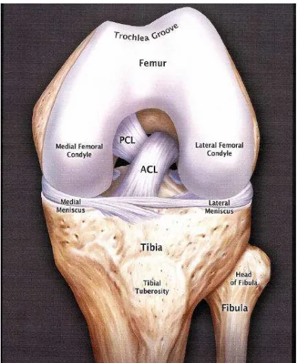
ANATOMY
Anterior cruciate ligament is a strong extra synovial ligament but it
is intra articular. It has a multi fascicular structure which runs from
anterior part of the tibia posteriorly and laterally to the medial aspect of
lateral femoral condyle. The ligament is 31 to 35 mm in length and 31.3
It has two bundles
• Anteromedial bundle
• Posterolateral bundle lying deep to the former
FEMORAL ATTACHMENT
The anterior cruciate ligament gets attached in the medial
aspect of lateral femoral condyle well posteriorly but around 15 mm
anterior to the posterior cortex of the lateral femoral condyle(21).The
posterior part of the ligament is seperated from the intermuscular septum
by the posterior capsule
1) Anteromedial bundle attaches posteriorly and superiorly over the
lateral condyle.
2) Posterolateral bundle attaches anteriorly and inferiorly over the
lateral condyle.
On a notch view of the knee joint, the entire femoral attachment of
the ACL is lateral to the midline of the intercondylar notch and occupies
the superior 66% of the notch. It is from just inferior to the postero
superior quadrant of the lateral femoral condyle. The center of the
10
Tibial Attachment
The tibial attachment is a large area of around 23mm in the anterior
tibial plateau anterolateral to the anterior tibial spine and medial to the
attachment of anterior horn of lateral meniscus
1. The anteromedial band inserts on the medial surface of the
intercondylar eminence.
2. The posterolateral band attaches lateral to the midline of the eminence.
The width of ACL averages 11.1 mm , length 31 to 38 mm.ACL
consist of longitudinally oriented fascicles, a different portion of which is
taut throughout the range of motion.
The important concepts of the normal ACL are that each fibre has a
unique origin and insertion and that all fibres are not parallel and not of
the same length and that do not have the same tension at any one point.
The anteromedial bundle becomes taut in flexion and the posterolateral
Figure 1: Anatomy of ACL
HISTOLOGY (22)
The ACL is made up of multiple fascicles, which are surrounded
by connective tissue called the paratenon. Each fascicle ranges from
several micrometers to several millimeters and consists of multiple
sub-fasciculi, which are enclosed by an epitenon. The subfasciculi appear to
have an undulating course, arranged in various directions. They consist
of groups of sub fascicular units (100 – 250 µm in diameter), which are
composed of fibers (1 – 20 µm in diameter) and surrounded by loose
connective tissues, the endotenon. Each fiber is made up of collagen
fibrils and interlace to form complex networks.Cells and elastic
12
BLOOD SUPPLY (23)
The blood supply of the ligament is by branches of middle
genicular artery that enters through the intercondylar notch near the
femoral attachment.The tibial site is supplied by the branches from the
patellar pad of fat which is from the medial and lateral inferior geniculate
artery.
There is little or no blood supply to ACL from its bony
attachments. In spite of the above sources of blood supply, ACL receives
[image:26.595.113.486.382.670.2]its predominant supply by diffusion from synovial fluid.
NERVE SUPPLY AND NEURAL RECEPTORS(24)
Posterior articular nerve, branch of posterior tibial nerve innervates
the anterior cruciate ligament. Most neural structures have been found in
the subsynovial layer and near the insertions of the ACL. Histologic
studies have demostrated nerves of sizes consistent with transmitting pain
in the intrafasicular spaces. Mechanoreceptors are also identified in the
surface of the ligament mostly near the femoral attachment site. The
receptors found are primarily ruffini receptors and free nerve-endings
that are thought to function as stretch receptors and nociceptors,
respectively.
FUNCTION (25)
Anterior cruciate ligament along with other intra articular and extra
articular ligaments, functions in maintaining static and dynamic
equlibrium of the knee joint.
The prime function of the anterior cruciate is restraining the
anterior movement of tibia over femur. The anteromedial bundle is taut in
flexion and is the prime restraint to anterior drawer in 90 degree flexion
14
which becomes taut in extension provides principle resistance for
hyperextension.
From the extensive histologic research and studies which had
demonstrated receptors and free nerve endings in ACL there are evidence
BIOMECHANICS
The cruciate ligaments form the pivot and nucleus of the knee joint
kinematics. On internal rotation the cruciates twist around each other and
in external rotation they unwind. The anterior cruciate ligament exerts
visco elastic properties like any other ligaments with its ultimate strength
of 1725+/-269 N(26).
The range of mobility is enhanced by the orientation of fibers.
Since the femoral origin of the cruciate ligaments lie on a line, it produces
normal mobility of 5-0-145 degrees.
The anteromedial fibers of anterior cruciate ligaments are tense
principally in flexion while the posterolateral fibers are in increasing
tension as the knee is extended. The reciprocal relationship of this bundle
constitutes a twist within the anatomy of this single ligament and
provides for stability throughout the entire arc of knee motion assisting
the rolling and gliding movement of the femoral condyle over the tibial
plateau in the sagittal plane. Anterior cruciate ligament also assist in the
screw home movement of the femoral condyle in the terminal extension.
In ACL tear, the femur rolls up onto the meniscus and its posterior
16
MECHANISM OF INJURY
Knee joint is inherently unstable without the strong capsulo
ligamentous structures supporting the joint in extension and flexion.
Varus and valgus stability is provided by the medial and lateral structures
along with cruciates. The cruciate ligaments provide anteroposterior and
rotatory stability along with other capsuloligamentous structures.
Depending on the position of the knee some acts as primary stabiliser and
some as secondary stabiliser. Most ligamentous injuries occur with flexed
knee when the capsule and ligaments are relaxed and femur is allowed to
rotate on the tibia.
The commonest mechanism of injury is a non-contact deceleration
with a valgus and twisting movement(28).In isolated ACL injury the
mechanism of injury was mostly deceleration, internal or external rotation
and hyperextension as with landing from a jump or sudden turning of
direction while running.
Valgus movements do not cause a serious injury until medial
collateral ligament is intact, but once it is injured the ACL ruptures as the
valgus thrust continues. When this is associated with a rotational
component the medial meniscus is torn caught between the articulating
Regarding rotatory violence, on continued extension of the knee
from flexion the femur rotates medially on tibia or tibia rotates externally
to lock the knee which is called as screw home mechanism. On sudden
block to screw home mechanism the ACL goes for stress and ruptures.
Also a direct posterior thrust on femur on fixed tibia as in dash
board injury can cause avulsion injury of ACL especially in young
individuals.
Various intrinsic and extrinsic factors are identified contributing to
the anterior cruciate ligament tear. The intrinsic factors include factors
which cannot be changed like the size of the ligament, notch width,
physiological alignment of the joint and physiological laxity, hormonal
influences, inherited skills and coordination. The extrinsic factors are
those that can be modified like strength, conditioning and motivation.
Many factors like coordination, proprioception, position sense and
balance require both intrinsic and extrinsic factors. The most important
factor contributing to ACL tear is the dynamic movement pattern rather
18
Figure 3: Mechanism of ACL injury
NATURAL HISTORY(28)
The course that an anterior cruciate ligament deficient knee follows
or the natural history of anterior cruciate ligament tear is varied and there
are many controversies regarding this, as all ACL tears are not reported,
not all ACL injuries are symptomatic, and due to varied patient
requirements and extent of injury. But it is very well documented that a
ACL deficient athlete with a unstable knee will acquire a meniscal tear
and arthrosis if he continues to involve in high demand activities in spite
of repeated episodes of instability. It has been reported that around 50 to
70% of ACL injuries are associated with meniscus injury. Studies reveal
within 5 years(29). The incidence of chondral damage is reported to be
twice in the chronic ACL tears than the acute tears.
Most patients are comfortable with their daily activities and have
limitations only in vigorous sports. Only a few patients are purely
asymptomatic. The persons with high pre injury activities like children,
athletes are the persons most affected. It is the challenge to the surgeon
and his responsibility to evaluate, understand and predict how an ACL
injury will affect different individuals based on their requirement. The
events following the anterior cruciate ligament injury is described as ACL
cascade.
ACL CASCADE
ACL INJURY
↓
JOINT SUBLUXATION
↓
MENISCAL TEAR
↓
CLINICAL EVALUATION
PATIENT HISTORY
A good clinical examination of ACL injured patient starts with a
good history taking. A twisting injury to the knee is usually the most
common history. An audible pop during the injury, inability to walk after
the injury and swelling of the knee joint over few hours are suggestive of
anterior cruciate ligament tear. With the associated heamarthrosis the
possibility of ACL tear is around 70%. Pain and sense of giving way of
the knee joint are the usual symptoms at the presentation. Non-contact
injuries are commonly associated with ACL tear while contact injuries
are commonly associated with multi ligament injuries. With valgus
violence and internal rotation injury the medial structures and collaterals
are initially disrupted and with continued violence ACL is torn. In varus
violence the lateral structures are disrupted first followed by the cruciates.
In hyperextension injuries ACL is torn first and with continued violence
posterior capsule and posterior cruciate ligament is torn. History of
locking episodes, click and clunk are suggestive of associated meniscal
injury. History of patient socioeconomic status, occupational and personal
requirements of the patient are important in individualizing patient
21
PHYSICAL EXAMINATION
General examination of the patient with inspection, palpation,
measurements and movements of the knee joint are done which is
followed by various test to accomplish the diagnosis and plan the
treatment. The tests for cruciate ligaments, collateral ligaments and
meniscus are done.
Lachmans test
Anterior drawer test
Slocum test
Pivot shift test
Mcmurrays test for meniscus
Valgus varus stress test for collateral ligaments
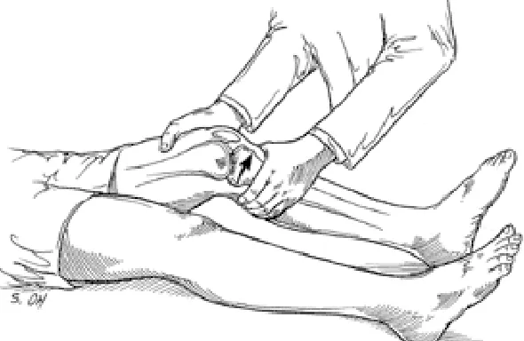
Lachman Test(30)
This test is done with patient supine and relaxed. The side affected
is placed towards the examiner or examiner stands by the affected side.
Patient is asked to relax and the limb is externally rotated to relax the
limb. Distal part of the thigh is grasped with one hand and proximal leg is
grasped with the other so that the thumb of the hand holding the leg is in
applied in an attempt to move the proximal tibia anteriorly and
posteriorly. The anterior translation of the tibia can be palpated with the
thumb of the hand holding the leg. Any anterior translation of the tibia
with a mushy end point signifies ACL tear.
When patients presents with heamarthrosis knee cannot be flexed
to 90 degrees and anterior drawer test is difficult to do. In those situations
Lachmans test is useful. Also Lachmans test is more sensitive than
[image:37.595.151.446.361.553.2]anterior drawer test in testing anterior cruciate ligament integrity.
Figure 4: Lachmans test
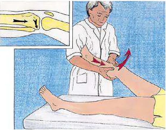
ANTERIOR DRAWER TEST (31)
This test is done with patient supine. The hip is flexed to 45
23
fingers of both the hands behind the knee and the proximal tibia is gently
pulled forward. Any movement of tibia over femur is noted and compared
with the opposite knee.
Pull of 6-8 mm more than the opposite knee with a mushy end
point indicates anterior cruciate ligament tear.
False positivity can occur in inherited ligament laxity, posterior
cruciate ligament tear. False negativity can occur in obese patients,
[image:38.595.194.404.359.545.2]hamstring spasm, heamarthrosis and mechanical block.
Figure 5: Anterior drawer test
SLOCUM TEST
Slocums modification of the anterior drawer test is performing the
anterior drawer test in neutral, 30 degree external rotation and 15 degree
indicates anteromedial instability and increased translation in internal
rotation indicates anterolateral instability.
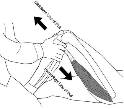
PIVOT SHIFT TEST(32)
The foot is lifted with the knee extended and internally rotated.
Valgus stress applied to the lateral side of the leg in the region of the
fibular neck with the opposite hand and when the knee is slowly flexed
while valgus and internal rotation being maintained the anteriorly
subluxated knee relocates at around 30 degrees of flexion. This test is
difficult to do with muscle spasm and it can be demonstrated easily with
[image:39.595.159.438.401.619.2]the patient anaesthetised.
Figure 6: Pivot shift test
RADIOGRAPHIC EVALUATION
25
haziness in Hoffa’s fat, a joint effusion, and may reveal subtle fractures of
the posterior tibial plateau, impaction of the lateral sulcus, or an avulsion
of the lateral tibial rim called as Segonds fracture. Tibial attachment
avulsions are more commonly detected on plain radiograph and are seen
more commonly in younger patients. Stress views are taken to
demonstrate the ACL injury radiographically. Anterior drawer sign is
elicited and lateral views are taken with and without stress. An anterior
translation of more than 5 mm is significant to call it abnormal. A
difference of more than 3 mm with the contralateral knee is significant.
MAGNETIC RESONANCE IMAGING
Magnetic resonance imaging offers direct, noninvasive
visualization of the ACL and other soft tissue structures, improving the
preoperative assessment of internal derangement. The accuracy of MR
imaging for evaluation of ACL pathology is high. Using direct signs,
sensitivities are as high as 92% to 94% and specificities are as high as
95% to 100%(33).
The patient is placed supine with the knee within the extremity
coil, avoiding excessive extension or flexion. Sagittal images are the
most useful for evaluation of ACL fiber orientation and the femoral and
sagittal views, then additional sequences may be supplemented with thin
cuts through the intercondylar notch and oblique images plotted parallel
to ACL on coronal or axial views. Images in the coronal plane are useful
for evaluation of the collateral ligaments and for assessing the signal
characteristics of the ACL within the intercondylar notch in equivocal
cases. On coronal views, the ACL appears coursing posterior
superolateral to anterior inferomedial.
Axial views are useful for assessment of the ACL and posterior
cruciate ligament (PCL) in the notch, bone contusions, para-articular fluid
collections, and the joint capsule. T1-weighted images, although useful
for delineation of bony anatomy, are not adequate for evaluation of
edema. Inversion recovery and / or T2-weighted images with fat
suppression are sensitive for visualization of marrow edema and
fractures. Either fast spin-echo or conventional spin-echo imaging can be
used to assess the ACL. On sagittal views, the normal ACL (with knee in
extension) should have a taut, straight anterior margin, with low signal
intensity of its fibers on all pulse sequences and fiber striation visible at
27
Figure 7: MRI showing normal ACL in sagittal plane
Figure 8: MRI showing normal ACL in coronal plane.
In this plane, the normal ACL often appears much more attenuated
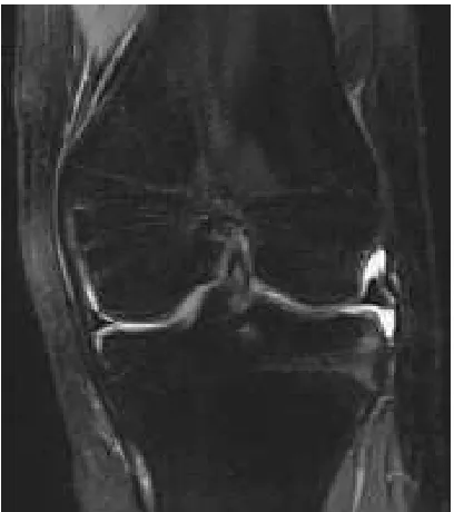
[image:42.595.196.400.365.595.2]INJURED ACL IN MRI
Direct MR imaging signs of acute ACL tear include poor or
non-visualization of the ACL on sagittal images, an amorphous edematous
mass with focally increased signal on T2-weighted images, irregular
contour with wavy redundant fibers, or interruption of fibers with tears
[image:43.595.124.473.317.575.2]seen midsubstance or at the tibial or femoral attachments.
29
TYPES OF ACL RUPTURE
Partial
Complete
Avulsion
SITE OF RUPTURE
Inferior attachment site which is usually avulsion fracture of the
tibial spine
At or about the superior attachment site
o Within the body of the ligament. This is the most common
site of tear. The anteromedial and posterolateral bundles can
MANAGEMENT
The aim of ACL reconstruction treatment is to attain functional
stability of the knee. An isolated rupture of the ACL is an extremely
common injury and most commonly results in instability of the knee. For
an active, healthy person who wishes to reach some state where he/she
can perform athletic activities, reconstruction of the ACL is
recommended. A minimum ROM of 5-90⁰ is usually desired and should
be achieved before surgical intervention.
There are three types of procedures for ACL insufficiency.
1. Extra articular reconstruction
2. Intra articular reconstruction
3. Combined
EXTRA ARTICULAR TECHNIQUES
1.Iliotibial band tenodesis
2.Biceps plasty
INTRA ARTICULAR TECHNIQUES
1. Transtibial technique
2. Transportal technique
31
GRAFT SELECTION(34)
The success of ACL reconstruction does not depend on type of
graft but on the technique used for the surgery such as placement of graft
and rehabilitation methods. The various types of grafts are :
1) Patellar tendon graft
Advantages:
• High initial tensile strength
• Good bone to bone healing
• Acceptable harvest morbidity
Disadvantages:
• Patellofemoral pain
• Tendinitis
• Patella fracture
• Quadriceps weakness
2) Hamstring tendon graft
Advantages:
Disadvantages:
• Good tensile strength only when folded double or triple
• late soft tissue to bone healing
3) Allograft Advantages:
• Improved cosmesis
• Less operative time
• Eliminate donor site morbidity
• Unlimited graft supply
Disadvantages: • High cost
• High disease transmission
• Later graft incorporation
• Alteration of graft pain
4) Synthetic grafts Advantages:
• Good postoperative results
MATERIALS
AND
MATERIALS AND METHODS
The Retrospective and prospective study of 60 patients treated with
arthroscopic anterior cruciate ligament reconstruction with quadrupled
hamstring graft with endobutton as the femoral fixation device and
titanium interference screw (no=30) and bioabsorbable interference screw
(no=30) as tibial fixation device respectively between May 2012 and
November 2013 at institute of orthopaedics and traumatology, Rajiv
Gandhi government general hospital, Chennai
INCLUSION CRITERIA
Patients with closed growth plate
Primary ACL surgery
No evidence of multiple ligament injury
No previous knee surgeries
No ligamentous injury to contralateral knee
EXCLUSION CRITERIA
Additional ligamentous laxity in affected knee
Previous ACL surgery of either knee
34
Any co-existing local conditions in the form of
-Active articular infection
-Inflammatory joint disease
Metabolic bone disease
Neoplastic disease
INSTRUMENTATION
Many specialized instruments are required for arthroscopic anterior
cruciate ligament reconstruction. An arthroscopic system which consist of
1. Television monitor
2. Camera
3. Light source and fibre optic light source cable
4. Arthroscope (30 degree)
5. Shaver system and hand piece
6. Tourniquet set (Pneumatic)
Equipments needed for surgery are
2.4mm drill tip guide pins
Trocar, canula, ACL probe
Meniscus punch
3.5 and 4.5 shaver blades
Cannulated headed reamers (size 5mm to 10mm)
Transtibial femoral ACL drill guide (usually 7 – mm offset
tip)
Extra long 2.4 mm guide pin with suture eye (Beath – type
guide pin)
4.5 mm cannulated reamer for passage of endobutton
Depth gauge
Sizing block
Cannulated interference screws
Endobutton
1.5 mm guide wire with screw driver for passage of
bioabsorbable interference screw
IMPLANTS
The fixation options for soft tissue grafts in femur can be direct
devices like interference screws and washers. The indirect devices like
endobutton, femoral cross pins, suture discs and anchors are also
available. Fixation options in tibia are interfernce screws, staples, screw
and washer(Washerloc).We used endobutton for femoral fixation and
titanium interference screws for 30 patients and bioabsorbable
36
ENDOBUTTON
Endobutton is preferred by most of the surgeons nowadays. It
ensures most of the graft in the tunnel. Endobutton has 4 holes of which
central two holes are used to create the loop for quadrupling the graft.The
peripheral two holes are for passing wires which are used to flip the
endobutton. Endo button was stronger than RCI screw and bio screw in
withstanding cyclical loads and has a greater advantage of not lacerating
the soft tissue graft.
Figure 10: Endobutton
INTERFERENCE SCREW
Interference screws are direct fixation device which hold the graft
to bone having inserted between the graft and the bone tunnel. These are
made of variety of materials. Round contoured interference screws, bio
absorbable interference screws, titanium interference screws are
available. We used regular titanium interference screws for 30 patients
and bioabsorbable interference screws for 30 patients. These interference
knee joint than the implants which suspend the graft or fix the graft at the
surface of the joint. However studies have proved that interference screws
to be inferior to the endobutton and the bone mulch screw. One another
concern was the laceration that interference screw can cause to the soft
tissue graft. But in spite of the concerns interference screw fixation of
soft tissue grafts have shown comparable results with that of interference
screw fixation of bone patellar tendon bone grafts.
Figure 11: Titanium Interference screw Figure 12: Bioabsorbable Interference screw
Bioabsorbable screw is made of poly-L-lactide and degradable over two
to six years. The advantages of the screw are
• Straightfoward technique
• graft will not be damaged ( as seen in mettallic)
38
METHODS
PREOPERATIVE WORK UP
Patients with ACL tear proven clinically and radiologically are
admitted in Institute of Orthopaedics and Traumatology. Routine
investigations like heamoglobin, total and differential counts, platelet
count, ESR, blood sugar, renal parameters, chest X-ray, ECG were taken
and anesthetist assessment for regional and general anesthesia was done.
Static and dynamic quadriceps exercise was taught to patients while
awaiting surgery.
ANAESTHESIA AND PATIENT POSITIONING
All patients are operated under spinal anesthesia. In supine position
under anesthesia anterior drawer test, posterior drawer test, Lachmans
test, pivot shift test are done. With patient supine knees are flexed to 90
degrees and a removable side support is placed in the side of the table to
support the ipsilateral thigh, a foot stopper is placed beneath the foot after
flexing the knee to 90 degrees. In all the cases a pneumatic tourniquet is
used which is placed in the upper thigh after soft padding. The limb is
shaved around the knee joint and prepared with betadine pre scrub. Limb
is draped exposing the knee joint lower thigh and upper leg after painting
is given before inflating the tourniquet and limb is held upright for 3
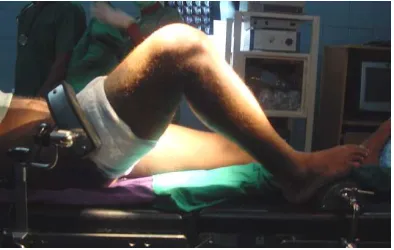
[image:55.595.200.397.148.273.2]minutes to exanginuate the limb before inflating the tourniquet.
Figure 13: Patient positioning
ARTHROSCOPIC PROCEDURE
An anterolateral portal is established 1 cm lateral to the patellar
tendon midway between the inferior pole of the patella and the upper end
of the tibia. Trocar and canula inserted with knee extended in to the
suprapatellar pouch. Inflow of normal saline from 3 liter saline bottles is
maintained through the TURP set. After adequate inflation of the joint
space scope is introduced and a diagnostic arthroscopy is done visualising
suprapatellar pouch, lateral gutter, intercondylar notch, articular surface
of patella, medial gutter and articular surfaces of femur and tibia. An
anteromedial portal or the working portal is established 1 cm medial to
the patellar tendon midway between the inferior pole of patella and the
40
amount of tear. If unstable meniscal injuries are found they are treated
with partial menisectomy and debridement depending on the site and type
of the tear.
GRAFT HARVEST AND PREPARATION
A 2 to 3 cm oblique incision is placed over the anteromedial aspect
of tibia exactly over the pes anserinus which is identified by palpating the
semitendinosus and gracilis tendon by running the fingers from above
downwards in the anteromedial aspect of the upper tibia. The tendons slip
under the finger during this gentle palpation. Skin subcutaneous tissue is
incised along the incision and blunt dissection is done to expose the
sartorius fascia which is lifted up with a forceps and cut with a number 11
scalpel.
After incising the sartorius fascia the gracilis and semitendinosus
tendons are indetified and localised using a right angle forceps. The
tendons are freed from all soft tissue attachments in the anteromedial tibia
and around their insertions. Then the tendons are secured with 1 vicryl
near their insertions and the tendons are detached from their insertions
one by one as long as possible. Holding the vicryl tied to the tendon a
closed tendon stripper is inserted encircling the tendon and the tendon
stripper is advanced with a minimal countertration. The stripper is
70 degree flexion and undue care is taken to prevent the amputation of
the graft. The stripper is advanced until the tendon muscle junction is cut
[image:57.595.204.394.181.250.2]and the tendon comes out through the incision.
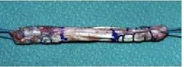
Figure 14: Quadrupled Hamstring Graft
The tendons are cleared of the muscle attachments and free ends of
the tendons are stitched together with a running whip stitch 4 to 5 cm
from the free ends with polybraided nonabsorbable number 2 suture
material (Ethibond). Mannual tensioning of the tendon is done and the
tendons are passed through the loop made in the endobutton with number
5 non-absorbable suture material (ethibond) or through the loop of the
endobutton CL ultra so that the tendons are quadrupled for
reconstruction. The free ends of the combined gracilis semitendinosus
tendons are again whip stitched with a number 2 nonabsorbable suture
material.Then the graft size is mesured with a sizer by pulling the graft
through the sizer and the graft is kept aside rolled in a moist cotton gauze
42
Figure 15: Graft harvest
INTRAARTICULAR PREPARATION
The arthroscope is introduced through the anterolateral portal and
the 4.5 or 3.5 shaver blade is inserted through the anteromedial portal and
the joint is debrided of the ligamentum plicae, some pad of fat and some
synovial reflections which hinder a through visualization of the medial
surface of lateral femoral condyle, the over the top position and the tibial
foot print of the anterior cruciate ligament.The medial surface of the
lateral femoral condyle is shaved of the native ACL remanents and the
over the top position is identified without misinterpreting the students
ridge. Then the ACL foot print in the tibia is prepared. Throughout this
joint debridement undue care is taken to avoid injury to the native
TIBIAL TUNNEL
The tibial guide or the guide pin targeting tibial jig is used to create
the tibial tunnel. The guide is set at 55 degree or by N+7 rule where N is
the effective length of the tendon. With the guide set in 55 degrees the tip
of the guide pin is positioned in the ACL foot print in the posterior half.
The guide can also be placed using various land marks like posterior rim
of the anterior horn of the lateral meniscus, anterolateral part of the
medial tibial intercondylar eminence, 8 mm anterior to the posterior
cruciate ligament.
After establishing the proper position of the guide tip the guide pin
sleeve is inserted and advanced to the anteromedial part of the tibia. The
guide pin sleeve is flushed with the anteromedial cortex of the upper tibia
midway between the tibial tuberosity and the posterior border of the
proximal tibia. The guide pin sleeve is inserted and advanced through the
incision made for harvesting the graft by retracting the skin edges. Before
drilling the tibial tunnel the arm of the tibial guide is ensured to be
parallel with the tibial plateau. Then the 2.4mm drill tip guide wire is
drilled through the tibial cortex to exit intraarticularly which is visualized
with the arthroscope. When the 2.4 mm drill tip guide wire had been
44
removed. Serial reaming of the tibial tunnel over the guide pin is done
with cannulated calibrated reamers up to the desired size of the graft.
During all these drilling a small curved curette is placed intraarticularly to
prevent the tip of the guide pin or the reamers from damaging the
articular surface of the joint. Once the tibial tunnel has been created the
posterior end or the intraarticular exit of the tibial tunnel is shaved of the
soft tissues and bone particles from obstructing the graft passage. Even a
sharp dissection can be used for this purpose and a rasp is used to
smoothen the tunnel walls for easy graft passage and to avoid graft
damage.
FEMORAL TUNNEL
The femoral tunnel is created by trans tibial technique in most of
the patients and transportal technique in rest of the patients in our study.
In transtibial technique, femoral tunnel is drilled through the tibial tunnel
and in transportal technique, femoral tunnel is drilled through a separate
medial portal with the help of femoral offset. The femoral aimer is placed
in the intercondylar notch at 1’O clock position for the left knee and 11’O
clock position for the right knee. The 7 mm offset aimer is placed so that
it is placed over the posterior edge of the notch to avoid blow out and to
leave atleast 2mm of intact posterior cortex. If the graft diameter is
anteriorly to avoid posterior blow out. Having placed the aimer the long
drill tip guide wire is drilled through the lateral femoral condyle to exit in
the anterolateral aspect of the lower thigh.
The intrarticular length of the graft is measured and the lateral
femoral condyle is drilled with 4.5 mm reamer until the anterolateral
cortex is breached to create a passing tunnel for the endobutton.
After reaming the lateral condyle the length of the femoral condyle
is measured with a depth gauge. Having known the intra articular length
of the graft and the whole length of the graft, the length of the graft to be
in the femoral condyle can be desired and marked, which is usually the
half the length of the remaining graft after subtracting the intraarticular
length from the total length. Having known the length of the femoral
condyle and the desired graft length in the femur, the loop length to be
adjusted in the endobutton is calculated and the loop is created or a
adequate length looped endobutton CL ultra is chosen. The femoral
condyle is reamed with a appropriate size reamer as of the graft to a
length of around 5 to 6 mm greater than the desired graft length for the
turning radius of the endobutton. The tunnel is smoothened with a rasp or
the shaver blade and the soft tissue interposition for the graft passage is
46
GRAFT PASSAGE AND FIXATION
In the peripheral holes of the endobutton two 5 number suture
material is passed and taken through the eyelet of the guide pin so that it
can used as a leading suture and as a toggle suture. The guide pin is
passed through the tunnel and pulled through the tunnel and extracted
along with the suture material in the anterolateral aspect of the distal
thigh. The leading suture is pulled so that the graft is pulled through the
tunnel headed by the end of the endobutton to which the leading suture is
passed. The graft is pulled until the desired length of the graft is pulled in
to the femoral condyle and the trailing suture is pulled to flip the
endobutton. Once the endobutton is flipped and confirmed by arthroscope
in the anterolateral aspect of the femur, the distal part of the graft is
pulled down to seat the endobutton so that the femoral fixation is done.
With manual tension to the distal graft the knee is taken through range of
motion to cyclically tension the graft and to look for impingement. If
there is impingement of the graft the notch is slightly enlarged to avoid
impingement. After tensioning the graft the tibial site is fixed with
appropriate size titanium interference screw or bioabsorbable
interferencescrew depending on the study group and ensured
Figure 16: Femoral tunnel fixed by endobutton
CLOSURE
The wound is closed in layers after through wash. The portals are
closed with single sutures with nonabsorbable suture material after
placing a intraarticular suction drain. Sterile dressing applied over the
wound and knee brace applied in extension after tourniquet is released.
[image:63.595.97.501.513.656.2]48
POST OPERATIVE MANAGEMENT
Immobilisation in knee brace and limb elevation immediate
post operatively
Intravenous antibiotics for 3 days
Drain removal on 2nd Post operative day
Wound inspection on 2, 5, 7 Post operative day.
Suture removal on 12th Postoperative day
Gradual physical rehabilitation
Follow up at 4, 8 weeks and 3, 6 months
POST OPERATIVE REHABILITATION
The general post operative protocol for anterior cruciate ligament
reconstruction is followed and progression of the rehabilitation is
individualized for each patient. Emphasis on arthrofibrosis, joint
contracture and joint laxity has been made.
Goals: Full range of motion (ROM), normal gait pattern, stability
1st Postoperative day
Rest in extension in long knee brace
Static quadriceps exercise
Ankle and foot movement and limb elevation.
0 – 2 Weeks
Full knee extension ROM
90 degrees knee flexion ROM
Strong QS/SLR without extention lag
Emphasize normal gait pattern
Passive, active, and active – assisted ROM knee flexion
Partial weight – bearing 50% to 75% with walker or
weight-bearing to tolerance with knee immobilizer with a walker
2 – 4 weeks
Full extension to 120 degrees flexion
Full weight bearing without
Progress SLR with weights
Walking, emphasis on normal gait.
4 – 10 Weeks
Progress to full ROM by 6 weeks
50
12-14 Weeks
Initiate full range knee extension exercises, light weight and
high repetition.
Initiate jogging program
16 –18 weeks
Isokinetic strength test for quadriceps and hamstrings
EVALUATION
All the patients are subjected for post-operative anteroposterior and
lateral radiographs to determine the tunnel placement and position of
endobutton in femur and interference screw in the tibia. Patients are
followed at 4 weeks, 8 weeks, 3months, 6 months and once in 6 months
thereafter.
All patients are evaluated with Lysholm & Gillquist scoring.
KNEE SCORING SCALE OF LYSHOLM & GILLQUIST
Limp
None 05
Slight /periodic or both 03
Constant or severe or both 00
Support
None 05
Cane or crutch 02
Weight bearing impossible 00
Locking
No locking or catching sensations 15
52
Locking - Frequently 02
Locked on examination 00
Instability / Giving Way
Never 25
Rarely during athletic activity or any other heavy exertion 20
Frequently during athletics or any other heavy exertion 15
Rarely in daily activities 10
Frequently in daily activities 05
At every step 00
Pain
None 25
Inconstant or slight during heavy exertion 20
Marked during heavy exertion 15
Slight during a walk >2 km 10
Marked during a walk <2 km 05
Constant 00
Swelling
None 10
Mild on exertion 06
Marked on exertion 02
Stair Climbing
No problems 10
Slightly impaired 06
One step at a time 02
Impossible 00
Squatting
No problems 05
Slightly impaired 04
Knee flexion possible only up to 90 degrees 02
Impossible 00
STATISTICAL ANALYSIS :
Data reported as mean and significant difference between the two
OBSERVATIONS
&
OBSERVATION AND RESULTS
60 Cases of arthroscopy assisted Anterior cruciate ligament
reconstruction with quadrupled hamstring tendon graft using endobutton
as the femoral fixation device and titanium interference screw (no=30)
and bioabsorbable interference screw (no=30) as tibial fixation device
respectively was followed for 6 months to 1.5 years. The mean follow up
55
AGE DISTRIBUTION
Minimum age was 20 years and maximum age was 55 with a mean
[image:72.595.93.505.228.730.2]age of 31.6 (Table 1 and Chart 1)
Table 1: Age distribution
AGE PATIENTS PERCENTAGE
15-20 3 5
21-25 16 26.66
26-30 14 23.33
31-35 11 18.33
36-40 6 10
41-45 7 11.66
46-50 2 3.33
51-55 1 1.66
SEX DISTRIBUTION
In this study, 51 patients were males and 9 patients were females
[image:73.595.90.533.247.643.2](table 2 and chart 2)
Table 2: Sex distribution
SEX PATIENTS PERCENTAGE
MALE 51 85
FEMALE 9 15
57
SIDE INVOLVED
In this study, 40 patients had injury in the right knee and 20
[image:74.595.92.505.447.712.2]patients had injury in the left knee (Table 3 and Chart 3)
Table 3: Side involved
SIDE INVOLVED PATIENTS PERCENTAGE
RIGHT 40 66.66
LEFT 20 33.33
MODE OF INJURY
MODE OF INJURY PATIENTS PERCENTAGE
SPORTS 10 16.6
FALL 22 36.66
RTA 28 46.66
59
DURATION OF INJURY
DURATION AFTER INJURY PATIENTS PERCENTAGE
<6 WEEKS 12 20
6-3 MONTHS 10 17
3-6 MONTHS 14 23
6-12 MONTHS 14 23
>12 MONTHS 10 17
ASSOCIATED INJURY
ASSOCIATED INJURY PATIENTS PERCENTAGE
MEDIAL MENISCUS TEAR 13 22
LATERAL MENISCUS TEAR 3 5
BOTH 2 3
NIL 42 70
61
OBSERVATION
Greater number of our patients was seen in the younger age group
of 20-40 years.
Male preponderance was noticed in our study
Right side was involved more commonly than left side
Road traffic accident was the most common cause accounting for
ACL injury.
Medial meniscus injury was involved more than the lateral
meniscus.
Most of the patients returned to their pre-functional level at 4
months.
SCORING ANALYSIS
60 patients of arthroscopic acl reconstruction with quadrupled
hamstring graft was followed for a minimum period of 6 months and
maximum period of 1.5 years.All patients are evaluated with Lysholm
and Gillquist scoring at the end of 6 months. The maximum score
Outcome Points
Good 84 - 100
Fair 65 - 84
Poor < 65
Two patients in titanium interference group and one patient in
bioabsorbable interference screw group lost to followup.
Outcome Titanium screw gp.no
of patients(28)
Percentage Bio-abs screw gp. No
of patients (29)
Percentage
Good 23 82.14 24 82.75
Fair 3 10.71 3 10.34
Poor 2 7.14 2 6.89
By Yates corrected Chi-Square Test,
X2 = 0.06 P = 0.97
63
GOOD RESULTS
In our study 23 patients in titanium interference screw group and
24 patients in bioabsorbable interference group had good results and the
patients had no limp, were able to walk without support, there was no
locking except for a few with mild instability during athletics or heavy
exertion. There was no pain or swelling of the knee joints. There was no
difficulty in climbing stairs or squatting.
FAIR RESULTS
In both the groups, 3 patients had fair results with the following
clinical findings. There was slight limping, occasional locking, with mild
instability during daily activities. There was anterior pain and swelling on
exertion. squatting and stair climbing were slightly impaired.
POOR RESULTS
In both the groups, 2 patients had poor results, with painful weight
bearing. The patient walked with support, and felt the knee giving way in
daily activities. There was constant swelling and pain of anterior knee
joint. Squatting and climbing stairs was painful.
The above 4 patients with poor results had lachmans and anterior
drawer test positive with restricted knee movements. These may be due to
rehabilitation.2 of the 4 patients had infection and septic arthritis 10 days
following which subsided with arthrotomy and joint lavage and
antibiotics.
COMPLICATIONS
One patient had post-operative infection and patient presented on
10th post-operative day with fever, pain and inability to move the limb.
Septic arthritis was suspected and patient treated with open arthrotomy
and joint debridement and antibiotics for 4 weeks and infection subsided.
The most common intraoperative complication proposed for
bioabsorbable interference screw were screw breakage , graft injury and
aseptic effusion or synovitis of knee joint, but we did not encounter such