DISSERTATION
A STUDY OF EVALUATION AND MANAGEMENT OF PATIENTS WITH
HEADACHE
By
DR.N.KUMAR
Dissertation Submitted to
The Tamil Nadu Dr. M.G.R. Medical University, Chennai, Tamilnadu In partial fulfillment
of the requirements for the degree of
MASTER OF SURGERY (OTORHINOLARYNGOLOGY) Thanjavur Medical College, Thanjavur
APRIL 2014
Under the Guidance of
Prof.Dr.A.RAVINDRAN, M.S.,D.L.O
Associate Professor
Department Of Otorhinolaryngology Thanjavur Medical College
CERTIFICATE
I certify that the dissertation titled
A STUDY OF EVALUATION AND
MANAGEMENT OF PATIENTS WITH HEADACHE
submitted byDr. N. Kumar for Degree of Master of Surgery (Otorhinolaryngology) to The
Tamilnadu Dr.M.G.R. Medical University,Chennai is the result of original research
work undertaken by him in the Department of Otorhinolaryngology, Thanjavur
Medical College,Thanjavur.
Prof.Dr.A.Ravindran, M.S.,D.L.O Prof.Dr.T.Ramanathan,M.S.,D.L.O
Associate Professor Head of the Department
Department Of Otorhinolaryngology Department of Otorhinolaryngology Thanjavur Medical College Thanjavur Medical College
Thanjavur Thanjavur
Place: Thanjavur DEAN
Date: Thanjavur Medical College
DECLARATION
I hereby declare that the dissertation titled A STUDY OF EVALUATION AND MANAGEMENT OF PATIENTS WITH HEADACHE,a clinical study submitted by me is the result of original work carried out by myself under the guidance of
Prof.Dr.A.RAVINDRAN,M.S.,D.L.O, Department Otorhinolaryngology,Thanjavur Medical College,Thanjavur.I further declare that the result of research have not been submitted previously
by myself or other persons in any conferences or journals.
ACKNOWLEDGEMENT
I thank the Dean Prof. Dr.Mahadevan M.S. Thanjavur Medical College,Thanjavur for permitting me to use the clinical material of this hospital for my study.
I express my sincere thanks to Associate Prof. Dr.A.Ravindran M.S.,D.L.O., my guide for his constant guidance,encouragement and untiring help during this study.
I express my deep sense of gratitude to Head of the Department
Prof.Dr.T.Ramanathan,M.S.,D.L.O., for his guidance and suggestions in preparing this dissertation.
My sincere thanks and gratitude to Assistant Professors Dr.Prince Peter Dhas M.S., Dr.M.Kavitha M.S., Dr.Ganeshkumar M.S.,D.L.O and Dr.Rameshbabu M.S. for their valuable suggestions in this study.
I express my sincere thanks to all the patients who in spite of their physical and mental
sufferings have co-operated and obliged my request to continue the study and regular follow up
without whom my study could not be fulfilled.
I am very grateful to all my fellow Post Graduates and other employers for their
Your digital receipt
This receipt acknowledges thatTurnitin received your paper. Below you will find the receipt information regarding your submission.
Paper ID 380866862
Paper title A STUDY OF EVALUATION AND MANAGEMENT OF
PATIENTS WITH HEADACHE
Assignment
title Medical
Author 22112221 . M.s. Ent KUMAR N . NEELAKANDAN
E-mail [email protected]
Submission
Total words 15067
First 100 words of your submission
INTRODUCTION History of headache Headache had been a mankind troubler since the rise of civilization. Neolithic human skulls dating 3000-7000 BC (1) were discovered with signs of perforation with an instrument known as TREPANATION, performed originally to release demons and evil spirits but recently evident to be carried out for medical reasons2 TREPANATION still practiced in African tribes for headache relief and fracture line removal following head injury(3). In ancient Egypt Headache
prescriptions were written on papyrus. The Eber papyrus dated circa 1200 BC describes migraine, neuralgia and shooting pains(4)in medical documents from 2500 BC.
ABSTRACT
Aim- To study about clinical evaluation of headache, its management and about effectiveness of
radiological investigations to find out type of headache.
Study Design-Prospective Clinical Study
Methodology- Patients presenting with headache for more than 1 month of all age groups and sexes
were selected. Selected patients were subjected to detailed history and complete examination according
to a defined proforma. According to clinical diagnosis,radiological investigations are done.Patients with
Migraine and Tension headache features were consulted with neurologist and psychiatrist.Treatment is
given accordingly.
Results-Most common type of headache is Tension headache. Majority of the patients were of age group
21-50 years and it is more predominant in males. Next Most common type of headache is migraine. It’s
is more common in females in 1:2 ratio. Third common type of headache is sinus headache and it is
more predominant in males. Headache was localized more than one site (59%) in majority of cases and 22% in forehead region. Patients who were having mucosal contact points headache, underwent
FESS and 88.89% of patients were relieved from headache. T.Propranalol 80 mgs and Amitriptyline 50 mgs were given for migraine patients. Most of the patients had subjective relief of headache. Patients
diagnosed with tension headache were treated with relaxation techniques, physical exercises and
Amitriptyline 10 mgs once a day in evening and most of the patients had subjective relief of headache.
Conclusion- Headache is nearly a universal human experience. The lifetime incidence of headache
is estimated to be at least 90%. A carefully taken history is key to accurate diagnosis and the
majority of patients will not have sinogenic pain. Accurate diagnosis will be helpful for
successful treatment.
LIST OF CONTENTS
S.NO
CONTENTS
PAGE NO
1
Introduction 12
Aim and Objectives of Study 43
Anatomy and Pathobiology of Head and Face Pain 54
Classification of Headache 115
Primary Headaches A] MigraineB] Tension Type Headache C] Cluster Headache
15 15 22 24
6
Secondary Headaches 307
Review of Literature 608
Materials and Methods 679
Observation 6910
Discussion 8111
Summary 8612
Conclusion 8813
Bibliography 8914
Proforma 9615
Abbreviations 101LIST OF TABLES
S.NO TABLES PAGE
NO
1 Intracranial Pain sensitive Structures 6
2 History Taking In Patients With Headache 10
3 Clinical Features Suggesting Serious Cause For Headache 10 4 Headache Society Classification of Headache and Facial Pain 11
5 Diagnostic ICHD Criteria for Migraine 18
6 Dose of Triptans 21
7 Diagnostic Criteria for Episodic Tension-Type Headache 23
8 Diagnostic ICHD Criteria for Cluster Headache 26
9 Major and Minor Factors Associated with the Diagnosis of
Rhino sinusitis 41
10 Possible Indications for Functional Endoscopic Sinus Surgery 51 11 1 year prevalence of Migraine from population-based studies 61
12 Age distribution of Headache 69
13 Sex distribution of Headache 71
14 Sex distribution of Headache-other causes 72
15 Etiology of Headache with Respect to Clinical Findings 73
16 Localization of Headache 74
17 Patients who underwent DNE 75
18 Patients who underwent FESS due to mucosal contact Point 76 19 Patients who underwent FESS due to causes other than
Contact Points 77
20 MIDAS Grade 79
21 Treatment of Migraine 79
LIST OF FIGURES
S.NO FIGURES PAGE
NO
1 Location of Headache in Migraine 16
2 Migraine with Aura 17
3 Location of Pain in Tension Headache 23
4 Patient of Cluster Headache 25
5 X Ray PNS Showing Spur 44
6 X Ray PNS showing Anterior deviation of Septum 44
7 X Ray PNS showing DNS with spur with Bilateral Maxillary
Sinusitis 45
8 CT PNS showing Bilateral Sphenoid and Ethmoid sinusitis 45 9 CT PNS showing Bilateral Ethmoidal and Frontal sinusitis 46
10 CT PNS showing Bilateral Sphenoid sinusitis 46
11 X Ray PNS showing Anterior deviation of Septum Towards
Left 47
12 Instruments Used in FESS 48
13 Endoscopic Picture Of Maxillary Sinus Ostium 48
14 Position of Instrument in Case of 00Endoscope 52
15 00Endoscope Showing Right Middle Meatus 54
16 Right Middle Meatus and Frontal Recess 54
17 Septal Spur Impinging into Inferior Turbinate 54
18 Gender and age specific prevalence of migraine 62
19 Age distribution of Headache 70
20 Sex distribution of Headache 71
21 Sex distribution of Headache (Other causes) 72
22 Etiology of Headache with Respect to Clinical Findings 73
23 Localization of Headache 74
24 Patients who underwent DNE 75
25 Patients who underwent FESS due to mucosal contact point 76 26 Patients who underwent FESS due to Non mucosal contact
point 77
27 Treatment Of Migraine 79
A STUDY OF EVALUATION AND
MANAGEMENT OF PATIENTS
WITH
INTRODUCTION
History ofheadache-Headache had been a mankind troubler since the rise of civilization.
Neolithic human skulls dating 3000-7000 BC(1) were discovered with signs of
perforation with an instrument known as TREPANATION, performed originally to
release demons and evil spirits but recently evident to be carried out for medical
reasons2 TREPANATION still practiced in African tribes for headache relief and
fracture line removal following head injury(3).
In ancient Egypt Headache prescriptions were written on papyrus.
The Eber papyrus dated circa 1200 BC describes migraine, neuralgia and shooting
pains(4)in medical documents from 2500 BC. Compression and cooling the scalp (5)
method using strip of linen with clay and grains (5,6) were practiced then for
headache relief.
Hippocrates 470-410 BC described headache as a shining light in
the right eye with violent pain arising in the temples and spreading to the entire head
and neck area being triggered by exercise and intercourse(6) and relieved by
vomiting.Aretaeus in second century AD discovered migraine headache based on
descriptions of migraine by Celsus 215-300 AD .Galen in 200 AD introduced the
term ‘migraine’ from greek word ‘hemicrania’.Abbess Hildegrade of Bingen in 12 th
century described in terms of migraine aura. Thomas Williams in 1683 described
prodromal symptoms associated with migrainous headache.Tissot in 1783(8)
differentiated migraine from common headaches stating it to be a supraorbital
neuralgia Dubois Raymond,Mollendorf and Eulenberg put forth different vascular
Erasmus Darwin in 18th century believed headache caused by
vasodilatation could be treated by centrifugation. In 1778, Fothergill termed
migrainous aura as ‘fortification spectra’(9,10).Liveing in 1873 originated the neural
theory of migraine in the first monograph entitled On Megrim,Sick-headache,and
Some Allied Disorders: A Contribution to the Pathology of Nerve storms William
Gowers in 1888 in his neurology textbook, A Manual of the Diseases of the Nervous
system recommended lifestyle modifications and use of Gower’s mixture
[nitroglycerin 1% alcohol solution] and marijuana for treating headaches.
In the line of treatment of migraine, Ergot being the mainstay ,the
history accounts since the middle ages in greek and roman ancient writings.
Epidemics of ergot poisoning broke out with the disease known as ‘Ignis sacer’ or
‘Holy Fire” or ‘St. Anthony’s Fire’. The term ‘ERGOT’ is derived from French
word ‘argot’ meaning ‘rooster spur ‘describing the small banana shaped sclerotium
(11) of the fungus.Louis Rene Tulasne of Paris in 1853 established that ergot is a
fungus named ‘Claviceps purpurea’with three stages in one life cycleHeinrich
Wiggers in 1831 tested ergot in animal models and studied the physiological
properties of ergot by the ‘rooster comb test’.Woakes in 1868 reported the use of
ergot of rye in neuralgia(12)treatment.
Eulenberg of Germany in 1883 ,Thomson of United states in
1894 ,Campbell of England in 1894 and Stevens in 1907(13)reported the use of ergot
for migraine early in the history of medical literature. Stoll in 1918 isolated the first
pure ergot alkaloid,ERGOTAMINE,used mainly in obstetrics and gynaecology until
1925.Rothlin used subcutaneous ergotamine tartrate, a vasoconstrictor to treat
(14) in 1938 proved the vascular theory of migraine stating ergotamine to be a
vasoconstrictor.
The modern approach treating headaches began with the
development of TRIPTANS by Humprey and his colleagues(15)with the belief that
migraine is due to excess serotonin. Sicuteri(16) developed methylsergide ,a
serotonin antagonist for migraine and cluster headache prophylaxis.Currently many
newer drugs are being tested and developed with concomitant development in basic
sciences and the renewed dedication of the clinicians in diagnosis and treatment of
headache .
Pain in the head, face, throat, and upper neck is a major cause of
care seeking that leads to a primary or secondary referral to otolaryngology. The
dramatic advances in epidemiology, diagnosis, diagnostic testing, and treatment of
headache have made case finding more satisfying. Evidence-based medicine has
presented challenges to long-held beliefs. The most important of these is the
recognition that migraine underlies most of the common headaches in clinical
practice, including sinus headache. Improvements in diagnosis and treatment have
begun to affect care delivery and patient satisfaction. The International
Classification of Headache Disorders, 2nd edition (17) divides the head and face
pains into primary or secondary disorders. The primary headaches include
migraine, tension type, trigeminal autonomic cephalalgias, and other primary
headaches, including common or unusual but benign headaches.Secondary
headache results from either pathology of the head and its surrounding structures
or from systemic disease and effects on cranial nociception. The task of the
practicing specialist is to differentiate those headaches requiring urgent attention
AIMS AND OBJECTIVES
• To study about clinical evaluation of headache
• To study about effectiveness of radiological investigations to find out type of headache
• To find out the effective management of each type of headache and comparison with previous studies
Anatomy and Pathobiology of Head and Face Pain
The parenchyma of the brain is insensitive to direct noxious
stimuli. The trigeminal nerve is the largest of all the cranial nerves. It is a mixed
nerve situated on the ventrolateral aspect of the pons. The sensory root conveys
impulses from the face and scalp, parts of the external ear and acoustic meatus, the
nasal and oral cavities, teeth, temporomandibular joint (TMJ), nasopharynx, and
most of the meninges of the anterior and middle fossa. It passes outward and
forward over the petrous temporal bone near the apex, extending into the
trigeminal ganglion. These pseudounipolar cells pass peripherally to become the
three major divisions: ophthalmic, maxillary, and mandibular.
The ophthalmic division is joined by projections of the internal
carotid sympathetic plexus and communicates with the oculomotor, trochlear, and
abducens nerves as it courses along the lateral wall of the cavernous sinus. The
main termination exits the skull through the superior orbital fissure. Other
branches include the tentorial, lacrimal, frontal, and maxillary nerves. The
maxillary division also carries sympathetic and parasympathetic fibers. Its
meningeal branch (middle cranial fossa) travels in the inferior part of the lateral
wall of the cavernous sinus to leave the skull through the foramen rotundum,
entering the pterygopalatine fossa where it communicates with the pterygopalatine
ganglion and enters the orbit through the inferior orbital fissure. The infraorbital
nerve then emerges on the face, where smaller branches (inferior palpebral,
external and internal nasal, and superior labial branches) supply the lower eyelid,
nasal alae, and upper lip. Additional branches go to the zygoma, upper teeth, and
gums as the superior alveolar and superior dental nerves. The mandibular nerve
through the foramen spinosum and travels along the middle meningeal artery to
supply the meninges of the middle and anterior cranial fossae, calvaria, and
mucous membranes of the mastoid air cells. The buccal nerve innervates the
mucous membranes and buccinator muscle. Other sensory branches of the
mandibular nerve include the auriculotemporal and lingual nerves, which
innervate the TMJ and tongue as well as the dental and mucosal surfaces of the
lower jaw.
In the primary head and face pains, abnormal activation of
peripheral neurons or central structures results in distinctive pain description,
phenomenology, and associations (18).These may then refer to areas of the head,
neck, or face at distance from the area of activation, making diagnosis more
challenging. The central connections of second-order sensory neurons of the
trigeminal system project to the thalamus and thence to the primary sensory cortex
of the parietal lobe. Descending inhibitory projections from periaqueductal gray
and paracentral aminergic nuclei modify the pain experience and have become
increasingly important in our understanding of the primary headaches.
Table
1-INTRACRANIAL PAIN SENSITIVE STRUCTURES
Major arteries in brain base
Artery-Internal carotid, Ophthalmic artery
Vertebrobasilar system and major branches
Circle of Willis
Proximal segments of cerebral arteries
Major venous sinuses and adjacent major venous tributaries
Cranial nerves V, VII, IX, and X
Floor of the pituitary fossa
Upper three cervical nerves (pain referral to occiput)
Secondary headaches rarely are initially seen with distinctive pain
characteristics. Many secondary headaches mimic migraine, tension-type, cluster,
or short-duration headache. It is therefore necessary for the clinician to assess the
patient's history carefully for clues to diagnosis of more urgent or serious
headache. Some well-known examples of classical presentations include
monocular eye pain with pupillary dysfunction in optic neuritis, sinus disease, or
jaw claudication in temporal arteritis.
First-division pain typically refers to the scalp or face anterior to
the coronal suture, although it can be felt deep inside the head or in the occiput.
Lesions in the middle fossa can refer to the apex of the head or in the midnasal
bridge, as can pain arising from the deep ethmoid or sphenoid sinuses or the sella
turcica. With lesions involving posterior fossa structures, pain is referred to the
back half of the head and upper neck. Pain coming from diffuse intracranial
disease, including meningitis, can mimic primary headache of the tension type, or
more severe diffuse head pain with meningismus.
Extracranial Patterns of
Referral-Orbital pathology usually is first seen with monocular pain and
often has associated visual disturbances. Unilateral eye pain should be considered
with distinctive patterns of referral. Disease in the frontal sinus may appear with
frontal pain or pain radiating behind the eyes or to the vertex. Maxillary pain is
typically infraorbital in location, and ethmoidal disease appears between and
behind the eyes or to the vertex, as does the sphenoid. In our experience,
mastoid disease may first be seen as unilateral occipital pain or with cluster-like
headache. Other mimics of cluster headache (CH) include carotid disease and
dissection. The TMJ and the muscles of mastication can be a source of pain that
radiates to the head and the ear. The TMJ and its articulations are innervated by
the auriculotemporal nerve as it passes behind the condyle and then upward in
front of the ear. Pain can be reported as a diffuse temporal headache or as
ipsilateral ear pain. Tooth pain is poorly localized and can be confused with
disorders occurring outside the mouth. It can appear as diffuse facial or ear pain.
Pulpal death results in localization of the pain to the offending tooth. Cervical
disease including spondylosis can be seen with unilateral head pain, sharing
features of migraine. Vascular structures of the anterior neck can cause pain that
radiates into the lower face. A clinically recognized complication of carotid
dissection is cluster-like headache and complete or incomplete Horner syndrome.
Headache Diagnosis and
Testing-A detailed history and physical examination are the keys to efficient
assessment of headache patients . Particular attention should be paid to pain
characteristics, including rate of onset, rate of offset, intensity, quality, location,
Pain localization may be helpful, although referral patterns are not
diagnostic of secondary headaches in general. Establishing the temporal profile,
including duration, is essential to good diagnosis. Exacerbating and relieving
factors and associated nonpainful symptoms should be noted. In primary care
practice, 94% of patients with recurrent headache have migraine or migrainous
headache (19). Migraine is most frequently misdiagnosed as sinus headache or
tension-type headache (20).In the emergency department, 95% of patients with a
headache have migraine (21). Recognized features of clinical history and physical
examination can alert the physician to underlying organic diseases of a more
serious nature.
Magnetic resonance imaging (MRI) is the gold-standard radiographic
study for headache diagnosis. Only 0.18% with migraine and a normal neurologic
examination will have significant pathology. Computed tomography (CT) is still
the test of choice for acute, thunderclap, or new-onset headache in the emergency
department to rule out hemorrhage. CT does miss multiple vascular malformations
including aneurysms; meningeal, pituitary, and intracranial neoplasm; Chiari
malformations; posterior fossa tumors and malformations; paranasal sinus disease;
encephalitis; meningitis; and cerebritis. Low or high cerebrospinal fluid (CSF)
pressure with headache and complications also may be missed with routine CT
scans. Electroencephalography (EEG) has minimal sensitivity for headache
diagnosis. Lumbar puncture is indicated for acute headache with a negative CT
Table
2-DIAGNOSIS HISTORY IN PATIENTS WITH HEADACHE History of present illness
Age at onset, progression to the present Frequency timing and duration of attacks Quality of the pain, including severity
Associated symptoms including warning symptoms or aura Precipitating and/or relieving factors including medications
Medical history
Prior surgery or trauma to head or neck Allergies, diabetes, hypertension,
psychiatric illness, recurrent infections, Current medications including over-the-counter analgesics, birth control pills, herbal medications, topical agents
Family and social history
Migraine, tension or sinus headache, psychiatric illness, substance abuse, suicide Cerebrovascular or cardiovascular disease, especially young
Marital status and occupation Stress factors
Tobacco, alcohol, and drug use
Review of systems
Changes in weight, appetite, sleep, mood, or somatic sensation including pain and limitations to activity or range of motion
Trauma including traumatic life events
Symptoms suggestive of endocrine disturbance
Table
3-DIAGNOSIS CLINICAL FEATURES SUGGESTING SERIOUS CAUSE FOR HEADACHE
Crescendo headache in any age group
Headaches exclusively triggered by cough or exertion Early-morning headache
Vomiting without significant premonitory nausea Projectile vomiting or persistent hiccoughing Associated fever, stiff neck, lethargy, confusion
Associated seizures, syncope, diplopia, visual obscurations
Sudden-onset of worst headache of my life,Known malignancy or chronic infection including human immunodeficiency virus,Endocrine disturbance of any kind,Focal neurologic symptoms or signs,Papilledema, visual field loss
Classification
Understanding headache and facial pain is essential to facilitate
diagnosis and treatment. To this end, definitions and features of clinical syndromes
were organized by the Headache Society- International.
This classification, with inclusion of diagnostic criteria for headaches, cranial
neuralgias and facial pain, was created in 1988 and has facilitated the diagnostic
approach and management of craniofacial pain across many medical fields.
Table
4-Headache Society Classification of 4-Headache and Facial Pain Migraine type
Without aura (common migraine) With aura (classic migraine)
With prolonged aura (complicated migraine) Ophthalmoplegic
Retinal
Tension type
Episodic (muscle contraction headache) Chronic (chronic daily headache)
Oromandibular dysfunction (myofascial pain dysfunction syndrome) (temporomandibular joint pain dysfunction syndrome)
Cluster (Horton’s cephalalgia) Post-traumatic headache Vascular intracranial disorder
Transient ischemic attack-associated headache Haematoma -Intracranial
Acute arterial hypertension
Pheochromocytoma
Malignant hypertension (accelerated) Pre-eclampsia and eclampsia
Non-vascular intracranial disorder
Pseudo tumor cerebri
Post-lumbar puncture headache Cerebrospinal fluid fistula headache
Intracranial infection
Meningitis Brain abscess Subdural empyema
Intracranial neoplasm
Headache from substance exposure or withdrawal Acute exposure
Nitrate or nitrite-induced headache (hotdog headache)
Monosodium glutamate-induced headache (Chinese restaurant syndrome) Carbon monoxide-induced headache
Alcohol-induced headache Chronic exposure
Analgesics
Ergotamine
Caffiene
Narcotics
Headache with extracranial infection Viral
Bacterial
Headache from metabolic disorder Hypoxia
High-altitude headache Sleep apnea headache Hypoglycemia
Headache or facial pain associated with craniofacial disorder Cranial disorder
Osteomyelitis Multiple myeloma Paget’s disease
Cervical spine disorder (cervicogenic headache) Eye disorder
Acute glaucoma Refractive errors Sinonasal disorder Acute sinus headache Rhinogenic headache Odontomandibular disorder
Pulpitis
Glossitis (burning mouth syndrome)
Temporomandibular joint disease Cranial neuralgia
Compression of cranial nerve or cervical root 1, 2 or 3 Inflammation of cranial nerves
Acute herpes zoster
Chronic postherpetic neuralgia Tolosa-Hunt syndrome
Gradenigo’s syndrome
Trigeminal neuralgia (tic douloureux)
Compression of trigeminal ganglion Vascular Tumor
Aneurysm
Multiple sclerosis
Glossopharyngeal neuralgia Occipital neuralgia Anesthesia dolorosa
Postsurgical after trigeminal rhizotomy
Epidemiology-Eighty to ninety percent of world population have headache in a
lifetime. 78% is lifelong prevalence of tension-type headache. Compared with this
is the less than 1% likelihood of serious intracranial pathology including tumors,
and the 15% lifetime prevalence of acute nasal or sinus headache. A patient
seeking care for recurrent or episodic headache has an extremely high (94%)
likelihood of having migraine(22).Two-thirds of this care is given in the primary
setting(23). Nevertheless, because of cranial autonomic symptoms or seasonal
periodicity associated with some of the primary headaches, the specialist in allergy
or diseases of the ear, nose, and throat may be the first contact a patient makes in
the search for good care and correct referral. In addition, serious pathology of the
skull, sinuses, or upper dental ridge may be first seen with face and head pain,
making the otolaryngologist the most important arbiter of diagnosis and decision
making.
Primary
Headaches-
Migraine-Migraine is a disabling disorder with a 1-year prevalence of 18%
among females and 6% among males in world (24). Fifty-two percent of
migraineurs remain undiagnosed. Fifty percent of migraineurs miss work 2 days
per month and have reduced work efficiency for 6 days per month. Two-thirds of
migraineurs recognize that this disorder has adversely affected their family life
(25).Migraine was considered a vascular disorder until the mid-1980s, when
models, he demonstrated that electrical stimulation of the trigeminal nucleus
caudalis in the pons causes plasma protein extravasation from Dural blood vessels.
He concluded that the generation of migraine depended on serotonin mediated
increases in neuronal excitation, not primary vascular reactivity. In humans,
trigeminal activation results in the expression of recognized inflammatory and
pro-nociceptive substances, including calcitonin gene related peptide, neurokinin A,
and substance P. Genetic studies suggest autosomal dominant transmission with
more than 70% of patients having a first-degree relative with migraine. Familial
hemiplegic migraine localizes to defects on chromosome 19p13, which encodes
voltage-gated P/Q-type calcium channels, and to the ATP1A2 gene on
chromosome 1, resulting in dysfunction of the Na-K pump. Genetic polymorphism
studies have implicated alterations in the gene for serotonin receptors in migraine
[image:27.595.227.421.463.674.2]without aura and in dopamine receptors in migraine with aura.
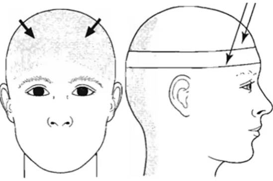
Figure 1- LOCATION OF HEADACHE IN MIGRAINE
[Supraorbital,Facial ,Frontal, Parietal,Occipital ,Temporal pain,photophobia,Pallor
Migraine with and without
Aura-Migraine aura occurs consistently in 15% to 25% of migraineurs.
Aura is a localizable and fully reversible neurologic deficit preceding head pain,
which results from progressive neuronal dysfunction spreading across the cerebral
cortex. Studies including single-photon emission computed tomography (SPECT),
positron-emission tomography (PET), functional MRI, magnetic resonance
spectroscopy (MRS), and magneto encephalography have bolstered this
hypothesis. Serotonergic, noradrenergic, and dopaminergic pathways; hormonal
regulation, especially estrogen; and hypothalamic and deep brainstem structures
are involved in the ultimate expression of migraine. Thus migraine can be
described as neuronal sensitization and neurogenic inflammation in a milieu of
multiple neurobiologic influences. The goal of migraine treatment is to attenuate
neuronal irritability and neurogenic inflammation while targeting central
[image:28.595.207.440.465.666.2]mechanisms to abort or prevent headaches and associated disability.
Table
5-DIAGNOSIS ICHD CRITERIA FOR MIGRAINE 1.1 Migraine without aura
A]Headache attacks lasting 4 to 72 h (untreated or successfully treated)
B]Headache has at least two of the following characteristics
Unilateral location
Pulsating quality
Moderate or severe intensity
Aggravation by or causing avoidance of routine physical activity (e.g., walking or
climbing stairs)
C]During headache, at least one of the following:
Nausea or vomiting
Photophobia and phonophobia
D]Not attributed to another disorder
1.2 Migraine with typical aura
A]At least two attacks
B]Aura consisting of at least one of the following but no motor weakness:
Fully reversible visual symptoms including positive features (e.g., flickering
Fully reversible sensory symptoms including positive features (i.e.,
pins-and-needles) and/or negative features (i.e., numbness)
Fully reversible dysphasic speech disturbance
At least two of the following:
Homonymous visual symptoms and/or unilateral sensory symptoms
C]At least one aura symptom develops gradually over 5 min, and/or different aura
symptoms occur in succession over 5 min
D]Headache fulfilling criteria for 1.1 Migraine without aura begins during the aura
or follows within 60 min
E]Not attributed to another disorder
Migraine and Sinus
Headache-Among patients diagnosable with migraine, only 19% identify their
diagnosis correctly. Twenty-eight percent of patients report sinus as their cause for
headache, and 34% called them tension. In one study of migraineurs, 42%
received an incorrect diagnosis of sinus headache (23)and 32% reported being told
they were tension type.
The potential confusion with sinus headache stems from long-held
beliefs about location of pain. Nasal, paranasal, or periorbital pain merges with
percent of patients complaining of recurrent sinus headache have migraine, and the
response to specific migraine therapies, including the triptans, is excellent.
Headache is a minor criterion of the American Association of Otolaryngologist
Head and Neck Surgeons (AAO-HNS) criteria for sinusitis ,making it more
important for the otolaryngologist to recognize the episodic, recurrent, and
treatable migraineur before assessing for definitive or ongoing treatment(27).
Migraine Treatment: Acute and
Preventive-Treatment of migraine begins with clear diagnosis and
knowledge of its pathophysiology. Acute treatment of headache should be
stratified to the level of disability in the patient. Nonspecific therapies include
nonsteroidal antiinflammatory drugs (NSAIDs), sympathomimetic including
caffeine, and analgesics. Frequent use of many of these has been associated with
substance-withdrawal headache and analgesic rebound, now termed
medication-overuse headache. For moderate to severely disabling headache, specific
medication should be used. The most specific of these are triptans. They bind to
serotonin receptors (5HT1D/B) on trigeminal nerve endings to halt neurogenic
inflammation in addition to binding to dural blood vessels to reduce painful
swelling. No class effect of triptans exists, and if one is not effective, then another
may be tried. Side effects include chest symptoms, nausea, and asthenia. They are
contraindicated in patients with uncontrolled hypertension or a history of cerebral
or coronary artery disease but are generally extremely safe and effective. A
growing body of evidence supports early treatment during the mild phase of
migraine headache. Cutaneous allodynia, a lowering of pain thresholds and
full-blown migraine attack, may be a marker for response to specific migraine
medications. Triptans are more costly than nonspecific therapy, but economic
analyses point to cost savings and decreased disability when triptans are used in
patients with moderate to severe headache(28).
Table -6
Almotriptan 12.5 mg Pills
Eletriptan 20/40 mg Pills
Frovatriptan 2.5 mg Pills
Naratriptan 2.5 mg Pills
Rizatriptan 5/10 mg Pills/melts
Sumatriptan 50/100 mg Pills
5/20 mg Nasal spray
6 mg Injection
Zolmitriptan 2.5/5 mg Pills and melts
5 mg Nasal spray
Dihydroergotamine 1 mg IV/IM
4 mg IN
Start low and increase dose slowly by using long-acting preparations or once-daily
dosing to enhance compliance.
Use an adequate trial of 2 to 3 months at an appropriate dosage.
Avoid interfering, overused, and contraindicated medications.
Continue for 6 to 24 months.
Attempt to taper and discontinue when headaches are well controlled.
Be aware of drug interactions.
Give special concern for women during childbearing years.
Tension Type Headache
Episodic tension-type headache is the most common head-pain
syndrome. It is characterized by a gradual onset of bilateral, nonthrobbing, aching
pain over the frontal and temporal regions, often spreading to involve the occipital,
posterior cervical, and trapezius musculature. The pain worsens as the day goes
on. Associated symptoms such as nausea and vomiting are rare, and patients
usually can continue activities of daily living during the headache. The headaches
are not seasonal and do not wake the patient from sleep. Adults rarely seek
medical care for occasional tension headaches (29).Chronic tension-type headache
is responsible for 15% of daily headaches. The patients seeking medical care for
chronic, persistent, or recurrent headaches most often are first seen with
transformed migraine. The most commonly encountered problem in this group is
analgesic rebound headache and depression. Combination therapy, including
limited NSAIDs and antidepressants, is the most effective treatment.
The causes of tension-type headache are poorly understood
despite its well-described symptom profile. No firm evidence exists for the role of
muscle contraction or spasm in these patients. Recent experimental evidence
implicates dopamine, endocannabinoids, and the endorphins/ enkephalins in
tension-type headache. Muscle relaxants, massage, and biofeedback can be very
require antiinflammatory medications, antidepressants, or both.
Figure 3-TENSION HEADACHE
Table
7-Diagnostic Criteria for Episodic Tension-Type Headache
A. At least 10 previous headache episodes fulfilling criteria B through D;
Number of days with such headaches: less than 180 days per year
B. Headache lasting from 30 minutes to 7 days C. At least two of the following pain characteristics:
1. Pressing or tightening (nonpulsating) quality
2. Mild or moderate intensity
3. Bilateral location
4. No aggravation by walking stairs or similar routine physical activity D. Both of the following:
1. No nausea or vomiting (anorexia may occur)
Trigeminal Autonomic Cephalalgia Including Cluster
Headache-Uncommon and unfamiliar except for CH, the trigeminal
autonomic cephalalgias (TACs) typically are initially seen with shorter-duration,
focal, and side-locked head pain associated with ipsilateral autonomic features.
This suggests significant but abnormal activation of the cranial autonomics and
trigeminal-parasympathetic. These may mimic pathology in orbital structures or
the sinuses. For this reason, delay in correct diagnosis of CH may range from 7 to
16 years. Early recognition spares the patient years of untreated and often
extremely severe pain. These groups of headaches appear in episodic and chronic
forms. All forms of the TACs have been associated with trigeminal neuralgia in
case reports and case series(30).
Cluster
Headache-CH is not common disorder. It is abrupt in onset , unilateral,
blinding, and less-duration attacks .Among the primary headaches, CH pain is the
most severe pain, and the accompanying autonomic features may confuse the
diagnosis with pathology in sinuses orbital or periorbital areas. Awakening the
patient from sleep at a time when dreaming is presumed to occur leads to sleep
evaluations, and CH's seasonal or circannual occurrence implies botanical allergy.
Secondary causes of CH can include cranial, cervical, or vascular disorders. The
carotid artery, cavernous sinus, and various brain structures, including the
periaqueductal gray matter, appear to participate in pain generation and
modulation associated with this disease(31).
CH occurs in 0.2% to 0.6% of the population, with a
ratio of CH may be changing, the overall prevalence is stable for the last 3 to 4
decades. Genetic studies suggest autosomal dominant transmission with a low
frequency of the susceptibility allele. During active periods, most commonly in the
spring and autumn, clusters of individual headaches lasting between 30 and 180
minutes occur daily, with episodes lasting weeks or months. The majority of
patients have episodic, recurrent attacks of unilateral, temporal, or periorbital pain
associated with autonomic features including rhinorrhea, ptosis, lacrimation,
miosis, and ipsilateral stuffiness. In a chronic disorder, daily attacks of typical CHs
persist.
Episodic cluster is gaps in time between painful periods.
Chronic cluster is unremitting. Episodes occur once or twice per year in 75% of
patients, with a typical episode lasting approximately 2 months. Attacks typically
last between 72 and 159 minutes, with attack frequency between two per week and
five per day, and 73% of patients have a predictable onset of attacks at night. Of
episodic patients, 43% describe a seasonal onset, and chronic sufferers also
[image:36.595.172.476.540.743.2]describe seasonal exacerbations.
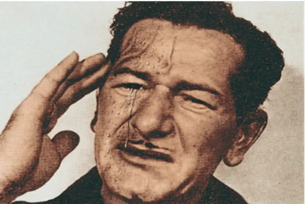
A patient of Bayard T. Horton. Horton is called as ‘father of cluster headache’. He
described features of cluster headache and he used oxygen for treatment. The
patient has some of the typical ‘leonine facies’ features recognized in cluster
headache: deep nasolabial folds, peau d’orange skin and squared jaw.
Table
8-DIAGNOSTIC ICHD CRITERIA FOR CLUSTER HEADACHE
Cluster headache
A]At least five attacks fulfilling criteria
B]Severe or very severe unilateral orbital, supraorbital, and/or temporal pain
lasting 15 to 180 min if untreated
C]Headache accompanied by at least one of the following:
Ipsilateral conjunctival injection and/or lacrimation
Ipsilateral nasal congestion and/or rhinorrhea
Ipsilateral eyelid edema
Ipsilateral forehead and facial sweating
Ipsilateral miosis and/or ptosis
A sense of restlessness or agitation
D]Attacks have a frequency of from one every other day to eight per day
Episodic cluster headache
A]Attacks fulfilling criteria A to E for 3.1 Cluster headache
B]At least two cluster periods lasting 7 to 365 days and separated by pain-free
remission periods of 1 month
Chronic cluster headache
A]Attacks fulfilling criteria A to E for 3.1 Cluster headache
B]Attacks recur over >1 year without remission periods or with remission periods
lasting <1 month
Paroxysmal
Hemicrania-These consists of daily attacks of severe unilateral orbital,
supraorbital, or temporal pain. The attacks are shorter, lasting 2 to 30 minutes, and
they occur 5 or more times per day. They are accompanied by ipsilateral eye
tearing and redness, eyelid edema, ptosis, nasal congestion or rhinorrhea, or a
combination of these. A major difference from CH is the female predominance
and the diagnostic response to therapeutic doses of indomethacin, up to 150
mg/day. The chronic form, which lasts more than 1 year without remission, was
first to be recognized, although episodic cases do occur.Paroxysmal hemicrania is
rare and appears in the middle years (mean, 33 years; range, 3 to 81 years). In
patients presenting with this unusual short duration headache, MRI is indicated. In
the presence of a normal study, a diagnostic trial of indomethacin should be given.
Short lasting Unilateral Neuralgiform Headache Attacks with Conjunctival Injection and Tearing
(SUNCT)-This rare syndrome is a diagnosis of exclusion. SUNCT is seen
unilateral orbital, supraorbital, or temporal pain lasting 5 to 240 seconds occur
with a frequency from 3 to 200 per day. They are accompanied by prominent
lacrimation and redness of the ipsilateral eye. Workup includes MRI scan with MR
angiography (MRA). No adequate therapy has been found for the treatment of
SUNCT. Trials have included carbamazepine, valproate, lamotrigine, azathioprine,
prednisone, nifedipine, and oral sumatriptan. It can be worsened by verapamil and
omeprazole. Mimics include vascular and compressive lesions affecting the
trigeminal nerve and posterior fossa or craniocervical lesions or malformations.
Other Primary Headaches
This collection of odd headaches includes many paroxysmal head pains that occur
spontaneously and often in odd circumstances.
Primary stabbing headache or ice-pick pains are transient and
localized stabs of pain in singles or series, predominantly in the distribution of the
first trigeminal division, lasting up to a few seconds in the absence of another
disorder. They are more common in patients with migraine or CH.
Cough headache is sudden and may be instantaneous or last up
to 30 minutes. It is brought on by coughing, straining, or Valsalva maneuver and
requires MRI scanning because it may be present with Arnold Chiari type I
malformations, carotid or vertebrobasilar disease, or aneurysms.
Primary exertional headache is a pulsating headache lasting
from minutes up to 2 days and is predictably precipitated by exercise. Heavy
exertion, such as that done by weight-lifters also is recognized as a cause. Workup
is the same as for cough headache. Indomethacin may be used in both of these.
Headache associated with sexual activity occurs during
orgasmic headache is explosive, occurs at orgasms, and with its first occurrence
mandates CT scan to rule out subarachnoid hemorrhage. Prevention of headache
may be attempted by using indomethacin, triptans, or standard antimigraine drugs.
Hypnic headache is a rare headache that occurs only during sleep,
lasts 15 minutes or more, and first occurs after age 50 years. Unlike CH, no
autonomic features are present. Caffeine and lithium are effective treatments.
Thunderclap headache resembles subarachnoid hemorrhage, with
sudden onset of maximal pain. CT is mandatory to rule out hemorrhage.
Hemicrania continua is a chronic and continuous unilateral headache
of moderate intensity lasting more than 3 months with conjunctival injection, nasal
congestion or rhinorrhea, or ptosis/miosis associated with episodic severe pain. It
is distinguished by its unique response to indomethacin.
New-daily persistent headache is a difficult and controversial entity
in which chronic continuous bilateral headache with a pressing or tightening
characteristic and no other classic migraine features starts one day and does not
end. The importance of ruling out secondary headaches, including high or low
CSF pressure, posttraumatic, infection, or new-onset chronic migraine, is
Secondary
Headaches-The International Classification of Headache Disorders recognizes
myriad secondary headaches. The majority of these do not have distinctive
features of pain in terms of location, duration, or quality, and yet for some,
associations make diagnosis and evaluation relatively straightforward. It is
recognized that a preexisting primary headache may worsen in frequency or
severity in relation to a secondary headache, making diagnosis and treatment more
difficult. In cases involving legal complications, this may present additional
challenges to the treating clinician. A selected group of these is presented for
review, as they may be seen by otolaryngologists.
Headache Attributed to Head and Neck
Trauma-Headache after head trauma becomes a problem most often when
it occurs in association with the other manifestations of posttraumatic syndrome:
depression, somatic preoccupation, and sleep disturbance. The headache can have
phenomenology similar to that of any of the primary headaches. Most patients
describe pain resembling tension-type headache. The key to treating this group of
disorders, especially in the chronic form, is to assess the patient as if he or she
were describing a primary headache and then to treat what you see and hear. The
presence of site of injury pain may be particularly difficult, and the treating
physician should be vigilant for signs and symptoms of CSF leak, intracranial
hypertension, or hydrocephalus.
Treatment consists of reassurance and medications used in
primary headache disorders. Unfortunately, acquired headache may not respond
with the same predictability as primary headache with the same phenomena.
and head position at the time of impact or whiplash.
A fascinating negative correlation exists between severity of
head trauma and complications of headache. Chronic posttraumatic headache
merges with the posttraumatic syndrome, which includes disturbances of
equilibrium, decreased concentration and work ability, depression, sleep
disturbances, and irritability. The role of litigation and ongoing legal
complications remains controversial. The treating clinician should establish a
positive relationship with the patient and assess early in the course the likelihood
of recovery. Additional workup and psychological or rehabilitative consultation
should be recommended when appropriate.In adolescents and young adults,
syncope may accompany the other changes. Episodes of altered consciousness
must be differentiated from posttraumatic epilepsy.
Headache and
Hematomas-Headache with epidural hematoma appears rapidly and resolves
within 3 months of evacuation. Headache associated with subdural hematoma
develops within 24 to 72 hours and may become chronic, even after evacuation. In
patients with acute and subacute hematomas, headache occurs in 11% to 53%.
After a chronic subdural hematoma, headache may occur in up to 80%. For an
elderly patient with new-onset headache, for which the trauma history may not be
elicited, the only other clue may be progressive cognitive impairment or subtle
neurologic signs. Treatment is symptomatic. Caution should be exercised when
using sedating or CNS-active medications in elderly or more-infirm patients with
cognitive sequelae.
Postoperative
surgeon or consultant. Premorbid primary headache, psychological and surgical
complications, or accompaniments of the primary illness or procedure complicate
diagnosis, testing, and treatment. Large series of outcome data for routine sinus
procedures are not readily available, but serious complications of major surgeries,
including combined procedures with neurosurgery, are addressed.
Postcraniotomy
Headache-In approximately 80% of patients undergoing craniotomy for
causes other than trauma, head pain develops, with resolution in most. In
one-fourth of those surgical patients, more chronic pain may develop. Posterior fossa
procedures, especially suboccipital craniotomies and retromastoid surgeries
performed for acoustic neuromas, may result in chronic headache. Headache is not
a common presenting complaint or symptom of acoustic neuroma. Patients report
headache as a chronic complication in 10% of patients who have surgery for an
acoustic neuroma (32). Additional complaints include hearing loss, facial
weakness, difficulty with balance, orbital pain, and changes in vision. In patients
with small tumors, headaches and balance problems predominate. The etiology of
the postcraniotomy headache appears to be related to chronic meningeal
inflammation, nerve entrapment, neuroma formation, and adhesions. Osteoplastic
cranioplasty may reduce the incidence of long-term complications by preventing
adhesion of muscle and fascia to the underlying dura.
Headache Attributed to Cranial or Cervical Vascular
Disorder-Headache is not a reliable indicator of ischemic
cerebrovascular disease or its results. Approximately one-fourth of patients with
cerebral infraction have head pain as a prominent acute symptom, with a greater
lacunar infarction or transient ischemic attacks. Headache is more common with
intraparenchymal hemorrhages. In those cases, emesis and focal signs of
neurologic dysfunction are almost always more prominent.
Acute Subarachnoid Hemorrhageand Thunderclap
Headache-The sudden onset of severe generalized headache, either the first or
the worst, should alert the practitioner immediately to the possibility of acute
subarachnoid hemorrhage (ASAH) and its dismal outcome, in which 50% may die
and an additional 50% of the survivors sustain significant long-term disability.
Patients may first be seen with focal and unilateral head pain accompanied by
nuchal rigidity, nausea, reduction in levels of consciousness, fever, and rarely
cardiac dysrhythmia. Grade 1 ASAH may appear with only mild to moderate
headache. Localization of the pain to the face is uncommon, although retroorbital
and supraorbital pain is often reported. If acute noncontrasted CT or
fluid-attenuated inversion recovery (FLAIR) MRI fails to demonstrate a cause for
hemorrhage, then a lumbar puncture is indicated. CSF remains xanthochromic for
2 weeks and is found in 70% of patients with ASAH 3 weeks after hemorrhage has
occurred. A third-nerve palsy associated with new-onset or thunderclap headache
may be the only sign of impending aneurysmal rupture.
Other vascular malformations, including ateriovenous
malformations (AVMs), are less specific and may appear with features of primary
headaches, including the trigeminal autonomic cephalalgias. Migraine with aura
has been reported in more than half of women with AVMs. A more usual
presentation is a seizure or strokelike symptoms.
In arterial dissections, headache is a prominent symptom and may
retinal signs, acute painful Horner syndrome, or tinnitus, carotid dissection should
be suspected, and duplex carotid sonography, MRA, and angiography should be
performed urgently. Nonsurgical treatment includes acute heparin followed by 3 to
6 months of warfarin with repeated studies, based on original presentation and
recovery.
Cerebral venous occlusions and sinus thrombosis appear with
headache as their most prominent feature and should be suspected in patients with
prothrombotic conditions including pregnancy and the postpartum, malignancy,
and primary or acquired blood diseases.
A last unique headache occurs in pituitary apoplexy, in which
patients may initially have severe acute, retroorbital, frontal, or diffuse headache
accompanied by nausea and vomiting, fever, diminished level of conscious,
hypopituitarism, hypotension, and ophthalmoplegia or impaired visual acuity. MRI
is more sensitive for detecting pathology in the area of the sella turcica, and the
headache and other signs usually resolve within 1 month. Treatment of the
headache should be symptomatic.
Giant Cell (Temporal)
Arteritis-Ninety-five percent of patients with temporal arteritis (giant cell
arteritis) are older than 60 years. They appear with complaints of daily moderate to
severe headache, scalp sensitivity, generalized fatigue, and feelings of being
unwell in nonspecific ways. Unilateral head pain is the rule, but the pain can be
bilateral or in the occipital region exclusively, reflecting the highly regional
distribution of the disease. Brief episodes of sharp, shooting head pain are
sometimes superimposed on a baseline of continuous dull, aching pain. Carotid
tender scalp arteries are found on palpation of the scalp in about half the cases.
This condition shares the same pathology as polymyalgia rheumatica, and many
patients have overlapping symptoms of extremity pain. The sedimentation rate is
elevated in all but the rarest of cases. Although scalp artery biopsies in the region
of the pain can confirm the diagnosis, skip lesions do occur, leading to
false-negative results. Patients respond with a dramatic reduction in head pain within
days of starting high-dose (60 mg) daily prednisone. Lack of a definite clinical
response to prednisone within several weeks makes the diagnosis much less secure
without a positive biopsy. Prednisone should be tapered after the initial response
and the sedimentation rate returns to normal. The disease may last 1 to 2 years,
during which maintenance steroids are continued. Visual loss is the major
complication and may occur in 30% of untreated cases. Other primary or
secondary intracerebral angiitides appear with fewer classic or diagnostic features
and are usually accompanied by signs attributable to stroke or more diffuse
encephalopathy, although headache occurs in 50% to 80%. In patients with a
history of immune or infectious disorders including human immunodeficiency
virus (HIV), MRI and MRA may be diagnostic, and spinal fluid may show an
increase in white cells and protein.
Headache Attributed to Nonvascular Intracranial Disorders-Intracranial Hypertension (Pseudo tumor Cerebri)
This syndrome is defined as a daily, diffuse, or constant
nonpulsating pain aggravated by coughing or straining in an alert patient with
papilledema, an otherwise normal neurologic examination (with the exception of
an enlarged blind spot, subtle visual field defect, or sixth nerve palsy), a normal
than 200 mm H2O in the nonobese (>250 mm H2O in the obese), and normal CSF
chemistries and cultures. MRI may show Chiari malformation. Intracranial
hypertension shares many of the symptoms that accompany a brain tumor,
including intermittent headache with variable or increasing intensity. Associations
may include predisposing mastoid or inner-ear infection; menstrual irregularity or
other endocrine abnormality; recent weight gain; or exposure to steroids, vitamin
A, tetracycline, or nalidixic acid. Tinnitus or transient visual obscurations or
dimming may be the first clinical symptom.
In a study of 85 patients with transformed migraine or chronic
daily headache, 12 patients were found to have increased intracranial pressure,
transient visual obscurations, or visual field defects. Their acute headaches
responded to ergots, dihydroergotamine or sumatriptan, and preventive
medications including acetazolamide or furosemide were effective. I recommend
spinal tap for all patients with chronic headache. Treatment includes weight
reduction, low-salt diet, and medications directed at reducing CSF production
(acetazolamide and furosemide, used separately or together). In chronic cases with
visual field loss not responding to diet and medications, CSF diversionary
procedures (including lumbo peritoneal or ventricular shunting or optic nerve
sheath fenestration) may be necessary. Sleep apnea should be considered, and
detailed sleep history and overnight sleep study are strongly recommended.
Headache Attributed to Low Cerebrospinal Fluid
Pressure-Low-pressure headache (LPH) is a symptomatic, secondary headache that can
appear hours and days after a surgical procedure, trauma, or spinal tap. In rarer
cases, it can occur spontaneously with a long clinical course. Clinical findings can
including those complaints typically associated with migraine. Diagnostic findings
on MRI consist of diffuse meningeal enhancement with or without Chiari
malformation(33).
Spinal tap may reveal low, absent, or negative pressure, with
normal fluid more the rule than not. Improvement with epidural blood patch
should be expected even if cause is unknown, although repeated treatments may be
necessary and incomplete recovery does occur. Spontaneous LPH may occur in
connective tissue disorders, and further evaluation should be considered in those
with historic or clinical signs of hyperextensible joints, Marfan syndrome, or
disorders of the large vessels including carotid or abdominal dissection.
Symptomatic treatment with nonspecific analgesics also is indicated. Spinal-tap
headache may occur in more than 20%, with the highest incidence in thin female
patients. No specific positioning of the patient after lumbar puncture (e.g., supine,
prone, or lateral decubitus) has been shown to reduce the occurrence of LPH.
Treatment of headache is symptomatic by using oral or intravenous caffeine or
standard analgesics. Blood patch performed by a trained anesthesiologist can be
curative. In chronic, severe LPH, radionuclide CSF flow studies and MRI or
standard myelography should be repeated. LPH represents the outcome of
hydrodynamic changes and mechanical forces influencing craniocervical
nociception and thus is a true secondary headache, no matter what its features.
Headache that worsens with standing and improves with recumbency remains an
important consideration in the evaluation of a patient with recent-onset or, in rarer
cases, chronic headache.
Infection of the Nervous System
problem with associated systemic illness, stiff neck, and fever. However,
nonspecific headache without the other findings can be part of the picture in
epidural abscess; fungal, tuberculous, or luetic meningitis; central nervous system
acquired immunodeficiency syndrome (AIDS); and meningeal sarcoidosis.
Diagnostic lumbar puncture with culture and antigen studies should be performed
after CT or MRI studies of the head have ruled out an intracranial mass.
Recommended studies include CSF VDRL, cryptococcal antigen, and
angiotensin-converting enzyme levels in addition to bacterial, viral, and tuberculosis cultures.
AIDS patients may have chronic headache in the absence of meningitis or cerebral
abscess. The headache is nonspecific and is most often a tension-type headache.
Any patient with immunocompromise and new-onset or subacute progressive
headache should be evaluated for infection. The patient with preexisting primary
headache and change in pattern, with systemic signs of infection, should be
evaluated as if no prior history existed. Nonspecific findings on CSF, including
low-grade lymphocytic pleiocytosis or increases in protein, may occur without
establishing a firm diagnosis. A rare but benign syndrome of migrainous headache
and CSF pleiocytosis has been recognized.
Headache or Facial Pain Attributed to Disorder of Cranium, Neck, Eyes, Ears, Nose, Sinuses, Teeth, Mouth, or Other Facial or Cranial
Structures-This large category of head, neck, and face pains remains
controversial, and many specialists in different areas disagree on the history of
these disorders. It must be remembered that the majority of patients with these
complaints will have primary headache, especially migraine. A detailed history
helps to rule out serious pathology of these vital structures. Monocular eye
retroorbital pain, increased intraocular pressure and conjunctival injection,
clouding of the cornea, or visual disturbance and resolves with treatment.
Refractive errors rarely cause adult headache. Ocular inflammatory disease such as
iritis, cyclitis, infection, trauma, or granulomatous infiltration may be
distinguished by anatomic site, temporal course, or type of inflammation.
Resolution follows proper treatment of the underlying cause.
Headaches attributed to disorders of the ear are always
accompanied by otalgia. Lesions of the outer ear, tympanic membrane, and middle
ear may give rise to primary otalgia and headache. Referred otalgia may result
from fifth, seventh, ninth, and tenth cranial nerves, all of which project to the
auricle, external auditory canal, tympanic membrane, and middle ear. Evaluation
should include physical examination and imaging with MRI.
Sinus
Headache-Sinus headache is accompanied by pain in the face, ears, or teeth
accompanied by clinical, endoscopic, and radiologic or laboratory evidence of
acute or acute-on-chronic rhinosinusitis that develops simultaneously and resolves
within 7 days after infection remits or is successfully treated. Clinical symptoms
include purulence in the nasal cavity, nasal obstruction, hyposmia, anosmia, and
fever. Chronic sinusitis is not validated as a cause of headache or facial pain unless
relapsing into an acute stage (34). Deviation of the nasal septum, hypertrophy of
turbinates, atrophy of sinus membranes, or mucosal contact points have not been
validated as causes of headache. Disease in the sinuses may appear with frontal
pain or pain radiating behind the eyes or to the vertex (frontal), over the antral or
(ethmoid), or between or behind the eyes or the vertex (sphenoid). In the absence
of rhinosinusitis, the recurrent, if even occasional headache sufferer, should be
considered for migraine diagnosis, referral, and treatment.
Sinusitis-Sinusitis is a clinical diagnosis based largely on history and
physical examination findings. In 1997, the Rhinosinusitis Task Force of the
American Academy of Otolaryngology – Head and Neck Surgery (AAO-HNS)
attempted to create a uniform diagnostic paradigm for sinusitis by organizing
common sinonasal symptoms and signs into major and minor factors. The
presence of two or more major factors or one major and two minor factors is
Table
9-Major and Minor Factors Associated with the Diagnosis of Rhino sinusitis Major factors
• Facial pain/ pressure
• Facial congestion/ fullness
• Nasal obstruction/ blockage
• Nasal discharge/ purulence/ discolored postnasal discharge
• Hyposmia/ anosmia
• Purulence in nasal cavity on examination
• Fever (acute rhinosinusitis only).
Minor Factors
• Headache
• Fever (in non-acute cases)
• Halitosis
• Fatigue
• Dental pain
• Cough
• Ear pain/ pressure/ fullness.
Acute rhinosinusitis is defined, in part, as having a duration of less than or
equal to 4-weeks.The next category is “sub-acute”, which is defined by a
[image:52.595.101.532.84.590.2]“Recurrent acute” is defined as four or more episodes of rhinosinusitis per year
with complete resolution between episodes.
Ultimately, however, the common pathway of acute sinusitis is
thought to be presence of bacteria in a sinus cavity with an obstructed ostium.
In addition, abnormalities of the quantity or consistency of the sinonasal
secretions ca