FORMULATION AND COMPARATIVE EVALUATION OF
PRONIOSOME HYDROCORTISONE GEL WITH MARKETED
FORMULATION
Dissertation Submitted to
The Tamilnadu Dr. M.G.R Medical University, Chennai
in partial fulfillment for the requirement of the Degree of
M
M
A
A
S
S
T
T
E
E
R
R
O
O
F
F
P
P
H
H
A
A
R
R
M
M
A
A
C
C
Y
Y
(
(
P
P
h
h
a
a
r
r
m
m
a
a
c
c
e
e
u
u
t
t
i
i
c
c
s
s
)
)
MARCH 2009
D
D
e
e
p
p
a
a
r
r
t
t
m
m
e
e
n
n
t
t
o
o
f
f
P
P
h
h
a
a
r
r
m
m
a
a
c
c
e
e
u
u
t
t
i
i
c
c
s
s
P
P
S
S
G
G
C
C
O
O
L
L
L
L
E
E
G
G
E
E
O
O
F
F
P
P
H
H
A
A
R
R
M
M
A
A
C
C
Y
Y
P
PEEEELLAAMMEEDDUU,, C
Prof. A.K. CHANDRASEKHARAN, M.Pharm.,
Principal,
PSG College of Pharmacy, Peelamedu
Coimbatore - 641 004. (T.N)
CERTIFICATE
This is to certify that the dissertation work entitled “FORMULATION AND COMPARATIVE EVALUATION OF PRONIOSOME HYDROCORTISONE
GEL WITH MARKETED FORMULATION” submitted by Mr. C. Praveen, is a bonafide work carried out by the candidate under the guidance of Mr. V. SANKAR M. Pharm., and submitted to the Tamil Nadu Dr. M.G.R. Medical University, Chennai, in partial fulfillment for the Degree of Master of Pharmacy in
Pharmaceutics at the Department of Pharmaceutics, PSG College of Pharmacy, Coimbatore, during the academic year 2008-2009.
Dr.
C. VIJAYARAGHAVAN, M. Pharm, Ph.D.,Head of the Department, Department of Pharmaceutics, PSG College of Pharmacy, Coimbatore.
CERTIFICATE
This is to certify that the dissertation work entitled “FORMULATION AND COMPARATIVE EVALUATION OF PRONIOSOME HYDROCORTISONE
GEL WITH MARKETED FORMULATION” submitted by Mr. C. Praveen, is a bonafide work carried out by the candidate under the guidance of Mr. V. SANKAR M. Pharm., and submitted to the Tamil Nadu Dr. M.G.R. Medical University, Chennai, in partial fulfillment for the Degree of Master of Pharmacy in
Pharmaceutics at the Department of Pharmaceutics, PSG College of Pharmacy, Coimbatore, during the academic year 2008-2009
DECLARATION
I do hereby declare that the dissertation work entitled “FORMULATION AND COMPARATIVE EVALUATION OF PRONIOSOME HYDROCORTISONE
GEL WITH MARKETED FORMULATION” submitted to the Tamil Nadu Dr. M.G.R. Medical University, Chennai, in partial fulfillment for the Degree of
Master of Pharmacy in Pharmaceutics, was done by me under the guidance of
Mr. V. SANKAR, M. Pharm., at the Department of Pharmaceutics, PSG College of Pharmacy, Coimbatore,during the academic year 2008-2009.
EVALUATION CERTIFICATE
This is to certify that the dissertation work entitled “FORMULATION AND COMPARATIVE EVALUATION OF PRONIOSOME HYDROCORTISONE
GEL WITH MARKETED FORMULATION” submitted by Mr. C. Praveen,
University Reg. No.26074658 to the Tamil Nadu Dr. M.G.R. Medical University, Chennai in partial fulfillment for the Degree of Master of Pharmacy in Pharmaceutics
is a bonafide work carried out by the candidate at the Department of Pharmaceutics, PSG College of Pharmacy, Coimbatore and was evaluated by us during the academic year 2008-2009.
Examination Center: PSG College of Pharmacy, Coimbatore.
Date:
Internal Examiner External Examiner
ACKNOWLEDGEMENT
I would like to express my most sincere gratitude to God’s grace, which has enabled me to finish my project work.
I would like to express my most sincere gratitude to Mr. V.Sankar, M.Pharm. Assistant professor, Department of pharmaceutics, for his critical comments, constructive suggestions, the effective training of my logical ideation and efficient communication skills during the whole research period
I greatly thank to Dr. C. Vijayaraghavan., M.Pharm., Ph.D., Vice principal and Head of department of pharmaceutics, for excellent instructions and selfless support in the research work.
I would like to express my respect and most sincere gratitude to Prof. A.K. Chandra sekharan, M.Pharm., for his excellence of support and providing facilities to me in our college and also in sister institutions.
My special thanks go to Mrs. S. Durgaramani, Assistant Professor, Mr. S. Subramanian, Lecturer, Department of Pharmaceutics, Mr. K.G. prasanth,
Assistant Professor, Department of Pharmacology, PSG College of Pharmacy,
and Mr. Balaji, Research scholar for their generosity, and kindly support for my dissertation to finish good and successful manner.
I would like to thank other teaching staff members in P.S.G College of pharmacy, and lab technicians, and other working staff, library for their self less support to me in research work.
And I would like to express my sincere gratitude to P.S.G College of sons and charity for providing all facilities to finish my dissertation successfully.
Happy to say thanks to my friends and beloved juniors for their precious help and joyful communication with me.
Last but not least, a huge thankfulness to my dear Mom and Dad for raising me well, always being there for me and always having faith in me.
Index
S.No Contents Page
No.
1 Introduction 1
2 Literature review 15
3 Drug profile 31
4 Polymer profile 35
5 Objective of work 46
6 Plan of work 48
7 Materials Methods 49
8 Experimental work 51
9 Results and discussion 90
10 Conclusion 96
11 Bibliography 97
INTRODUCTION
Dermal and transdermal delivery
The skin covers 1,4,5,9,13a total surface area of approximately 1.8m2 and provides the contact between the human skin and its external environment. This large and outermost layer of the human body is easily accessible and hence attractive as a non-invasive delivery route for selected drug compounds. Dermal and Transdermal drug delivery can have many advantages as compared to other routes of drug administration. (22)
Figure 1: Skin Structure
Dermal drug delivery is the topical application of drugs to the skin in the treatment of localized skin diseases. The advantage of dermal drug delivery is that high concentration of drugs can be localized at the site of action, reducing the systemic drug levels and therefore also reducing the systemic side effects (22). Transdermal drug delivery, on the other hand uses the skin as an alternative route for the delivery of systemically acting drugs. This drug delivery route for systemic therapy can have several advantages as compared to conventional oral drug administration. First of all, it circumvents the variables that could influence gastrointestinal absorption. Such as pH, food intake and gastrointestinal motility. Secondly it circumvents the first pass hepatic metabolism and is therefore suitable for drugs with a low bio-availability. Thirdly transdermal drug delivery can give a constant, controlled drug input. This would reduce the need for frequent drug intake, especially of drugs with a short biological half-life. Furthermore, variations in drug plasma levels can be avoided, reducing the side effects in particular of drugs with a narrow therapeutic window. Finally, transdermal drug delivery is easy and painless, which in turn will increase patient compliance.
Routes of drug penetration:
Drugs applied to the skin surface can serve two purposes. Dermal delivery is aimed at treating localized skin diseases. In this case, it is required that the drug penetrates the outer skin layers it reach its site of action within the skin, with little or no systemic uptake. On the other hand, transdermal delivery systems are designed to obtain therapeutic systemic blood levels. Hence, it is required that the drug reaches the dermal or transdermal drug delivery, the drug has to cross the outer layer of the skin, the stratum corneum. Since this layer is the main barrier of the skin, transport across the stratum corneum is the rate-limiting step in both dermal and transdermal; drug delivery.
There are two potential pathways for a molecule to across the stratum corneum: (a) the transappendagel route and (b) the transepidermal route (22). The transappendagel route involves transport of drugs via the sweat glands and the pilosebaceous units. This route bypasses the intact stratum corneum and is therefore also known as a “shunt”
Vesicle as skin delivery systems:
Vesicular Drug Delivery System is a novel approach having small spherical vesicles in which one or more aqueous compartments are completely enclosed by molecules that have hydrophilic and hydrophobic functionality such as phospholipids and cholesterol. These are varying in properties like composition, size, surface charge and method of preparation. They can be formed as single lipid bilayer or in multiple bilayer.These vesicular novel drug delivery systems are as a tool for the Dermal and Transdermal drug delivery.
Figure 2: Vesicle Formation
One of the methods to enhance drug transport across the skin is the use of vesicles. Vesicles are hollow colloidal particles, consisting of amphilic molecules. These amphiphilic molecules consist of a polar hydrophilic head group and a polar hydrophobic tail. Due to their amphiphilic properties, these molecules can form in the presence of excess of water one (uni lamellar vesicles/0 or more (multilamellar vesicles) concentric bilayer that surround an equal number of aqueous compartments. Both water-soluble and water-insoluble drugs can be entrapped into the vesicles. Hydrophilic drugs can be entrapped into the internal aqueous compartment. While lipophillic drugs can be entrapped in the vesicle bilayer or partition between the bilayer and the aqueous phase (22).
Figure 3: Vesicle structure
A wide variation of lipids and surfactants can be used to prepare vesicles. Most commonly, the vesicles are composed of phospholipids or non-ionic surfactants. These are referred to as liposomes and niosomes or non-ionic surfactants vesicles. The composition of the vesicles influences their physicochemical characteristics such as size, charge, phase state, lamellarity, and bilayer elasticity. These physicochemical characteristics in turn have a significant effect on the behaviour of the vesicles and hence also on their effectiveness as a drug delivery system
The rationale for using vesicles in dermal and transdermal drug delivery is manifold: ¾ Vesicles might act as drug carriers to deliver entrapped drug molecules into or
across the skin.
¾ Vesicles might act as penetration enhancers owing to the penetration of the individual lipid components into the stratum corneum and subsequently the alteration of the intercellular lipid lamellae within the skin layer.
¾ Vesicles might serve as a depot for sustained release of dermal active compounds.
¾ Vesicles might serve as a rate-limiting membrane barrier for the modulation of systemic absorption, hence providing a controlled transdermal delivery system.
To pursue optimal drug action, functional molecules could be transported by a carrier to the site of action and released to perform their task. Non-ionic surfactant vesicles known as niosomes are microscopic lamellar structures formed on admixture of a non-ionic surfactant, cholesterol and dicetyl phosphate with subsequent hydration in aqueous media. Niosomes are unilamellar or multilamellar vesicles of entrapping hydrophilic and hydrophobic solutes. These Niosomes can entrap solutes are quite stable, and require no special conditions and lack of many disadvantages associated with liposomes.
PRONIOSOMES AS A VESICULAR DRUG DELIVERY SYSTEM
A novel approach to minimize the physical instability of niosomes is
Proniosomes - derived niosomes for the delivery of poorly soluble drugs. This is based on liposome production method. These Proniosomes consist of maltodextrin powder coated with surfactant or a surfactant/drug mixture to yield a dry powder. Upon addition of hot water and brief agitation, the maltodextrin dissolves and the surfactant forms a suspension of multilamellar vesicles (niosomes) containing the poorly soluble drug. The niosomes slowly release drug in to solution (3). The proniosome powder can also be mixed with hydrogel powder. Adding hot water to the mixed powders allow formation of a hydrogel in which niosomes spontaneously form. The niosome-containing hydrogel can be formed as a gel that will degrade and release intact niosomes or as a stable gel, which slowly releases the drug from niosomes that remain in side the gel matrix.
Figure 4: Proniosomes Methodology
Proniosomes minimizes problems of niosomes physical stability such as aggregation, fusion and leaking and provide additional convenience in transportation, storage and dosing. Transdermal therapeutic system has generated an interest as this system provides the considerable advantage of a non-invasive parenteral route for drug therapy, avoidance of first pass gut and hepatic metabolism, decreased side effects and relative ease of drug input termination in problematic cases (2).
Dermatitis:
Dermatitis is a blanket term meaning any “inflammation of the skin” (e.g.
rashes, etc.) there are several different types of dermatitis. The different kinds usually have in common an allergic reaction to specific allergens. The term may be used to refer to eczema, which is also known as dermatitis eczema or eczematous dermatitis. A diagnosis of eczema often implies childhood or dermatitis, but without proper context, it means nothing more than a “rash”
Types of Dermatitis
Spongiotic dermatitis:
This pattern of skin reaction includes many other subtypes - irritant dermatitis, seborrheic dermatitis, atopic dermatitis, allergic contact dermatitis, thermal induced dermatitis, and drug induced dermatitis.
Childhood eczema:
Also known as atopic dermatitis or atopic eczema. This can be immunologic
Seborrhoeic dermatitis:
Seborrhoeic dermatitis is also known as dandruff. A rash of the scalp, face, and occasionally chest and groin. It is associated with a common yeast, Pityrosporum. It is treated with either an antiinflammatory or an antifungal agent, or both.
Psoriasis:
Psoriasis or psoriatic dermatitis is a pattern of dermatitis with distinct relationship to a defined entity, psoriasis. It can be familial, and is associated with arthritis.
Dyshidrotic dermatitis:
Dyshidrotic dermatitis is also known as Pompholyx. It is a pattern of spongiotic dermatitis presenting as small fluid filled or pus filled bumps on the hands and feet. The cause is unknown, but it has been highly associated with contact dermatitis (see Allergic Contact Dermatitis). Some cases are due to a food intolerance to nickel.
Urticaria:
Vesicular or bullous dermatitis:
This can be caused by drug reaction, or auto immune diseases. Examples includes Steven Johnson Syndrome, bullous erythema multiforme, bullous pemphigoid, and pemphigus vulgaris. Athlete foot fungus can also cause bullous dermatitis of the foot.
Papular urticaria:
A pattern of dermatitis often presenting after insect bite reactions. Flea bite dermatitis are often grouped around the ankles in a walking adult. In a crawling infant, it can be anywhere on the body.
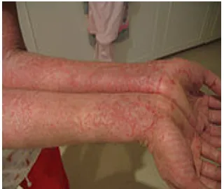
Eczema is a form of dermatitis, or inflammation of the epidermis. The term
eczema is broadly applied to a range of persistent skin conditions. These include
dryness and recurring skin rashes which are characterized by one or more of these symptoms: redness, skin edema (swelling), itching and dryness, crusting, flaking, blistering, cracking, oozing, or bleeding. Areas of temporary skin discoloration may appear and are sometimes due to healed lesions, although scarring is rare. In contrast to psoriasis, eczema is often likely to be found on the flexor aspect of joints.
Eczema
Figure - 5
Medications:
Dermatitis is often treated by glucocorticoid (a corticosteroid) ointments, creams or lotions. They do not cure eczema, but are highly effective in controlling or suppressing symptoms in most cases. Dermatitis is often treated by glucocorticoid (a corticosteroid) ointments, creams or lotions. They do not cure eczema, but are highly effective in controlling or suppressing symptoms in most cases.
Prolonged use of topical corticosteroids is thought to increase the risk of possible side effects, the most common of which is the skin becoming thin and fragile (atrophy).[6] Because of this, if used on the face or other delicate skin, only a low-strength steroid should be used.
LITERATURE REVIEW
¾ Yi-Hung Tsai a., et al., investigated the estradiol skin permeation from various proniosome gel formulations across excised rat skin in in-vitro studies. The encapsulation efficiency and size of niosomal vesicles formed from Proniosomes upon hydration were also characterized. The encapsulation (%) of Proniosomes with Span surfactants showed a very high value of ¥100%. Proniosomes with Span 40 and Span 60 increased the permeation of estradiol across skin. Both penetration enhancer effect of non-ionic surfactant and vesicle-skin interaction may contribute to the mechanisms for Proniosomes to enhance estradiol permeation. Niosome suspension (diluted Proniosomal formulations) and proniosome gel showed different behavior in modulating transdermal delivery of estradiol across skin. Presence or absence of cholesterol in the lipid bilayers of vesicles did not reveal difference in encapsulation and permeation of the associated estradiol. The types and contents of non-ionic surfactant in Proniosomes are important factors affecting the efficiency of transdermal estradiol delivery.
separation method. The formulated systems were characterized in vitro for size, vesicle count, drug entrapment, drug release profiles and vesicular stability at different storage conditions. Stability studies for proniosomal gel were carried out for 4 weeks. The method of proniosome loading resulted in an encapsulation yield of 66.7 - 78.7%. Proniosomes were characterised by transmission electron microscopy. In vitro studies showed prolonged release of entrapped captopril. At refrigerated conditions, higher drug retention was observed.It is evident from this study that proniosomes are a promising prolonged delivery system for captopril and have reasonably good stability characteristics.
optimization of drug encapsulation in the final formulation based on the type and amount of maltodextrin. This formulation of proniosomes is a practical and simple method of producing niosomes at the point of use for drug delivery.
¾ Jain. N.K., et al., investigated on development of proniosome based transdermal drug delivery system of levonorgestrel (LN) and extensively characterized both in vitro and in vivo. The proniosomal structure was liquid crystalline-compact niosomes hybrid which could be converted into niosomes upon hydration. The system was evaluated in vitro for drug loading, rate of hydration (spontaneity), vesicle size, polydispersity, entrapment efficiency and drug diffusion across rat skin. The effect of composition of formulation, amount of drug, type of Spans, alcohols and sonication time on transdermal permeation profile was observed. The stability studies were performed at 48C and at room temperature. The biological assay for progestational activity included endometrial assay and inhibition with the formation of corpora lutea. The study demonstrated the utility of proniosomal transdermal patch bearing levonorgestrel for effective contraception.
provided a higher ketorolac flux across the skin than did those prepared with Tween 20 (7- and 4-fold the control, respectively). A change in the cholesterol content did not affect the efficiency of the proniosomes, and the reduction in the lecithin content did not significantly decrease the flux (PO0.05). The encapsulation efficiency and size of niosomal vesicles formed by proniosome hydration were also characterized by specific high performance liquid chromatography method and scanning electron microscopy. Each of the prepared niosomes achieved about 99% drug encapsulation. Vesicle size was markedly dependent on the composition of the proniosomal formulations. Proniosomes may be a promising carrier for ketorolac and other drugs, especially due to their simple production and facile up
Contour plots were constructed to show the effects of X1, X2 and X3 on the PDE. A model was validated for accurate prediction of the PDE by performing checkpoint analysis. The computer optimization process and contour plots predicted the levels of independent variables X1, X2, and X3 (0, -0.158 and –0.158 respectively), for maximized response of PDE with constraints on vesicle size. The Box-Behnken design demonstrated the role of the derived equation and contour plots in predicting the values of dependent variables for the preparation and optimization of piroxicam proniosomes.
¾ El-laithy. H.M., et al., investigated on development of Novel approach for the preparation of controlled release proniosome-derived niosomes, using sucrose stearate as non-ionic biocompatible surfactants for the nebulisable delivery of cromolyn sodium. Conventional niosomes were prepared by a reverse phase evaporation method followed by the preparation of proniosomes by spraying the optimized surfactant–lipid mixture of sucrose stearate, cholesterol and stearylamine in 7:3:0.3 molar ratios onto the surface of spray dried lactose powder. Proniosome-derived niosomes were obtained by hydrating proniosomes with 0.9% saline at 50 ◦C and mixing for approximately 2 min. All vesicles were evaluated for their particle size, morphological characteristics, entrapment efficiency, in vitro drug release,
achieved with the proniosome-derived niosomes, where the t50% value of
the release profile was 18.1 h compared to 1.8 h. Moreover, high nebulisation efficiency percentage and good physical stability were also achieved. The results are very encouraging and offer an alternative approach to minimize the problems associated with conventional niosomes like degradation, sedimentation, aggregation and fusion
effective topical immunization. The proposed system is simple, stable and cost effective compared to liposomes.
their elasticity values were 124.4±4.2, 29.3±2.4 and 21.7±1.9, respectively. In vivo study revealed that topically given TT containing transfersomes, after secondary immunization, could elicit immune response (anti-TT-IgG) that was equivalent to one that produced following intramuscularly alum-adsorbed TT-based immunization. In comparison to transfersomes, niosomes and liposomes elicited weaker immune response. Thus transfersomeshold promise for effective non-invasive topical delivery of antigen(s).
diffusion cell where the experimental set up simulates in vivo application of the proliposomes under an occlusive condition. The nicotine flux from proliposomes was initially retarded compared with that of nicotine powder. The flux from proliposomes appeared to remain constant throughout the experimental period compared with that of nicotine powder, indicating that nicotine may be delivered across the skin in a sustained manner at a constant rate from proliposomes. These results, therefore, indicate that sustained transdermal delivery of nicotine is feasible using proliposomal formulations if the formulations are topically applied under occlusive conditions.
¾ Katare. O.P., et al., investigated the Dithranol is one of the mainstays in
drug-leakage study carried out at different temperatures of 4–8, 25_2 and 37 °C for a period of two months affirms that the drug leakage increased at a higher temperature. The in vitro permeation study using mouse abdominal skin shows significantly enhanced permeation with vesicles as indicated by flux of dithranol from liposomes (23.13 _g/cm2/h) and niosomes (7.78 _g/cm2/h) as compared with the cream base (4.10 _g/cm2/h).
¾ Barry. B.W., et al., studied on optimization of drug delivery through human skin is important in modern therapy. This review considers drug– vehicle interactions (drug or prodrug selection, chemical potential control, ion pairs, coacervates and eutectic systems) and the role of vesicles and particles (liposomes, transfersomes, ethosomes, niosomes).We can modify the stratum corneum by hydration and chemical enhancers, or bypass or remove this tissue via microneedles, ablation and follicular delivery. Electrically assisted methods (ultrasound, iontophoresis, electroporation, magnetophoresis and photomechanical waves) show considerable promise. Of particular interest is the synergy between chemical enhancers, ultrasound, iontophoresis and electroporation.
encapsulated enoxacin had been observed after selecting the appropriate formulations. The optimized formulations could also reserve a large amount of enoxacin in the skin. A significant relationship between skin permeation and the cumulative amount of enoxacin in the skin was observed. Both permeation enhancer effect and direct vesicle fusion with stratum corneum may contribute to the permeation of enoxacin across skin. Formulation with niosomes demonstrated a higher stability after 48 h incubation compared to liposomes. The inclusion of cholesterol improved the stability of enoxacin liposomes according to the results from encapsulation and turbidity. However, adding negative charges reduced the stability of niosomes. The ability of liposomes and niosomes to modulate drug delivery without significant toxicity makes the two vesicles useful to formulate topical enoxacin.
proniosome-derived niosomes are as good as or better than conventional niosomes.
¾ Işık Sarıgüllü Özgüney, Hatice Yeşim Karasulu., et al. Study was to evaluate and compare the in vitro and in vivo transdermal potential of w/o microemulsion (M) and gel (G) bases for diclofenac sodium (DS). The effect of dimethyl sulfoxide (DMSO) as a penetration enhancer was also examined when it was added to the M formulation. To study the in vitro potential of these formulations, permeation studies were performed with Franz diffusion cells using excised dorsal rat skin. To investigate their in vivo performance, a carrageenan-induced rat paw edema model was used. The commercial formulation of DS (C) was used as a reference formulation. The results of the in vitro permeation studies and the paw edema tests were analyzed by repeated-measures analysis of variance. The in vitro permeation studies found that M was superior to G and C and that adding DMSO to M increased the permeation rate. The permeability coefficients (Kp) of DS from M and M+DMSO were higher (Kp = 4.9 × 10⁻³ ± 3.6 × 10⁻⁴ cm/h and 5.3 × 10⁻³ ± 1.2 × 10⁻³ cm/h, respectively) than the Kp of DS from C (Kp = 2.7 × 10⁻³ ± 7.3 × 10⁻⁴ cm/h) and G (Kp = 4.5 × 10⁻³ ± 4.5 × 10⁻⁵ cm/h). In the paw edema test, M showed the best permeation and effectiveness, andM+DMSO had nearly the same effect as M. The in vitro and in vivo studies showed that M could be a new, alternative dosage form for effective therapy.
permeability to tritiated water measured to establish the integrity of the skin before the phthalate esters were applied to the epidermal surface. Absorption rates for each phthalate ester were determined and a second tritiated water permability assessment made to quantify any irreversible alterations in barrier function due to contact with the esters. Rat skin was consistently more permeable to phthalate esters than the human skin. As the estersbecame more lipophillic and less hydrophilic, the rate of absorption was reduced. Contact with the esters caused little change in the barrier properties of human skin, but caused marked increases in the permeability to water of rat skin. Although differences were noted between species, the absolute rates of absorption measured indicate that the phthalate esters are slowly absorbed through both human and rat skin. ¾ Sushama talegaonkar., et al., investigated on development of novel drug
were developed. Advances have since been made in the area of vesicular drug delivery, leading to the development of systems that allow drug targeting, and the sustained or controlled release of conventional medicines. The focus of this review is to bring out the application, advantages, and drawbacks of vesicular systems.
PROFILE OF THE DRUG USED IN THIS STUDY
HYDROCORTISONE
Hydrocortisone Short-acting glucocorticoid that depresses formation, release, and activity of endogenous mediators of inflammation including prostaglandins, kinins, histamine, liposomal enzymes, and complement system. Also modifies body's immune response (24)
Hydrocortisone is a topical corticosteroids constitute of primarily synthetic steroids used as anti-inflammatory and anti-pruritic agents
SYNONYM: Pregn-4-ene-3, 20-dione, 11, 17, 21-trihydroxy-, (11ß)-)
Molecular formula: C21H30O5
Molecular weight : 362.46
Potency : 0.004-0.005 µg/ml Melting Point : 211-214 °C (lit.)
CLINICAL PHARMACOLOGY:
Topical corticosteroids share anti-inflammatory, antipruritic and vasoconstrictive actions.
The mechanism of anti-inflammatory activity of the topical corticosteroids is unclear. Various laboratory methods, including vasoconstrictor assays, are used to compare and to predict potencies and/or clinical efficacies of the topical corticosteroids. There is some evidence to suggest that a recognizable correlation exists between vasoconstrictor potency and therapeutic efficacy in man (25).
Pharmacokinetics:
The extent of percutaneous absorption of topical corticosteroids is determined by many factors including the vehicle, the integrity of the epidermal barrier, and the use of occlusive dressings. Topical corticosteroids can be absorbed from normal intact skin. Inflammation and/or other disease processes in the skin increase percutaneous absorption. Occlusive dressings substantially increase the percutaneous absorption of topical corticosteroids.
INDICATIONS AND USAGE
Topical corticosteroids are indicated for the relief of the inflammatory and pruritic manifestations of corticosteroid-responsive dermatoses.
DIFFERENT DERMATITIS
Figure 6
CONTRAINDICATIONS
ADVERSE REACTIONS
The following local adverse reactions are reported infrequently with topical corticosteroids, but may occur more frequently with the use of occlusive dressings.
These reactions are listed in approximate decreasing order of occurrence:
Burning, itching, irritation, dryness, folliculitis, hypertrichosis, acneiform eruptions, hypo pigmentation, perioral dermatitis, allergic contact dermatitis, maceration of the skin, secondary infection, skin atrophy, striae and miliaria (25).
OVERDOSAGE
Topically applied corticosteroids can be absorbed in sufficient amounts to produce systemic effects.
DOSAGE AND ADMINISTRATION
Topical corticosteroids are generally applied to the affected area as a thin film from 2 to 4 times daily depending on the severity of the condition.
Polymer profile
Lecithin biochemistry also called
Phosphatidyl Choline:
Any of a group of phospholipids (phosphoglycerides) that is important in cell structure and metabolism. Lecithins are composed of phosphoric acid, cholines, esters of glycerol, and two fatty acids; the chain length, position, and degree of unsaturation of these fatty acids vary, and this variation results in different lecithin with different biological functions.
Pure lecithin is white and waxy and darkens when exposed to air. Commercial lecithin is brown to light yellow, and its consistency varies from plastic to liquid.
The term lecithin is also used for a mixture of phosphoglycerides containing principally lecithin, cephalin (specifically phosphatidyl ethanolamine), and phosphatidyl inositol.
Commercial lecithin, most of which comes from soybean oil, contains this mixture and, commonly, about 35 percent neutral oil. It is widely used as a wetting and emulsifying agent and for other purposes. Among the products in which it is used are animal feeds, baking products and mixes, chocolate, cosmetics and soaps, dyes,
insecticides, paints, and plastics.
Scientific names: 1,2-diacyl-sn-glycero-3-phosphatidylcholine
Lecithin is usually used as synonym for phosphatidylcholine, a phospholipid which is the major component of a phosphatide fraction which may be isolated from either egg yolk or soy beans. It is commercially available in high purity as a food supplement and for medical uses.
Lecithin is regarded as a well tolerated and non-toxic emulsifier. It is approved by the United States Food and Drug Administration for human consumption with the status "Generally Recognized As Safe". Lecithin is an integral part of cell membranes, and can be totally metabolised, so it is virtually non-toxic to humans. Other emulsifiers can only be excreted via the kidneys.
Lecithin is used commercially for anything requiring a natural emulsifier and/or lubricant, from pharmaceuticals to protective coverings. For example, lecithin is the emulsifier that keeps chocolate and cocoa butter in a candy bar from separating.
Sorbitan Esters (Sorbitan Fatty Acid Esters)
Functional Category
Emulsifying agent, non-ionic surfactant; solubilizing agent, wetting and dispersing/suspending agent (26).
Span 20
Non-proprietary Name
: Sorbitan monolaurateSynonym:
Arlacel 20; Armotan ML; Crill 1; Dehymuls SML; E493; Glycomul L; Hodag SML; Liposorb L; Montane 20; Protachem SML; Sorbester P12; Sorbirol L; sorbitan laurate; Span 20; Tego SML.Chemical name
: Sorbitan monododecanoateStructure:
Empirical formula
: C18H34O6Molecular weight
: 346Colour and form
: Yellow viscous liquid Safety: LD50 (rat, oral): 33.6 g/kg.Span 40
Non-proprietary Names
: Sorbitan monopalmitateSynonym
: 1,4-Anhydro-D-glucitol, 6-hexadecanoate; Ablunol S-40; Arlacel 40; Armotan MP; Crill 2; Dehymuls SMP; E495; Glycomul P; Hodag SMP; Lamesorb SMP; Liposorb P; Montane 40; Nikkol SP-10; Nissan Nonion PP-40R; Protachem SMP; Proto-sorb SMP; Sorbester P16; Sorbirol P; sorbitan palmitate; Span 40.Chemical name
: Sorbitan monohexadecanoateStructure:
Span 60
Non-proprietary Name
: Sorbitan monostearateSynonym
: Ablunol S-60; Alkamuls SMS; 1,4-Anhydro-D-glucitol, 6-octadecanoate; anhydrosorbitol monostearate; Arlacel 60; Armotan MS; Atlas 110K; Capmul S; Crill 3; Dehymuls SMS; Drewmulse SMS; Drewsorb 60K; Durtan 6O; Durtan 60K; E491; Famodan MS Kosher; Glycomul S FG; Glycomul S KFG; Hodag SMS; Lamesorb SMS; Liposorb S; Liposorb SC; Liposorb S-K; Montane 60; Nissan Nonion SP-60R; Norfox Sorbo S- 60FG; Polycon S60K; Protachem SMS; Prote-sorb SMS; S-Maz 60K; SMaz 60KHS; Sorbester P18; Sorbirol S; sorbitan stearate; Sorgen 50; Span 60; Span 60K; Span 60 VS; Tego SMS..Chemical name
: Sorbitan mono-octadecanoateStructure:
Empirical formula
: C24H46O6Molecular weight
: 431Colour and form
: Cream solidSafety
: LD50 (rat, oral): 31 g/kg.Span 80
Non-proprietary Name
: Sorbitan monooleateSynonym
: Ablunol S-80; Arlacel 80; Armotan MO; Capmul O; Crill 4; Crill 50; Dehymuls SMO; Drewmulse SMO; Drewsorb 80K; E494; Glycomul O; Hodag SMO; Lamesorb SMO; Liposorb O; Montane 80; Nikkol SO-10; Nissan Nonion OP-80R; Norfox Sorbo S-80; Polycon S80 K; Proto-sorb SMO; Protachem SMO; S-Maz 80K; Sorbester P17; Sorbirol O; sorbitan oleate; Sorgen 40; Sorgon S-40-H; Span 80; Tego SMO.Chemical name
: (Z)-Sorbitan mono-9-octadecenoateStructure:
Empirical formula
: C24H44O6Molecular weight
: 429Polyoxyethylene Sorbitan Fatty Acid Esters
Functional Category
Emulsifying agent, non-ionic surfactant, solubilizing agent, wetting, dispersing / suspending agent (26).
Tween 40
Non-proprietary Names
: Polysorbate 40Synonym
: Crillet 2; E434; Eumulgin SMP; Glycosperse S-20; Hodag PSMP-20; Lamesorb SMP-20; Liposorb P-20; Lonzest SMP-20; Montanox 40; poly(oxy- 1,2-ethanediyl) derivatives; Protasorb P-20; Ritabate 40; sorbitan monohexadecanoate; Sorbax PMP-20; Tween 40.Chemical name: Polyoxyethylene 20 sorbitan monopalmitate
Structure
Empirical formula
: C62H122O26Molecular weight
: 1284Colour and form
: Yellow oily liquidTween 60
Non-proprietary Names: Polysorbate 60
Synonym
Atlas 70K; Atlas Armotan PMS 20; Capmul POE-S; Cremophor PS 60; Crillet3; Drewpone 60K; Durfax 60; Durfax 60K; E435; Emrite 6125; Eumulgin SMS; Glycosperse S-20; Glycosperse S-20FG; Glycosperse S-20FKG; Hodag PSMS-20; Hodag SVS-18; Lamsorb SMS-PSMS-20; Liposorb S-PSMS-20; Liposorb S-20K; Lonzest SMS-20; Nikkol TS-10; Norfox SorboT-60 Montanox 60; Polycon T 60 K; polyoxyethylene 20 stearate; Ritabate 60; Protasorb S-20; Sorbax PMS-20; sorbitan monooctadecanoate poly(oxy-1,2-ethanediyl) derivatives; T-Maz 60; T-Max 60KHS; Tween 60; Tween 60K; Tween 60 VS (26).
Chemical name
: Polyoxyethylene 20 sorbitan monostearateStructure:
Emprical formula
: C64H126O26Molecular weight
: 1312Colour and form
: Yellow oily liquidTween 80
Non-proprietary Names
: Polysorbate 80Synonym
: Atlas E; Armotan PMO 20; Capmul POE-O; Cremophor PS 80; Crillet 4; Crillet 50; Drewmulse POE-SMO; Drewpone 80K; Durfax 80; Durfax 80K; E433; Emrite 6120; Eumulgin SMO; Glycosperse O-20; Hodag PSMO-20; Liposorb O-20; Liposorb O-20K; Montanox 80; polyoxyethylene 20 oleate; Protasorb O-20; Ritabate 80; (Z)-sorbitan mono-9-octadecenoate poly(oxy1,2- ethanediyl) derivatives; Tego SMO 80; Tego SMO 80V; Tween 80(26).Chemical name
: Polyoxyethylene 20 sorbitan monooleateStructure:
Emprical formula
: C64H124O26Molecular weight
: 1310Colour and form
: Yellow oily liquidSolubility:
Soluble in water and ethanol. Insoluble in mineral oilsSafety
: moderately toxic by IV route. Mildly toxic by ingestion. Eye irritation. Experimental tumorigen, reproductive effects.
Cholesterol
Non-proprietary Name
: CholesterolSynonyms
: Cholesterin; cholesterolum.Chemical Name
: Cholest-5-en-β-ol [57-88-5]Empirical Formula
: C27H46OMolecular Weight
: 386.67Structure
Functional Category
Emollient; emulsifying agent.
Applications in Pharmaceutical Formulation or Technology
Description
Cholesterol occurs as white or faintly yellow, almost odourless, pearly leaflets, needles, powder, or granules. On prolonged exposure to light and air, cholesterol acquires a yellow to tan colour.
Solubility
: Chloroform1 in 4.5 Acetone SolubleEthanol (95%) 1 in 78 (slowly) 1 in 3.6 at 80°C Ether 1 in 2.8
Methanol 1 in 294 at 0°C
Stability and Storage Conditions
Cholesterol is stable and should be stored in a well-closed container, protected from light.
Safety
Objective of the study
Hydrocortisone is given by topical application for its anti-inflammatory effect in allergic rashes, eczema and certain other inflammatory conditions.
Hydrocortisone is synthetic carticosteriod and it is also a proved anti-inflammatory drug. Hydrocortisone is available in different dosage forms for topical treatment.
Hydrocortisone is available with salt formations like hydrocortisone acetate, hydrocortisone butyrate,hydrocortisone sodium sucinate and other salt formations.
The available in market having trade names like 1. CUTISOFT cream 1 % w/w INNOVA (IPCA)
2. ENTOFORM cream CIPLA
3. HYDROCORT cream PFISCAR
4. TENDRONE cream YASH PHARMA
5. WYCORT ointment 2.5 % WYETH
Hydrocortisone is found to have 10% absorption through topical route. It is also affected by pharmacokinetic parameters like plasma half life, plasma protein binding. To improve the absorption of caroticosteroid thruough skin. Proniosome approach was tried using a less potent hydrocartisone.
¾ The objective of the study is to explore proniosomes for the delivery of hydrocortisone through transdermal route.
¾ To enhance the transport / permeation of drug through skin without side effects.
PLAN OF WORK
1. Construction of Standard Curve
2. Formulation of proniosome hydrocortisone gel in 1 % and 2.5 % by coacervation phase separation.
3. Determination of Vesicle size by optical microscopy. 4. Encapsulation efficiency by ultra centrifugation. 5. Drug content uniformity.
6. In-vitro drug release using sigma dialysis membrane.
7. Ex In-vivo studies using rat skin. 8. Anti inflammatory activity in mice Paw
9. Drug Permeation study in Human Skin by Prick test
Materials and Methods
Hydrocortisone USP from SAMARTH LABS
Soya lecithin (phosphatidyl choline) from Hi-Media laboratories Cholesterol from LOBA CHEMIE
Span 20,40,60,80 from LOBA CHEMIE Tween 40, 60, 80 from LOBA CHEMIE
Dialysis Membrane was purchased from Hi-Media Laboratories (Mumbai, India).
Instruments
Magnetic stirrer 2MLH by REMI EQUIPMENTS Rotary evaporator
Dialysis membrane 50 Himedia (Molecular weight cut-off ranges 12000 – 14000)
UV spectrophotometer 1650 PC Shimadzu Eppendroff Centrifuge 5415
pH Meter ELCO, LI 120
Experimental Work
Preparation of Standard Stock Solution
Accurately, weighed 10 mg of Hydrocortisone was dissolved in 5 ml of absolute alcohol to get a drug concentration (2 mg/ml). From this stock solution 1 ml solution was taken and transferred to 100 ml volumetric flask and made up the volume up to the mark with PBS pH 7.4 to obtain a standard stock solution of a drug concentration, 20 µg /ml.
Selection of Analytical Wavelength for Hydrocortisone
:The λmax of Hydrocortisone is determined by appropriate dilution of the standard stock solution with PBS pH 7.4, the solution was scanned using the double beam UV visible spectrophotometer (Model: UV- 1650 PC, SHIMADZU) in the spectrum mode between the wavelength range of 400 nm to 200 nm. The λmax of Hydrocortisone is found to 248 nm as the wavelength for further analysis.
Table 1
: Wavelength of spectrum
Standard Plot of Hydrocortisone:
Standard stock solution was further diluted to get the different concentrations like 2, 4, 6, 8, 10, 12, 14, 16, 18 and 20 µg/ml to determine the linearity range. Linearity was obtained in the above concentration at 248 nm using UV Spectrophotometer was shown in Table 1 & fig. 2.
Table 2: Standard Curve for Hydrocortisone
S.No. Concentration of Drug (µg / ml)
Absorbance at 248nm
Figure 8:
Standard Graph for hydrocortisone
Straight line equation Y = 0.06759 x + 0.01742 Correlation co-efficient r2 = 0.99917
0
0.2
0.4
0.6
0.8
1
1.2
1.4
1.6
0
2
4
6
8
10
12
14
16
18
20
Concentration (µg/ml)
A
Pre Formulation study:
Proniosomal gel was prepared by a coacervation-phase separation method .Precisely weighed amounts of surfactant, lecithin, cholesterol were taken in a clean and dry wide mouthed glass vial of 5.0 ml capacity and ethyl alcohol and water was added to it. After warming, all the ingredients were mixed well with a glass rod; the open end of the glass bottle was covered with a lid to prevent the loss of solvent from it and warmed over water bath at 60-70°C for about 5 min until the surfactant mixture was dissolved completely. Then the aqueous phase (0.1% glycerol solution) was added and warmed on a water bath till a clear solution was formed which was converted into Proniosomal gel on cooling (2).
Table 3:
Formulation using different ratios of Non ionic surfactant: S.No. Surfactant
Type
Ratio Soya lecithin (mg) Cholestero l (mg) Ethanol (ml) Water (ml)
1 S20:S40
1:9 1:1 9:1
100 100 2 0.5
2 S20:S60
1:9 1:1 9:1
100 100 2 0.5
3 S20:S80
1:9 1:1 9:1
Formulation procedure:
The proniosome hydrocortisone gel was prepared with 1 % and 2.5 % drug concentration with the same procedure described above using appropriate ratio of surfactants. The proniosome hydrocortisone gel formulation compositions are given in Table 4. (2)
Table 4: Composition of hydrocortisone gel formulation
S.No. Formulation Type Drug conc. Surfactant Type Ratio Lecithin (mg) Cholesterol (mg) Ethanol (ml) Water (ml)
1 PHG 1 1% S20:S40 1:9 100 100 2 0.5
2 PHG 2 1% S20:S60 1:9 100 100 2 0.5
3 PHG 3 1% S20:S80 1:9 100 100 2 0.5
4 PHG 4 1% S20:T40 1:9 100 100 2 0.5
5 PHG 5 1% S20:T60 1:9 100 100 2 0.5
6 PHG 6 1% S20:T80 1:9 100 100 2 0.5
7 PHG 7 2.5% S20:S40 1:9 100 100 2 0.5
8 PHG 8 2.5% S20:S60 1:9 100 100 2 0.5
9 PHG 9 2.5% S20:S80 1:9 100 100 2 0.5
10 PHG10 2.5% S20:T40 1:9 100 100 2 0.5
11 PHG 11 2.5% S20:T60 1:9 100 100 2 0.5
[image:63.612.106.569.254.627.2]Characterization of Proniosomal Gel
Vesicle Size Analysis:
Proniosomal hydrocortisone gel (100 mg) was hydrated in saline solution (0.9% solution) in a small glass vial with occasional shaking for 10 min. The dispersion was observed under optical microscope. The size of 50 vesicles was measured using a calibrated ocular and stage micrometer fitted in the optical microscope. Vesicle size is calculated using Equation 1. (2)
Number of divisions of stage micrometer
Size of each division = --- X 10 Æ (1)
[image:64.612.112.521.353.728.2]Number of divisions of eye piece micrometer
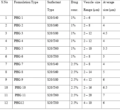
Table 5: Vesicle Size Determination by Optical Microscope S.No Formulation Type Surfactant
Type Drug conc. Vesicle size Range (µm) Average size (µm)
1 PHG 1 S20:S40 1% 2 – 6 3
2 PHG 2 S20:S60 1% 2 – 8 4
3 PHG 3 S20:S80 1% 2 – 12 4.5
4 PHG 4 S20:T40 1% 2 – 12 4
5 PHG 5 S20:T60 1% 2 – 10 3.5
6 PHG 6 S20:T80 1% 2 – 8 3
7 PHG 7 S20:S40 2.5% 2 – 8 4
8 PHG 8 S20:S60 2.5% 2 – 14 5
9 PHG 9 S20:S80 2.5% 4 – 12 6
10 PHG 10 S20:T40 2.5% 2 – 16 6.5
11 PHG 11 S20:T60 2.5% 2 – 20 7
Proniosome vesicle pictures for 12 different formulations are shown below.
Figure 9 Figure 10
PHG 1 (S20: 40 1%) in 40X PHG 2 (S20:60 1 %) in 40 X
Figure 11 Figure 12
PHG 3 (S20:80 1%) in 40X PHG 3 (S20:80 1%) in 10X
Figure 13 Figure 14
PHG 4 (S20:T40 1 %) in 40X PHG 4 (S20:T40 1 %) in 10X
Figure 15 Figure 16
PHG 5 (S20:T60 1%) in 40x PHG 6 (S20:T80 1%) in 40X
Figure 17 Figure 18
PHG 7 (S20:40 2.5 %) in 40X PHG 8 (S20:60 2.5 %) in 40X
Figure 19 Figure 20
PHG 9 (S20:80 2.5 %) in 40X PHG 10 (S20:T40 2.5 %) in 40X
Figure 21 Figure 22
PHG 11 (S20:T60 2.5 %) in 40X PHG 12 (S20:T80 2.5 %) in 40X
Encapsulation Efficiency
:Encapsulationof hydrocortisone drug in Proniosomal gel was evaluated by dispersing the Proniosomal hydrocortisone gel (100 mg) in distilled water and the dispersion was warmed gently for the formation of niosomes. Then the dispersion was centrifuged at 13000 rpm for 1hr at 50C. The supernatant was taken for the determination of free drug at 248 nm spectrophotometrically. (2)
The percentage encapsulation efficiency was calculated from Equation 2. % Encapsulation Efficiency = [(Ct - Cr)/Ct] X 100 Æ (2)
Where,
Ct – Concentration of total Hydrocortisone.
Table 6: Entrapment efficiency of Hydrocortisone
Drug content uniformity:
Formulated Proniosomal gel was mixed well and 100 mg of gel was weighed and transferred into vial. The gel was dissolved in 25 ml of phosphate buffer saline (pH 7.4) with vigorous shaking, and the solutions were assayed for hydrocortisone content at 248 nm. Amount Drug content present in 100 mg gel was calculated by Equation 3. (27).
Amount of drug = [(concentration) x (1) x (100) / 1000] Æ (3)
Table 7: Drug content uniformity of proniosome hydrocortisone gel
S.No. Formulation Type
Surfactant Type
Absorbance Concentration (mg)
Amt. of Drug (25ml)
% of drug
In vitro
release studies:
Table 8: In-vitro Release for S20:40 1 % S.No TIME
in min
Absorbance Concentration (µg/ml)
Concentration (mg)
Amt. release 50 ml
Cumulative % release 1 30 0.10266 1.224889 0.001225 0.061244 4.71111
2 60 0.22437 2.973847 0.002974 0.148692 11.43787
3 90 0.3417 4.659865 0.00466 0.232993 17.92256
Table 9: In-vitro Release for S20:60 1 % S.No TIME
in min
Absorbance Concentration (µg/ml)
Concentration (mg)
Amt. release
50 ml
Cumulative % release
1 30 0.03979 0.321454 0.000321 0.016073 1.236362
2 60 0.16846 2.170427 0.00217 0.108521 8.347795
3 90 0.24219 3.229918 0.00323 0.161496 12.42276
4 120 0.32214 4.37879 0.004379 0.21894 16.8415
Table 10: In-vitro Release for S20:80 1 % S.No TIME
in min
Absorbance Concentration (µg/ml)
Concentration (mg)
Amt. release
50 ml
Cumulative % release
Table 11: In-vitro Release for S20:T40 1 % S.No TIME
in min
Absorbance Concentration (µg/ml)
Concentration (mg)
Amt. release 50v ml
Cumulative % release
1 30 0.06445 0.675815 0.000676 0.033791 2.448607
Table 12: In-vitro Release for S20:T60 1 % S.No TIME
in min
Absorbance Concentration (µg/ml)
Concentration (mg)
Amt. release
50 ml
Cumulative % release
1 30 0.09998 1.186377 0.001186 0.059319 4.329844
2 60 0.42407 5.843512 0.005844 0.292176 21.32669
3 90 0.55322 7.699382 0.007699 0.384969 28.09993
Table 13: In-vitro Release for S20:T80 1 % S.No TIME
in min
Absorbance Concentration (µg/ml)
Concentration (mg)
Amt. release
50 ml
Cumulative % release
1 30 0.07598 0.8415 0.000842 0.042075 3.048914
2 60 0.32407 4.406524 0.004407 0.220326 15.96567
3 90 0.45332 6.263831 0.006264 0.313192 22.69504
Table 14: In-vitro Release for S20:40 2.5 % S.No TIME
in min
Absorbance Concentration (µg/ml)
Concentration (mg)
Amt. release
50 ml
Cumulative % release
1 30 0.06812 0.728553 0.000729 0.036428 2.464658
2 60 0.37146 5.087513 0.005088 0.254376 17.2108
3 90 0.41748 5.748814 0.005749 0.287441 19.44795
Table 15: In-vitro Release for S20:60 2.5 % S.No TIME
in min
Absorbance Concentration (µg/ml)
Concentration (mg)
Amt. release
50 ml
Cumulative % release
1 30 0.09131 1.06179 0.001062 0.05309 3.604177
2 60 0.39355 5.404943 0.005405 0.270247 18.34672
3 90 0.48633 6.738181 0.006738 0.336909 22.8723
Table 16: In-vitro Release for S20:80 2.5 % S.No TIME
in min
Absorbance Concentration (µg/ml)
Concentration (mg)
Amt. release
50 ml
Cumulative % release
1 30 0.05258 0.505245 0.000505 0.025262 1.766591
2 60 0.16797 2.163386 0.002163 0.108169 7.564285
3 90 0.27319 3.675384 0.003675 0.183769 12.85099
Table 17: In-vitro Release for S20:T40 2.5 % S.No TIME
in min
Absorbance Concentration (µg/ml)
Concentration (mg)
Amt. release
50 ml
Cumulative % release
1 30 0.09778 1.154764 0.001155 0.057738 4.037635
2 60 0.41675 5.738324 0.005738 0.286916 20.06407
3 90 0.63184 8.829142 0.008829 0.441457 30.87113
4 120 0.63184 8.829142 0.008829 0.441457 30.87113 5 150 0.76648 10.7639 0.010764 0.538195 37.63602
6 180 0.86035 12.1128 0.012113 0.60564 42.35246
Table 18: In-vitro release for S20:T60 2.5 % S.No TIME
in min
Absorbance Concentration (µg/ml)
Concentration (mg)
Amt. release
50 ml
Cumulative % release
1 30 0.0387 0.305791 0.000306 0.01529 1.0692
2 60 0.18958 2.473919 0.002474 0.123696 8.650065
3 90 0.25867 3.466734 0.003467 0.173337 12.12145
4 120 0.36536 4.999856 0.005 0.249993 17.48202
5 150 0.40076 5.50855 0.005509 0.275428 19.26066 6 180 0.43848 6.050582 0.006051 0.302529 21.15588 7 210 0.47278 6.543469 0.006543 0.327173 22.87926 8 240 0.49939 6.925851 0.006926 0.346293 24.21626
9 300 0.51819 7.196005 0.007196 0.3598 25.16086
Table 19: In-vitro Release for S20:T80 2.5 % S.No TIME
in min
Absorbance Concentration (µg/ml)
Concentration (mg)
Amt. release
50 ml
Cumulative % release
1 30 0.04968 0.463572 0.000464 0.023179 1.67961
2 60 0.39453 5.419026 0.005419 0.270951 19.63415
3 90 0.48633 6.738181 0.006738 0.336909 24.4137
Table 20: In-vitro Release for marketed 1 % Hydrocortisone cream S.No TIME
in min
Absorbance Concentration (µg/ml)
Concentration (mg)
Amt. release
50 ml
Cumulative % release
1 30 0.05273 0.5074 0.000507 0.02537 2.537002
2 60 0.53845 7.487139 0.007487 0.374357 37.43569
3 90 0.53308 7.409973 0.00741 0.370499 37.04986
Table 21:
Release comparison of 1 % PHG preparations & Marketed Hydrocortisone 1 % Cumulative % release of 1 % proniosome formulation S.No
.
Time in min
S20:S40 S20:S60 S20:S80 S20:T40 S20:T60 S20:T80
Hydrocort isone CREAM
Figure23:
Release of 1 % PHG Formulation comparing with Marketed -10 0 10 20 30 40 50 60 70
0 30 60 90 120 150 180 210 240 270 300 330 360 420 480
Time in min
[image:86.612.114.499.96.285.2]C u m u la ti ve % r e lease S20: 40 S20:60 S20: 80 S20: T40 S20: T60 S20: T80 Mark eted
Table 22: Release comparison of 2.5 % PHG preparations
Cumulative % release of 1 % proniosome formulations S.No Time in
mins S20:S40 S20:S60 S20:S80 S20:T40 S20:T60 S20:T80
Figure 24:
% release 2.5 % proniosome fromulations
0 10 20 30 40 50 60
0 30 60 90 120 150 180 210 240 300 360 420
Time in min
C u m u la ti v e % r e le a s e S20:S40 S20:S60 S20:S80 S20:T40 S20:T60 S20:T80
Kinetics for S20: 80 1 % PHG formulation:
Figure 25:Zero order reaction of S20:80 1% PHG formulation
0 20 40 60 80 100 120
0 30 60 90 120 150 180 210 240 300 330 360 420 480
Time in min
% r e ma in in g
[image:87.612.112.465.385.572.2]Figure 26:
First order reaction of S20: 80 1 % PHG formulation 0 0.5 1 1.5 2 2.5
0 30 60 90 120 150 180 210 240 270 300 330 360 420 480
Time in min
lo g o f % r e m a in in g
Regression coefficient r2 = 0.9631
Figure 27:
Higuchi plot of S20: 80 1 % PHG Formulation
0 10 20 30 40 50 60 70
0 5.4 7.7 9.4 10.9 12.2 13.4 14.4 15.4 16.4 17.3 18.1 18.9 20.4 21.9
Square root of time
cu m u la ti ve % rel eas e
[image:88.612.116.464.122.324.2]Figure 28:
PEPPAS plot of S20: 80 1% PHG formulation
0 0.2 0.4 0.6 0.8 1 1.2 1.4 1.6 1.8 2
0 1.4 1.7 1.9 2 2.1 2.2 2.3 2.43 2.47 2.5 2.55 2.62 2.68
Log Time L o g cu m u la ti ve % r e le a s e
[image:89.612.112.467.145.362.2]Ex In-vivo studies:
Institutional animal ethics committee (PSG institute of medical sciences and research) has grant approval for animal usage Reg No: 158 / 1999 / CPCSEA on 5th January, 2009.
Table 23:
Release comparison of 1 % PHG formulations using diffusion cell in rat skin
Cumulative % release of 1 % proniosome formulations S.No. TIME
in MINS
S20:40 S20:60 S20:80 S20:T40 S20:T60 S20:T80
Figure29:
1% PHG FORMULATION RELEASE IN DIFFUSION CELL
0 1 2 3 4 5 6 7 8
0 30 60 90 120 150 180 210 240 270 300 330 360 420 480 540
Time in min
C
u
m
u
la
ti
ve %
r
e
le
ase
S20: 40
S20: 60
S20: 80
S20: T40
S20: T60
Table 24:
Release comparison of 2.5 % PHG formulations using diffusion cell in rat skin
Cumulative % release of proniosome formulations S.No. TIME
in MINS
S20:40 S20:60 S20:80 S20:T40 S20:T60 S20:T80
Figure 30:
2.5% PHG FORMULATION RELEASE IN DIFUSSION CELL 0 1 2 3 4 5 6
0 30 60 90 120 150 180 210 240 270 300 330 360 420 480 540
Time in min
C u m u la ti v e % r e leas e S20: 40 S20: 60 S20: 80 S20: T40 S20:T60 S20: T80
Statistical analysis:
Comparison between the invitro and invivo results in S20:80 1 % proniosome hydrocortisone gel formulation was performed by analysis of variance (one way ANOVA with turkeys multiple camparision post test) with graph pad prism (version 3.0) software.
Anti inflammatory action:
Figure 31: Injecting carrageenan to induce inflammation in mice paw
Table 25: Animal Group Type
S.No Group Type No. of Animals
1 I 1 mice
2 II 3 mice
3 III 3 mice
Table 26: Comparative Anti-inflammatory activity in Mice
S.No. Group Type
Animals in group
Paw size before inflammation(mm) 0 hr(mm) 1hr (mm) 2hr (mm) 3hr (mm) 4hr (mm)
1 I 1 2.65 3.5 4.7 5.0 5.1 4.0
2 II 1 3.05 3.90 3.64 3.6 3.45 3.4
3 2 2.75 3.55 3.67 3.46 3.3 3.25
4 3 2.80 3.60 3.38 3.2 3.1 3.1
5 III 1 3.25 4 3.72 3.29 3.1 3.05
6 2 2.90 3.75 3.84 3.6 3.05 2.8
7 3 2.64 3.49 3.55 3.28 3.09 2.75
Drug permeation testing in human volunteers:
Prick test
: Anti inflammatory activity were evaluated in human volunteers using skin prick test. It was done for Control, Marketed formulation and Proniosomal formulation.The histamine wheal suppression was measured using scale. The ability of histamine wheal suppression of proniosome hydrocortisone gel 1% was compared with marketed hydrocortisone cream 1% and control. (26)
Table 27: Histamine Wheal Size using prick test in Human Volunteers
Histamine wheal size S.No. Formulation Type
Sample I (mm)
Sample II (mm)
Sample III (mm)
Average mean (mm)
1 Control 4 5 4 4.6
2 Marketed hydrocortisone 1%
4.5 6 4.5 5