“MANAGEMENT OF SECRETORY OTITIS MEDIA
– A COMPARATIVE STUDY”
Submitted in partial fulfillment of the requirements for
M.S. DEGREE BRANCH -IV OTORHINOLARYNGOLOGY
of
THE TAMILNADU DR. M.G.R. MEDICAL UNIVERISTY,
COIMBATORE MEDICAL COLLEGE
COIMBATORE
DECLARATION
I solemnly declare that the dissertation entitled “MANAGEMENT OF SECRETORY OTITIS MEDIA –A COMPARATIVE STUDY” is done by me at the Coimbatore Medical College Hospital, Coimbatore during 2007-2009 under the guidance and supervision of Prof.K.B.MOTHILAL , M.S., D.L.O.
This dissertation is submitted to The Tamilnadu Dr. M.G.R Medical University, towards partial fulfillment of regulation for the award of M.S. DEGREE (BRANCH–IV) in Otorhinolaryngology.
Place:
Date:
DR.D.Y.RAJ PRAKASH M.S. (E.N.T) post graduate, Coimbatore medical College,
ACKNOWLEDGEMENT
I am immensely grateful to Prof. K.B.MOTHILAL, M.S. D.L.O., Head of the department Department of ENT, for his valuable guidance, suggestions, encouragement and help in conducting this study.
I am greatly indebted to DR. V.ARAVINTHAN M.S., (E.N.T),DNB., Associate Professor, Coimbatore medical college, who encouraged and helped me throughout this study.
I would like to express my sincere gratitude to PROF.V.KUMARAN M.ch, The DEAN, Coimbatore Medical College, for having permitted me to use the hospital material in this study.
I express my sincere thanks to all the Assistant Professors, Dr. S. Dhanalakshmi, Dr. A.R. Ali sulthan, Dr.S. Muthuchitra, Dr.V. Saravanan, Dr.M. Vasudevan, Dr. P. Ezhilarasu for their
thoughtful guidance throughout the work.
I thank Mr.Narendiran, M.Sc., (Speech & Hearing ) our Audiologist & Speech therapist for his valuable assistance in this project.
I thank all my colleagues and friends for their constant encouragement and valuable criticism.
Last but not least, I express my gratitude for the generosity shown by all the patients who participated in the study.
CERTIFICATE
This is to certify that this dissertation entitled “MANAGEMENT
OF SECRETORY OTITIS MEDIA-A COMPARATIVE STUDY ” submitted by Dr. D.Y.RAJ PRAKASH, appearing for M.S. (E.N.T) Degree (Branch IV) examination in March 2010 is a bonafide record of work done by him under my guidance and supervision in partial fulfillment of regulations of the Tamil Nadu Dr. M.G.R. Medical University, Chennai. I forward this to the Tamil Nadu Dr.M.G.R. Medical University, Chennai, Tamil Nadu, India.
PROFESSOR & HOD, DEPARTMENT OF ENT,
COIMBATORE MEDICAL COLLEGE, COIMBATORE.
THE DEAN
CONTENTS
S.no Contents Page no.
1. INTRODUCTION 1
2. OBJECTIVES OF THE STUDY 3
3. REVIEW OF THE LITERATURE 4
4. MATERIALS AND METHODS 35
5. RESULTS AND ANALYSIS 39
6. DISCUSSION 51
7. SUMMARY 55
8. CONCLUSION 56
9. BIBLIOGRAPHY 57
10. PROFORMA 68
11. MASTER CHART 74
INTRODUCTION
Secretory otitis media is the most common cause of hearing impairment in children. It is defined as the persistence of serous or mucoid middle ear effusion for 12 weeks or more.1 It is also called as otitis media with effusion, catarrhal otitis media, exudative otitis media, seromucinous otitis media, non-suppurative otitis media. The term secretory is appropriate as it reflects a particular aspect of pathological process. The term otitis media with effusion allows differentiation of the type of effusion and facilitates distinction into serous and mucinous, acute and chronic forms.
OBJECTIVES:
1) To identify the distribution of age and sex in cases of secretory otitis media.
2) To find out the commonest predisposing factor.
3) To evaluate the different clinical parameters of secretory otitis media.
4) To compare the efficacy of medical management with various surgical procedures.
HISTORY
The problem of fluid in middle ear has been recognised for hundred of years. In 1869, Politzer described a condition that he termed ‘otitis media catarrhalis’in his classic book ‘The diseases of ear’. He recognised secretory and adhesive forms of the condition. The treatment that he advocated consisted of insufflation of air and paracentesis of the middle ear, which were intended to equalize atmospheric pressure on both sides of tympanic membrane. The principles of both ventilation and drainage of middle ear have remained the same in the management of middle ear effusion till date. This received little attention till second world war.
The clinical challenge of diagnosing this disorder was described by Hooper (1950). He elucidated the various clinical features of middle ear effusion.
EPIDEMIOLOGY:
The epidemiology of Secretory otitis media has been studied in several countries like Belgium, Holland , Denmark, Spain, U.K, U.S.A, and India.
PREVALANCE:
The prevalence of a condition is the proportion of a population that has a condition at any one time. It is an indicator of potential clinical workload. In children the main determinants are age of the child and season of the year.
AGE OF THE CHILD:
Secretory otitis media shows a bimodal prevelance with first peak at 2 year when the child attends a playgroup or nursery school, second peak at around 5years of age when most child attend a primary school.3 By the age of 7or 8 years, prevelance falls.
[
SEASON OF THE YEAR:
greater chances of passing them among children in winter.The prevelance of secretory otitis media in Mediterranean8,9 and subtropical countries 10,11 does not appear to be different overall from those in temperate countries.
HEREDITARY FACTORS :
In a same sex twin /triplet prospective cohort study Casselbrant et al 12 look at sets where zygosity was known. In children who had OME during the first two years of life, there was greater concordance in monozygotic twins in the number and duration than in dizygotic twins.
RACE:
GENDER:
Some multivariate studies report that there is no difference in boys and girls. In some study more risk in boys14,15 and others in girls.16
BREAST FEEDING:
In numerous studies, breast-feeding has been reported as reducing the risk of ear and respiratory infections, and hence, of OME . Maternal immunity has been proposed as the mechanism underlying this protective effect. Breast-fed infants carry reduced numbers of bacteria in their nasopharynx. A two-fold increase in risk of first episodes of AOM or OME was found in infants exclusively formula-fed as opposed to those breast fed for 6 months.
DAY CARE:
Attending day-care centres increases the risk of OME up to three times than for children cared for at home or by a child-care giver in a small group. The risk is also higher in families with a large number of siblings at home. The influences of season, day-care attendance and family size are likely to be inter-related variables, the common element being increased exposure to both viral and bacterial respiratory pathogens.
PARENTAL SMOKING:
Maternal, but not paternal smoking was shown to be associated with presence of middle ear effusion in the children (8–18 years) of British servicemen . In a large, longitudinal cohort study, a similar dependence on parent gender was found, suggesting a direct relation to dose18 . Controlling statistically for further influential factors, a systematic quantitative review from the UK (Strachan and Cook 1998) concluded that there is likely to be a causal relationship between parental smoking and both acute and chronic middle ear disease in children.
ANATOMY OF THE EUSTACHIAN TUBE & MIDDLE EAR CLEFT:
The Eustachian tube, middle ear cavity proper and the aditus with antrum, mastoid air cell system form the middle ear cleft.The eustachian tube lumen is wider at both the proximal (nasopharyngeal) and distal (middle ear) ends than in the midportion. The isthmus is the narrowest party of the eustachian tube.On the lateral wall of the nasopharynx, a prominence, the torus tubarius, protrudes into the nasopharynx. This prominence is formed by the abundant soft tissue overlying the cartilage of the eustachian tube. Anterior to this is the triangular nasopharyngeal orifice of the tube. From the torus, a raised ridge of mucous membrane, the salpingopharyngeal fold, descends vertically. On the posterior wall of the nasopharynx lie the adenoids, or pharyngeal tonsil, composed of abundant lymphoid tissue. Above the tonsil is a variable depression within the mucous membrane called the pharyngeal bursa. Behind the torus lies a deep pocket, extending to the nasopharynx posteriorly along the medial border of the eustachian tube. This pocket, the fossa of Rosenmuller, varies in height from 8 to 10 mm and in depth from 3 to 10 mm. Adenoid tissue usually extends into this pocket, giving soft-tissue support to the tube.
Nasopharynx & middle ear cleft
ear. The junction of the osseous tube and the epitympanum lies 4 mm above the floor of the tympanic cavity.
The osseous (protympanic or middle-ear) portion of the tube has a course that is linear anteromedially, following the petrous apex and deviating little from the horizontal plane. The lumen is roughly triangular, measuring 2 to 3 mm vertically and 3 to 4 mm along the horizontal base. The healthy osseous portion is open at all times, in contrast to the fibrocartilaginous portion, which is closed at rest and opens during swallowing or when forced open, such as during the Valsalva manoeuvre. The osseous and cartilaginous portions of the eustachian tube meet at an irregular bony surface and form an angle of about 160 degrees with each other. The medial wall of the bony portion of the eustachian tube consists of two parts, posterior (labyrinthine) and anterior (carotid) whose size, shape, and relation depend on the position of the internal carotid artery. The average thickness of the anteromedial portion is 1.5 to 3 mm, and in 2% of persons, the wall is absent, exposing the carotid artery.
cartilaginous tube is attached firmly at its posterior end to the osseous orifice by fibrous bands and usually extends some distance (3 mm) into the osseous portion of the tube. At its inferomedial end, it is attached to a tubercle on the posterior edge of the medial pterygoid lamina .
cartilage of the tube presses against the pharyngeal wall to form a prominent fold, the torus tubarius, which measures 10 to 15 mm thick . The torus is the site of origin of the salpingopalatine muscle and is the point of origin of the salpingopharyngeal muscle, which lies within the inferoposteriorly directed salpingopharyngeal fold .
DIFFERENCES BETWEEN PAEDIATRIC AND ADULT EUSTACHIAN TUBE
Infant Adult Length
Direction
Angulation with isthmus
Bony vs cartilagenous part
Tubal cartilage
Density of cartilage at hinge
13-18mm More horizontal. Forms an angle of 100 with horizontal.
Absent
Bony part longer than cartilaginous part
Flaccid
Less. Tubal closure less efficient
31-38mm
Slopes downwards forwards & medially. Forms angle of 450 with horizontal.
Present
Bony part-1/3 Cartilagenous -2/3
Rigid
AETIO –PATHOLOGY:
Anterior and inferior part of the middle ear cavity and Eustachian tube is lined by the ciliated, pseudostratified columnar epithelium of the respiratory tract .These cells along with goblet cells, mucous glands secrete mucus.This mucous is removed by mucociliary transport into the nasopharynx via the eustachian tube. Secretory otitis media is primarily caused by factors resulting in an overproduction of mucus, an impaired clearance of mucus or both. Both viral and bacterial infection can lead to the increased production and viscosity of secretions from the middle ear mucosa. Infection also leads to inflammatory edema of the mucosa, which may obstruct the eustachian tube. Temporary paralysis of cilia by bacterial exotoxins further impedes the clearance of an effusion.
INFECTION:
In a small study of middle ear effusions that produced “sterile” cultures, polymerase chain reaction (PCR) confirmed the presence of bacteria (like Haemophilus influenzae, Streptococcus pneumoniae and
Moraxella catarrhalis) in apparently sterile middle ear fluid.
The incidence of pathogens was higher in younger children (less than two years) and those with recurrent upper respiratory infections and recurrent attacks of otitis media.
The following table shows a positive bacteriological culture of middle ear aspirate of OME fluid (more than two months duration) in children with a wide spectrum of organisms21
Confocal laser scanning microscopy and vital dye examination of middle ear tissue biopsy samples taken at the time of ventilation tube insertion have demonstrated biofilm colonies in 80% of ears .
Koch’s postulates, using traditional methods of culturing bacteria as proof of infectious aetiology, is no longer a tenable approach to the confirmation of bacterial infection. More recent studies of culture-negative middle ear effusions found that PCR techniques could demonstrate bacterial mRNA as a marker for metabolically active organisms in culture-sterile middle ear effusions. In the context of a biofilm infection, the fulfilment of middle ear effusions, the inference is that bacteria, in a form that could not be previously demonstrated, are present even in “sterile” effusions, cultured by traditional techniques.
of biofilm-resistant surfaces, probiotic treatment, chemical or mechanical disruption of the protective glycocalyx of the biofilm and ultimately, modulation of the biofilm phenotype are all modalities of treatment for secretory otitis media that have been proposed.
.
EUSTACHIAN TUBE DYSFUNCTION:
Eustachian tube dysfunction can lead to poor aeration of middle ear cleft. Most common cause of damage to epithelium of Eustachian tube is secondary to viral upper respiratory tract infection, the other reasons can be secondary to an allergic reaction or pollutants (cigarette smoke), chronic nasopharyngeal infection in the adenoid or gastroesophageal reflux, disorder in palatine muscles.
ALLERGY:
media that looked for risk factors for persistence did not find atopy/allergy to be a significant factor. 24,25,26,27,28.
CRANIOFACIAL ABNORMALITIES:
Secretory otitis media is invariably seen in every child with cleft palate due to deficient palatal muscles and resultant poor Eustachian tube function.Surgical correction of cleft palate does not influence the incidence of secretory otitis media.Downs and Turner syndrome are also more likely to have secretory otitis media, but bifid uvula do not appear to predispose it.
TUMORS OF NASOPHARYNX :
All benign and malignant tumors of nasopharynx can lead to Eustachian tube blockade and cause secretory otitis media .
GASTRO-OESOPHAGEAL REFLUX: [
1000-fold greater than those in serum.30,31 Inflammation allows the establishment of a biofilm colony, resulting in persistence of the middle ear effusion. Inflammation secondary to acid reflux also promotes bacterial colonization.
SYMPTOMS AND SIGNS:
DIAGNOSIS: HISTORY:
History is not a reliable indicator of the current presence of secretory otitis media or the degree of hearing impairment. In a cohort study of 216 children followed from birth to 27 months of age at 3 months intervals the sensitivity of parental report of current OME and hearing improvement was poor. 32
Clinical diagnosis following a full history is made by examination of the ears and age-appropriate audiological testing.
[[[
OTOSCOPY:
secretory otitis media, and are not absolute indications for urgent surgical intervention.
Otoscopy alone is poorly predictive of the hearing loss associated with the presence of middle ear fluid, and while pneumatic otoscopy may aid diagnosis, audiometry is essential to assess the degree of hearing loss also serves as a marker for the wider impact and likelihood of resolution.
PNEUMATIC OTOSCOPY:
This can be done with either a hand held otoscope or with a siegle pneumatic speculum viewed with headlight illumination or microscope. Inability to achieve a seal with the available speculum can occur in upto 20% of children over 18 months.34 Overall there is a relative improvement in the diagnosis with pneumatic otoscopy.
American clinical practice guidelines have strongly recommended the use of pneumatic otoscopy as the primary diagnostic method for
OME.35,36,37 .
[
AUDIOLOGY:
sound field or with headphones and can also cooperate with speech testing using the toy identification test.
TYMPANOMETRY:
Tympanometry alone is a useful screening tool in the investigation of secretory otitis media. It is easy to use, provides reproducible results, is inexpensive, and is widely tolerated by patients—even young children. By measuring the compliance of the middle ear transformer mechanism, it provides an objective assessment of the status of the middle ear. Tympanometry produces a peak (ie, maximal compliance) when the pressure in the external ear canal equals that of the middle ear. By varying the pressure in the external ear, the tympanometer is able to provide information on the status of the middle ear. If there is an effusion in the middle ear, then compliance does not vary with changes in canal pressure, and a flat (Type B) tympanogram is produced. If the air in the middle ear is at or near atmospheric pressure, then a normal (Type A) tympanogram is produced. Negative middle ear pressure results in a Type C tympanogram, with the compliance peak being at less -99 da pa.
TYPE DESCRIPTION PEAKED
A between +200 and -99 dapa C1 between -100 and -199 dapa
C2 between -200 and -399 dapa
NON PEAKED
B no observable peak between +200 and -600 dapa
TREATMENT:
Any treatment regimen should take into account not only the severity of disease, but also the persistence of constant or intermittent disease, whether the condition is symptomatic.Such symptoms may concern hearing loss or its secondary effect on speech, language, cognition and behaviour.
Treatment should aim to relieve the symptoms caused by the middle ear condition, to resolve the underlying pathophysiological changes and to prevent recurrence and development of sequelae.
MEDICAL MANAGEMENT:
Medical treatment will be of immense value if it speeds the resolution of an episode of secretory otitis media. Most trials follow up children for one or two weeks after therapy. If at this point the therapy is ineffective, there is no reason for further follow up as it is unlikely to be of benefit thereafter. However, if it is effective after one or two weeks, then follow up for the recommended watchful waiting period of 12 weeks is necessary to see if it is of benefit in the longer term and might be used to reduce the proportion of children being considered for surgery.
ANTIBIOTICS:
were significantly greater than in the control groups. However, the magnitude of the difference was not great.43,44 The AHCPR 45 recommended a course of antibiotics (optional for children with asymptomatic OME) followed by at least a 1-month observation period. If signs of improvement are noted by otoscopy or tympanometry, additional observation may be warranted. Surgical treatment may be considered if the effusion persists and is associated with hearing loss.
ANTIHISTAMINES AND NASAL DECONGESTANTS:
To determine whether systemic medical therapy is of value in patients with secretory otitis media, Bluestone, Cantekin, and Beery 48 randomly assigned 553 infants and children with secretory otitis media to receive either a decongestant-antihistamine or placebo. The clearance of effusion did not differ between the two groups. As a consequence of this study, the routine use of decongestants in children with secretory otitis media has been abandoned.
CORTICOSTEROIDS:
weeks) even combined with antibiotics.51,52,53,54 Also steroid treatment has the potential complication of adrenal suppression and the development of varicella.Systemic steroids cannot be recommended at present for childhood secretory otitis media.
ADENOIDECTOMY
Adenoidectomy is being increasingly used for the treatment of secretory otitis media because recent studies have confirmed its effectiveness.55,56,57 Adenoidectomy was once the principal surgical treatment for OM. With the widespread use of tympanostomy tubes, however, adenoidectomy was used far less in certain countries. Probable reasons for this difference were the following: adenoidectomy takes longer time than tympanostomy tube insertion, adenoidectomy carries a risk of hemorrhage and other complications such as hypernasality, the mechanism of its effect on the middle ear is not well understood, and early studies did not demonstrate effectiveness.58,59,60
tube reflux. Although the proportion of children with otitis media caused by reflux is unknown, this rationale for adenoidectomy is logical. The association of enlargement with abnormality has been called into question, however, because adenoid and tonsil enlargement result from clonal expansion of immunocompetent cells.62 This suggests that the large adenoid may be more immunocompetent than the small adenoid because chronic infection is associated with cellular depletion. Brandtzaeg and Berdal63 demonstrated a general decline in immunocytes in the tonsils of patients with recurrent tonsillitis. Thus, current knowledge suggests that adenoid enlargement, a common phenomenon in 4- to 7-year-old children,64 does not necessarily indicate abnormality. Basing the rationale for adenoidectomy in children with otitis media on size alone, therefore, has little scientific basis. Further, clinical evidence from three separate studies 65,66,67 indicates that the effect of adenoidectomy is independent of adenoid size. Removal of a large adenoid is a clinically attractive option because it offers symptomatic relief to a compromised airway. The removal of a small, chronically infected adenoid offers no obvious benefit except in controlling otitis media.
of positive middle ear pressure after adenoidectomy but no change in the ability to equilibrate negative pressure and no change in the static opening pressure of the tube. Obstruction of the eustachian tube, either anatomic or functional, is a logical rationale for the procedure. Bluestone and others69 showed that obstruction of the eustachian tube is unusual in most children with secretory otitis media. The obstruction is functional in most cases. Eustachian tube function tests are not available except in research centers, and thus eustachian tube function is not routinely tested preoperatively. In children with hypercompliant eustachian tubes, adenoidectomy may increase reflux, particularly if the tissue in Rosenmüller's fossa is removed.70The third and most current rationale is removal of the chronically infected adenoid to eliminate a nasopharyngeal source of infection.71Adenoidectomy should result in a smooth lining of the nasopharynx, which should decrease bacterial colonization of the nasopharynx and, indirectly, the middle ear. Further research is needed to develop the methodology to identify subgroups of children who might benefit the most from adenoidectomy for control of infection.
the treatment groups that may have been clinically significant. Other older studies of adenoidectomy78,79,80 are not reviewed here because of severe flaws in experimental design.
MYRINGOTOMY:
Myringotomy with aspiration has not been shown to be effective in restoring the hearing levels in children with secretory otitis media.
Evacuation of the middle ear effusion by myringotomy and suction aspiration has been studied as one type of therapy. However, the results from this simple procedure have been disappointing.81 Most investigators and clinicians agree that if a child is to receive an anesthetic for such treatment, then tympanostomy tubes should be inserted or an adenoidectomy performed (or both) because the cost-benefit ratio for myringotomy and aspiration is too low to justify myringotomy as an independent procedure.
[
TYMPANOSTOMY TUBES
permanent perforation, focal drum atrophy, and recurrent effusion), while minor, are a cause of concern. The goal of using tympanostomy tubes is prolonged ventilation of the tympanum. Removal of the middle ear effusion and restoration of an aerated tympanum results in prompt return of hearing to preinfection levels in the vast majority of patients. Experimental evidence suggests that the mucosal hyperplasia of the tympanum will revert to a more normal condition with aeration.83Once the tubes are extruded, however, the clinical benefit appears to end.84
than adenoidectomy. They reserve adenoidectomy for cases of recurrent secretory otitis media.
SEQUELAE OF SECRETORY OTITIS MEDIA :
TOS CLASSIFICATION OF ATTIC RETRACTION POCKETS:
Stage I Mild retraction, with air still present between the pocket
and malleus neck.
Stage II Pocket touches malleus neck with or without erosion of
neck.
Stage III Pocket begins to expand: limited erosion of outer attic
Wall.
Stage IV More severe erosion ofouter attic wall ,pocket attached
to malleus head. [
SADE CLASSIFICATION OF TYMPANIC MEMBRANE ATELECTASIS :
Grade Description
Grade 1 The “retracted drum”
Grade 2 The drum is touching or adhering to the incus or stapes.
Grade 3 The drum is touching the promontory.
MATERIALS AND METHODS
[
This study was done in the Department of ENT, Coimbatore Medical College, Coimbatore. It consists of 50 patients and their age ranges from 4years to 50 years.
INCLUSION CRITERIA:
1. Patients with complaints of hard of hearing/discomfort or blocking sensation of the ear for more than 3 months.
2. Otoscopic evidence of secretory otitis media
3. An impendance audiometry with type ‘B’ or ‘C’ curve. [
Patients with combination of two or more of these criteria were included in the study.
EXCLUSION CRITERIA:
1. Patients with acute ear pain,ear discharge.
2. Patients deaf since childhood or with family history of hard of hearing.
A detailed history was recorded for all patients. Past surgical history was also noted. All patients were examined with a pneumatic otoscope and the findings were recorded in 3 formats as
1. Normal tympanic membrane
2. A thin semitransparent tympanic membrane with air bubble or fluid level.
3. A dull or yellowish/opaque,retracted/bulging lustreless
tympanic membrane with distorted or absent cone cone of light
Other clinical examination like tuning fork tests, nasal, oral cavity examination were performed. Patients with allergy, chronic sinusitis, chronic adenotonsilitis were segregated.
All patients were subjected to pure tone audiometry and graphical recording of their hearing threshold were made and the pure tone average in both ears were recorded. Tympanometry was done in all patients.
Examination under microscope was done to confirm the otoscopic findings. The predisposing factors for otitis media with effusion if present were noted.
One group of patients ( 25 Nos ) were given medical treatment for 6 weeks which included
1. Antibiotics,anti inflamatory drugs,antihistamines,nasal decongestants.
2. They were instructed to perform valsalva manoeuvre ,3-5 times a day.
Improvement in medical treatment is considered if 1. Patients have symptomatic relief.
2. Otoscopically tympanic membrane become normal
3. Pure tone audiometry showed good improvement in hearing air bone gap shows <10 dB
If patients satisfy these criteria, improvement was further confirmed by tympanometry.
Other group of patients (25 Nos ) were subjected to Surgical management which includes :
1. Myringotomy and grommet insertion
These patients were followed regularly.Success of the procedure was assessed by following means
1. Symptomatic improvement
2. Otoscopic evidence of improvement.
3. Air bone gap <10dB in postoperative audiogram done after 3 weeks.
RESULTS AND ANALYSIS
This study is the prospective analysis of the incidence, predisposing factors, clinical profile and the treatment outcome of medical and surgical management conducted in the Department of ENT, Coimbatore Medical College Hospital, Coimbatore.
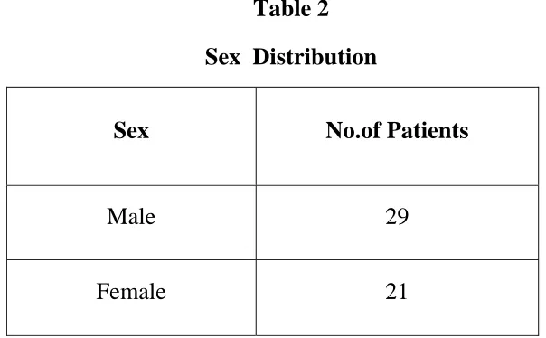
The demographic profile shows the most common age group affected was between 5 to 15 years. Of the 50 patients studied 58% (29 patients ) were male and 42% (21 patients ) were female. In the various age groups there was no significant difference between male and female patients.
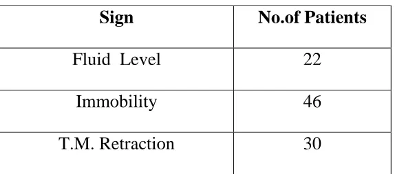
The most common symptom was hard of hearing which was seen in 68% of patients followed by ear fullness (54%), otalgia (48%), nasal symptoms (26%). On pneumatic otoscopic examination, most common sign was impaired tympanic membrane mobility which accounted for 92% of patients, followed by retracted tympanic membrane (60%), fluid level (44%).
Though cleft palate is a risk factor for secretary otitis media, we did not encounter any patient with cleft palate.
Majority of the patients had hearing loss in the range of 20-40 db (69%). About 77% of patients had B curve, 23% patients had ‘C’ curve.
Our patients were randomized into medical treatment arm and surgical treatment arm, and the results were analyzed in terms of symptomatic relief, pure tone audiogram results and pneumatic otoscopy. Out of 25 patients, who were taken up for medical treatment 60% of patients showed a significant reduction in the air bone gap with air bone gap less than 10 db as compared to the pretreatment values. 52% of the patients had their tympanic membrane returned to normal appearance. Only 20% of the patients had symptomatic relief.
TABLES & CHARTS
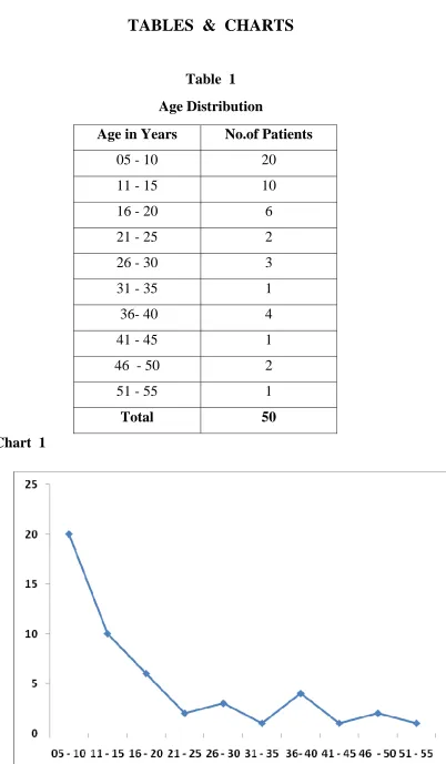
Table 1 Age Distribution
Age in Years No.of Patients
05 - 10 20
11 - 15 10
16 - 20 6
21 - 25 2
26 - 30 3
31 - 35 1
36- 40 4
41 - 45 1
46 - 50 2
51 - 55 1
Total 50
[image:48.612.116.518.54.743.2]Table 2 Sex Distribution
Sex No.of Patients
Male 29
Female 21
Table 3
Age Group /Sex Distribution
Age Male Female
1 – 10 10 10
11 – 20 9 7
21 – 30 4 1
31 – 40 3 2
41 – 50 1 2
51 – 60 1 0
Table 4 Symptomology
Symptoms No.of Patients
Hard of Hearing 34
Otalgia 24 Ear Fullness 27
Nasal Symptoms 13
Table 5 Signs
Sign No.of Patients
Fluid Level 22
Immobility 46 T.M. Retraction 30
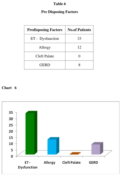
Table 6
Pre Disposing Factors
Predisposing Factors No.of Patients ET - Dysfunction 33
Allergy 12 Cleft Palate 0
GERD 8
Table 7
Medical management-Results Results of Medical
Treatment No.of Patients Symptomatic Relief 7
TM Normal 13
Air bone gap < 10 db 15
Table 8
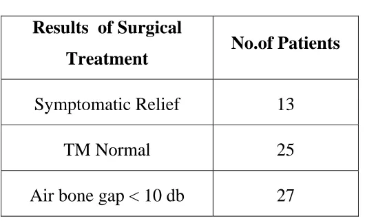
Surgical Management-Results Results of Surgical
Treatment No.of Patients Symptomatic Relief 13
TM Normal 25
Air bone gap < 10 db 27
Table 9 PTA Evaluation
Hearing Loss in db
No.of Patients
R L
20 – 30 13 14
31 – 40 22 20
41 – 50 10 7
51 – 60 3 0
61 - 70 0 0
Table 10
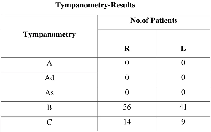
Tympanometry-Results
Tympanometry
No.of Patients
R L
A 0 0
Ad 0 0
As 0 0
B 36 41
[image:56.612.147.499.418.638.2]DISCUSSION
Zielhius et al3 reviewed about 23 studies which used tympanometry as one of the diagnostic tool to give age specific prevalence rates and found that the prevalence is bimodal with first peak, at 2 years. When child attends a play group or nursery school, second peak, at 5 years of age when the child attends a primary school. But in our study there was no bimodal prevalence. About 60% of the patient were in the age group 5-15 years.
According to Tos et al,4 Rovers et al,5 secretory otitis media shows a increased prevalence in temperate climate when compared to summer, probably due to increased incidence of upper respiratory tract infection. Engels et al,14 Rovers et al,15 has suggested that there is less more risk in boys when compared to girls. In our study also incidence was more in boys (58%)
Bluestone et al has proposed that there might be a casual relationship between parental smoking and both acute and chronic middle ear disease in children. But in some study by Engel, has found that when the other factors have no effect of parental smoking is detected.
Muscles. In our study, ET dysfunction was the major predisposing factor seen in 66% of patients followed by allergy (12%), GERD (8%).
American academy of paediatrics has strongly recommended the use of pneumatic otoscopy as the primary diagnostic method. Tympanometry alone is a useful screening tool in the investigation secretory otitis media.36
Wafters GW, Jones JE in published in clinical otolaryngology about the predictive values of tympanometry in the diagnosis secretory otitis media. A type B tympanogram has a high sensitivity (0.91) in predicting middle ear effusion with good specificity (0.3) type ‘C’ increases the sensitivity of predicting dry middle ear to 0.79. In our study, type B curve was found in 77% and type ‘C’ curve was found in 23%.
Lambert et al, Rovers proposed that there is a short term benefit with Prednisolone (1mg/kg) clearance of the effusion is sling and termporary
Basing the rationale for adenoidectoimy in children with Otitis media on size alone, has little scientific basis. But Gates et al, paradise et al, have demonstrated the effectiveness of adenoidectoimy in the management of secretary otitis media. Further clinical evidence from the above studies indicates the effect of adenoidectomy is independent of adenoid size. The other classic rationale is improvement in Eustachian tube function. Honjo 68 showed improvement in equilibration of positive middle ear pressure after adenoidectomy but no change in the ability to equilibrate negative pressure and no change in the static opening pressure of the tube. Obstruction of the Eustachian tube either anatomic or functional is a logical rationale for the procedure.
SUMMARY
CONCLUSION
From our study it is evident that secretory otitis media is a treatable cause of conductive hearing loss in children. In children, Eustachian tube dysfunction was secondary to functional or mechanical obstruction was the common precipitating factor for secretory otitis media. GERD was found to be associated with almost all adult patients. It has to be studied whether control of acid reflux can have any effect in these patients. From our study it was evident that medical management helps in the control of acute episodes of secretory otitis media which is associated with frequent relapse and recurrence.
BIBLIOGRAPHY
1. Bluestone CD. State of the art: definitions and classifications. In : Liu DJ, Bluestone CD, Klien JO, Nelson JD. (eds). Recent advances in otitis media with effusion. Proceedings of the 3rd International Conference. Ontario: Decker and Mosby; 1984.
2. Bennet Ke, Haggard MP ( 1999) Behaviour and Conginire outovells in middle ear duair . Arch Dis childhood 80:28-35.
3. Zielhuis GA, Rach GH, Van den Basch A, Van den Broek P. The prevalence of otitis media with effusion : a critical review of the Literature. Clinical Otolaryngology. 1990; 15:283-8
4. Tos M, Holm-Jensen S, Sorensen CH, Mogensen C. Spontaneous course and frequency of secretory otitis in 4-year-old children.
Archives of Otolaryngology. 1982; 108: 4-10.
5. Rovers MM, Straatman H, Zielhuis GA, Ingels K, van der Wilt GJ. Seasonal variation in the prevalence of persistent otitis media with effusion in one-year-old infants. Paediatric and Perinatal
Epidemiology. 2000; 14: 268-74.
6. Rovers MM, Stratman H, Ingels K, van der Wilt GJ, van den Broek P, Zielhuis GA. The effect of ventilation tubes on language development in infants with otitis media with effusion: A randomized trial. Pediatrics. 2000; 106: E42.
7. Midgley EJ, Dewey C, Pryce K, Maw AR. ALSPAC study team. The frequency of otitis media with effusion in British pre-school children: a guide for treatment. Clinical Otology. 2000; 25: 485-91. 8. Apostolopoulos K, Xenelis J, Tzagaroulakis A, Kandiloros D,
effusion among school children in Greece. International Journal of
Pediatric Otorhinolaryngology. 1998; 44: 207-14.
9. Marchisio P, Principi N, Passali D, Salpietro DC, Boschi G, Chetri G
et al. Epidemiology and treatment of otitis media with effusion in
children in the first year of life. Acta Otolaryngologica. 1998; 118: 557-62.
10. Saim A, Siam L, Siam S, Ruszymah BHI, Sani A. Prevalence of otitis media with effusion amongst pre-school children in Malaysia.
International Journal of Pediatric Otorhinolaryngology 1997; 41:
21-8.
11. Rushton HC, Tong CF, Yue V, Wormald PJ, van Hasselt CA. Prevalence of otitis media with effusion in multicultural schools in Hong Kong. Journal of Laryngology and Otology. 19.97; 111: 804-6. 12. Casselbrant ML, Mandel EM , Fall PA, Rockette HE, Kurs Lasky M. Bluestone CD et al. The heritability of otitis media ; a twin and triplet study. Journal of the American Medical Association. 1999; 282 :2125-30
13. Dewey C, Midgeley E, Maw R. The ALSPAC study team. The relationship between otitis media with effusion and contact with other children in a British cohort studied from 8 months to 3.5 years of age. International Journal of Pediatric Otorhinolaryngology.
2000; 55: 33-45. Good multivariate analysis.
14. Engel J, Anteunis L, Volovics A, Hendriks J, Marres E. Risk factors of otitis media with effusion during infancy. International Journal of
Pediatric Otolaryngology. 1999b; 48: 239-49. Good multivariate
15. Rovers MM, Hofstad EA, vann den Brand Kl, Ingels K, Vanden Wilt GJ, Zielhius GA. Prognostic factors for otitis media with effusion. Clinical Otolaryngology. 1998:23 543-6
16. Sassen ML, Brand R, Grote JJ. Risk factors for otitis media with effusion in children 0 to 2 years of age. American Journal of
Otolaryngology. 1997; 18: 324-30.
17. Bluestone CD. State of the art: definitions and classifications. In : Liu DJ, Bluestone CD, Klien JO, Nelson JD. (eds). Recent advances in otitis media with effusion. Proceedings of the 3rd International Conference. Ontario: Decker and Mosby; 1984.
18. Bluestone CD. State of the art: definitions and classifications. In : Liu DJ, Bluestone CD, Klien JO, Nelson JD. (eds). Recent advances in otitis media with effusion. Proceedings of the 3rd International Conference. Ontario: Decker and Mosby; 1984.
19. Engel J, Anteunis L, Volovics A, Hendriks J, Marres E. Risk factors of otitis media with effusion during infancy. International Journal of
Pediatric Otolaryngology. 1999b; 48: 239-49. Good multivariate
analysis including 19 pertinent risk factors.
20. Jero J, Karma P. Bacteriological findings and persistence of middle ear effusion in otitis media with effusion. Acta Otolaryngologica. 1997:529:22-6.
21. Alho Op, OJa H, Koivu M. Sorri M. Risk factors for Chronic otitis media with effusion in infancy. Archives of Otolaryngology- Head and Neck Surgery. 1995:112 695-9
23. Van Balen FAM, de Melker RA, Persistent otits media with effusion; can it be predicted ? A family practice follow - up study in Children aged 6 months to 6 years. Journal of Family practice 2000 ;49: 605 -11
24. Medical Research council Multicentre Otitis Media Study Group. Risk factors for persistence of bilateral otitis media with effusion. Clinical Otolaryngology 2001; 27:147-56
25. Medical Research Council Multicentre Otitis Media Study Group. Selecting persistent glue ear for referral in general practice; a risk factor approach. Bbritish journal of General practice. 2002; 52:549-53
26. Medical Research Council Multicentre Otitis Medial Study Group. Pars tensa and pars flaccid retraction in persistent otitis media with effusion. Otology and Neurtology. 2001 22:291-8
27. Medical Research Council Multicentre Otitis Media Study Group. Surgery for persistent otitis media with effusion; generalizability of results from the UK trial (TARGET).Clinical Otolaryngology. 2001 :26:417-24.
28. Sheanan, P. Blaney AW, Sheanan NJ, Earley MJ. Sequelae of otitis media with effusion among children with cleft lip and /or palate. Clinical Otolaryngology. 2002:27 494-500
29. Taker A, Dettmar PW, Panetti M, Koufman JA, Birchall JP, pearson JP. Reflux of gastric juice and glue ear in children. Lancet. 2002:359:493
31. Lie JEC, Nuthappan BS, Uppaluri R. Association of reflux with otitis media in children. Otolaryngology- Head and Neck Surgery. 2005:133: 360-65
32. Anteunis LIC, Engel JAM, Hendriks JJT, Manni JJ.A Longitudinal Study of the Validity of parental reporting in the detection of otitis media and related hearing impairment in infancy. Audiology. 1999:38:75-82.
33. Steeart MG, Friedman EM, Sulek M. Duncan No, Fernandex AD, Bautista MH. Is parental perception an accurate predictor of childhool hearing loss A Prospective Study . Otolaryngology and Head and Neck Surgery. 1999: 120:340- 4.
34. Cavanaugh RM. Obtaining a seal with otic specula : Must we rely on an air of uncertainty? Pediatrics. 1991:87 :114-6.
35. American Academy of Pediatrics . Otitis media with effusion ; clinical practice guideline. Pediatrics.2004:113: 1412-29 OME guidelines.
36. Rosenfeld RM, Culpepper L, Doyle Kl. Grundfast KM, Hoberman A, kenna MA et al. Clinical practice guideline: Otitis media with effusion. Otalaryngology and head and Neck Surgery. 2004:130: S95-118.
37. Preston K. Pneumatic Otoscopy : a review of the Literature . issue incomprehensive Pediatric Nursing . 1998:21 117-28 . Good Literature review.
38. Liu YS: Microorganisms in chronic otitis media with effusion.
Ann Otol Rhinol Laryngol 1976; 85:245-249.and others
40. Healy GB: Antimicrobial therapy of chronic otitis media with effusion. Int J Pediatr Otorhinolaryngol 1984; 8:13-17.
41. Mandel EM: Efficacy of amoxicillin with and without decongestant-antihistamine for otitis media with effusion in children. N Engl J Med 1987; 316:432-437.and others
42. Thomsen J: Antibiotic treatment of children with secretory otitis media. Arch Otolaryngol Head Neck Surg 1989; 115:447-451.and others
43. Rosenfeld RM, Post JC: Meta-analysis of antibiotics for the treatment of otitis media with effusion. Otolaryngol Head Neck Surg 1992; 106:378-386
44. Williams RL: Use of antibiotics in preventing recurrent acute otitis media and in treating otitis media with effusion: a meta analytic attempt to resolve the brouhaha. JAMA 1993; 143:1414-1418.and others
45. Stool SE: Otitis media with effusion in young children. Clinical Practice Guideline Technical Report No. 12 AHCPR Pub No
94-0622, Rockville, Maryland: Agency for Health Care Policy and Research, Public Health Services, U.S. Department of Health and Human Services; 1994:192-208.
46. Presswood G: Effect of artificial airway on ear complications from hyperbaric oxygen. Laryngoscope 1994; 104:1383-1384.and others 47. Perrin JM: Sulfisoxazole as chemoprophylaxis for recurrent otitis
media. N Engl J Med 1974; 291:664-667.and others
48. Bluestone CD, Cantekin EI, Beery QC: Certain effects of adenoidectomy on eustachian tube ventilatory function.
49. Lambert PR: Oral steroid therapy for chronic middle ear effusion: a double-blind crossover study. Otolaryngol Head Neck Surg 1986; 95:193-199.
50. Rovers MM: The effect of ventilation tubes on language development in infants with otitis media with effusion: a randomized trial. Pediatrics 2000; 106:E42.and others
51. Paradise JL, Smith GC, Bluestone CD: Tympanometric detection of middle ear effusion in infants and children.
Pediatrics 1976; 58:198-210
52. Perrin JM: Sulfisoxazole as chemoprophylaxis for recurrent otitis media. N Engl J Med 1974; 291:664-667.and others
53. Pichichero ME: Diagnostic accuracy, tympanocentesis training performance, and antibiotic selection by pediatric residents in management of otitis media. Pediatrics 2002; 110:1064-1070
54. Pichichero ME, Berghash LR, Hengerer AS: Anatomic and audiologic sequelae after tympanostomy tube insertion or prolonged antibiotic therapy for otitis media. Pediatr Infect Dis J 1989; 8:780-787.
55. Gates GA: Effectiveness of adenoidectomy and tympanostomy tubes in the treatment of chronic otitis media with effusion. N Engl J Med 1987; 31:1444-1451.and others
56. Maw AR: Chronic otitis media with effusion (glue ear) and adenotonsillectomy: a prospective randomized controlled study. Br
Med J 1983; 127:1586-1588
57. Paradise JL: Efficacy of adenoidectomy for recurrent otitis media in children previously treated with tympanostomy-tube placement. Results of parallel randomized and nonrandomized trials.
58. Fiellau-Nikolajsen M, Felding J, Fischer H: Adenoidectomy for eustachian tube dysfunction: long-term results from a randomized
controlled clinical trial. In: Lim DL, ed. Recent advances in otitis
media with effusion, Philadelphia: BC Decker; 1983. and others
59. . Roydhouse N: Adenoidectomy for otitis media with mucoid effusion. Ann Otol Rhinol Laryngol 1980; 89:312-315.
60. Widemar L: The effect of adenoidectomy on secretory otitis media: a 2-year controlled prospective study. Clin
Otolaryngol 1985; 10:345-350.and others
61. Bluestone CD, Cantekin EI, Beery QC: Certain effects of adenoidectomy on eustachian tube ventilatory function.
Laryngoscope 1975; 85:113-127.
62. Fujiyoshi T: Functional architecture of the nasopharyngeal tonsil.
Am J Otolaryngol 1989; 10:124-131.and others
63. Brandtzaeg Jr LS, Berdal P: Immunoglobulin system of human tonsils I. Control subjects of various ages: quantification of Ig producing cells, tonsillar morphometry and serum Ig concentration.
Clin Exp Immunol 1978; 31:367-387.
64. Fujioka M, Young LW, Girdany BR: Radiographic evaluation of adenoidal size in children: adenoidal-nasopharyngeal. Am J
Radiol 1979; 133:401-404.
65. Gates GA: Effectiveness of adenoidectomy and tympanostomy tubes in the treatment of chronic otitis media with effusion. N Engl J Med 1987; 31:1444-1451.and others
66. Maw AR: Chronic otitis media with effusion (glue ear) and adenotonsillectomy: a prospective randomized controlled study. Br
Med J 1983; 127:1586-1588
Results of parallel randomized and nonrandomized trials.
JAMA 1990; 263:2066-2073.and others
68. Honjo I: Eustachian tube and middle ear diseases, Tokyo, Spring-Verlag, 1988.
69. Bluestone CD, Cantekin EI, Beery QC: Certain effects of adenoidectomy on eustachian tube ventilatory function.
Laryngoscope 1975; 85:113-127.
70. Bluestone CD: Eustachian tube function as related to adenoidectomy for otitis media. Trans Am Acad Ophthalmol
Otolaryngol 1972; 76:1325-1354.and others
71. Gates GA, Avery CA, Prihoda TJ: Effect of adenoidectomy upon children with chronic otitis media with effusion.
Laryngoscope 1988; 98:58-63.
72. Gates GA: Effectiveness of adenoidectomy and tympanostomy tubes in the treatment of chronic otitis media with effusion. N Engl J Med 1987; 31:1444-1451.and others
73. Maw AR: Chronic otitis media with effusion (glue ear) and adenotonsillectomy: a prospective randomized controlled study. Br
Med J 1983; 127:1586-1588.
74. Paradise JL: Efficacy of adenoidectomy for recurrent otitis media in children previously treated with tympanostomy-tube placement. Results of parallel randomized and nonrandomized trials.
JAMA 1990; 263:2066-2073.and others
75. Fiellau-Nikolajsen M, Felding J, Fischer H: Adenoidectomy for eustachian tube dysfunction: long-term results from a randomized
controlled clinical trial. In: Lim DL, ed. Recent advances in otitis
media with effusion, Philadelphia: BC Decker; 1983. and others
77. McKee WJE: The part played by adenoidectomy in the combined operation of tonsillectomy and adenoidectomy. Second part of a controlled study in children. Br J Prev Soc Med 1963; 17:133-140. 78. Mawson SR, Adlingon P, Evans M: A controlled study of
adenotonsillectomy in children. J Laryngol Otol 1967; 81:777-790. 79. McKee WJE: A controlled study of the effects of tonsillectomy and
adenoidectomy in children. Br J Prev Soc Med 1963; 17:49-69. 80. McKee WJE: The part played by adenoidectomy in the combined
operation of tonsillectomy and adenoidectomy. Second part of a controlled study in children. Br J Prev Soc Med 1963; 17:133-140 81. Mandel EM, Bluestone CD, Paradise JL: Myringotomy with and
without tympanostomy tube insertion in the treatment of chronic otitis media with effusion. Arch Otolaryngol Head Neck Surg 1989; 115:1217-1224.
82. Armstrong BW: A new treatment for chronic secretory otitis media.
Arch Otolaryngol 1954; 9:849.654
83. Schneider ML: Bacteriology of otorrhea from tympanostomy tubes.
Arch Otolaryngol Head Neck Surg 1989; 115:1225-1226
84. Gates GA, Avery CA, Prihoda TJ: Effect of adenoidectomy upon children with chronic otitis media with effusion.
Laryngoscope 1988; 98:58-63.
85. Kilby D, Richards SH, Hart G: Grommets and glue ears. Two-year results. J Laryngol Otol 1972; 86:881-888.
86. Leek JH: Middle ear ventilation in conjunction with adenotonsillectomy. Laryngoscope 1979; 89:1760-1763.
87. Lildholdt T: Unilateral grommet insertions and adenoidectomy in bilateral secretory otitis media: preliminary report of 91 children.
88. Klein JO: Otitis media with effusion during the first three years of
life and development of speech and language.
In: Lim DJ, ed. Recent advances in otitis media with effusion, Philadelphia: BC Decker; 1983. and others
89. Mandel EM, Bluestone CD, Paradise JL: Myringotomy with and without tympanostomy tube insertion in the treatment of chronic otitis media with effusion. Arch Otolaryngol Head Neck Surg 1989; 115:1217-1224.
90. Paradise JL: On tympanostomy tubes: rationale, results, reservations, and recommendations. Pediatrics 1977; 60:86-90.
91. Paradise JL: Efficacy of adenoidectomy for recurrent otitis media in children previously treated with tympanostomy-tube placement. Results of parallel randomized and nonrandomized trials.
JAMA 1990; 263:2066-2073.and others
92. Gates GA: Adenoidectomy and chronic otitis media (letter). N Engl
J Med 1988; 318:1470-1471.and others
PROFORMA
NAME: AGE: SEX:
COMPLAINTS: Hard of hearing / blocking sensation of ears.
Tinnitus.
Otalgia.
Associated symptoms-nasal obstruction
-nasal discharge.
HISTORY OF PRESENTING ILLNESS:
1.HARD OF HEARING :
• Side
• Duration
• Onset-sudden/insidious/progressive
• Ototoxic drugs
• Fluctuant deafness
• Paracusis willisii
2.TINNITUS:
• Side
• Duration
• Type
• Onset- sudden insidious/progressive
•
Character-intermittent/pulsatile/clicking
• Aggravating /relieving factors
3.OTALGIA:
•Side
•Duration
•Onset-sudden/gradual/progressive
•Aggrevating factors/relieving factors
4.VERTIGO:
• Duration
5.NASAL OBSTRUCTION:
• Side
• Duration
• Onset-sudden/gradual/progressive
• Associated with mouth breathing
• Continous/intermittent
6.NASAL DISCHARGE:
• Side
• Duration
• Onset
• Type
HISTORY OF PREVIOUS ILLNESS:
• Allergy • Trauma
• Asthma • Irradiation
• ASOM • Previous Surgery
• Hypertension • Travel by flight
• Diabetes • Hill travel
• Pulmonary TB
SOCIO ECONOMIC HISTORY:
GENERAL EXAMINATION:
Build, Anaemia, Jaundice, Cyanosis, Generalised Lymphadenopathy.
Cardiovascular system:
Respiratory system:
Central nervous system:
EXAMINATION OF EAR: RIGHT LEFT
Pinna
Preauricular region
Postauricular region
External auditory canal
Tympanic membrane
Pars flaccida
Handle of malleus
Lustre
Cone of light
Retraction
Movement
TUNING FORK TESTS :
Rinne test:
Weber test:
Absolute bone conduction tests:
EXAMINATION OF NOSE:
Anterior rhinoscopy:
EXAMINATION OF THROAT:
• Oral hygiene
• Dental formula
• Tongue
• Tonsil
• Tonsillar pillar
• Palate(hard/soft)
• Posterior pharyngeal wall.
Indirect laryngoscopy:
INVESTIGATIONS:
Pure tone audiometry
Impedance audiometry
X-ray PNS
X-ray Nasopharynx lateral view for adenoids
Diagnostic nasal endoscopy
CT- SCAN PNS
FOLLOW –UP: Otoscopy / Audiogram / Tympanometry.
T-Tube (12mm) Silicone 1.10 x 9.00 x 12.00 mm
T-Tube (9 mm) Silicone 1.10 x 7.50 x 9.00 mm
T-Tube (6 mm) Silicone 1.10 x 7.50 x 6.00 mm
Paparella Tube I Silicone 1.20 x 2.15 x 1.19 mm
Paparella Tube II Silicone 1.50 x 4.50 x 1.10 mm
Shepard Grommets with tab
Silicone 1.10 x 2.30 x 1.50 mm
Shepard Grommets with wire
Fluoroplastic 1.10 x 2.30 x 1.50 mm
Donaldson Silicone 1.14 x 2.30 x 0.80 mm
Donaldson with tab Silicone 1.14 x 2.30 x 0.80 mm
Shah without wire Fluoroplastic 1.10 x 3.15 x 1.55 mm
Shah with wire Fluoroplastic 1.10 x 3.15 x 1.55 mm
Straight Tube Fluoroplastic 1.14 x 2.40 x 7.00 mm
Armstrong Fluoroplastic 1.10 x 2.60 x 3.80 mm
Armstrong (plain end)
Silicone 1.15 x 2.70 x 10.00 mm
Shepard with tail Silicone 1.20 x 2.40 x 1.50 mm
Shepard Grommets without wire
Fluoroplastic 1.20 x 2.40 x 1.50 mm
Donaldson Fluoroplastic 1.10 x 2.30 x 0.90 mm
Reuter Bobbin Fluoroplastic 1.00 x 2.50 x 0.90 mm
X‐RAY
PNS
X‐RAY SKULL LAT
VIEW FOR
ADENOIDS
A B C AS AD A B C AS AD
1 SUDHARSAN 13 MALE + ‐ ‐ ‐ DULL + ‐ ‐ ‐ ‐ ‐ ‐ + ‐ + 48 42 ‐ + ‐ ‐ ‐ ‐ + ‐ ‐ ‐ + ‐ ‐ ‐ ‐ ‐ ‐ ‐ ‐ + + + +
2 ARUNACHALAM 9 MALE + ‐ ‐ ‐ DULL + ‐ + ‐ ‐ ‐ ‐ + ‐ + 18 38 ‐ + ‐ ‐ ‐ ‐ + ‐ ‐ ‐ + ‐ ‐ ‐ ‐ ‐ ‐ ‐ ‐ + ‐ + +
3 NITHYA 14 FEMALE + ‐ ‐ ‐ DULL + ‐ ‐ ‐ ‐ ‐ ‐ + ‐ + 18 18 ‐ + ‐ ‐ ‐ ‐ + ‐ ‐ ‐ + ‐ ‐ ‐ ‐ ‐ ‐ ‐ ‐ + + ‐ +
4 NANDAKUMAR 20 MALE + + + ‐ AMBER + ‐ ‐ ‐ ‐ ‐ + ‐ ‐ ‐ 38 48 ‐ ‐ + ‐ ‐ ‐ + ‐ ‐ ‐ ‐ + ‐ ‐ + ‐ + ‐ ‐ ‐ ‐ ‐ ‐
5 INDIRANI 48 FEMALE ‐ + + ‐ AMBER ‐ ‐ ‐ ‐ ‐ + ‐ ‐ ‐ 34 40 ‐ + ‐ ‐ ‐ ‐ + ‐ ‐ ‐ + + ‐ ‐ + + + ‐ ‐ ‐ ‐ ‐ ‐
6 SRIMATHI 10 FEMALE + + + ‐ AMBER + ‐ ‐ ‐ ‐ ‐ + ‐ ‐ ‐ 46 40 ‐ ‐ + ‐ ‐ ‐ ‐ + ‐ ‐ + ‐ ‐ ‐ + + ‐ ‐ ‐ ‐ ‐ ‐ ‐
7 VISWA 6 MALE + ‐ + + DULL + ‐ + + ‐ ‐ ‐ + ‐ + 50 20 ‐ ‐ + ‐ ‐ ‐ + ‐ ‐ ‐ + ‐ ‐ ‐ ‐ ‐ ‐ ‐ ‐ + + + ‐
8 KEERTHANA 8 FEMALE + + + ‐ DULL ‐ + + ‐ ‐ ‐ ‐ + ‐ + 27 10 ‐ + ‐ ‐ ‐ ‐ + ‐ ‐ ‐ ‐ ‐ ‐ + ‐ ‐ ‐ ‐ ‐ + ‐ + +
9 NIVETHA 12 FEMALE + ‐ + + DULL + ‐ ‐ ‐ ‐ ‐ ‐ + ‐ + 53 15 ‐ + ‐ ‐ ‐ ‐ + ‐ ‐ ‐ ‐ ‐ ‐ + ‐ ‐ ‐ ‐ ‐ + + ‐ +
10 DEIVANAI 30 FEMALE + + ‐ ‐ DULL + ‐ ‐ ‐ ‐ ‐ + ‐ ‐ ‐ 45 18 ‐ + ‐ ‐ ‐ ‐ + ‐ ‐ ‐ + ‐ ‐ ‐ ‐ ‐ ‐ ‐ + ‐ ‐ + +
11 BEULA 10 FEMALE ‐ + ‐ + AMBER ‐ ‐ + ‐ ‐ ‐ ‐ + ‐ + 38 28 ‐ + ‐ ‐ ‐ ‐ ‐ + ‐ ‐ ‐ ‐ ‐ ‐ ‐ + ‐ ‐ ‐ + ‐ + +
12 NANDHINI 10 FEMALE + ‐ ‐ + AMBER ‐ ‐ + ‐ ‐ ‐ ‐ + ‐ ‐ 38 46 ‐ + ‐ ‐ ‐ ‐ + ‐ ‐ ‐ + ‐ ‐ ‐ ‐ + + ‐ ‐ ‐ ‐ ‐ ‐
13 SUBASH 11 MALE + + ‐ ‐ DULL + ‐ ‐ ‐ ‐ ‐ ‐ + ‐ + 23 23 ‐ ‐ + ‐ ‐ ‐ + ‐ ‐ ‐ ‐ ‐ ‐ ‐ ‐ ‐ ‐ ‐ ‐ + + + +
14 KISHORE 9 MALE + + + ‐ DULL + ‐ ‐ ‐ ‐ ‐ ‐ + ‐ + 38 17 ‐ + ‐ ‐ ‐ ‐ ‐ + ‐ ‐ ‐ ‐ ‐ ‐ ‐ ‐ ‐ ‐ ‐ + ‐ + +
15 MALINI 16 FEMALE + ‐ ‐ ‐ DULL + ‐ ‐ ‐ ‐ ‐ ‐ + ‐ + 32 36 ‐ + ‐ ‐ ‐ ‐ + ‐ ‐ ‐ ‐ ‐ ‐ ‐ ‐ ‐ ‐ ‐ ‐ + ‐ + +
16 KUMARAN 38 MALE + ‐ + + AMBER + + + ‐ ‐ ‐ + ‐ ‐ ‐ 44 38 ‐ ‐ + ‐ ‐ ‐ + ‐ ‐ ‐ + + ‐ ‐ + ‐ + ‐ ‐ ‐ ‐ ‐ ‐
17 RAVIKUMAR 53 MALE ‐ + + ‐ DULL ‐ + ‐ ‐ ‐ + ‐ ‐ ‐ 40 30 ‐ + ‐ ‐ ‐ ‐ + ‐ ‐ ‐ + + ‐ + ‐ ‐ ‐ ‐ ‐ ‐ ‐ ‐ ‐
18 VELUSAMY 35 MALE + + + ‐ AMBER + ‐ + ‐ ‐ ‐ + ‐ ‐ ‐ 58 17 ‐ ‐ + ‐ ‐ ‐ + ‐ ‐ ‐ + ‐ ‐ ‐ ‐ ‐ ‐ ‐ + ‐ ‐ + ‐
19 MOHAMMED 10 MALE + ‐ ‐ ‐ AMBER + ‐ + ‐ ‐ ‐ ‐ + ‐ + 27 30 ‐ + ‐ ‐ ‐ ‐ + ‐ ‐ ‐ + ‐ ‐ ‐ ‐ ‐ ‐ ‐ ‐ + ‐ + +
20 SUJITHA 9 FEMALE + ‐ + ‐ AMBER ‐ ‐ + ‐ ‐ ‐ ‐ + ‐ ‐ 38 32 ‐ ‐ + ‐ ‐ ‐ + ‐ ‐ ‐ + ‐ ‐ ‐ ‐ ‐ ‐ ‐ ‐ + ‐ ‐ ‐
21 YASMINE 8 FEMALE + + + + AMBER ‐ ‐ ‐ ‐ ‐ ‐ ‐ + ‐ + 40 44 ‐ + ‐ ‐ ‐ ‐ ‐ + ‐ ‐ + ‐ ‐ ‐ ‐ ‐ + ‐ ‐ + ‐ ‐ ‐
22 RAJESH 28 MALE ‐ + + ‐ AMBER ‐ ‐ + ‐ ‐ ‐ + ‐ ‐ ‐ 48 46 ‐ + ‐ ‐ ‐ ‐ + ‐ ‐ ‐ + ‐ ‐ ‐ ‐ ‐ + ‐ + ‐ + ‐ +
23 EVAJANLINE 7 FEMALE ‐ + + ‐ DULL + ‐ ‐ ‐ ‐ ‐ ‐ + ‐ + 36 30 ‐ + ‐ ‐ ‐ ‐ + ‐ ‐ ‐ ‐ ‐ ‐ ‐ ‐ ‐ ‐ ‐ ‐ + + ‐ +
24 KUMAR 10 MALE + ‐ ‐ ‐ AMBER ‐ ‐ ‐ ‐ ‐ ‐ ‐ + ‐ + 38 40 ‐ + ‐ ‐ ‐ ‐ + ‐ ‐ ‐ + ‐ ‐ ‐ ‐ + ‐ ‐ ‐ ‐ ‐ ‐ ‐
25 THAMESH 6 MALE ‐ + + ‐ AMBER ‐ ‐ + ‐ ‐ ‐ ‐ + ‐ + 30 37 ‐ + ‐ ‐ ‐ ‐ + ‐ ‐ ‐ ‐ ‐ ‐ ‐ ‐ ‐ ‐ ‐ ‐ + ‐ ‐ +
26 DHINESH 11 MALE ‐ ‐ + ‐ DULL ‐ ‐ + ‐ ‐ ‐ ‐ ‐ ‐ ‐ 20 38 ‐ + ‐ ‐ ‐ ‐ ‐ + ‐ ‐ ‐ ‐ ‐ ‐ ‐ ‐ ‐ ‐ + ‐ ‐ + ‐
27 SREJITH 8 MALE ‐ + + ‐ DULL + ‐ ‐ ‐ ‐ ‐ ‐ ‐ ‐ ‐ 30 36 ‐ ‐ + ‐ ‐ ‐ + ‐ ‐ ‐ + ‐ ‐ ‐ ‐ ‐ ‐ ‐ + ‐ + + +
28 FAHAD 14 MALE + ‐ ‐ ‐ DULL ‐ ‐ ‐ ‐ ‐ ‐ ‐ ‐ ‐ ‐ 28 34 ‐ + ‐ ‐ ‐ ‐ + ‐ ‐ ‐ ‐ ‐ ‐ ‐ ‐ ‐ ‐ ‐ + ‐ ‐ + +
29 RAMESH 10 MALE + + ‐ + AMBER + ‐ + ‐ ‐ ‐ ‐ + ‐ + 40 42 ‐ ‐ + ‐ ‐ ‐ + ‐ ‐ ‐ + ‐ ‐ ‐ ‐ ‐ ‐ ‐ ‐ + ‐ + +
30 VIJAY 8 MALE ‐ + + ‐ AMBER + ‐ ‐ ‐ ‐ ‐ ‐ + ‐ + 38 36 ‐ + ‐ ‐ ‐ ‐ + ‐ ‐ ‐ + ‐ ‐ ‐ ‐ ‐ + ‐ ‐ + + + ‐
31 SHANTHI 9 FEMALE + ‐ ‐ + AMBER + ‐ ‐ ‐ ‐ ‐ ‐ + ‐ + 42 38 ‐ + ‐ ‐ ‐ ‐ + ‐ ‐ ‐ + ‐ ‐ ‐ + + ‐ ‐ ‐ ‐ ‐ ‐ ‐
32 PANKAJAM 42 FEMALE + ‐ + ‐ AMBER + ‐ ‐ ‐ ‐ ‐ + ‐ ‐ ‐ 17 30 ‐ ‐ + ‐ ‐ ‐ ‐ + ‐ ‐ + + ‐ + ‐ ‐ ‐ ‐ ‐ ‐ ‐ ‐ +
33 LAVANYA 8 FEMALE + ‐ + + AMBER + ‐ ‐ + ‐ ‐ ‐ ‐ + ‐ 36 20 ‐ + ‐ ‐ ‐ ‐ + ‐ ‐ ‐ ‐ ‐ ‐ ‐ ‐ + + ‐ ‐ ‐ ‐ ‐ ‐
34 JASMINE 16 FEMALE + + ‐ ‐ DULL ‐ ‐ + ‐ ‐ ‐ ‐ ‐ ‐ ‐ 38 30 ‐ + ‐ ‐ ‐ ‐ + ‐ ‐ ‐ + ‐ ‐ ‐ ‐ ‐ ‐ ‐ + ‐ ‐ + +
35 SHARMILA 8 FEMALE + ‐ ‐ ‐ DULL ‐ ‐ ‐ ‐ ‐ ‐ ‐ + ‐ + 30 28 ‐ ‐ + ‐ ‐ ‐ ‐ + ‐ ‐ + ‐ ‐ ‐ ‐ + ‐ ‐ ‐ ‐ ‐ ‐ +
36 RANI 12 FEMALE + + ‐ ‐ DULL ‐ ‐ + ‐ ‐ ‐ ‐ + ‐ + 17 40 ‐ + ‐ ‐ ‐ ‐ + ‐ ‐ ‐ ‐ ‐ ‐ ‐ + + + ‐ ‐ ‐ ‐ ‐ ‐
37 MEENA 38 FEMALE ‐ + + ‐ DULL + ‐ ‐ ‐ ‐ ‐ + ‐ ‐ ‐ 30 38 ‐ ‐ + ‐ ‐ ‐ + ‐ ‐ ‐ + + ‐ + ‐ ‐ ‐ ‐ + ‐ ‐ ‐ ‐
38 RAJA 18 MALE + ‐ + + DULL ‐ ‐ + ‐ ‐ ‐ + ‐ ‐ ‐ 32 42 ‐ + ‐ ‐ ‐ ‐ + ‐ ‐ ‐ ‐ + ‐ + ‐ ‐ + ‐ ‐ ‐ ‐ ‐ +
39 SENTHIL 20 MALE ‐ ‐ + ‐ AMBER ‐ ‐ ‐ ‐ ‐ ‐ + ‐ ‐ ‐ 44 38 ‐ + ‐ ‐ ‐ ‐ + ‐ ‐ ‐ + + ‐ ‐ ‐ ‐ + ‐ ‐ ‐ ‐ ‐ +
40 GAYATHRI 16 FEMALE ‐ + + ‐ AMBER + ‐ ‐ ‐ ‐ ‐ ‐ ‐ ‐ ‐ 46 18 ‐ + ‐ ‐ ‐ ‐ + ‐ ‐ ‐ + ‐ ‐ ‐ ‐ ‐ ‐ ‐ + ‐ + + ‐
41 MUTHU 10 MALE + ‐ + ‐ AMBER ‐ ‐ + ‐ ‐ ‐ ‐ + ‐ ‐ 30 38 ‐ + ‐ ‐ ‐ ‐ + ‐ ‐ ‐ + ‐ ‐ ‐ ‐ ‐ + ‐ ‐ + ‐ ‐ +
42 SADHASIVAM 12 MALE ‐ ‐ ‐ ‐ DULL + ‐ ‐ ‐ ‐ ‐ ‐ + ‐ ‐ 40 23 ‐ ‐ + ‐ ‐ ‐ + ‐ ‐ ‐ ‐ ‐ ‐ ‐ + + ‐ ‐ ‐ ‐ ‐ ‐ ‐
43 AMUTHAN 28 MALE + ‐ ‐ ‐ DULL ‐ ‐ + ‐ ‐ ‐ + ‐ ‐ ‐ 37 38 ‐ + ‐ ‐ ‐ ‐ ‐ + ‐ ‐ + + ‐ + ‐ ‐ ‐ ‐ ‐ ‐ ‐ ‐ ‐
44 SARAVANAN 24 MALE ‐ + + + DULL ‐ ‐ ‐ + ‐ ‐ ‐ ‐ ‐ ‐ 38 30 ‐ + ‐ ‐ ‐ ‐ + ‐ ‐ ‐ + ‐ ‐ ‐ ‐ ‐ ‐ ‐ + ‐ ‐ + ‐
45 CHARLES 13 MALE + ‐ ‐ ‐ AMBER ‐ ‐ ‐ ‐ ‐ ‐ ‐ + ‐ + 53 17 ‐ + ‐ ‐ ‐ ‐ + ‐ ‐ ‐ + ‐ ‐ ‐ ‐ + ‐ ‐ ‐ ‐ ‐ ‐ ‐
46 ASHOK KUMAR 36 MALE + ‐ + ‐ AMBER + ‐ + ‐ ‐ ‐ + ‐ ‐ ‐ 45 40 ‐ + ‐ ‐ ‐ ‐ + ‐ ‐ ‐ + + ‐ ‐ ‐ + + ‐ ‐ + ‐ ‐ ‐
47 MUTHUSAMY 22 MALE + ‐ ‐ + DULL ‐ ‐ + + ‐ ‐ ‐ ‐ ‐ ‐ 38 40 ‐ + ‐ ‐ ‐ ‐ + ‐ ‐ ‐ ‐ ‐ ‐ ‐ ‐ ‐ ‐ ‐ + ‐ ‐ ‐ ‐
48 PRIYA 14 FEMALE ‐ ‐ ‐ ‐ AMBER ‐ ‐ + ‐ ‐ ‐ ‐ + ‐ + 38 20 ‐ ‐ + ‐ ‐ ‐ ‐ + ‐ ‐ ‐ ‐ ‐ ‐ ‐ + ‐ ‐ ‐ + ‐ ‐ +
49 SARASWATHI 40 FEMALE + + ‐ ‐ DULL + ‐ + ‐ ‐ ‐ + ‐ ‐ ‐ 23 17 ‐ + ‐ ‐ ‐ ‐ + ‐ ‐ ‐ + + ‐ + ‐ ‐ + ‐ ‐ ‐ ‐ ‐ ‐
50 RAMAN 48 MALE + + ‐ + DULL + ‐ ‐ ‐ ‐ ‐ + ‐ ‐ ‐ 38 30 ‐ + ‐ ‐ ‐ + + ‐ ‐ ‐ + + ‐ ‐ ‐ ‐ ‐ ‐ ‐ ‐ ‐ ‐ ‐
ADENOTON SILECTOMY WITH
GROMMET
ADENOID HYPERTROPY
HISTORY OTOSCOPIC FINDINGS DIAGNOSTIC NASAL ENDOSCOPY
ALLERGIC RHINITS SINUSITIS DNS MOBI LITY EAR FULLNESS AGE NAME S. NO MYRINGOTOMY
WITH GROMMET
RIGHT SYMPTAMAT
IC
RELEIF
TM ‐
NORMAL AB‐GAP <10DB
SEX
OTALGIA
HOH ET ‐ ALLERGY
DYSFUNCTION
CLEFT
PALATE GERD
AIR BUBBLES/
FLUID LEVEL CONE OF
LIGHT RETRACTION COLOUR
OF TM NASAL
SYMPTOMS
SURGICAL TREATMENT RESULTS
LEFT
RIGHT
ADENOID
HYPERTROPHY
SINUSITIS LEFT
TYMPANOMETRY ‐ CURVE PREDISPOSINGFACTORS MEDICAL
TREATMENT SURGICAL TREATMENT DONE
SYMPTAMATIC
RELEIF
TM ‐
NORMAL
AB‐GAP
<10DB MYRIN GOTOMY PTA ‐CONDN
HEARING LOSS