RADIOLOGICAL AND HISTOLOGICAL CORRELATION OF
ULTRASOUND GUIDED FINE NEEDLE ASPIRATION OF
FOCAL LIVER LESIONS
Dissertation submitted in partial fulfillment of the
requirements for the degree of
M.D. (Pathology) – Branch III
THE TAMILNADU DR.M.G.R.MEDICAL UNIVERSITY
CHENNAI
CERTIFICATE
This is to certify that this dissertation entitled “RADIOLOGICAL
AND HISTOLOGICAL CORRELATION OF ULTRASOUND
GUIDED FINE NEEDLE ASPIRATION OF FOCAL LIVER
LESIONS”
is a bonafide work done by Dr.CHITRAKALA
SUGUMAR
, in partial fulfillment of the requirements of The TAMIL
NADU DR.M.G.R. MEDICAL UNIVERSITY, Chennai for the award of
M.D. Pathology Degree.
DIRECTOR
GUIDE
Prof. Dr.G.LEELA, M.D., Director and Professor Institute of Pathology Madras Medical College, Chennai – 600 003.
Prof. Dr.P.KARKUZHALI,M.D., Professor of Pathology,
Institute of Pathology Madras Medical College, Chennai – 600 003.
DEAN
Prof.Dr.T.P.KALANITI., M.D., Dean
DECLARATION
I declare that this dissertation entitled “
RADIOLOGICAL AND
HISTOLOGICAL CORRELATION OF ULTRASOUND GUIDED
FINE NEEDLE ASPIRATION OF FOCAL LIVER LESIONS
” has been done by me under the guidance and supervision of Prof.Dr.P.KARKUZHALI, M.D., It is submitted in partial fulfillment of the requirements for the award of the M.D., Pathology degree by The Tamilnadu Dr. M.G.R. Medical University, Chennai. This has not been submitted by me for the award of any degree or diploma from any other University.ACKNOWLEDGEMENT
I express my sincere thanks to Prof. Dr. T.P.KALANITI M.D., Dean,
Madras Medical College, for permitting me to utilize the facilities of the
institution.
I express my heartfelt thanks to Prof. Dr. G. LEELA. M.D., Director
and Head of the Department, Institute of Pathology, Madras Medical College
for her encouragement.
I wish to express my sincere gratitude to Prof Dr. P.KARKUZHALI
M.D., Professor of Pathology, Institute of Pathology, Madras Medical College,
for her expert guidance, continuous support, encouragement, valuable
suggestions and constructive criticism during every stage of this study.
I also express my special and sincere thanks to the faculty, postgraduates
and staff of the departments of Medical Gastroenterology, Surgical
Gastroenterology and Barnard Institute of Radiology, Government General
Hospital for all their valuable help and support.
My thanks to all the Additional Professors and Assistant professors of
the Department of Pathology for their continuous support.
I am extremely thankful to my co- post graduates and friends for
I also thank the technical staff of the cytology and histopathology
laboratories of the Department of Pathology in preparing the slides for this
study.
My heartfelt thanks to my husband and children for all their kindness
and support to carry out this dissertation work successfully.
I also express my gratitude to all the patients who were subjects of this
CONTENTS
S.NO TITLE PAGE NO.
1.
INTRODUCTION
1
2.
AIM OF THE STUDY
4
3.
REVIEW OF LITERATURE
5
4.
MATERIALS AND METHODS
31
5.
RESULTS
38
6.
DISCUSSION 56
7.
CONCLUSION
63
8.
BIBLIOGRAPHY
9.
MASTER CHART
INTRODUCTION
Fine needle aspiration (FNA) has proven to be a very effective means
of obtaining tissue from many different body sites for diagnosis. Fine needle
aspiration (FNA) of liver in diagnosing hepatocellular carcinoma and liver
metastases is proven to be a safe, sensitive and specific method when guided by
ultrasound (US) or computed tomography (CT). Numerous studies have
reported a sensitivity between 67% and 100% and accuracy rate as high as
96%.1 This diagnostic method was first applied to the liver as early as 1895.
FNA is used predominantly for diagnosing mass lesions when there is a
question of a neoplastic process, either primary or metastatic. The procedure,
however, has not been successful in identifying diffuse liver disorders, such as
hepatitis or cirrhosis. The risk of malignancy growing along the biopsy tract is
small but real, with a reported incidence up to 1:1,000 in abdominal biopsies.
Severe complications and mortality rate are low, and was reported in 0.04% to
0.05% and 0.004% to 0.008% respectively in two large reviews which included
a combined total of more than 65,000 abdominal biopsies.2
In most cases, the diagnosis presents no significant challenges to the
pathologist. Problems tend to occur when the lesion is a very well
differentiated hepatocellular process, which the pathologist must identify as
benign or malignant or a poorly differentiated neoplasm that arises in a patient
without any other known malignancy, for which the pathologist must determine
Hepatic masses are increasingly being detected on radiography with the
use of sophisticated abdominal imaging studies. Specific diagnoses can often
be suspected based on sensitive radiographic imaging techniques (computed
tomography, magnetic resonance imaging) coupled with clinical data and blood
investigations. Except for hemangiomas, however, histopathological diagnosis
remains the gold standard in determining tumor classification and appropriate
clinical treatment.
The varied array of primary benign and malignant masses and the high
rates of metastases to the liver account for much of the diagnostic difficulty
encountered. Primary tumors can be solid or cystic and can arise from
epithelium (hepatocyte, bile duct epithelium, neuroendocrine cells) or
mesenchymal cells (principally endothelium), or heterotopic tissues. The
majority of malignant hepatic neoplasms in normal liver represent metastatic
carcinoma derived from virtually any primary site, whereas in patients with
cirrhosis, hepatocellular carcinoma (HCC) is more common.
Although diagnosis of the primary hepatic neoplasms is often
straightforward in resection specimens, definitive classification of a biopsy
specimen (core or fine-needle aspiration) showing evidence of
benign-appearing hepatocytes can be quite difficult. The most common problem
encountered in biopsy specimens is in making the distinction between HCC
and metastatic carcinoma. The selective use of immunohistochemistry can be
Since fine-needle aspiration (FNA) has assumed a primary diagnostic
role in the evaluation of hepatic masses, this prospective study has been done
focussing on the value of percutaneous FNA in the diagnosis of focal liver
AIM OF STUDY
1. To investigate the value of percutaneous FNA in the diagnosis of liver
tumors.
2. To evaluate the correlation of FNA diagnosis of focal liver lesions with
that of radiological and histopathological diagnosis.
3. To predict the possible primary site in cases of metastatic neoplasm to
the liver.
4. To confirm the diagnosis of metastases from a known primary site.
5. To evaluate the role of immunohistochemistry on selected problematic
cases.
REVIEW OF LITERATURE
HISTORY
Hepatic aspiration was performed as long ago as in 1833, when Robert
and Biet reported its use in the treatment of hepatic suppuration and hydatid
disease.3,4
Needle biopsy using aspiration was first employed in 1883 by Paul
Ehrlich (cited in Schupfer 1907) in a study of glycogen content of diabetic
liver. 5
Aspiration using very fine needle to evaluate cytological specimens was
first used by Lucatello in 1895 (cited in Lundquist 1971).6 At the beginning of
20th century , needle biopsy was accompanied by a high mortality rate. In 1935
Frola in France tried to reduce complications by using a needle which
measured 0.5mm in diameter. Since then in 1939 Iverson and Roholm from
Denmark, Baron from USA and other workers from northern continental
Europe investigated on cytological methods.7
In 1966, Nils So Derstrom 8 published a series of samples in which his
observation on metastatic carcinoma and myeloid metaplasia was helpful in
clinical diagnosis. Lundquist published several papers including a thesis on his
experience of intrahepatic tumors, acute hepatitis, cirrhosis, iron overload, fatty
In 1967, Sherlock et al 9 proved that more neoplasms are detected when
cytological examination is performed in addition to histology. This included
fluid from needles and syringes and touch preparations of biopsy tissue.
In 1972, Rasmussen et al10 described a method for FNA of liver
metastases under direct guidance by ultrasonic scanning. They found that FNA
cytology had a higher diagnostic rate than routine liver biopsy using the
Menghini method.
In 1976, Haaga et al 11 described a method for precise localization of
lesion by US/CT. This allowed accurate positioning of needle when lesions
were very small and reduced the rate of false negativity. Over the last 15 to 20
years of the 20th century, it became increasingly clear that percutaneous FNA of
single or multiple focal liver lesions demonstrated by palpation, nuclear scan ,
U/S or CT is both accurate and safe .12,13
Caution should be exerted when taking a biopsy in a patient with an
obstructive biliary tree due to the increased risk of bile leakage. Ascites has
also been considered a relative contraindication to biopsy. However, in a
comparative study, Murphy et al (1988) concluded that the risk is not higher
ALGORITHMIC APPROACH TO FOCAL LIVER LESIONS
1. Establish category of clinical presentation
2. Establish category of radiological findings
3. Establish nature of FNA findings
4. Further confirm nature of FNA findings by Histopathological
examination(HPE)
5. Establish final diagnosis based on multidisciplinary approach
1. CLINICAL DIAGNOSIS
The clinical diagnosis of a patient presenting with a liver mass rests on
clinical examination of the patient and investigations like hematological
analysis including coagulation profile, urine tests, liver function tests, viral
markers, serum alpha-fetoprotein (AFP) and evaluation for cirrhosis and biliary
tract disease. The clinical diagnosis of malignancy was 58% according to D.K.
Das.14
2. RADIOLOGICAL FINDINGS
The clinical diagnosis of malignancy improved with imaging.14
Radiological correlation of liver masses by various imaging techniques like
Resonance Imaging (MRI) have assumed a primary role in the evaluation of
hepatic masses. The imaging findings of various common focal liver lesions
are discussed below . These may be unifocal or multifocal and solid or cystic.
Hepatocellular carcinoma
Ultrasound shows focal form of HCC as a rounded or lobular lesion with
often high level echoes and becoming heterogenous with enlargement. Invasion
of hepatic veins or portal veins are demonstrated as echogenic foci within the
vessel. On non-contrast CT, HCC appears as a solitary mass or multiple masses
that are hypodense relative to normal hepatic parenchyma. Calcification is seen
in less than 10%. Following administration of intravenous contrast, HCC is
normally hyperdense in arterial phase due to its vascularity and hypo or
isodense compared to hepatic parenchyma in portal phase. Multifocal HCC
appears as low density lesion in unenhanced CT, showing peripheral
enhancement and heterogenous internal density on contrast.15
Fibrolamellar hepatocellular carcinoma:
On CT, appears as large well defined low attenuation mass. The central
stellate scar shows lower attenuation appearance with calcification occurring
within the scar. After IV contrast administration, enhancement of tumor occurs
because of its perivascularity. A distinguishing feature from HCC is its lack of
Intrahepatic cholangiocarcinoma:
It is an adenocarcinoma arising from small intrahepatic ducts.
Ultrasonography demonstrates mass with irregular margins that is slightly
hyperechoeic due to fibrotic tissue. CT shows a hypo attenuating mass with
irregular margins that shows mild peripheral enhancement. Slow diffusion of
contrast medium from vascular to interstitial space results in delayed and
prolonged enhancement.
Metastases:
The liver is second in frequency to the lungs as a site of involvement by
distant metastases. Although presence of multiple hepatic masses is suggestive
of metastatic disease, a variety of benign hepatic lesions can be multiple like
cysts, hemangiomas, biliary hamartomas, fungal abscesses and multicentric
HCC. On ultrasound, they may be echopoor or echogenic, while mixed patterns
as well as fluid regions following necrosis also occur. Metastases are
exclusively supplied by hepatic artery. Echogenic lesions are typical of
secondaries from urogenital and gastrointestinal tract.
On CT, most metastases are hypervascular and appear hypodense
relative to normal liver, that shows rim enhancement representing vascularized
viable tumor periphery. Centrally low attenuation may be present if a lesion has
central necrosis or cystic change .The borders of metastases may be sharply
intravenous contrast administration. Hyperdense metastases are usually hyper
vascular in nature that appears as a hyper attenuating lesion.16 Some metastases
may have a cystic appearance as seen with mucinous adenocarcinoma of the
colon and cystadenocarcinoma of the ovary. In many instances, a preoperative
diagnosis can be achieved with a high degree of accuracy based on
non-invasive imaging techniques and close clinical correlation. The solid or cystic
nature of the lesion, number, size and location of the lesions, absence or
presence of hepatomegaly, cirrhosis, steatosis, regional lymphadenopathy and
calculi and status of the biliary tract are important clues to the final diagnosis.
FNA is useful in defining those lesions without characteristic imaging
appearance.
Hepatic adenoma
Ultrasound appearance is often non-specific and mimics other benign
and malignant lesions. It appears as a well demarcated hyperechoeic mass.
Heterogenous echogenicity may result from hemorrhage or necrosis. Non
contrast CT shows well demarcated, hypodense lesions, although hemorrhage
and necrosis result in hyperdense lesion. On contrast, early phase peripheral
enhancement with subsequent centripetal contrast flow is seen.
3. FINE NEEDLE ASPIRATION FINDINGS
Liver aspirates can come from malignant or benign conditions of
FNA of normal/reactive liver
The liver parenchyma comprises a heterogeneous population of
hepatobiliary and related cells, namely, hepatocytes, bile duct epithelium,
Kupffer, endothelial, mesothelial and inflammatory cells.17 Hepatocytes often
contain intracytoplasmic inclusions such as fat vacuoles, Mallory bodies and
hyaline bodies; as well as intranuclear cytoplasmic inclusions. Pigments such
as lipofuscin, bile and iron may be present.
FNA of liver cell dysplasia
Hepatocytes with large cell change, exhibit both cell and nuclear
enlargement with nuclear atypia but retaining the normal nuclear-cytoplasmic
ratio (N/C) of ≤ 1/3. On the other hand, in small cell change, with precancerous
link to HCC, the hepatocytes are small and monotonous with subtle increase in
N/C ratio.
FNA of Hepatocellular carcinoma
With regard to HCC, FNA is accurate with a sensitivity rate of 80 to
95% and a specificity rate of 100%.18,19,20
Needle aspiration biopsy may occasionally be used as an additional
staging procedure to distinguish tumor invasion in the portal vein from simple
The sensitivity of guided FNA for diagnosing hepatic malignancy in most recent series is 90% to 96%, with a specificity of 90% to 100%. False-negative diagnoses of HCC are related either to very well differentiated tumors that are difficult to identify on the basis of cytology as being neoplastic or to poorly differentiated tumors that are difficult to distinguish as hepatocellular in origin.
The presence of at least two of three criteria (polygonal cells with centrally placed nuclei, malignant cells separated by sinusoidal endothelial
cells and bile) was considered by Bottles et al21 to be 97% sensitive and 100%
specific for HCC compared with other malignancies. Cohen et al22 found that
the presence of the following three features was 87% specific and 100% sensitive for the diagnosis of HCC versus non neoplastic conditions: an increased nuclear to cytoplasmic ratio, a trabecular pattern and atypical naked nuclei.
Classic HCC is usually graded into well, moderately or poorly differentiated lesions. Histologic patterns comprise trabecular-sinusoidal, pseudoacinar and solid types; combinations are frequent.
CYTOLOGICAL FEATURES
• Hypercellular smears with uniformly granular pattern of spread of the
cells.
• Cohesive clusters of malignant hepatocytes with arborizing, tongue-like
• Rows of transgressing endothelium in larger aggregates, “sinusoidal
capillarization".23
• Pseudoacini containing bile or pale secretions .
• Hepatocytic characteristics include polygonal cells with well-defined
borders, ample granular cytoplasm, central round nucleus,
well-delineated nuclear membrane, prominent nucleolus and fine, irregularly
granular chromatin. Mitoses increase with nuclear grade.
• Well differentiated HCC cells tend to be conspicuous by their small size,
monotony, subtle increase in N/C ratio and nuclear crowding. Poorly
differentiated HCC cells tend to be pleomorphic.
• Atypical naked hepatocytic nuclei are seen.
• Bile may be present within tumor cells or in canaliculi or pseudoacini.
• Intracytoplasmic fat and glycogen vacuoles are common.
Intracytoplasmic inclusions include hyaline, pale and Mallory bodies.
Intranuclear cytoplasmic inclusions are seen.
• Bile duct epithelial cells, if present, are few and far apart. Kupffer cells
FNA of variants of hepatocellular carcinoma
The variants of HCC include:
HCC with fatty change; HCC- clear cell type; HCC- small cell type;
HCC- undifferentiated type; HCC-spindle cell type; HCC- giant cell type; HCC
with biliary differentiation.
Fibrolamellar HCC:
This occurs in non-cirrhotic livers of young patients and has a good
prognosis. It comprises large, discohesive polygonal hepatocytes with abundant
oncocytic cytoplasm and lamellar fibrosis. Pale bodies are common.
Combined hepatocellular-cholangiocarcinoma (CHCC-CC):
This is a rare tumor containing unequivocal elements of HCC and CC
that are intimately admixed with a transitional component. The HCC cells are
expected to be AFP and Hep Par 1-positive and show polyclonal CEA (pCEA)
canalicular staining. The CC cells are AE1/3-positive and show brush
border/diffuse cytoplasmic pCEA reactivity. The intermediate cells exhibit
hybrid features with equivocal immunoprofiles.
FNA of cholangiocarcinoma
Intrahepatic Cholangiocarcinoma are rare and usually well to
desmoplasia. Smears are variably cellular and shows sheets or clusters or
tubular arrangement of cuboidal to columnar cells with eccentric large regular
nuclei & prominent nucleoli. The cytoplasm shows fine vacuolization. The
tumor cells are usually loosely cohesive ,and form acini. Hepatocytes are
absent.
FNA of metastatic carcinoma
The liver is a common target for metastases. This makes the separation
between primary and secondary malignancies all the more difficult, especially
when the particular histologic subtype can arise in the liver as well.
• Adenocarcinoma: Most are metastases from stomach, colorectum,
pancreas, breast and lungs. Colorectal metastases have much tumor
diathesis. Signet-ring cell adenocarcinomas are likely to be gastric in
origin. Pancreaticobiliary tract adenocarcinomas can have squamous
components. For any adenocarcinoma in hepatic aspirates, CC, HCC
with pseudoacini and CHCC-CC have to be considered.
• Squamous cell carcinoma: Most are metastatic or arise in the
pancreaticobiliary tract. Large, spindly, "tadpole-shaped" or bizarre cells
with dense cytoplasm, keratinization and much necrosis may be seen.
• Spindle cell malignancy: Well-differentiated spindle cell tumors
fibroblastic/stromal tumors including gastrointestinal stromal tumor
(GIST). At the poorly differentiated end, Leiomyosarcoma, malignant
fibrous histiocytoma, undifferentiated sarcoma or even sarcomatoid
HCC or CC with a spindle cell component, have to be considered.
• Others include Small/intermediate round cell malignancy, Pleomorphic
cell malignancy and Clear cell malignancy
FNA of Hepatic Adenoma:
The smears are moderately cellular with monotonous cells resembling
normal hepatocytes. The cells are uniform, polygonal with central round nuclei,
with low nuclear cytoplasmic ratio. The cytoplasm is usually pale or
vacuolated. The absence of bile duct epithelium is of diagnostic significance.
FNA of Hepatoblastoma:
Distinctive finding of FNA of fetal epithelial type includes highly
cellular smears with small malignant cells in clusters, rosettes or trabeculae.
The nuclei are round to oval and hyperchromatic with occasional nucleoli and
scant cytoplasm. The embryonal type shows small oval to spindled cells with
round to oval nuclei with prominent nucleoli, high N/C ratio and mitotic
activity. Malignant mesenchymal tissue may be present. Extramedullary
4. HISTOPATHOLOGICAL FINDINGS (HPE)
Histopathology is the gold standard for diagnosis of any malignancy.
The histopathological findings of common focal liver lesions are discussed
below;
Hepatocellular Carcinoma
Malignant epithelial tumors account for about 98% of all primary
hepatic malignancies, with HCC representing by far (about 85–90%) the single
most common histologic type. The male-to-female ratio is 3:1 to 6:1. Patients
usually show symptoms in the sixth or seventh decade of life.Virtually any
condition associated with chronic hepatic injury (usually cirrhosis) may
predispose toward HCC; hepatitis B, hepatitis C and alcohol are the other
etiologic factors associated with an increased risk of HCC. HCC in the normal
liver may also arise from hepatic adenoma or nodular regenerative hyperplasia.
Periodic screening of patients with chronic liver disease for HCC, using
a combination of ultrasonography and serum levels of AFP has become an
accepted practice by hepatologists and has led to the diagnosis of many small
(less than 2 cm) asymptomatic HCCs.
Serum AFP levels remain the most useful marker for HCC. The level of
serum des-U-carboxy prothrombin (DCP) has been suggested as an useful
of AFP-seronegative patients. Serum AFP levels are elevated (more than 10 to
20 ng/ml) in about 70% to 80% of patients(specificity 90%). Sustained AFP
increases suggest HCC, but HCC can develop in the absence of elevated serum
AFP. Malignant neoplasms often associated with very high levels (more than
1,000 ng/ml) of serum AFP include HCC, HBL, and germ cell tumors
containing a yolk sac component.
Small Hepatocellular Carcinoma
Virtually all tumors less than 1 cm consist of Well differentiated HCC
with relatively thin trabeculae (less than or equal to three cells thick) of small
hepatocytes showing little atypia. WD-HCC is distinguished from borderline
foci/nodules, from which it may arise (nodule in a nodule), by a nuclear density
greater than twice normal and by mild but definite nuclear atypia and
inconspicuous nucleoli. Fatty change is noted in 40% of cases, sometimes with
Mallory bodies. Stromal and portal tract invasion may occur, but vascular
invasion is quite rare.
Advanced Hepatocellular Carcinoma
The tumor cells resemble that of normal hepatocytes typically arranged
in a trabecular pattern outlined by sinusoids. Histological grading of HCC was
devised by Edmundson and Steiner nearly 50 years ago; subsequently, other
similar systems have been proposed. Most tumors are moderately differentiated
malignant epithelial tumor in the liver should be regarded as a poorly
differentiated carcinoma that is most likely metastatic.
HCC is typically associated with little tumor-induced stroma. Significant
fibrosis occurs in about 5% of cases of scirrhous and fibrolamellar variants of
HCC. As HCC progresses from a small to an advanced type, the extent of
sinusoidal capillarization increases. The World Health Organization (WHO)
recognizes five histological patterns and four cytological variants of HCC.
Histological Patterns; These patterns are frequently found together in the
same tumor. Only the fibrolamellar type appears to have prognostic
significance. The patterns are
1. Trabecular or sinusoidal
2. Compact or solid
3. Pseudoglandular (acinar, adenoid)
4. Fibrolamellar
5. Scirrhous
Cytological Appearance; The tumor cells are usually polygonal and have
(a) distinct cell membranes (b) a higher nuclear to cytoplasmic ratio compared
with normal hepatocytes, (c) abundant, finely granular eosinophilic cytoplasm
irregular nuclear membrane. Although nucleoli are often prominent, this is not
a consistent finding. The cytological variants of HCC include:
1. Pleomorphic or giant cell
2. Clear cell
3. Oncocyte-like
4 .Sarcomatoid or spindle cell
Several different types of eosinophilic hyaline globules, both intra- and
extracellular, have been described in 10% to 15% of HCCs. They often display
immunoreactivity for AFP, A1AT, or alpha1-antichymotrypsin (A1ACT). The
finding of a hepatic tumor with immunoreactivity for AFP is very suggestive of
HCC and its presence in poorly differentiated tumors may be of particular
diagnostic utility. However, other neoplasms (such as HBL; adenocarcinomas
of the pancreas, stomach and lung and yolk sac tumor) may demonstrate this
antigen. Measuring serum AFP by modern techniques is more sensitive than
finding immunohistochemical evidence of AFP in tumor tissue.
Fibrolamellar Hepatocellular Carcinoma
The tumor consists of large polygonal cells with abundant granular
eosinophilic cytoplasm (oncocytes), sharply defined cell borders and a large
separated into nests, columns or variably sized sheets by parallel, hyalinized
bands of relatively acellular collagen (thus the term “fibrolamellar”) that may
contain small, thick-walled arteries. Mitoses are infrequent.
Combined Hepatocellular Carcinoma–Cholangiocarcinoma
Less than 5% of primary hepatic carcinomas demonstrate an intimate
admixture of both unequivocal HCC and cholangiocarcinoma (hence combined
HCC-CC), the latter characterized by cells with a cuboidal to columnar shape,
less abundant and more amphophilic cytoplasm, less conspicuous nucleoli,
gland formation and mucin production. Separate HCC and CC, no matter how
closely situated in the liver are best considered “collision tumors” rather than
combined HCC-CC. A tumor that has foci only suggestive but not diagnostic of
both HCC and CC should be considered an undifferentiated carcinoma and is
likely a metastasis. A “biliary type” CK profile has been suggested as helpful in
defining the cholangiocarcinoma component.
Hepatoblastoma
HBL represents the most common primary hepatic tumor in children.
The serum AFP level is elevated in up to 90% of cases, usually with very high
titers. HBLs may be classified as either epithelial (56%) or mixed epithelial–
mesenchymal (44%). The epithelial component is usually divided into
irregular lobules by collagenous septa. Foci of extramedullary hematopoiesis
1. Fetal pattern (31%): In this pattern the hepatocytes are similar in size to
or smaller than those seen in the adjacent non neoplastic liver. They
have a slightly higher nuclear to cytoplasmic ratio and inconspicuous
nucleoli. The tumor cells are arranged in trabeculae two to three cells
thick, separated by sinusoids lined by endothelial cells. Portal tracts, bile
ducts and ductules are absent.
2. Embryonal pattern (19%): Compared with the fetal pattern, the tumor
cells have more poorly defined cell borders, more basophilic cytoplasm,
a higher nuclear to cytoplasmic ratio, coarser chromatin, and more
prominent nucleoli.
3. Macrotrabecular pattern (3%): This pattern is characterized by
trabeculae that are ten or more cells.
4. Small-cell undifferentiated pattern (3%).
5. Mixed epithelial and mesenchymal pattern (44%):The primitive
mesenchymal component has oval to spindle-shaped cells with little
cytoplasm, often located within or adjacent to the neoplastic epithelial
component.
Intrahepatic Cholangiocarcinoma
Microscopic Features: Most cases of CC demonstrate a variable degree
moderate amount of densely fibrotic stroma. In well-differentiated cases, the
glands are lined by cuboidal to low columnar cells that contain a moderate
amount of pale sometimes slightly granular cytoplasm. The size of the cells and
nuclei is generally smaller and the nucleoli less prominent than in HCC. Bile is
not produced by cholangiocarcinomas. A trabecular pattern may be found
simulating HCC, but collagenous stroma, rather than sinusoids surround the
cords of tumor cells; bile canaliculi as well as bile are absent.
Making the distinction between cholangiocarcinlma and metastatic
adenocarcinoma, particularly from the gallbladder, pancreas, extrahepatic
biliary tree and breast is impossible on histological grounds. At present, there
are no specific tumor markers useful in distinguishing cholangiocarcinoma
from other forms of adenocarcinoma.
Metastatic tumors in the liver
Metastatic tumor accounts for about 98% of all hepatic malignancies
and is found in nearly 4% of all liver biopsies. Forty percent of patients dying
from cancer have hepatic metastases. In the cirrhotic liver, however, primary
hepatic malignancies (nearly always HCC) are more common than metastatic
tumors representing 77% and 23% of all hepatic malignancies respectively .
The sensitivity of ultrasonography and CT for detecting metastatic disease is
about 85% but it is considerably lower when lesions are few and smaller than 2
overwhelming majority of hepatic metastases in adults, whereas metastatic
neuroblastoma, Wilms’ tumor, and rhabdomyosarcoma are most common in the
pediatric age group. Carcinomas of the pancreas, stomach and lung are the
tumors most likely to be found in adults in conjunction with hepatic metastases
and an inapparent primary site. In general, patients with hepatic metastases die
within 1 year, but notable exceptions include patients with metastatic
neuroendocrine neoplasms and neuroblastoma and a select subgroup
(approximately 5%) of patients with metastatic colon carcinoma. In the latter
instance, 5-year survival rates of 25% to 39% have been reported after
resection of hepatic metastases.
Hepatic Adenoma (HCA)
Microscopic Features include normal-sized or slightly enlarged
hepatocytes in cords that are one to two cells thick. Bile ducts, ductules and
portal tracts are absent within HCA. The hepatocytes of HCA possess
acidophilic, clear or vacuolated cytoplasm. The nuclei are bland with
inconspicuous nucleoli. The so-called oncocytic liver cell adenoma may
represent an oncocytic variant of HCC.
The absence of a classic trabecular pattern, a relatively low nuclear to
cytoplasmic ratio and the absence of vascular invasion aid in making the
5. FINAL DIAGNOSIS BASED ON MULTIDISCIPLINARY
APPROACH
Close clinicopathological correlation is mandatory for enhancing the
yield of FNA diagnoses and the reduction of indeterminate reports. A benign
cytodiagnosis obviates unnecessary surgery. Surgical resection is indicated for
any resectable malignant hepatic mass be it primary or secondary. In
unresectable malignant lesions, a precise cytohistological typing is crucial for
appropriate alternative therapy. There is no reliable data to establish the risk of
needle track seeding. Only 0.006% has been regarded by many authors.24,25,26
Tissue procurement by FNA under radiological guidance and cytological
interpretation of the aspirated material has improved the diagnosis of
malignnacies of the liver.
FINE NEEDLE ASPIRATION VERSUS CORE NEEDLE BIOPSY
Fine needle aspiration :
Fine needle aspiration is useful for (i) cirrhotic patients with poor liver
function with risk of bleeding; (ii) liver masses with obstructive jaundice and
risk of bile leakage, those near big vessels, or where there is need to go through
bowel; (iii) small (<2 cm diameter), deep-seated and difficult to approach
nodules that require close patient co-operation during the procedure; (iv)
representative sampling of sizeable lesions by re-direction of the needle and
provisional diagnosis, as well as for appropriate triage of tissue specimens for
ancillary studies (e.g. microbiology, flow cytometry, genetic testing, molecular
diagnostics, cell block preparation and electron microscopy).
Core needle biopsy:
Core needle biopsy, with the availability of more material, provides
tissue for histological and immunohistochemical studies, especially in two
major areas of diagnostic difficulties namely in the (i) differentiation of well
differentiated HCC from benign hepatocellular nodules; and (ii) separation of
HCC from Cholangio carcinoma and metastases.
Consensus: The diagnostic accuracy in terms of sensitivity, specificity
and positive predictive value of FNA for HCC is almost similar to that of core
needle biopsy. The accuracy rate is highly operator-dependent and increases
with both techniques combined. The specificity and positive predictive value of
FNA in the diagnosis of malignant hepatic lesions has been shown to be close
to 100% in most studies.27,28,29,30 These results are comparable to the accuracy
of core needle biopsy. In a comparative study, it was reported that both
procedures FNA and core needle biopsy, had the same diagnostic accuracy of
78% when considered separately and of 88% when considered in
combination.31 The conclusion was that the great advantage of combining the
two techniques was the reduction in false negative results. Using larger caliber
complications.30 Many studies have shown improved diagnostic yield and
accuracy of FNA using the combined cytohistological approach.32,33
FNA can provide rapid on-site diagnosis when the smears are stained
with Diff-Quik or Ultra-fast Papanicolaou stain.34 In the era of rising costs in
medical practice and higher patient/practitioner/institution expectations of
efficiency and faster turn-around time, FNA can obviate the need to wait for
tissue processing if accurate cytological diagnoses can be rendered. Another
cost-saving advantage, especially for less developed countries is that smears
are cheap, convenient and easy to prepare as long as there is an experienced
person to interpret them.
Considering the overall advantages and cost-analysis, FNA can be
suggested as the initial method of choice for evaluation of focal liver lesions in
most clinical settings.
Diagnostic utility of immunohistochemistry
There are two major applications for immunohistochemical markers in
the diagnostic workup of focal liver lesions. One is to decipher the exact
histogenetic origin of obvious tumor nodules i.e the histological typing and the
primary site. It may not always be possible to distinguish between the poorly
differentiated entities of HCC, cholangiocarcinoma and metastatic carcinomas.
Adenocarcinomas occurring in the liver may be metastatic or primary in origin.
extrahepatic hepatoid/non-hepatoid carcinomas that have a propensity for
vascular invasion and liver metastases. The immunoprofile of these tumors,
originating mostly in the GIT and lungs, is almost identical to that of HCC.
Serum AFP levels tend to be very high. For ascertainment of malignancy in
hepatocellular nodules, the antibody panel should comprise at least AFP, pCEA
or CD10, and CD34.35,36,37 The panel should comprise at least AFP, pCEA or
CD10, and CD34.35,36,37
• CD10 should be included if the histogenesis of the tumor is to be
studied. The sensitivity of CD10 (68.3%) is far better than
immuno-staining for AFP (23.8%) but less sensitive than pCEA (95.2%) in the
diagnosis of HCC
• AFP is fairly specific but not sensitive for HCC. Tissue AFP
immunoreactivity is expected in HCC but it may be patchy and minimal.
Sensitivity is about 50% (range, 20–75%) and is low at both ends of the
histologic spectrum of HCC. A study of 56 patients with small HCC (<2
cm diameter) showed AFP-positivity in 44.6% of the tumors. A variable
staining pattern may be encountered with CHCC-CC.
• pCEA. There are two patterns of staining in HCC – canalicular and/or
diffuse cytoplasmic staining. Bile located within neoplastic cells or
tubular lumina is pathognomonic of HCC. Routine
antiserum or certain monoclonal CEA (m-CEA) antibodies, each of
which cross-reacts with canalicular biliary glycoprotein 1, demonstrates
bile canaliculi (canalicular pattern) in 70% to 80% of HCCs.
Canalicular CEA staining remains the most useful and most thoroughly
investigated immunohistochemical marker in the differential diagnosis of HCC,
although one drawback is that about 50% of poorly differentiated tumors lack
immunoreactivity.
• Hep Par 1 (Hepatocyte antigen) Hep Par-1 is a recently described
monoclonal antibody that reacts with a hepatocyte-specific epitope, the
exact nature of which is unknown. Its staining pattern suggests organelle
localization, possibly mitochondrial . Studies from the University of
Pittsburgh have shown performance characteristics similar to p-CEA
with 82% sensitivity and 90% specificity. Drawbacks to the use of this
antibody are that it is not commercially available, occasional staining of
non-HCC malignancies has been described and that there are false
positives due to staining of trapped non neoplastic hepatocytes and
insensitivity of identification of poorly differentiated HCC (50%)
However, not all HCC stain uniformly and not all Hep Par 1-positive
tumors are of hepatocellular origin or arise in the liver. MRN, DN, FNH
and LCA tend to exhibit 100% positivity. Hence, this antibody has no
discriminant value in the evaluation of the biological status of
• Cytokeratins (CK 7, 8, 18, 19, 20; CAM 5.2; AE1/AE3). Mature hepatocytes stain with CK 8 and 18 and CAM 5.2 but not with CK 7, 19 or 20 or AE1/AE3.Bile ducts express CK 7 and 19. CAM 5.2 is the most reliable cytokeratin antibody for HCC. AE1/AE3 negativity is expected in hepatocellular lesions. Focal CK 7 and 19 positivity can be seen in high-grade HCC. HCC is generally CK 20 negative. HCCs (up to 60%, particularly moderate and poorly differentiated tumors) and even non neoplastic hepatocytes have been found to frequently modify their CK expression and express non hepatocyte CK (other than CK 8 and 18) therefore limiting their diagnostic utility
• CD34 highlights regions of sinusoidal capillarization where there is
basement membrane material deposition. Diffuse sinusoidal CD34 reactivity is seen in HCC, even in small WD-HCC. However, significant reactivity is also seen in LCA and some FNH.
• Erythropoiesis-associated antigen (ERY-1; not commercially
available) was found in 89% of HCCs in one studyis a sensitive marker
for hepatocytic differentiation and is part of the antibody panel for distinguishing HCC from CC and metastases
MATERIALS AND METHODS
This prospective study was undertaken in the Institute of Pathology,
Madras Medical College from June 2006 to July 2008. Fifty two patients who
were detected to have focal liver lesions by US/CT imaging were chosen and
subjected to FNA followed by trucut biopsy under US guidance. The
aspirations were performed either to confirm or exclude suspected primary or
metastatic liver malignancy based on clinical findings in symptomatic patients.
All patients signed informed consent prior to aspiration and the study protocol
conformed to the ethical guidelines of the Declaration of Government General
Hospital, as reflected in a prior approval by the Hospital’s Human Research
Committee.
Inclusion and exclusion criteria were used to select the patients for
interventional procedure.
INCLUSION CRITERIA:
Candidates for liver biopsy must be carefully selected, as this procedure,
by nature, is invasive. In all cases, noninvasive imaging studies such as CT
scan or ultrasound are obtained first. Though there are many indications for
liver biopsy, this prospective study focusses on the radiologically (CT/US)
EXCLUSION CRITERIA
1. Impaired hemostasis with prothrombin time more than 3 seconds over
control, PTT more than 20 seconds over control, thrombocytopenia and
markedly prolonged bleeding time (Mahal et al38 in 1979 noted 22
bleeding episodes in3800 percutaneous liver biopsies)
2. Severe anemia (Hb <8 g/dL)
3. Local infection near needle entry site, such as right sided pleural
effusion or empyema, right lower lobe pneumonia, local cellulitis,
infected ascites or peritonitis
4. Tense ascites (low yield technically, risk of leakage)5. High-grade
extrahepatic biliary obstruction with jaundice (increased risk of bile
peritonitis)
5. Septic cholangitis
6. Possible hemangioma
7. Possible echinococcal (hydatid) cyst
8. Uncooperative patient
9. Poor performance status
PATIENT PREPARATION:
Procedures and risks of the procedure were explained and informed
consent was obtained. Procedure entailed overnight hospitalization and the
patients needed to stay in the hospital for 1-2 days post biopsy for observation.
All aspirin products and non steroidal agents were discontinued at least 5 days
beforehand. Injection vitamin K was given in jaundiced and liver failure
patients. The patients were kept in empty stomach after midnight, the day prior
to the procedure. Screening laboratory studies including CBC, PT/PTT, BUN,
bleeding time, coagulation time and typing and crossmatching for possible
transfusion, electrolytes and liver function tests, viral markers and serum alpha
feto protein were done 24-48 hours in advance.
EQUIPMENT: Disposable automated Trucut biopsy gun –18 Gauge
needle with 2 cm throw length, designed to cut out cores of tissue. Specimens
obtained with this needle were less fragmented, even in the cirrhotic liver and
thus a high success rate. Specimen was obtained using suction/aspiration into a
10 ml syringe. Trucut needle is a modernized Vim-Silverman needle.
TECHNIQUE:
Patient was laid supine in bed with right hand behind his head. Liver
margins were estimated by ultrasound. Two approaches are popular,
transthoracic (intercostal) or subcostal (anterior). With the former, biopsy site is
the eighth or ninth intercostal space.This approach avoids other abdominal
organs but always penetrates the pleura.With the subcostal approach, the biopsy
site lies below the bottom rib anteriorly and is used when a liver mass is easily
palpable below the right costal margin. The risk of visceral laceration is higher
and this approach is infrequently used.
A wide area was prepped and draped in sterile fashion. The skin was
anaesthetized with 1% lidocaine, then deeper structures such as subcutaneous
tissue, intercostal muscles and diaphragm were infiltrated in that order. A small
superficial incision was made with a No 11 blade at the needle entry site to
facilitate needle insertion. The first needle pass should sample the centre of the
lesion since this will reduce contamination by cells from surrounding normal
liver. The centres of large lesions may occasionally be necrotic and hence may
not render diagnostic material. If the first pass yielded only necrotic debris
and/or inflammatory cells, the second pass should be made close to the edge
but well within the target. Under US guidance, an outer guide needle of larger
diameter and 10 cm long was first introduced through the superficial layers.
This outer needle will not only ensure needle stability, but will also allow
multiple passes of the needle without inconvenience to the patient. The fine
needle of 20 gauge was attached to a disposable syringe and was passed
through it. When the tip of the fine needle was correctly located within the
lesion by US, negative pressure was applied and the needle advanced steadily
pressure was released and needle withdrawn. The patient was asked to suspend
respiration during advancement of the needle. Usually several passes of the
needle were performed in slightly different directions to ensure representative
sampling. The material in the needle was expelled on to glass slides and
smeared immediately.
Through the outer needle, 18 gauge automated biopsy gun of 2 cm throw
length was inserted and patient asked to suspend respiration. The position of
the stylet was confirmed by US and then the device was fired. A 2.5 cm core of
liver was aspirated and needle withdrawn. Several passes of the biopsy needle
(2-3) were performed to minimize sampling bias.
SPECIMEN:
At least two to three liver cores, each more than 2 cm in length was
routinely fixed in 10% buffered formalin, specimen processed and the tissues
stained with hematoxylin and eosin.
Cytological preparation - fluid from aspirating syringe was smeared on
clean microscope slides and sent to Cytology Laboratory.Smears were air-dried
and stained with May-Grunwald-Giemsa as well as fixed in 95% alcohol and
AFTERCARE:
Patients were monitored in a recovery area with frequent examination of
vital signs (blood pressure, pulse) post biopsy. If no complications were
apparent, they were transferred back to ward in stretcher. Strict bed rest was
enforced for 24 hours. For the first 2 hours, patient was positioned on his right
side. Vital signs were checked frequently. Diet was restricted to clear liquids for
several hours, then full liquids as tolerated. Acetaminophen was usually
sufficient for pain control.
COMPLICATIONS:
Based on several large series, serious morbidity has been estimated at
0.1% to 0.2%. Fatality rates have ranged from 0% to 0.17%, both figures being
derived from studies involving >20,000 biopsies each. The more commonly
seen complications are:
1. Pain was the most common adverse event, noted in almost all the cases.
2. Hemorrhage - minor episodes were common. Self-limited oozing from
the puncture site persisted for approximately 1 minute, but with loss of
only 5-10 ml blood. Significant hemorrhage was less frequent. But is the
most common cause of death from liver biopsy. Several series have
estimated an incidence of approximately 0.2%, but Sherlock (1984)
bleeding.39 Bleeding usually results from a tear of a distended portal or
hepatic vein and vascularized tumor. In our study we did not encounter
any massive bleeding episodes.
3. Bile leakage with peritonitis - associated with severe obstruction of the
larger bile ducts. This is felt to result from laceration of a small,
distended duct or from puncture of the gallbladder.
4. Laceration of internal organs and viscera
RESULTS
This prospective analysis was done on fifty two patients, among which
39 were males accounting to 75% of our study population with focal liver
[image:44.612.89.449.270.324.2]lesions and 13 were females which was 25% ( Table 1)
Table 1 : STUDY POPULATION – SEX DISTRIBUTION
Male Female
Number of cases 39 13
Percentage of total 75% 25%
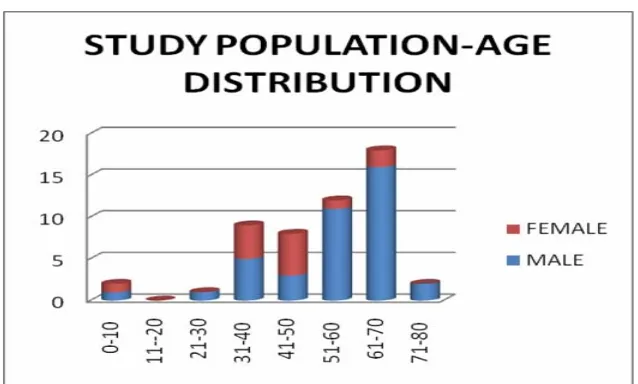
The peak incidence of focal liver lesions was highest in the age group of
61-70 years in the males and 41-50 years in the females as given in Table 2 and
[image:44.612.90.450.489.659.2]figure 2.
Table 2 : STUDY POPULATION – AGE & SEX DISTRIBUTION
AGE (YRS) MALE FEMALE TOTAL
1-10 1 1 2
11-20 0 0 0
21-30 1 0 1
31-40 5 4 9
41-50 3 5 8
51-60 11 1 12
61-70 16 2 18
71-80 2 0 2
Fig.1
M:F Ratio
MALE 75% FEMALE
25%
Fig.2
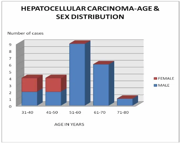
[image:45.612.112.427.140.295.2] [image:45.612.109.428.430.622.2]Males formed the majority of the cases reported as Hepatocellular
carcinoma contributing to 20 of the 24 cases of which 45% were in the sixth
decade as shown in table 3. The incidence of liver secondaries was also high in
males (14 cases) and in seventh decade, as that of hepatocellular carcinoma.as
[image:46.612.85.452.277.438.2]shown in table 4.
Table 3 : HEPATOCELLULAR CARCINOMA- AGE & SEX DISTRIBUTION
AGE (YEARS) MALE FEMALE TOTAL
31-40 2 2 4
41-50 2 2 4
51-60 9 0 9
61-70 6 0 6
71-80 1 0 1
TOTAL 20 4 24
Table 4 : LIVER SECONDARIES – AGE & SEX DISTRIBUTION
AGE (YEARS) MALE FEMALE TOTAL
31-40 3 2 5
41-50 1 3 4
51-60 2 0 2
61-70 8 1 9
TOTAL 14 6 20
[image:46.612.89.450.513.655.2]Fig 3
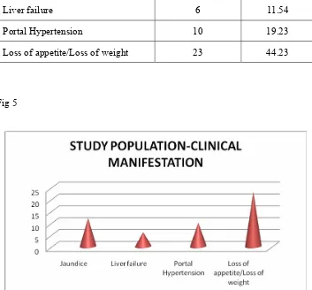
[image:47.612.123.413.137.366.2]The damage to the liver by various focal lesions was clinically
manifested as jaundice in 12cases (23%), liver failure in 6 cases (11.5%), portal
hypertension in 10 cases (19.2%), loss of weight and loss of appetite in 23
cases (44.2%). Similar results were found in both HCC and metastatic liver
[image:48.612.90.451.283.402.2]lesions.
Table 5 : STUDY POPULATION – CLINICAL MANIFESTATIONS
Clinical features Number of cases %
Jaundice 12 23.08
Liver failure 6 11.54
Portal Hypertension 10 19.23
Loss of appetite/Loss of weight 23 44.23
[image:48.612.92.445.329.657.2]Table 6 : CLINICAL MANIFESTATIONS IN HEPATOCELLULAR CARCINOMA
Clinical features Number of cases %
Jaundice 10 19.23
Liver failure 4 7.69
Portal hypertension 9 17.31
Loss of appetite/loss of weight 17 32.69
[image:49.612.84.451.175.297.2]Table 7 : STUDY POPULATION – LAB INVESTIGATIONS
Lab investigations Number of cases %
Increased bilirubin 7 13.4
HBS Ag 2 3.85
Increased SGOT/ SGPT 11 21.15
Increased SAP 7 13.4
HBS Ag- Hepatitis B surface antigen
SAP- Serum Alkaline Phosphatase
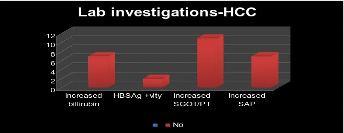
The abnormalities in liver function tests in our study population are
shown in table 7 and figure 7. Increased bilirubin was seen in 7 cases (13.5%),
increased SGOT/SGPT in 11 cases (21%) and increased serum alkaline
phosphatase in 7 cases (13.5%). Viral markers were done for all cases and 2
cases showed positivity. Similar results were found in both HCC and metastatic
liver lesions. Fig 7 0 2 4 6 8 10 12 Increased billirubin
HBSAg +vity Increased SGOT/PT
Increased SAP
Lab investigations-HCC
[image:50.612.99.444.517.651.2]Among the 52 cases of focal liver lesions subjected to US guided FNA
and biopsy, histopathological diagnosis was primary hepatocellular carcinoma
in 24 cases (46.15%), followed by secondary adenocarcinomatous deposits in
16 cases (30.77%) and hepatoblastoma in 2 cases (3.85%). Other interesting
cases were Cholangiocarcinoma (1.92%), hepatic adenoma (1.92%), secondary
synovial sarcomatous deposit (1.92%) and secondary squamous cell
carcinomatous deposits (1.92%) each contributed to one case. Definitive
typing of malignancy could not be done in 2 cases (3.85%), for which
immunohistochemistry was done. Biopsy material was inadequate and showed
no evidence of malignancy in 2 cases (3.85%). One case showed evidence of
liver cell dysplasia (1.92%) only. Another case which had definitive
radiological evidence of malignancy, proved to be an abscess (1.92%) by both
Table 8: DISTRIBUTION OF FOCAL LIVER LESIONS - HISTOPATHOLOGY
LESION NUMBER OF
CASES Percentage of total
%
HEPATOCELLULAR CARCINOMA 24 46.15
ADENOCARCINOMA 16 30.77
SQUAMOUS CELL CARCINOMA 1 1.92
SYNOVIAL SARCOMA 1 1.92
SECONDARIES NOT SPECIFIED 1 1.92
CHOLANGIOCARCINOMA 1 3.85
HEPATIC ADENOMA 1 1.92
HEPATOBLASTOMA 2 1.92
CARCINOMA NOT SPECIFIED 1 1.92
INADEQUATE 2 3.85
OTHERS 2 3.85
[image:52.612.85.454.134.611.2]TOTAL 52 100%
By FNA, 23 cases (44.23%) were diagnosed to be HCC, 15 cases (28.84%) were secondary adenocarcinomatous deposits and 2 cases (3.84%) were hepatoblastoma. Cholangiocarcinoma (1.92%), hepatic adenoma (1.92%), secondary synovial sarcoma deposit (1.92%) and secondary squamous cell
carcinomatous deposit (1.92%) each contributed to one case. As with
[image:53.612.84.449.430.662.2]histopathology, in FNA also definitive typing of malignancy could not be done in 2 cases (3.84%) and in one case (1.92%) the smear showed evidence of secondaries liver but could not be specified. Another 4 smears (7.69%) showed no evidence of malignancy, which might be due to non representative sampling. Another case (1.92%) which had definitive radiological evidence of malignancy, proved to be an abscess by both HPE and FNA, the details of which are shown in table 9.
Table 9 : DISTRIBUTION OF FOCAL LIVER LESIONS - FNA
LESION NUMBER OF
CASES PERCENTAGE OF TOTAL
HEPATOCELLULAR CARCINOMA 23 44.23%
ADENOCARCINOMA 15 28.84%
SQUAMOUS CELL CARCINOMA 1 1.92%
SYNOVIAL SARCOMA 1 1.92%
SECONDARIES NOT SPECIFIED 1 1.92%
CHOLANGIOCARCINOMA 1 1.92%
HEPATIC ADENOMA 1 1.92%
HEPATOBLASTOMA 2 3.84%
CARCINOMA NOT SPECIFIED 2 3.84%
UNREPRESENTATIVE/INADEQUATE 4 7.69%
OTHERS 1 1.92%
Considering histopathology as the gold standard for definitive diagnosis
of any lesion, of the 52 cases of our study, 47 cases correlated well with the
FNA. Thus in 90.38% of focal liver lesions, FNA findings were consistent with
that of HPE. Of the 24 cases diagnosed to be HCC by biopsy, 22 cases were
also diagnosed as HCC by FNA. The percentage of correlation with respect to
HCC was 91.67%. Of the 16 secondary adeno carcinomatous deposits
diagnosed by biopsy, 13 cases were found to have correlated well with that of
FNA (81.25%).The other cases of Cholangiocarcinoma, hepatic adenoma,
hepatoblastoma, secondary synovial sarcoma deposit and secondary squamous
cell carcinomatous deposit correlated well with respect to FNA and HPE.
Another case which had radiological evidence of malignancy, proved to be an
abscess by both HPE and FNA. For 2 cases for which definitive typing of
malignancy could not be done by biopsy, FNA was also not contributory and
IHC was done. Hep Par 1 was the marker used which showed positivity
indicating probable origin from the hepatocytes. In 2 cases both cytology and
histopathology were negative for malignancy inspite of radiological findings,
which might be due to non representative sampling technique. A case of liver
cell dysplasia was diagnosed by biopsy, though cytology showed evidence of
adenocarcinoma which probably could be non representative sample.The
Table 10 : FNA – HISTOPATHOLOGY CORRELATION
LESION HPE (n)
FNAC (n)
% of Correlation
HEPATOCELLULAR CARCINOMA 24 22 91.67
ADENOCARCINOMA 16 13 81.25
SQUAMOUS CELL CARCINOMA 1 1 100.00
SYNOVIAL SARCOMA 1 1 100.00
SECONDARIES NOT SPECIFIED 1 1 100.00
CHOLANGIOCARCINOMA 1 1 100.00
HEPATIC ADENOMA 1 1 100.00
HEPATOBLASTOMA 2 2 100.00
CARCINOMA NOT SPECIFIED 1 1 100.00
NEGATIVE 2 2 100.00
OTHERS 2 2 100.00
[image:55.612.134.400.429.677.2]TOTAL 52 47 90.38
Radiological diagnosis of focal liver lesions was unifocal in 22 cases
(42.31%) and multifocal in 30 cases (57.69%).With respect to HCC, unifocal
lesions accounted to 41.67% and multifocal 58.33% as given in table 11 and
12. The liver secondaries were unifocal lesion in 7 cases (35%) and multifocal
[image:56.612.87.449.280.353.2]in 13 cases (65%) as shown in table 13
Table 11 : RADIOLOGY OF FOCAL LIVER LESIONS
UNIFOCAL 22 42.31%
MULTIFOCAL 30 57.69%
TOATL 52 100%
Fig 10
[image:56.612.114.408.317.619.2]Table 12: RADIOLOGY OF HEPATO CELLULAR CARCINOMA
UNIFOCAL 10 41.67%
MULTIFOCAL 14 58.33%
TOTAL 24 100%
[image:57.612.100.434.198.477.2]Table 13 : RADIOLOGY OF SECONDARIES
UNIFOCAL 7 35%
MULTIFOCAL 13 65%
TOTAL 20 100%
Fig 12
Radiological correlation with the histological diagnosis was 57.69 %
with 30 cases of imaging diagnosis correlating well with HPE.
The liver metastases diagnosed by imaging studies (20cases) had 100%
correlation with histopathology, which also diagnosed them to be secondaries
of the liver. Out of 24 cases diagnosed by HPE, only 10 cases were diagnosed
[image:58.612.97.433.167.476.2]cases diagnosed as hepatoblastoma in FNA and HPE were also diagnosed as
[image:59.612.85.451.196.301.2]the same in CT imaging
Table 14 : HPE & IMAGING CORRELATION IN LIVER MALIGNANCY
LESION HPE IMAGING %
HEPATOCELLULAR CA 24 10 41.67
SECONDARIES 20 20 100.00
HEPATOBLASTOMA 2 2 100.00
Fig 13
[image:59.612.104.431.301.555.2]Table 15 : STATISTICAL ANALYSIS Lesion Sensitivity (%) Specificity (%) +ve predictive Value (%) - ve predictive Value (%) False +ve (%) False – ve (%) Focal liver lesion
95.7 80 97.8 66.7 20 4.3
HCC 91.66 96.42 95.65 93.5 3.57 8.34
Secondaries 85 93.75 89.5 90.9 6.25 15
The sensitivity of FNA in diagnosis of malignancy was 95.7% in our
study which is in accordance with the sensitivity rates of studies by various
authors like Pagani, Holm et al , Butler and Smith, Buscatine et al and Fornari
et al.The specificity in diagnosing malignancy was 80%. False positive rate
was 20% and false negativity was 4.3%.The low false negativity rate could be
attributed to the image guidance of the procedure.The positive predictive value
was 97.8% and negative predictive value was 66.7%.
FNA of HCC showed a sensitivity of 91.66% and a specificity of
96.42% of false positive rate was 3.57%, false negative rate was 8.34%,
positive predictive value was 95.65% and negative predictive value was
93.10%
FNA of secondaries showed a sensitivity of 85%, specificity of 93.75%,
false positive rate of 6.25%, false negative rate of 15%, positive predictive
RESULTS OF IMMUNOHISTOCHEMISTRY
Cytokeratin was applied for almost all cases .
Hep Par 1
Hep Par1 was done for 10 cases. HCC was taken as a control for
Hep Par1 that showed strong diffuse cytoplasmic granular positivity. The
cases of hepatic adenoma and hepatoblastoma also showed strong diffuse
positivity as that of HCC. Cholangiocarcinoma showed negativity for
Hep Par 1, thus confirming the tissue diagnosis. The usefulness of the
marker in selected cases is shown in table below;
Sl.
No. FNA Diagnosis HPE Diagnosis
Hep
Par 1 Interpretation 1. +ve for malignancy +ve for malignancy -ve -ve for HCC
2. HCC +ve for malignancy ++ HCC
HEPATOCELLULAR CARCINOMA –FNA SHOWING BILE PLUGS (100x)
HEPATOCELLULAR CARCINOMA –FNA SHOWING ENDOTHELIAL RIMMING (100x)
HEPATOCELLULAR CARCINOMA-FNA SHOWING ENDOTHELIAL RIMMING-PAP STAIN
HEPATOCELLULAR CARCINOMA-FNA(H&E) SHOWING INTRANUCLEAR INCLUSIONS (400x)
HEPATOCELLULAR CARCINOMA-HPE (100x)
SECONDARY ADENOCARCINOMA-FNA (40x)
SECONDARY ADENOCARCINOMA-FNA (100x)
SECONDARY ADENOCARCINOMA-HPE (100x)
HEPATIC ADENOMA-FNA –(100x) H&E
HEPATIC ADENOMA-FNA –(400x) H&E
HEPATIC ADENOMA-HPE (100x)
CHOLANGIOCARCINOMA-FNA (400x)
SECONDARY SYNOVIAL SARCOMA DEPOSIT-FNA (100x)
HEPATOBLASTOMA-FNA (100x)
HEPATOBLASTOMA-HPE (100x)
IMMUNOHISTOCHEMISTRY-Hep Par 1 (100x)
IMMUNOHISTOCHEMISTRY-Hep Par 1 (400x)
HEPATOCELLULAR CARCINOMA
UNIFOCAL LESION - CECT
SECONDARIES LIVER
UNIFOCAL LESION - CECT
HEPATIC ADENOMA
SPIRAL CT – SAGITTAL SECTION
TRUCUT BIOPSY GUN (18G) AND FINE NEEDLE ASPIRATION
DISCUSSION
Literature supports the usefulness of FNA in diagnosing benign and
malignant liver lesions. The overall sensitivity varies from 67-100% in
diagnosing malignant liver lesions. The specificity was 99%. The positive
predictive value was 99%, whereas the negative predictive value was 71%.
This was in accordance to our study with sensitivity of 95.7%, specificity of
80%, positive predictive value of 97.8% and the negative predictive value of
[image:77.612.89.449.442.693.2]66.7%. The experiences of some of the authors are given below:
Table 16 : COMPARISON WITH STANDARD STUDIES
Author No
o f pa tients Ty pe o f lesion Se ns
itivity (%)
Sp
ec
if
ic
ity
PVpos PV
ne
g
Montali et al 1982 108 M 92 100 100 70
Rosenblatt et al 1982 59 M 94 100 100 80
Whitlach et al 1984 86 M 87 100 100 76
Tatsuta et al 1984 41 M 94 96 94 96
Gabel et al 1986 854 M 88 100 - -
Servol et al 1988 175 M 80 100 100 76
Buscatine et al 1990 972 M 91 99 100 77
Au th or No of pa tients T yp e of lesi on Sens it iv ity (%) Sp eci fi ci ty PVp os PVneg
Jacobson et al 1983 55 S 100 100 100 100
Pagani 1983 100 S 95 100 100 56
Schwerk et al 1983 130 S 92 93 98 77
Droese et al 1984 100 S 94 100 100 89
Haubek 1985 380 S 91 100 100 65
Holm et al 1985 247 S 92 100 100 60
Bell et al 1986 197 S 67 100 100 45
Butler & smith 1989 40 S 98 100 100 88
Edoute et al 1991 321 S 86 98 99 76
Ohlsson et al 1999 178 S 89 67 98 27
Our study 52 S 95.7 80 97.8 66.7
The relationship between size of lesion and proportion in which a
correct diagnosis was made was studied by Reading et al (1988)40 and correct
diagnosis was made by FNA in 79% of lesions 1 cm or less in diameter . False
positive were due to sampling error or are were based on aspiration material
that often was scanty.With regard to HCC, FNA is accurate with a sensitivity
rate 80 to 95% and a specificity of 100%.41,42,43
Jacobsen et al 1983 , Droese et al 1984 , Hajdu et al 1989, Fornari et al
1990, Edoute et al 1991 were able to produce the cytological diagnosis which
corresponds closely to histology of the tumor.
There is no agreement as to the superiority of cytology or
[image:79.612.90.453.465.691.2]microhistology in the diagnosis of focal liver lesions
Table 17
Author No
of
patients Sens
itivity Cyt olo gy % Sens itivity Histolo gy %
Wittenberg et al 198244 65 81 73
Sangalli et al 198945 112 74 82
Buscarini et al 199046 969 91 94
Edoute et al 199147 34 32 62
Rapaccini et al 199448 73 80 61
Fang-Ying Kuo et al 200450 936 78.4 76.3
From the comparative studies shown above, it is evident that neither
method has clear advantages and the retrieval rates and tissue typing accuracies
are fairly similar.
The cytology may be inadequate in some patients , particularly in those
with vascular lesions, in fibrotic, dense tumors, in lymphomas and in well
differentiated primary liver


