STUDY AND ANALYSIS ON VERRUCOUS SKIN LESIONS OVER THE LOWERLIMBS
Dissertation submitted to
THE TAMILNADU DR. M.G.R. MEDICAL UNIVERSITY in partial fulfillment of the requirements for the award of
M.D. DEGREE (BRANCH-XII) IN
DERMATOLOGY, VENEREOLOGYAND LEPROLOGY
CERTIFICATE
This is to certify that the dissertation entitled “Study and analysis on verrucous skin lesions over the lowerlimbs” is the bonafide original work of Dr.N.S.Jayanthi in partial fulfillment of the requirements for MD
DERMATOLOGY, VENEREOLOGY AND LEPROLOGY BRANCH XII
examination of the Tamilnadu DR.M.G.R Medical University, Chennai to be
held in April 2013. The period of study was from August 2011 to July 2012.
Date : Professor & Head of the Department,
Department of Dermatology,
Coimbatore Medical College & Hospital,
Coimbatore.
Date : Dean,
Coimbatore Medical College & Hospital, Coimbatore.
DECLARATION
I Dr.N.S.Jayanthi solemnly declare that the dissertation entitled
“Study and analysis on verrucous skin lesions over the lowerlimbs” is a bonafide work done by me at Coimbatore Medical College Hospital during
the year August 2011 to July 2012 under the guidance & supervision of
Dr.P.P.Ramasamy M.D.,D.D., Professor & Head of Department, Department of
Dermatology, Coimbatore Medical College & Hospital.
The dissertation is submitted to Dr.MGR Medical University towards
partial fulfillment of requirement for the award of MD degree branch XII
Dermatology, Venereology and Leprology.
PLACE: Dr.N.S.JAYANTHI
DATE:
ACKNOWLEDGEMENT
I solicit my humble thanks to the Dean Dr.Vimala M.D. (Path),
Coimbatore Medical College Hospital, for allowing me to conduct the study in
this hospital. I am also immensely thankful to our Prof. Dr. P.P.Ramasamy M.D., D.D., Professor Head of the Department, Dermatology and Leprology for his invaluable guidance, motivation and help throughout the study.
I would like to express my gratitude and indebtness to our Prof. Dr. K. Mahadevan, M.D., D.V., Department of Venereology for his support.
I express my earnest gratitude to Assistant Professor, Department of
Dermatology and my guide Dr. R.Madhavan M.D.Without his help and guidance this work would not have been possible.
I owe great debt of gratitude to Dr. B.Eswaramoorthy M.D., Dr.R.Revathy M.D.,Assistant Professors, Department of Dermatology for their kind support and encouragement.
I sincerely thank Dr. S.Bharathi M.D., Assistant Professor, Department of Dermatology for her invaluable guidance and help.
I owe to my husband and kids who have always stood by me in my
career.
I duly acknowledge my juniors and roommates for their help and
favour.
I am very grateful to all patients for their co-operation and participation in
CONTENTS
S.NO CONTENTS PAGE
1 INTRODUCTION
2 REVIEW OF LITERATURE
3 AIM OF STUDY
4 MATERIALS &METHODS
5 OBSERVATION AND RESULTS
7 DISCUSSION
8 SUMMARY
9 CONCLUSION
ANNEXURES
BIBILOGRAPHY
PROFORMA
ABBREVIATION
MASTER CHART
KEY TO MASTER CHART
ETHICAL COMMITEE
CLEARANCE
ABSTRACT
Title
Study and analysis on verrucous skin lesions over the lowerlimbs.
BACKGROUND
Verrucous skin lesions of lower limbs often brings a
diagnostic dilemma to a treating Dermatologist . Much study has not been
done on this subject. A proper workup is needed to arrive at a diagnosis.
Verrucous skin lesions are more common on lower limbs than upper limbs.
Histopathological study is mandatory to differentiate these conditions. Usually
we need deep biopsy. At times, serial biopsies should be taken to arrive at the
diagnosis.
AIM
To study the clinical and histopathological patterns of various
verrucous skin lesions of lower limb and to find out the etiology of these
verrucous skin lesions.
Material and methods
During the study period of one year from 1 August 2011 to 31st July
2012 in our outpatient department, department of Dermatology Coimbatore
Medical College Hospital. All the patients with verrucous skin lesions of lower
patients underwent routine and certain special investigations to arrive at the
diagnosis.
Result
Out of hundred patients, the common causes of verrucous lesions over the
lowerlimbs were hypertrophic lichen planus, wart, lichen simplex chronicus,
psoriasis and lichen amyloidosis. The less common etiologies were
porokeratosis, hypertrophic discoid lupus erythematosus, callosity and
Elephantiasis nostros verrucosa cutis.
Conclusion
Hypertrophic lichen planus, Lichen simplex chronicus and wart were the common etiological factors for the occurrence of verrucous skin lesions over the lower limbs. Porokeratosis, hypertrophic discoid lupus erythematosus,
elephantiasis nostros verrucosa cutis were the least common cause of verrucous
skin lesions of lower limbs. Patients with phlebolymphedema, verrucous
carcinoma and verrucous skin lesions of neuropathy, males are mostly affected.
Key words
verrucous skin lesions, hypertrophic lichen planus, wart, lichen simplex
chronicus, psoriasis and lichen amyloidosis, phlebolymphedema, verrucous
INTRODUCTION
Verruca is a Latin word which means a wart like projection, over
the back of a toad or on some leaves. The term was first used by
Sennertus. He originally used this term for wart because ‘they appear on
the skin surface like eminence of little hill’ .1 Aulus Cornelius Celsus,
who lived during the reign of Tiberius Caesar, in discussing wart-like
lesions in his classical work on medicine 'De Medicina', mentioned three
types.2,3,4 Hippocratic writings mentioned about the wart in children.5
Verrucous skin lesions of lower limbs often brings a diagnostic
dilemma to a treating Dermatologist . Much study has not been done on this
subject. A proper workup is needed to arrive at a diagnosis. Verrucous skin
lesions are more common on lower limbs than upper limbs. From a clinical
standpoint, many verrucous lesions have diverse etiology , but closely resemble
each other. Most often they present as hyperpigmented verrucous plaques.
Verrucous skin lesions may be due to infectious and non infectious causes.
Some verrucous lesions may be the manifestation of different disease process .
For that, first step is to identify the underlying pathology. Histopathological
study is mandatory to differentiate these conditions. Usually we need deep
biopsy. At times, serial biopsies should be taken to arrive at the diagnosis. It is
difficult to differentiate verrucous lesions due to lymphedema and
phlebolymphedema clinically and also histopathologically. To differentiate
these two, we need further workup. Verrucous lesions due to neuropathy also
REVIEW OF LITERATURE
Verrucous skin lesions of lower limbs comprise a heterogenous
group of disorder. These disorders have been considered diagnostically
challenging for the dermatologist.
Verrucous lesions have jagged, undulating surface [warty] and
most often with histopathological findings of papillomatosis.6
Verrucous lesions of lower limbs are as follows
Infectious causes :
Viral – Wart 7
Bacterial – Tuberculosis verrucosa cutis6
Lupus vulgaris6
Fungal – Chromoblastomycosis8
Fixed cutaneous sporotricosis 9
Cutaneous rhinosporidosis10
Cutaneous blastomycosis11
Coccidioidomycosis 12
Ectoparasite – Crusted scabies6
Non infectious causes :
Papulosquamous – Hypertrophic lichen planus 6 ,
Eczema _ Lichen simplex chronicus 6
Prurigo _ Prurigo nodularis 7
Collagen vascular disorder – Hypertropic variant of Discoid lupus erythematosus6
Deposition disorders – Lichen amyloidosis7
Lipoid proteinosis. 6
Keratinization disorders – Porokeratosis6 ,
Acrokeratosis verruciformis6 ,
Darier’s disease7
Benign tumor – Seborrhoeic keratosis7
Stucco keratosis 7
Pigmentary disorder – Incontinentia pigmenti 6
Nevi – Verrucous epidermal nevi6
Disorder of lymphatics – Lymphangioma circumscriptum6,
Elephantiasis nostros verrucosa cutis 16
Disorders of blood vessel – Verrucous hemangioma13
Angiokeratoma circumscriptum14
Phlebolymphedema15
Acantholytic disorders – Warty dyskeratoma6
Malignancy – Verrucous carcinoma6,
Verrucous variant of malignant melanoma 20,
Kaposi sarcoma 21.
Neuropathy associated – Leprosy17
verrucous skin lesions Diabetes Mellitus18
Other Sensory neuropathies 18.
Bromoderma 6
HYPERTROPHIC LICHEN PLANUS: [Lichen planus verrucosus] 23
Lichen planus (Greek leichen, "tree moss" Latin planus, "flat") is a
common inflammatory disorder that affects the skin, nails, mucous membranes
and hair.22
Clinical features:
Hypertrophic lichen planus seen commonly over extremities especially
over shin, ankles, interphalangeal joints. Two-thirds of cases occur between the
ages of 30 and 60 years. There is no sexual predilection22. The lesions are often
pruritic, symmetrical, verrucous plaque with central depigmented area
surrounded by a hyperpigmented rim. These lesions heal with scarring. Most
often it is refractory to treatment. Chronic venous stasis is usually associated with hypertrophic lichen planus24. It can rarely transfer into squamous cell carcinoma25. Malignant transformation is more common in distal extremities.
From the diagnosis of hypertrophic lichen planus to malignancy needs atleast 12
years. 26, 27.
Histopathology:
Compact orthokeratosis, hypergranulosis, pseudoepitheliomatous
hyperplasia, irregular acanthosis, vacuolar degeneration of basal layer.
changes are discrete and often to the base of rete ridges. Necrotic keratinocytes
are present in lower epidermis and papillary dermis 28.
Lichen simplex chronicus : [ Circumscribed neurodermatitis]29
Definition :
It is an eczematous dermatosis which is characterized by heavily lichenified plaques more often with a single lesion. 29
Etiopathogenesis :
Patients with LSC are readily conditioned to scratch following an itch stimulus . This may occur as the manifestation of atopic state. Emotional state plays important role in development of LSC.
Clinical features :30
Lichen simplex chronicus is uncommon in childhood. Any age group from adolescent age is commonly affected. The most common age group is between 30 and 50 years of age. Women are more commonly affected than men. The common sites are the nape of the neck, lower legs ,ankles, sides of the neck, scalp, upper thighs, vulva, pubis or scrotum, and the extensor forearms. When excoriation continues for many years with lichenification and lax subcutaneous tissues, a solid, tumour-like plaques will be formed. This giant lichenification of Pautrier will be warty and have cribriform surface. This occurs commonly in the genitocrural region.
Hyperkeratosis, interspersing parakeratosis, acanthosis, papillomatosis with irregular elongation of rete ridges. Broadening of dermal papillae with spongiosis and increased number of fibroblast and vertical orientation of collagen bundles. With chronic rubbing, epidermal hyperplasia and fibrosis become marked.
Psoriasis verrucosa : 32
It is a rare variant of psoriasis in which two types are recognised. They
are dome shaped papules with keratotic plug and crater shaped papule with
central depression.
Clinical features:
These lesions occur along with typical psoriatic lesions elsewhere.
Histopathology : 33
Parakeratosis, epidermal acanthosis with elongation of rete
ridges, thin suprapapillary epidermal plates, epidermal hypogranulosis and
dilated, tortuous capillaries and a lymphocyte-predominant inflammatory
infiltrate, which may contain admixed neutrophils in the papillary dermis.
Neutrophil collections in the stratum corneum and stratum spinosum, called
‘Munro microabcesses’ and ‘spongiform pustules of Kogoj’, respectively, are
the most specific findings for psoriasis. Papillomatosis, epithelial buttressing,
VIRAL WART :
HPV is the causative organism of viral wart which infects keratinizing or non - keratinizing stratified squamous epithelium. Cutaneous warts occur at any age. Rarely seen in infancy and early childhood. Peak incidence is seen in school children, adolescence and early adulthood88,89.
Incubation period: Variable from few weeks to years90. Spreads by direct or indirect contact. Loss of the epithelial barrier function by trauma, maceration or by both leads to inoculation of the virus in the basal epidermal layer. Plantar warts are usually acquired from swimming pool or shower-room floors.
Papillomaviruses are double-stranded DNA viruses. These virus
will get integrate into host DNA. The size is 55 nm. More than 200 genotypes
of HPV are there which can affect skin and mucous membrane. They are highly
species-specific. The viruses infect the basal keratinocyte of the epidermis,
through disruptions of the skin or mucosal surface. At this location, the virus
remains latent in the cell as a circular episome in low copy numbers. As the
epidermal cells differentiate and migrate to the surface, the virus replicates and
matures. This results in cutaneous and mucosal wart. Most of the
papillomaviruses have different anatomic predilections either skin or genital
malignancies. They interact with E6 and E7 proteins of host cell function.
Clinically, Common warts91, 92(except plantar warts) are caused mainly due to HPV-2,1,4,27 and 57serotypes. Firm papules with a rough, horny surface, ranging in size from less than 1 mm to over 1 cm in diameter. Common site is on the backs of the hands and fingers. Single wart remains unchanged for months together. It exhibits Köebner-like isomorphic phenomenon at the site of trauma. Compared to plane wart, this phenomenon occurs rarely. Warts in adults heals slower, either with or without treatment. Common warts regresses asymptomatically. It will take several weeks to resolve without any pigmentation. Plantar warts93 are caused by HPV-1, 2, 4, 27 or 57. The deep ‘myrmecia’ form is due to HPV-1. Plantar wart starts as ‘sago-grain’ papule, then becomes a rounded lesion, which is sharply defined with a keratotic, rough surface surrounded by a smooth collar of thickened horn. While on paring, small bleeding points are seen. This helps to differentiate this wart from corn foot. Pain may be a common but variable symptom. Severity may change. Mosaic warts are often painless in children. Spontaneous regression occurs faster in children than in adults. It may be associated with hyperhidrosis or orthopaedic defects. Mosaic warts persist for longer time. Before the lesion separates, there may be blackening from thrombosed blood .94
The foci of koilocytes are located at stratum malphigii. Koilocytes posses small ,round deeply basophilic nuclei surrounded by a clear halo and pale staining cytoplasm.
LYMPHOEDEMA AND VENOUS DISEASE (PHLEBOLYMPHOEDEMA):
Phlebolymphedema is a type of secondary lymphedema in which mixed
venous and lymphatic insufficiency may present. It cannot be easily recognized
and treated. The lymphatic and venous systems are interrelated. When there is
venous hypertension, there may be increase in compensatory lymphatic flow.84.
This increased lymph load leads to failure of local lymphatics which leads to edema. Thrombosis of the major veins and deep vein incompetence affect the small initial and precollecting lymphatics of the skin and subcutaneous tissues of the lower leg which leads to chronic lipodermatosclerosis. Lymphoedema with venous disease leads to the gross swelling and skin changes.85, 86 Kaposi– Stemmer sign will be positive. Skin creases become enhanced and hyperkeratosis develops. Papillomatosis occurs as the consequence of dilatation of upper dermal lymphatics and fibrosis. As the disease progress this leads to elephantiasis.
leads to hard late-stage. Increased protein leads to fibrosis .The number of blood vessels greatly increases. Lymphostatic vasculopathy develop in the blood vessels. In the upper dermis, numerous newly formed vessels can be seen. Highly vascularized dermis develops due to angiogenesis. Complications are hyperpigmentation, stasis dermatitis, lymphostasis, verrucose cutis, lipodermatosclerosis, and ulceration.
Lichen amyloidosis 63:
Lichen amyloidosis presents as a pruritic eruption of multiple, discrete, scaly, hyperpigmented, hyperkeratotic papules. These papules coalesced to form verrucous plaque which resembles hypertrophic lichen planus or lichen simplex chronicus. It is commonly seen over shin. Other areas of involvement are ankles, calf and dorsum of feet. Abdomen, thigh, chest may be involved. Bullous forms are rarely reported. 64 It is the most common form of cutaneous amyloidosis. 66 This condition is common in Indian subcontinent.65
Histopathologically, amyloid deposition is restricted to upper dermis. The
amyloid deposits will expand the papillae and push the rete ridges laterally.
PRURIGO NODULARIS: [Hyde’s prurigo]
Described by Hyde in 1909 41. It is a group of skin diseases characterized by intensely pruritic papules or nodules. 42
Etiopathogenesis :
insect bite. In most of the cases, the cause could not be found out43. Chronic trauma by scratching leads to neural proliferation. Nerve growth factors and
receptors are expressed in the skin lesion of prurigo. Increased numbers of calcitonin gene related peptide and substance P immunoreactive neuropeptides may play role in intense pruritus. In 75% of cases, Merkel cells are expressed. These changes do not occur in the lesions of lichen simplex chronicus44, 45. Clinical features43:
All age group may be affected, but most commonly seen between 20 and 60 years. Male to female ratio is 1:1.
They are small, tiny papules to hard globular nodules, with a raised, warty surface of 1–3 cm in diameter with an irregular ring of hyperpigmentation immediately around the nodules. The nodules will be arranged in groups. Pigmentary changes, crusting and scaling may occur. Xeroderma present over the intervening skin. They usually seen over the distal parts of the limbs, especially over the extensor surfaces. The trunk, face, palms may be affected. Patient’s usual complaint is intense pruritus. New nodules develop over time and older nodules remain pruritic for a long time .Some lesions may regress spontaneously leaving a scar.
Histopathology 46,47 :
This is characterised by marked hyperkeratosis, irregular acanthosis,
pseudoepitheliomatous hyperplasia and papillomatosis. Papillary dermis shows
dermis. Dendritic cells and mast cells are enlarged with cytoplasmic granules
over the lesional dermis.
TUBERCULOSIS VERRUCOSA CUTIS: (WARTY TUBERCULOSIS) 48
A warty, indolent, plaque-like form of tuberculosis occurring due to the inoculation of Mycobacterium tuberculosis into the skin of a previously infected patient. This patient will have moderate or high degree of immunity. There are few organisms in these lesion (paucibacillary)49 .
Pathogenesis:
Organism may be inoculated in three ways.
1. Accidental super infection from exogenous sources: physicians, pathologists and post-mortem attendants are traditionally at risk (‘anatomist’s warts’, ‘prosector’s warts’, ‘verruca necrogenica’) 50.
2. Autoinoculation with sputum in a patient with active tuberculosis. 3. Already infected children and young adults, who are having some degree
of immunity may become infected from sputum by sitting on the ground or walking barefoot. 51,52
Clinical features:
inflammation. With gradual progression, a verrucose plaque is formed. Irregular extension at the edges leads to a serpiginous outline with finger-like projections to form a massive, infiltrated papillomatous growth. Involution of centre of the lesion may lead to a white atrophic scar. The colour is purplish, red or brown. The consistency is generally firm with few areas of softening. Pus may be expressed from these soft areas or from fissures. Exudation and crusting occur rarely. The lesions may resemble Lupus vulgaris. 54 But the sites are different. Psoriasiform or keloidal appearance may be seen. Sporotrichoid spread and tuberculous lymphadenitis are very rare.55 Anomalous forms are deeply destructive papillomatous and sclerotic forms, may cause deformity of the limbs56. In generalized form, papulonecrotic and lupoid lesions are seen. 57 This form occurs in patients with active disease, which may be due to haematogenous spread with a variable tissue response. An exuberant granulomatous form, Tumour-like forms was described.52, 53, 58 Without treatment, extension is extremely slow and lesions may remain inactive for months to years together. 56,60 ,61 Spontaneous remission may occur and usually results in atrophic scars. The condition responds to antituberculosis treatment. Bone,joints and lymphnodes should be examined properly52. Miliary tuberculosis rarely reported. 59
moderate amount of necrosis are seen. Tubercle bacilli are numerous in this disease when compared to lupus vulgaris.
LEPROSY WITH HYPERKERATOTIC AND VERRUCOUS SKIN LESION ON LOWER EXTREMITIES : 87
These lesions are characteristically seen on the anterior aspect of ankle joint in Indian leprosy patients. These lesions are seen only in male patients who use stiff plastic shoes with a long tongue to their uppers. Commonly occurs as unilateral lesion but can occur bilateral also. It can occur in any leprosy spectrum. The skin of affected area will be dry or hypoaesthetic. Morphologically, three types are described.
Type1: Hyperkeratotic lesions with thread or finger like projections which resembles filiform warts.
Type 2: Hyperkeratotic lesions had hornlike projections with deep fissures in between.
Type 3: Least common type with mild hyperkeratosis and there were no projections.
Histopathologically, compact hyperkeratosis and acanthosis can be made out. In type 1 and type 2 lesions, the acanthosis was massive with pseudoepitheliomatous hyperplasia of epidermis. In type 3 lesions, the hyperkeratosis and acanthosis are mild. The dermis revealed only a mild perivascular mononuclear cell infiltrate without any granuloma.
Verrucous carcinoma34:
The term was first coined by Ackerman in 1948. It is a slow-growing
neoplasm with a tendency for local recurrence. It rarely metastasizes. It is a
form of squamous cell carcinoma which is characterised by slow growing
exophytic tumor. Clinically it is characterized by cauliflower like appearance
at the site of chronic irritation. Four types has been described according to the
anatomical site of involvement (a) oral florid papillomatosis - verrucous
carcinoma of the oral cavity (b) giant condyloma of Buschke and Löwenstein -
verrucous carcnoma of the genitoanal region (c) epithelioma cuniculatum -
verrucous carcinoma of the plantar region (d) cutaneous verrucous carcinoma -
verrucous carcinoma occurring in other areas of the skin. 34 The pathogenesis of
common site of involvement is anterior weight-bearing area of sole of the foot. When tumour grows, it locally invades and plantar fascia may get affected.
When it is advancing toward the dorsal surface of the foot it may destroy
metatarsal bones 36.
Histopathology39,40:
Large deep biopsy is essential for diagnosing verrucous carcinoma. There
will be hyperkeratosis, parakeratosis, acanthosis, well differentiated
keratinocyte with a small nucleus. The tumor invades with broad strands and
contains keratin filled cyst in their centre, large ,bulbous downward
proliferation that compress the collagen bundles and push them aside .Thus the
tumor has bulldozing rather than stabling effect. In the deeper portions, nuclear
atypia , individual cell keratinisation &horn pearls are absent.
Verrucous skin lesions of neuropathy68, 69
Verrucous skin lesions of neuropathy is most commonly associated with
diabetes mellitus. In diabetic patients, it may present as verrucous skin lesions
on the feet and skin ulcers simultaneously or either ulcer or neuropathies
preceed. These associations may be due to neuropathy and diabetic ulcers are
interrelated in their aetiology and pathogenesis. Multiple treatment modalities
with foot care are necessary for this condition.
ELEPHANTIASIS NOSTROS VERRUCOSA CUTIS :
chronic lymphedema. It is due to lymphatic obstruction caused by recurrent bacterial infection. 71
In 1969, Castellani 70 classified elephantiasis into four subtypes: 1. Elephantiasis tropica - due to filariasis
2. Elephantiasis nostras - due to bacterial infection
3. Elephantiasis symptomatica - due to mycotic, syphilitic,
tuberculoid, neoplastic, or traumatic causes of lymphatic obstruction.
4. Elephantiasis congenita - inherited disorders such as
Milroy’s disease. Bacterial infection, lymphangioma, malignancy, lymphatic
fibrosis and prior surgery or trauma, radiation therapy, chronic venous
stasis, scleroderma and obesity can lead to lymphatic obstruction and
edema. Lymphatic vessels are an important pathway for immune cell
trafficking and antigen delivery72,73. The protein-rich interstitial fluid of
lymphedema leads to inflammation and an accumulation of fibroblasts,
adipocytes and keratinocytes that transform the initially soft swollen tissue
into a hard fibrotic tissue with stiff, thickened skin.74,75 Protein rich fluid
induces fibroblast proliferation and increases susceptibility to infection and
inflammation. Because of this ongoing process, further fibrosis of the
dermis and lymph channels can occur. In chronic venous stasis, activated
leukocytes may migrate out of the vasculature and release TGF-β1,
stimulating collagen production by dermal fibroblasts, which culminates in
include impairment of limb function, recurrent cellulitis which further
aggravates the lymphedema and accelerate skin changes. Rarely
malignancies such as lymphangiosarcoma (Stewart-Treves syndrome),
melanoma , squamous cell carcinoma, Kaposi’s sarcoma and lymphoma.80-82
Clinically it presents as fatigue and heaviness in the affected limb. The
edema increases towards the evening and relieved by bed rest. Early edema is
soft and pitting. But later become indurated and non-pitting. The skin over the
affected leg becomes thick and hyperkeratotic with pebbled appearance of the
skin surface. Squaring of the toes with difficulty in pinching the skin over the
second toe known as Kaposi -Stemmer’s sign is positive . Investigations like
Lymphoscintigraphy (isotope lymphography), Magnetic resonance imaging (MRI), X-ray contrast lymphography are useful.
Histopathologically, pseudoepitheliomatous hyperplasia, dilated lymphatic spaces, and fibrosis are characteristic of ENV. These findings may
not be present always during the advanced stage 83.
Hypertrophic Discoid lupus erythematosus 111 :
verrucous plaque with adherent scales. These patients usually have classical DLE lesions elsewhere in the body.112
Histologically, 113 two patterns are recognised. In the first pattern, hyperplastic papillomatous epidermis with hyperkeratotic scale with dyskeratotic keratinocytes are seen. Thickened basement membrane zone is seen in old lesions. Band like mononuclear infiltration is seen along the dermoepidermal junction which mimicks like lichen planus. Second pattern consists of deep dermal perivascular, periappendageal and interstitial infiltrate with mucin deposition.
POROKERATOSIS:
Porokeratosis was first described by Vittorio Mibelli
in 1893. Depending on clinical criteria five clinical variants are recognized:
classic porokeratosis of Mibelli, disseminated superficial (actinic and non
actinic type) PK, linear PK ,punctuate PK, porokeratosis Palmaris et plantaris
disseminata. Rare morphological forms like facial PK, giant PK, hypertrophic
verrucous porokeratosis, reticulate PK are also reported. 108 Except the punctate
type, all other types are characterized by a keratotic ridge .This feature
histologically correlates the presence of cornoid lamella.109,110 In the
disseminated superficial form, the lesions are smaller, more in number, with
LUPUS VULGARIS:
Lupus vulgaris is a paucibacillary form of cutaneous tuberculosis which has chronic and progressive course. It is seen in patient with moderate to high degree of immunity. The source of lupus vulgaris is contiguous, haematogenous or lymphatic spread.98 In European countries face, especially around the nose is commonly involved. In developing countries common sites are lower limbs and buttocks99, especially in children100. It starts as a small, reddish-brown, soft flat plaque with gelatinous consistency. These lesion extends by slow peripheral extension with areas of atrophy. Apple jelly nodules are seen on diascopy. Usually it manifests as a single lesion except in disseminated forms. Sporotrichoid-like spread can occur102. Five clinical forms are known depending on the local tissue response to the infection. Those types are tumour-like forms, vegetating form, ulcerative and mutilating form ,plaque form, papular and nodular forms. Histopathologically 97, the epidermis shows atrophy and ulceration. Hyperplastic lesions shows hyperkeratosis, acanthosis, papillomatosis and pseudoepitheliomatous hyperplasia. Tuberculoid granuloma with epithelioid giant cells are seen in the upper dermis. Caseation necrosis within the tubercle is slight or absent. Extensive fibrosis occurs during the healing process. Bacilli are seen very rarely.
Bromoderma is caused by anticonvulsant therapy with potassium bromide 124. The Skin lesions are multiple nodules and plaques with verrucous surface seen commonly on limbs and face. Histopathology 123 shows verrucous pseudoepitheliomatous hyperplasia with abscesses containing neutrophils and eosinophils in the epidermis, dense dermal infiltrate initially consists mainly of neutrophils and eosinophils and later lymphocytes, plasma cells and histiocytes. The abundant dilated blood vessels may show endothelial proliferation.
Iododermas are caused by oral intake(cough syrup) ,intravenous (radiographic contrast) and after throidectomy. The skin lesions are urticaria, papulopustules, bulla and vegetating masses. Histopathology shows marked ulceration but there is usually less epithelial hyperplasia. Both conditions must be differentiated from blastomycosis and coccidioidomycosis, and pemphigus vegetans. 124
Kaposi’s sarcoma (Granuloma multiplex haemorrhagicum) 125,126,127 Kaposi’s sarcoma is a multifocal, endothelial proliferative disorder predominantly involving the skin and other organs and associated with formation of vascular channels and proliferation of spindle-shaped cells.
Etiology:
which promotes the development of the lesions by activating cytokines and angiogenic factors.
Types :
1. Classic 2. Endemic 3. Iatrogenic 4. HIV associated
Classic type - It is found mainly in elderly males, particularly in Jews of Eastern European origin. The lesions begin around the ankle and slowly spread up the leg. It begins as blue red macule, evolve into a plaque/nodule later ulcerate. It has a chronic course. Mucosal and visceral involvement is rare.
Endemic type – It is endemic in Africa.It has two types.
1. Benign nodular variety which is similar to classic type.
2. Fulminant lymphadenopathic type which is fatal in 2-3 years. Iatrogenic type - It is seen in renal transplant patients. The lesions are chronic but aggressive than the classical type.
HIV associated type-It is common in homosexual men, especially in oro-anal intercourse. It may appear at any stage of HIV infection irrespective of CD4 count. The skin lesions occur along the cleavage lines in face and trunk. Mucosal and visceral involvement (GIT/LUNG) is common.
In the patch stage:
There is a proliferation of jagged, irregular, lymphatic-like vascular channels lined by a single layer of bland endothelial cells. These channels particularly surround the pre-existing blood vessels and adnexal structures. These structures are seem to be floating within the newly formed channels, the so-called “promontory sign”. There is prominent mononuclear inflammatory cell infiltrate in which plasma cells are seen.
In the nodular stage:
Well-circumscribed nodules of generally bland, spindle-shaped cells forming cleft like spaces that impart a typical sieve-like appearance. Extravasated red blood cells are plenty. Mitotic figures are common. The periphery of the nodules gives a angiomatous appearance. The lesions of Kaposi’s sarcoma that have regressed after chemotherapy or with angiogenesis inhibitors, including Col-3 or HAART, may be very difficult to interpret.
The Histopathological differential diagnosis are spindle cell haemangioma, tufted angioma, microvenular haemangioma, hobnail haemangioma, progressive lymphangioma, venous dermatitis (acroangiodermatitis). Angiosarcoma is also often considered in the differential diagnosis, but in the latter there is clear evidence of cytological atypia, mitotic activity and multi layering.
CHROMOBLASTOMYCOSIS :
Chromoblastomycosis is the chronic fungal infection which is characterized by slow growing exophytic lesions, seen commonly on the feet and legs103. Chromoblastomycosis is caused most commonly by Phialophora verrucosa, F. Compacta, Fonsecaea pedrosoi and Cladophialophora carrionii (carrionii) Rhinocladiellaaquaspersa.104 Feet, legs, arms, face and neck are sites which are commonly involved. It starts as warty painless papule slowly enlarges to form a verrucous plaque. Secondary ulceration may occur. Lymphatic spread occurs to adjacent areas. Rarely, Haematogenous spread can occur. Squamous cell carcinoma as has been reported in chronic cases105, 106.
Histology107 :
Marked pseudoepitheliomatous hyperplasia with transepidermal elimination of fungal cells are seen. Foreign-body granuloma with isolated areas of microabscess formation and muriform or sclerotic cells are the important features of chromoblastomycosis.
Blastomycosis is a chronic suppurative and granulomatous mycosis caused by Blastomyces Dermatitidis. It mainly affects the lungs. During dissemination it affects skin, central nervous system, bones and other sites. Three types of blastomycosis like primary cutaneous, pulmonary and disseminated form are reported. Primary cutaneous form which is very rare. Following trauma, entry of organism leads to an erythematous, indurated plaque appears within 1–2 weeks. This is associated with lymphangitis and lymphadenopathy. Histologically, B.dermatitidis produces broad based budding yeast in tissues. In disseminated forms, pseudoepitheliomatous hyperplasia with intra and subepidermal polymorphonuclear abscesses with granulomatous infiltrate are found in the dermis.
Seborrhoeic keratosis 114:
Seborrhoeic keratosis is a benign tumour, which is mainly composed of epidermal keratinocytes occurs commonly in elderly individuals. It is usually occurs in fifth decade of life. This lesions are common in tropical countries. It manifests as superficial verrucous plaque which appear to stuck to the epidermis. Histopathologically, hyperkeratosis, acanthosis, marked papillomatosis which gives church spire pattern with melanocyte proliferation in the immature keratinocytes. 115
Incontinentia pigmenti:
associated with developmental defects of the eye, teeth and central nervous system.116, 117 Early inflammatory changes can occur even at birth and do not progress after birth. Tense bulla will be arranged in the linear fashion is the most striking feature of incontinentia pigmenti.
Clinical stages are 118
Stage 1: inflammatory macules, papules, vesicles and pustules Stage 2: hyperkeratotic and verrucous lesions
Stage 3: grey - brown pigmentation
Stage 4: atrophic, hypopigmented and depigmented bands or streaks that are hairless and anhidrotic and fail to tan on sun exposure
Darier’s disease 119
Darier’s disease manifest as firm, brown , rough papule over the Seborrhoeic areas of the trunk and face especially over the the scalp margins, temples, ears and scalp.
In lower legs and arms multiple discrete papules coalesced to form warty plaques with fissures or papillomatous masses. It forms a vegetating and malodorous mass especially in the flexures, anogenital region, the groins and the natal cleft.
Acrokeratosis verruciformis :
Stucco keratosis
Stucco keratosis is a variant of Seborrhoeic keratosis in which whitish, keratotic plaques that can be removed easily with a fingernail without bleeding. It is seen commonly on extremities, especially around the ankle region.
Verrucous epidermal naevus 122
Verrucous epidermal naevus is a congenital, non inflammatory cutaneous hamartoma composed of keratinocytes .Two types are recognised.
1. Epidermolytic verrucous epidermal naevus:
Pigmented linear verrocous papules or plaques seen in young children. 2. Non-epidermolytic verrucous epidermal naevus :
AIM OF THE STUDY
To study the clinical and histopathological patterns of various verrucous skin
lesions of lower limb and to find out the etiology of these verrucous skin
MATERIALS AND METHODS
A study was conducted during the period from 1 August 2011 to 31st
July 2012 in our outpatient department, department of Dermatology Coimbatore
Medical College Hospital, among the patients attending the Dermatology
Department as well as those referred from other departments.
Type of study : Cross sectional study.
Inclusion criteria :
1. All the patients with verrucous skin lesions of lower limbs of all age
groups were included.
Exclusion criteria:
1. Those patients who are not willing for the study.
2. Pregnant women.
3. Patient with bleeding disorders.
The study was explained to eligible participants. Informed consent was
obtained. A detailed history was recorded regarding complaints, duration,
evolution of skin lesions, associated local and systemic complaints. Past history
of tuberculosis, leprosy, diabetes mellitus, injury, prior drug intake, and history
Clinical examination:
Clinical examination included morphology of the skin lesions, number of
lesions and their size, distribution of lesions, symmetry, tenderness, ulceration
and secondary infections. Other areas of skin, mucous membrane, palms and
soles, hair and nail were examined. A detailed general and systemic
examination was done.
Investigations:
1. Routine laboratory investigations included complete haemogram, urine
routine, renal function test and liver function test.
2. Slit skin smear for acid fast bacilli was done for the suspected patients of
Hansen’s disease.
3. Screening for HIV, Hepatitis B, VDRL were done for high risk patients with
history of sexual exposure or occupational exposure to blood and blood
products.
4. Mantoux test, X ray chest, ultrasonogram of abdomen were done in relevant cases.
5. Lower limb Doppler was done in patients with phlobolymphoedema and
Elephantiasis nostros verrucosa cutis.
6. Nerve Conduction Study was done in patients with sensory or motor neuropathy.
and fungus were done whenever required.
Analysis:
A descriptive analysis of clinical characteristics, laboratory parameters and
histopathological features of various verrucous skin lesions was done. These
data were analysed and compared with published literature.
OBSERVATION AND RESULTS
Among the one hundred and five patients with verrucous skin
lesions over the lower limbs who attended our outpatient department from
August 2011 to July 2012, five patients were excluded from the study due to
denial for the study(3) and refusal for biopsy(2).
Hundred patients with clinical features of verrucous
skin lesions over the lower limbs were included in the study. The common
causes of verrucous lesions over the lowerlimbs were hypertrophic lichen
planus (29), wart (10),lichen simplex chronicus(10), psoriasis (7) and lichen
amyloidosis(7). The less common etiologies were porokeratosis(2),
hypertrophic discoid lupus erythematosus (2), callosity (2) and Elephantiasis nostros verrucosa cutis(1).
Table 1
Clinical spectrum of verrucous skin lesions of lower limbs
Disease No.of patients Percentage
Hypertrophic lichen planus 29 29
Lichen simplex chronicus 10 10
Wart 10 10
Psoriasis 7 7
Lichen amyloidosis 7 7
Phlebolymphoedema with verrucous skin lesions
7 7
Leprosy with hyperkeratotic and verrucous skin lesion on lower extremities
6 6
Prurigo nodularis 5 5
Tuberculosis verrucosa cutis 5 5
Verrucous carcinoma 4 4
verrucous skin lesions of neuropathy 3 3
Others 7 7
7% 7% 7% 6% 5% 5% 4% 3% 7%
No.of patients ( In Percentage)
29%
10% 10%
7%
No.of patients ( In Percentage)
Hypertrophic lichen planus
Lichen simplex chronicus
Wart
Psoriasis
Lichen amyloidosis
Phlebolymphoedema
Leprosy with hyperkeratotic and verrucous skin lesion on lower extremities
Prurigo nodularis
Tuberculosis verrucosa cutis Hypertrophic lichen planus
Lichen simplex chronicus
Lichen amyloidosis
Phlebolymphoedema
Leprosy with hyperkeratotic and verrucous skin lesion on lower Prurigo nodularis
Table 2
Sex distribution
Male Female
Disease No. % No. %
Hypertrophic lichen planus 12 41 17 59
Lichen simplex chronicus 4 40 6 60
Wart 7 70 3 30
Psoriasis 5 71 2 29
Lichen amyloidosis 4 57 3 43
Phlebolymphoedema with verrucous skin lesions
7 100 - -
Verrucous skin lesion in Leprosy 3 50 3 50
Prurigo nodularis 1 20 4 80
Tuberculosis verrucosa cutis 2 40 3 60
Verrucous carcinoma 4 100 - -
verrucous skin lesions of neuropathy 3 100 - -
Others 5 71 2 29
TOTAL 57 57 43 43
In our study, 57 patients were male while 43 were female. Male to Female ratio
0 12
4 7 5 4 7 3
1 2 4 3 5
0 41 40 70 71 57 100 50 20 40 100 100 71 0 17 6
3 2 3
0 3 4 3 0 0 2
0 59 60 30 29 43 0 50 80 60 0 0 29 0 20 40 60 80 100 120
Sex distribution
Table -3
Age distribution
Age in years <10 10-20 21-30 31-40 41-50 51-60 >60 Total
Hypertrophic lichen
planus
- 3 4 3 6 7 6 29
Lichen simplex
chronicus
3 2 5 10
Wart - 5 1 1 2 - 1 10
Psoriasis - - - 2 2 2 1 7
Lichen amyloidosis 1 3 1 1 1 7
Phlebolymphoedema with verrucous skin lesion
1 2 2 2 7
Leprosy with hyperkeratotic and verrucous skin lesion on lower extremities
3 2 1 6
Prurigo nodularis 2 1 - 1 1 5
TBVC 2 1 - 1 1 - 5
Verrucous
carcinoma
1 2 1 4
Verrucous skin
lesions of
neuropathy
1 2 3
others 2 2 1 2 7
Majority of the patients were between the age of 41-80 yrs.
Mean age of presentation was 48.95 years.
Out of hundred cases, 86 patients had non infective etiology of verrucous skin
lesions. 14 patients had infectious etiology of verrucous lesions.
0 0 0
3 5 0 2 4 1 0 1 2 1 3 1 2 3 1 1 0 2 6 3 2 2 1 2 3 0
1 1 1
2 7 2 0 2 1 2 2 1 1 2 1 6 5
1 1 1
2 1 1 0 1 2 2 0 1 2 3 4 5 6 7 8
Age distribution
Distribution of cases - Verrucous skin lesions of infective etiology
Infective etiology Wart TBVC 0 10 20 30 40 50 60 70 80 Wart 10 71
Distribution of cases
of infective etiology
Table -4
Verrucous skin lesions of infective etiology
No.of cases Percentage
10 71
4 29
TBVC 4
29
Distribution of cases - Verrucous skin lesions
of infective etiology
No.of cases Percentage Verrucous skin lesions of infective etiology
Percentage
71
29
Verrucous skin lesions
Table -5
Distribution of cases- Verrucous skin lesions of non infective etiology
Non infective etiology No.of cases Percentage
Hypertrophic lichen planus 29 34
Lichen simplex chronicus 10 11.6
Psoriasis 7 8.1
Lichen amyloidosis 7 8.1
Phlebolymphoedema with verrucous skin lesions
7 8.1
Leprosy with hyperkeratotic and verrucous skin lesion on lower extremities
6 6.9
Prurigo nodularis 5 5.8
Verrucous carcinoma 4 4.7
verrucous skin lesions of neuropathy 3 3.5
0 5 10 15 20 25 30 35 29 10 7 34 11.6 8.1
Distribution of cases
No.of cases
7 7 6
5 4
3 7
8.1 8.1 6.9
5.8 4.7 3.5
8.1
Distribution of cases- Verrucous skin lesions of
noninfective etiology
No.of cases
Verrucous skin lesions of
Table- 6
Papulosquamous disorders with verrucous skin lesions
Distribution of cases
Papulosquamous disorder No.of cases Percentage
Hypertrophic LP 29 81
Psoriasis 7 19
Table -7
Rare verrucous skin lesions –distribution of cases
Rare verrucous skin lesions No.of cases Percentage
Porokeratosis 2 28.57
Callosity 2 28.57
Hypertrophic DLE 2 28.57
[image:52.612.91.490.398.555.2]DESCRIPTION OF CASES AND FINDINGS:
Hypertrophic Lichen planus
There were 29 cases of hypertrophic lichen planus.
Clinical profile:
The age group of patients presented with features of hypertrophic lichen
planus was between 16 and 85 years. The male to female ratio was 1:1.4. The
mean duration of the skin lesions was 1.9years. All the patients had
hyperpigmented verrucous lesions over the lower limbs. Twenty five (86%)
patients had hyperpigmented lesions with depigmentation in the centre of the
lesions. Three (10%) patients had associated nail involvement. Five (17%)
patients had associated mucous membrane involvement. Two (6.8%) patients
had secondary infection. One (3.4%) patient had ulceration of the lesion. Two
(6.8%) Patients had associated venous stasis.
Histopathology:
Among the 29 patients, twenty six (90%) showed the features of hyperkeratosis, pseudoepitheliomatous hyperplasia with irregular acanthosis, wedge shaped hypergranulosis, discrete interface vacuolar changes and pigmentary incontinence were seen. Dermal fibrosis was seen in two patients.
Table -8
Hypertrophic lichen planus clinical features
Laboratory investigations and histopathology
Age No.of
patients DSL
yrs
Asso.nail
invol
Asso.mucous
mem invol
Asso.inf Asso.
ulcer
Asso.venous
stasis
H/E
PEH
H/Edermal
fibrosis
<10 -
10-20 3 1.5 2
21-30 4 2 1 1 4
31-40 3 1.2 2 1 2
41-50 6 2.1 1 1 6
51-60 7 2.4 1 1 1 1 1 8 1
>60 6 2.2 1 8 1
Key-DSL=duration of skin lesions, Asso.inf –associated infection,
Asso.ulcer- Associated ulcer, PEH-pseudoepitheliomatous hyperplasia,
Asso.nail invol- associated nail involvement, H/E –hematoxylin and eosin
Lichen simplex chronicus (LSC)
There were 10 patients of LSC with verrucous morphology.
Clinical profile:
The age group of patients with LSC was between 41 and 70 years
the male to female ratio is 1:1.5.The mean duration of illness was 2.5 years.Six
(60%) patients had verrucous hyperpigmented plaques around the ankle. Four
(40%) patients had lesions over the lateral aspect of legs. Two (20%) patients
had secondary infections. One (10%) patient had associated depression.
Histopathology :
Among the ten patients of lichen simplex chronicus, eight (80%) patients
showed hyperplasia of all the components of epidermis. Vertically oriented
collagen bundles and increased fibroblasts was seen in seven (70%) patients.
Two (20%) patients showed mild spongiosis.
Table -9
Lichen simplex chronicus-clinical features
Laboratory investigations and histopathology
Age No.of patients
DSL years
Asso.disease Asso.inf H/E EH
Vertically oriented collagen
<10
10-20
21-30
31-40
41-50 3 1.5 1 3 2
51-60 2 2.6 2 2
>60 5 3.4 1 3 3
Key-DSL=duration of skin lesions, asso.inf –associated infection, asso.
Disease-associated disease, EH-epidermal hyperplasia, H/E-Hematoxylin and eosin
stain.
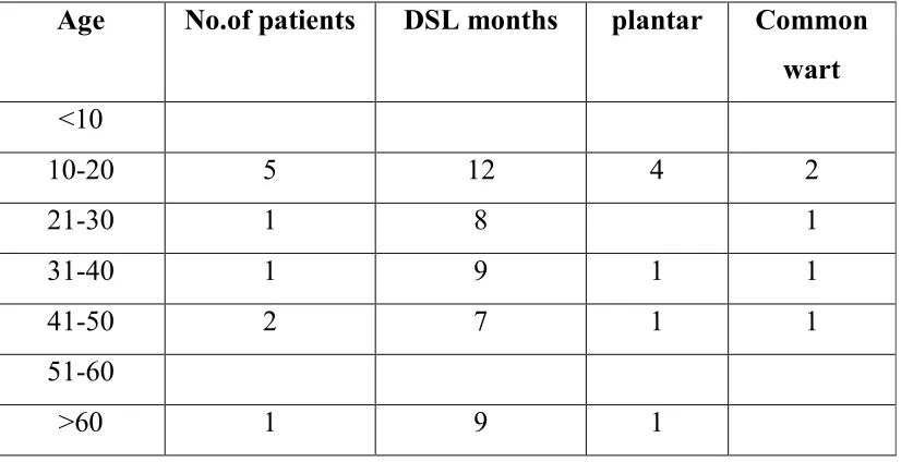
Viral Wart:
There was 10 patients with viral wart .
Clinical profile:
The age group of the patients with features of wart was between 12 and 61
years. The mean duration of illness was 9 months. Seven (70%) patients had
plantar warts. Two (20%) patients had common wart over the legs and knees.
One (10%) patients had single hyperpigmented verrucous nodule over lateral
aspect of right leg. This lesion was excised and confirmed with biopsy. Two
[image:57.612.90.503.464.676.2](20%) patients had associated lesions over the upper limbs and face.
Table- 11
Viral wart – Clinical features
Key-DSL=duration of skin lesions
Age No.of patients DSL months plantar Common wart
<10
10-20 5 12 4 2
21-30 1 8 1
31-40 1 9 1 1
41-50 2 7 1 1
51-60
Histopathology:
All the patients (100%) showed the features of hyperkeratosis, vertical tiers of parakeratosis, papillomatosis, incurving of rete ridges, the foci of koilocytes are located at Stratum Malphigii.
Psoriasis:
7 patients had psoriasis with verrucous morphology.
Clinical profile:
The age group of patients presented with the features suggestive of
Psoriasis with verrucous morphology was between 33 years and 71 years. The
male to female ratio was 2.5:1. The mean duration of illness was 2.5 years. Five
(71%) patients had associated lesions over the trunk and upper limbs. Two
(28%) patients had palmoplantar psoriasis. These patients had hyper pigmented
verrucous plaque over the lower limbs, which mimicked hypertrophic lichen
Histopathological findings:
Specimens of all the patients showed hyperkeratosis, parakeratosis,
acanthosis, regular elongation of rete ridges, papillomatosis, suprapappilary thinning, dilated capillaries. 1(14%) specimen showed Munro micro abcess.
Lichen amyloidosis :
There were 7(7%) patient with lichen amyloidosis showing verrucous
morphology.
Clinical profile:
The mean age of presentation was 57.5 years. Male to female ratio was
1.3:1. The mean duration of illness was 3.2 years. Hyperpigmented, bilaterally
symmetrical discrete, verrucous papules coalesced to form verrucous plaques
were seen over both anterior aspect of legs in six (85%) patients. One (14%)
patient had unilateral lesion over the right leg.
Histopathology :
All (100%) specimens showed the features of hyperkeratotic, hyperplastic
epidermis, rounded dermal papillae and homogenous deposits of amyloid filled
up the entire dermal papillae with pigmentary incontinence seen in all patients.
Phlebolymphoedema:
Clinical profile:
The age group of patients presented with features suggestive Phlebolymphoedema was between 40 and 70 years. The mean duration of illness was 5 years. All patients were males. Four (57%) patients had bilateral leg oedema and limb hypertrophy. Two (28%) patients had bilateral verrucous plaques over the second toe and third toe. Two (28%) patients had ulcer over the medial malleolus with verrucous plaque surrounding the ulcer. Venous Doppler was done for all patients.
Findings in venous Doppler:
Five (71%) patients showed incompetence of saphenofemoral system with extensive lymphedema. One (14%) patient had saphenopopliteal incompetence. One (14%) patient had isolated perforator incompetence.
Prurigo nodularis :
Five (5%) patients had prurigo nodularis.
Clinical profile:
The age group of the patients showing prurigo nodularis was between 20
and 72 years. The mean duration of skin lesions was 1.4 years. The male to
female ratio was 1:4. Four patients (80%) had hyperpigmented verrucous
nodules of right leg with depigmented plaque around the ankle. One (20%)
patient had secondary infection.
Histopathology :
All the patients (100%) showed the features of hyperkeratosis, acanthosis,
pseudoepitheliomatous hyperplasia, vertically oriented collagen bundles.
Tuberculosis verrucosa cutis (TBVC):
There were 5 patients presented with features suggestive of TBVC.
Clinical profile:
The age group of the patients showing TBVC was between 10 and 62
years. The mean duration of skin lesions was 1.5 years. The male to female ratio
was 1:1.5. One (20%) patient had verrucous plaque over the left foot extending
from dorsum of foot to plantar aspect of the foot laterally. One (20%) patient
had hyperpigmented verrucous plaque over the dorsum of foot. Three (60%)
patients had verrucous plaque around the ankle joints. Out of these, One (20%)
patient had pus discharge from the lesion.
Histopathology:
Hyperkeratosis, acanthosis, pseudoepitheliomatous hyperplasia, neutrophil
Investigatory findings:
All the patients were positive for Mantoux. One (20%) patient had
associated pulmonary tuberculosis.
Verrucous carcinoma:
In this study, 4 patients showed the features of verrucous carcinoma.
Clinical profile :
The age group of the patients showing verrucous carcinoma was
between 51 and 65 years. The mean duration of skin lesions was 4.2years. All
patients were males. One (25%) patient had cauliflower flower growth over the
left foot over the dorsum and plantar aspect. One (25%) patient had verrucous
growth over the right lateral malleolus and hyperkeratotic verrucous plaque over
the left lateral malleolus. One (25%) patient had pedunculated verrucous growth
just above the right knee. One (25%) patient had verrucous growth over left
thigh. Out of these, two (50%) patients were old case of Hansen’s disease and
had chronic ulcer. Two (50%) patients needed multiple deep biopsy to arrive at
the diagnosis. One (25%) patient had destruction of metatarsal bones.
Xray findings:
Histopathology :
On histopathological examination hyperkeratosis, parakeratosis,
acanthosis with keratin filled cyst. Well differentiated keratinocyte with a small
nucleus with nuclear atypia with downward proliferation which compress the
collagen bundles seen in all specimens.
Leprosy with hyperkeratotic and verrucous skin lesion on lower extremities:
Six (60%) patients had verrucous skin lesions associated with leprosy. Clinical profile:
The age group of patients with hyperkeratotic and verrucous skin lesion on lower extremities was between 43 and 65 years. The mean duration of disease was 4 years. Three (50%) patients had verrucous and hyperkeratotic lesion over anterior aspect of the ankle joints. Two (33%) patients had verrucous plaque around the trophic ulcer. One (16%) patient had verrucous plaque over the toes. One (16%) patient had associated foot drop. All patients completed their MDT.
Histopathology :
verrucous skin lesions with sensory or motor neuropathy:
Three (30%) patients had verrucous skin lesions with sensory or
motor neuropathy.
Clinical profile:
The age group of patients with features of verrucous skin lesions
with sensory or motor neuropathy was between 50 to 72 years. Mean duration
of illness was 3.5 years. All patients were male. Two (66%) patients had
depigmented verrucous plaque over right ankle joint. One (33%) patient had
depigmented verrucous plaque over the anterior aspect of left leg. Out of these,
one (33%) patient had diabetic neuropathy.
Nerve conduction study:
All the patient underwent nerve conduction study. Two (66%)
had severe demyelinating sensory and motor neuropathy. One (33%) patient had
pure sensory diabetic neuropathy.
Porokeratosis :
There were two (20%) patients with features suggestive of porokeratosis.
Clinical profile:
A 50 year old male patient presented with hyperkeratotic verrucous
from 2x1cm to 3x2cm for 3 years. Another patient, 45year old female with
hyperkeratotic depigmented verrucous plaque over the great toe extended to the
anterior aspect of foot with peeling of skin. On histopathological examination,
both the patients showed keratin filled invagination of epidermis with coronoid
lamella in the center and absent granular layer beneath the coronoid lamella.
Hypertrophic Discoid lupus erythematosus:
There were two patients with features suggestive of Hypertrophic Discoid
lupus erythematosus.
Clinical profile:
Two patients presented with the complaints of photosensitivity. A 39 year
old male patient presented with hyperpigmented verrucous plaques both thighs
and legs. Depigmented plaques with sorrounding hyperpigmentation. Another
patient, 45 year old male patient with hyperpigmented verrucous plaque over
the right leg with SCLE lesions over the face, upper trunk.
Histopathology:
Hyperkeratosis, acanthosis, papillomatous hyperplasia, basement
membrane degeneration and deep dermal infiltration of lymphocytes
perivascular, periappendageal infiltration with mucin deposition seen on
Callosity:
There were two (2%) cases of callosity.
Clinical profile:
The mean age of presentation was 63 years. The duration of skin lesions
was 5 years. Both the patients were males. Both of them had hyperpigmented
verrucous plaque over both knees.
Histopathology:
Hyperkeratosis, acanthosis, papillomatosis with increased dermal
collagen and fibrosis around the neurovascular bundles seen in both patients.
ELEPHANTIASIS NOSTROS VERRUCOSA CUTIS (ENVC)
One (10%) patient had the features suggestive of ENVC.
A 37 year old female patient had a hyperpigmented verrucous plaque
involving all the toes of right foot of size 12x7cm for the past 3 years.
Hyperkeratotic verrucous plaque present over the dorsum of right foot. Little toe
was completely encircled by verrucous plaque.
Biopsy showed the pappillomatosis, non specific chronic inflammation
with fibrosis . Doppler showed the features chronic of lymphedema with normal
saphenofemoral and saphenopopliteal valve.
DISCUSSION
The verrucous skin lesions of lower limbs are diverse group of skin
disorders with different aetiology. These lesions are morphologically similar,
characterised by jagged, undulating surface with papillomatosis on
histopathology. Detailed history, histopathological examination and certain
specific investigations are necessary to arrive at the diagnosis.
There are limited studies on verrucous skin lesions of lower limbs. We
discussed and compared with the available resources.
In our study, the age group of patients presented with features of
hypertrophic lichen planus was between 16 and 85 years. The male to female
ratio was 1:1.4. The mean duration of the skin lesions was 1.9years. Two
(6.8%) Patients had associated venous stasis. In a study by Dilip Kachhawa et al,
a clinicopathological study on lichen planus 128, the most common age group
with hypertrophic lichen planus was between 40 and 60 years of age. We also
observed the same incidence in our study group. Another study by Bhattacharya
M et al on Lichen planus, associated mucosal involvement was 16.8% and nail
involvement was 15.1%. In our study 10% patients had associated nail
In our study, the age group of patients with LSC was between 41 and 70
years. The male to female ratio is 1:1.5.The mean duration of illness was 2.5
years. Six (60%) patients had verrucous hyperpigmented plaques around the
ankle. Four (40%) patients had lesions over the lateral aspect of legs. One(10%)
had associated depression. In a study by Julius L et al 130 incidence of Lichen
Simplex Chronicus in Orientals and Caucasians was 3%. In a study by Thappa et
al131 on patterns of lower leg and foot eczema in south India, LSC was common
between 30-50 years of age. The age group affected by LSC in our study was
between 40-70 years.
The age group of the patients with features of wart in our study
were between 12 and 61 years. The mean duration of illness was 9 months.
Berth Jones and Hutchinson 133 in their study on 400 patients of warts,
found 54% patients in the age group of 11–25 years. In our study,
10-20 years old patients were commonly affected with wart. These findings
were comparable with our study. A study by Sudhakar Rao et al on clinical
study of wart132 , plantar wart were commonest presentation of wart on
lower limbs when compared to common wart. This is comparable with our
study in which 70% patients had plantar warts and 20% patients had
common wart over the legs and knees.
In our study, t