PART I
ANTI-MICROBIAL ACTIVITY OF
ILAVU VER CHOORANAM
(
Bombax ceiba
)
&
PART IIANTI-ULCER ACTIVITY OF
“HINGU CHOORANAM”
The dissertation Submitted by
K. ARIVUMANI
Under the Guidance of
Dr. A. KUMAR, M.D(s)
Dissertation submitted to
THE TAMILNADU DR. MGR MEDICAL UNIVERSITY
CHENNAI-600032
In partial fulfilment of the requirements
for the award of the degree of
DOCTOR OF MEDICINE (SIDDHA)
BRANCH-II-GUNAPADAM
POST GRADUATE DEPARTMENT OF GUNAPADAM
GOVERNMENT SIDDHA MEDICAL COLLEGE
CHENNAI – 106.
DECLARATION BY THE CANDIDATE
I hereby declare that this dissertation entitled “Anti – Microbial Activity of Ilavu Ver Chooranam (Bombax ceiba) and Anti - ulcer Activity of Hingu chooranam” is a bonafide and genuine research work carried out by me under the guidance of Prof.Dr.A.Kumar,M.D(s), Post Graduate Department of Gunapadam,
Govt.Siddha Medical College, Arumbakkam, Chennai-106 and the dissertation has not formed the basis for the award of any Degree, Diploma, Fellowship or other similar title.
CERTIFICATE BY THE GUIDE
This is to certify that the dissertation entitled “Anti – Microbial Activity of Ilavu Ver Chooranam (Bombax ceiba) and Anti - ulcer Activity of Hingu chooranam ” is submitted to the Tamilnadu Dr.M.G.R.Medical University in partial fulfillment of the requirements for the award of degree of M.D (Siddha) is the bonafide and genuine research work done by K.Arivumani Under my supervision and guidance and the dissertation has not formed the basis for the award of any Degree, Diploma, Associateship, Fellowship or other similar title.
Date: Seal & Signature of the Guide
ENDORSEMENT BY THE HOD, PRINCIPAL/HEAD OF THE
INSTITUTION
This is to certify that the dissertation entitled “Anti – Microbial Activity of Ilavu Ver Chooranam (Bombax ceiba) and Anti - ulcer Activity of Hingu chooranam is a
bonafide work carried out by K.Arivumani under the guidance of
Prof.Dr.A.Kumar,M.D(S).,Head of the Department, Post graduate department of
Gunapadam, Govt.Siddha Medical College, Chennai - 106.
Seal & Signature of the HOD Seal &Signature of the Principal
Date: Date:
ACKNOWLEDGEMENT
I would like to acknowledge and extent my cordial credit to the following persons who have made the completion of this dissertation study fruitful.
I wish to express my intense gratitude to prof. Dr.A.Kumar M.D (Siddha), Head of the Dept of PG Gunapadam, Govt Siddha Medical College, Chennai and my research guide for his valuable guidance, back-up for completion of my study.
I express my sincere thanks to principal incharge Prof. Dr.V.Banumathi M.D. (S), Govt. Siddha Medical College, Chennai for her permission to perform this study and also for her valuable ideas and support throughout the course of the study.
Words are not enough to to thank Dr.V.Velpandian, M.D. (S), lecturer, Govt. Siddha Medical College, Chennai-106., who has helped me at all stages of my work, and also for his guidance throughout the study.
I feel intensely grateful to Dr. S.Ayyasamy. Ph.D., Assistant lecturer, PG - Gunapaadam Department, for his passionate encouragement and valuable straight forward suggestions in pre clinical studies.
I feel pleasure to offer my deep sense of appreciation to Dr. M. Pitchiah kumar M.D (Siddha), Lecturer, for his valuable guidance, hopeful support for completion of my whole study.
I cordially register my humble thanks to Dr. Anbu, M.Pharm, Ph.D, and
I express my special thanks to Prof. P.Jayaraman, Director, Plant anatomical research centre, Chennai, E.Manikandan and I.Isaivani for doing pharamacognostical studies and other guidance to do the research work.
I acknowledge my thanks to Dr.Sasikala Ethirajulu, M.Sc., Ph.D., CRIS, Chennai- 106 , and Mr.Menon for their help in identify the plant material.
My sincere and heartful thanks to Mrs. Shagila, Research officer, CRIS, Chennai-106 for do quantitative studies of our research drug.
I express my thanks to Mrs. Girija Srinivasan, Assistant Professor in Medicinal Botany, Govt. Siddha Medical College Chennai-106 for her valuable suggestions and help towards the successful completion of the entire Study.
.I am also thankful to Dr. Prof. Selvaraj, HOD, Biochemistry dept, and Lab assistant Mrs. Rajalakshmi, for helping me to carry out the Chemical analysis studies of the trial drug.
I am also thankful to my college staffs for their kind co-operation for my stud
I should express my gratefulness to All My Classmates and PG Gunapaadam students for lending their helping hands whenever needed during the course of the study.
My special thanks to my lovable husband Mr. M.Balamurugan for giving me full co-operation and providing me all needed things during this study period. Especially I thank him for his utmost help.
My special thanks to my beautiful son B.Amudan for his lovable adjustments and co-operation during this study period .Espesially Ithank him for his dedication.
I heartfully thank my wonderful sister as well as dear friend
Mrs.R.Karuna Rajeshkannan for valuable encouragement and moral support. Lastly but most importantly, I dedicate this dissertation to my brother
CONTENTS
PART-I
S.No TITLE Page. No
1. INTRODUCTION 01
2. AIM AND OBJECTIVES 04
3. REVIEW OF LITERATURES 05
BOTANICAL ASPECT 05
GUNAPADAM ASPECT 09
MODERN ASPECT OF THE DISEASE 10 SIDDHA ASPECT OF THE DISEASE 17
LATERAL RESEARCH 23
4. MATERIALS AND METHODS 26 4.1 PREPARATION OF THE DRUG 26 4. 2 STANDARDIZATION OF THE DRUG 28 4.2.1 PHARMACOGNOSTIC ASPECT 28 4.2.2 PHYSIO-CHEMICAL ANALYSIS 29 4.2.3 PHYTOCHEMICAL ANALYSIS 32 4.2.4 CHEMICAL ANALYSIS 33 4.3 PHARMACOLOGICAL STUDY 35
4.4 CLINICAL STUDY 38
5. RESULTS & DISCUSSION 56
6. CONCLUSION 69
S.No TITLE Page. No
1. INTRODUCTION 71
2. AIM AND OBJECTIVES 73
3. REVIEW OF LITERATURES 74 3. 1 BOTANICAL ASPECT 74 3. 2 GUNAPADAM ASPECT 75 3. 3 MODERN ASPECT OF THE DISEASE 87 3.4 SIDDHA ASPECT OF THE DISEASE 92 4. MATERIALS AND METHODS 99 4.1 PREPARATION OF DRUG 99 4. 2 STANDARDIZATION OF DRUG 103 4.2.1 PHYSIO-CHEMICAL ANALYSIS 103 4.2.2 PHYTOCHEMICAL ANALYSIS 105 4.2.3 CHEMICAL ANALYSIS 107 4.3 TOXICOLOGICAL STUDY 109 4.4 PHARMACOLOGICAL STUDY 111
4.5 CLINICAL STUDY 114
5. RESULTS & DISCUSSION 133 6. CONCLUSION 151
7. SUMMARY 152
8. BIBILIOGRAPHY 153
1. 4.4.1 Age distribution
52
2. 4.4.2 Socio-Economic status53
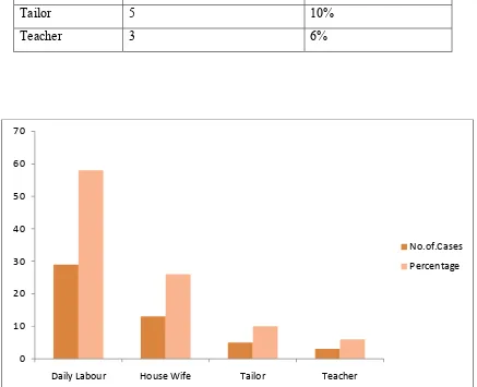
3. 4.4.3 Occupational status54
4. 4.4.4 Signs & Symptoms55
5. 4.2.2.1 Physico Chemical Report60
6. 4.2.2.2 TCL Report
61
7. 4.2.3. Phytochemical Report
63
8. 4.4.5. Improvement Signs & Symptoms66
9. 4.4.6.Gradation Result
67
PART-2
Sl.No.
Title of the Tables
Page
1. 4.2.2 Qualitative phytochemical report
136
2. 3.3.2Physio chemical report .
134
3. 4.2.1.2 TLC Estimation report
135
4. 4.3.1 Behavioral signs of toxicity
139
5. 4.3.2. Body weight of albino rats
139
6. 4.3.3.Food intake of rates
139
7. 4.3.4.Water intake of rats
140
8. 4.3.5.Hematological Parametors
140
9. 4.3.6. Biochemical parameters
141
10. 4.3.7.RFT
141
11. 4.3.8 Lipid profile
141
12. 4.3.9 Urine analysis
142
13. 4.3.10 Organ weight
142
14. 4.4.1 Behavioral signs of loxicity
147
15. 4.4.2 Ulcer index
147
16. 4.5.1 age wise distribution
129
17. 4.5.2 Sex distribution
130
18. 4.5.3 Socio economic status
130
19. 4.5.4 Occupational status
131
21. 4.5.6 Improvements in signs and symptoms
148
22. 4.5.7 Gradation results
149
NO TITIE
OF
FIGURE
4.1.1 Ilavu
ver
(Bombax ceiba)
4.1.2
Ilavu ver chooranam
4.2.1.1
T.S of root – Entire view
4.2.1.2
T.S of root – Enlarged section
4.2.1.3 Secondary
xylem
4.2.1.4 Secondary
phloem
4.2.1.5 Periderm
zone
4.2.1.6 Secondary
phloem & secondary xylem
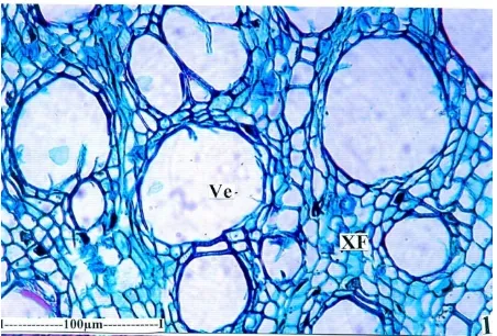
4.2.1.7 Vessels
&
Fibers
4.2.2.4
Scanning electron microscope (SEM)
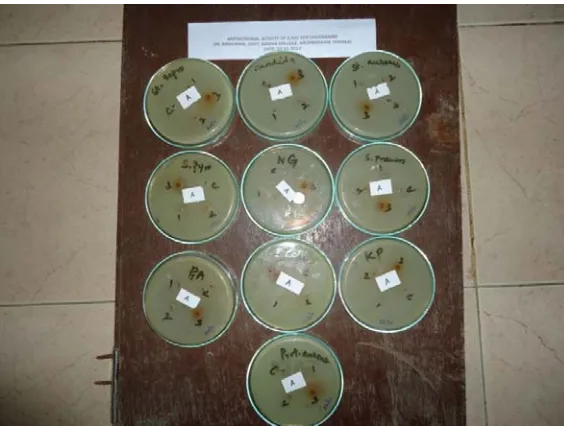
4.3
Anti microbial slide
4.1.1
Purified sodium chloride impure
4.1.2 Purified
asafoetida
4.1.3 Purified
zingiber
officinale
4.1.4 Purified
cuminum cyminum
NO
TITLE OF FIGURES
4.1.6
Purified plumbago zyelanica
4.1.7
Purified costus speciosus
4.1.8
Purified terminalia chebula
4.1.9 Acorus
calamus
4.1.10 Purified
acorus
calamus
4.1.11
Hingu chooranam
4.2.1.4
Scanning electron microscopecsem
4.3.1 to
4.3.12
Histopathological features
PART
–
I
ANTI
MICROBIAL
ACTIVITY
OF
ILAVU
VER
1.
INTRODUCTION
World’s first originated medicine is our traditional siddha system of
medicine. This system of medicine was founded by siddhars. In the Siddha
pharmacopeia, the Sidddars have used 4 typs of crud drugs which is crude drugs of plant
origin, animal origin, mineral origin and metal origin for preparing medicines. Herbs are extensive used for the preparation of medicine in siddha system of medicine.
“Ver paaru thazhai paaru minjinakkal mella mella Parpa chenduram paaru” .
Roots, leaves, flowers, fruits, seeds of plants are being used to alleviate the diseases. If the herbal drugs cannot control the severity of the disease, the next step is to
use the drugs of mineral & metal origin as parpam and chenduram. It is the line of
treatment in this system.
Roots are rich in phytochemicals. To illustrate the importance of roots, “Pathartha guna chinthamani” classified roots as Dasamoolam and panchamoolam.
The basic theory of siddha system is that the human body is made up of 96
thathuvas. The human body functions mainly by the three uyir thaathus (3 Humors ) are
named under mukkutram and these are related to arusuvaigal (6 Tastes ) and pancha
boodhangal (5 Basic elements).
Disease occurs in the human by means of decrease or increase in quantities of uyir thaathus.
Siddhars classified the total diseases in human body according to the
literature “Agathiyar rathina churukka naadi” as 4448 disease.
Vellai noi is one of the “Neer rogam” under these classification. The
aetiology,types and clinical features of vellai noi are mentioned in “Yugi chinthamani”.
In modern aspect the vellai noi is compared to that of Leucorrhoea. Abnormal
vaginal discharge (Leucorrhoea) is quite a frequent complaint of women in day to day life. It could be painful (dragging pain in abdomen, lumbar region pain) cause lot of discomfort (Intense itching, irritability),stress and even affect the sexual life and libido(Obstetrics and Gynecology for post graduates -S.S.Ratnam).
The survey conducted all over the world indicates that 75% of women will have at least one episode of candidal vaginitis during their life time and 40% -50% of women may experience a recurrence(Sobel J.D,1985). 90% of patients caused by candida albicans, trichomonas vaginalis, gardnerella vaginalis (Clayton et al., 1985).
The development of the nation partially depends on the development of the women who constitute half of its population. But woman is more prone to disease regarding her situation to come across various explanatory changes of life like puberty, menstruation, child bearing, lactation and menopause. If woman is healthy, she will give her ‘Best’ for the health and development of her child, family, society and nation.
So I have decided to choose one of the most common complaints of women “The
vaginal discharge” as my dissertation topic ”Vellai noi”.
Herbal medicine also called as phytomedicine, and it has a long tradition of use. It has a high value in treating and preventing diseases. The WHO estimated that 80% of people worldwide rely on herbal medicine for health care.
Current treatments for leucorrhoea include Metronidazole, Itrconazole, Imidazole, Ecanozole. The advers effect of these drugs are Neuropathy, Epileptiform seizures, Leucopenia, CNS & GI disturbances,Urticaria,Angioedema, Menstural irregularities, Increase in liver enzymes, Dark urine. So I have decided to take the herbal medicine “Ilavu ver (Bombax ceiba) since it will be less toxic and will not produce the adverse
effects like the modern medicines.
In the siddha literature “GUNAPADAM – MOOLIGAI VAGUPPU” the drug
“Ilavu ver chooranam”(Bombax ceiba)is indicated for leucorrhoea. So the drug has been
selected for the dissertation work in treating the disease leucorrhoea.
The root of Bombax ceiba is a modified root for the special function of
supporting and it is named as ROOT BUTTRESSES. This tree is widely distributed in tropical regions. It grows in alluvial sand near the banks of rivers and rainfall is well distributed throughout the year.
Since the above said conditions are conducive for the plantation of bombax ceiba.
So it is widely cultivated in the Theni district which lies in the hilly regions of western ghats. If the plant well growing in this alluvial soil, this root contains rich
So this root can be used as a cost effective medicine for the treatment of leucorrhoea, which in turn provides curing low economic status women.
Similar to the exploration of the underground gold to ornamental ones, I have
decided to use the roots of Bombax ceiba in an effective manner to cure the ailment and
2. AIM AND OBJECTIVES
AIM:
Herbal medicines also called phytomedicine. It has long tradition of use for long life with better health. The value of herbal medicine in the treating and preventing
disease. Root of Ilavu maram ( Bombax ceiba ) are very useful to treat a vellai noi (
Leucorrhoea ) which is mentioned in our literature. It will be less toxic and will not produce the adverse effects for leucorrhoea. Leucorrhoea is one of the most common complaints of women. These roots are abundant in quantity in root buttresses. So this root can be used as a cost effective medicine for the treatment of leucorrhoea, which in turn provides curing even for low economic status women.So I have decided to use the
roots of Bombax ceiba in an effective manner to cure the ailment and thereby creating a
healthy womenhood.
The ultimate aim of my dissertation work is to prove the ANTI – MICROBIAL
ACTIVITY of Ilavu ver chooranam OBJECTIVE:
In this dissertation work, the “ILAVU VER CHOORANAM” is analyzed to
assess the following aspects:
K To collect the literature review
K Get the authentication of the raw drug
K Pharmacognostic study for the raw drug
K Phytochemical and Chemical analysis for the trial drug to identify the
active components.
K Pharmacological study to evaluate the anti – microbial activity
K Clinical study to assess the efficacy of the drug through open clinical trial
3. REVIEW OF LITERATURE
3.1. Botanical aspect:
Bentham & Hooker’s classification:
Kingdom - Plantae
Sub kingdom - Tracheabionta
Super division - Spermatophyta
Division - Magnoliophyta
Class - Magnoliopsida
Sub class - Dilleniidae
Order - Malvales
Family - Bambacaceae
Genus - Bombax
Species - Bombax ceiba
Vernacular names:
English - Cotton Tree , Red Silk-cotton Tree
Tamil - Ilavam , Ilavu , Mullilavu , Kongu , Pulai
Sanskrit - Kantakadruma , Raktapushpa , Bahuvirya , Pichhala ,
Mochani - Tuliphala
Hindi - Kaantisenbal , Pagun , Rakatsenbal , Semul
Bengal - Pagun , Roktosimul , Simul , Tula
Gujarat - Sawar , Shimalo , Shimar , Shimul
Kanadam - Booruga , Kempubooruga , Mullilavau
Marathi - Kantesavar , Saur , Simalo , Tamari
Malayalam - Mullilabpoola , Mullilavau , Mocha , Unnamuriku , Purani
Oriya - Bouroh , Mochoroso , Salmali , Simuli
Telugu - Boorugachettu , Kondabooruga , Mundlaburaga ,
Pinnaburaga
Combodia - Roka
Chinese - Mu Mien
Burma - Didu , Lepanbin , Letpan
Ceylon - Parutti
French - Bambax de Malabar , Cotonnier Mapou
Distribution:
A lofty, deciduous tree buttressed at the base, up to 40m in height and 6m or more in girth, with a clear bole of 24-30m. Widely distributed throughout India, including the Andamans, up to 1,500m or even higher. The trees, in full bloom, present a striking blaze of colour and are grown in avenues. In peninsular India, the tree is very common in the dry as well as moist, mixed deciduous forests; in West Bengal and Assam, it is found in the mixed evergreen forests as well. The tree grows sporadically in the mixed deciduous forests in the sub-Himalayan region and lower valleys, and is typical of the alluvial-savannah-type forests, tending to be gregarious near the riverbanks
Description of the plant:
Branches horizontally spreading, more or less in whorls; young stems and branches covered with stout, hard prickles; bark pale ash to silver-grey, smooth in the early years, later becoming rough, with irregular vertical cracks.
Leaves large, spreading, glabrous, digitate, leaflets 5-7, lanceolate, 10-20cm long. Flowers numerous, large, 10-13cm in diam, fleshy, bright crimson, yellow or orange, clustered at the end of branches, bisexual, very rarely unisexual.
Capsules oblong-ovoid, woody, 10-19cm long; seeds many, obovoid, smooth,6-9mm long, oily, with dense silky hairs.
The buttresses are present only in trees of c30 years or more. The tree sometimes reaches very large dimensions, as much as 59m in height and 35m in girth around the buttress. It is eminently suited for afforestation of new ground and of grasslands in the riverain tracts. It is also useful in controlling soil erosion.
The tree is natural habitat, excluding the hills, the absolute maximum shade-temperature varies from 37.5 to 50, the absolute minimum from- 2.5 to 17.5, and the rainfall from 50 to 460cm or more; it thrives best in places where the rainfall is well distributed throughout the year.
Phenology:
seeds are irregular, obovoid, dark brown, smooth with a brittle testa. The seeds are collected from mid- March to mid- May and are separated from the floss. The weight of 100 dry fruits is c 2kg, of which the floss is c 600g and seeds c 500g, the rest being made up of the woody rind; 25,300-38,500 seeds weigh a kilogram. Although the seeds are oily, they remain viable for c 2 years, if properly stored. Fresh seeds show high germination percentage and are preferred for sowing.
Part used:
Root, Bark, Flower, Fruit, Gum
Actions:
Root - Astringent, demulcent, tonic, aphrodisiac, cooling, slightly
diuretic
Bark - Demulcent, diuretic, tonic, slightly astringent
Flower - Cooling, diuretic, astringent, tonic aphrodisiac, stimulant,
expectorant, alterative.
Young fruits - Expectorant, stimulant, diuretic
Gum - Astringent, styptic, tonic aphrodisiac, demulcent.
Chemical constituents: Leaves:
Crud protein, 18.69; crud fiber, 11.42; total minerals, 6.96; reducing sugar, 1.2; total sugars, 4.94; and starch, 12.09%.
Flowers: Calyces:
Moisture, 85.66; crud protein, 1.38; ethwr extr, 0.44; carbohydrates, 11.95; and mineral matter,1.09; calcium, 92.25; phosphorus, 49.0; and magnesium, 54.24 mg|100g.
Fresh petal:
Orang-red anthocyanin, pelagonidin-5β-D-glucopyranoside (C21 H21 CIO10 m p,
300◦) and cyaniding-7-methyl-ether-3 β-glucopyranoside (C22 H23 CIO11, m p 300◦)
Alcoholic extract of flowers:
Hentriacontane, hentriacontanol, β-sitosterol and its β-D-glucoside, quercetin,
kaempferol and an essential oil.
Stamens:
Young roots:
Moisture, 7.5; mineral matter, 2.1; pritein, 1.2; fat,0.9; starch, 71.2; pectic substances, 6.0; cellulose,2.0; phpsphatides(cephalin), 0.3; semul-red(a colouring matter), 0.5; tannins, 0.4; non-tannins, 0.1; and sugars(arabinose and galactose), 8.2%.
The proportion of the constituents varies with the age of the roots ; younger roots contain more sugars, starch, and pectic substances than the older roots, but contain less oil, colouring matter and cellulose.
Root bark:
Lupeol,β
-sitosterol,8-formyl-7-hydroxy-5-isopropyl-2-methoxy-3-methyl-1,4-nsphthaquinone(C16 H16 O5 ,m p 82◦ ), isohemigossypol-1-methyl ether(C16 H18 O4,m p
153◦),
isohemigossypol-1, 2-dimethyl ether(C17 H20 O4,m p 101-02◦),
7-hydroxycadalene (C15 H18O, m p 113-14◦) .
Bark:
Total water extr, 9.92; tannins, 3.01; and non-tannins, 6.91%. It also contains
lupeol, β-sitosterol and its D-glucoside.
Gum:
8.9% mineral matter and a considerable propotion of catechol tannin; on complete hydrolysis, it yiels a mixture of L-arabinose, D-galactose, D-galacturonic acid, and possibly rhamnose. The gum also contains tannic and gallic acids.
Seeds:
Moisture, 6.2; crude protein, 28.51; pentosans, 8.9; and mucilage, 2.0%. The seeds also contain n-hexacosanol, palmitic, gallic and tannic acids, octyl palmitate,
l-galloyl-β-glucose, ethyl-gallate, α-, β- and γ-tocopherols, carotenoids, and an unidentified
terpene.
MEDICINAL USES: Leaves & Flowers:
The paste of flowers, and also that of leaves, is employed as an application in cutaneous troubles.
Flowers:
These are good for haemorrhage, conjunctivitis, splenomegaly and haemorrhoids.
Root:
Bark:
It’s aqueous extract mixed with curd is used to check blood- dysentery. Externally it is used as a styptic, and also for fomenting wounds. The paste of the bark is applied to skin eruptions.
Gum:
It is used for dysentery, haemoptysis in pulmonary tuberculosis, influenza and menorrhagia, burning sensation, strangury and haemorrhoids.
The young fruits:
These are considered beneficial in calculous affections, chronic inflammation and ulceration of the bladder and kidneys.
Seeds:
It is used in gonorrhoea, chronic cystitis and other catarrhal affection. The hot, aqueous extract of the seeds possesses moderate oxytocic activity.
3.2. GUNAPADAM ASPECT : Part used:
Leaves, Flower, Seed, Bark,Gum, Cotton, Root.
Taste (suvai):
Sweet, Astringent, Bitter.
Character(thanmai): Thatpam
Classifaication(pirivu): Inippu
Gunam:
“ ந க்கடுப்புநெரrவுநண்ெடாழுகுேமகமும்ேபாம்
ஆ க்கும்விந்துேவாைடக்குமாண்ைமயுறும்- பா க்குள்
நிலவுமதிவதனேநrைழேயெவப்பாயம்
இலவுமரத்தாலியம்பு”
(AGATHIYAR GUNAVAGADAM)
The bombax ceiba tree cures Burning micturation and Leucorrhoea.Its strengthens the Sukkilam and Rasa thadhu.
The bark of Bombax ceiba is grainded and taken lemon size then mixed with
3.3. MODERN ASPECT OF DISEASE :
LEUCORRHOEA : Physiology of vagina:
It is a fibromuscular passage that connects the uterus to the introitus.The vaginal mucosa is lined by squamous epithelium which consists of a basal layer of cuboidal cells, a middle layer of prickle cells and a superficial layer of cornified cells. Vaginal secretion is small in amount in healthy women, and consists of white coagulated material. The vaginal contents are mostly derived from the squamous cells of the vaginal mucosa. Some contribution comes from endometrium.
Patho physiology:
The superficial cornified cells of the vaginal mucosa produce glycogen under oestrogen stimulation and are continuously desquamated. Subsequently, as a result of the breaking down of the cells, glycogen is liberated and ultimately converted into lactic acid.In the newborn, before the appearance of the Doderlein’s bacilli, glycogen is broken down into lactic acid and there is some evidence that the process is brought about by enzyme action. After the appearance of Doderlein’s bacilli, the production of the lactic acid is augmented by the action of bacilli on simple sugar.The vaginal secretion is acidic due to the precence of lactic acid and acidity inhibits the growth of pathogenic organisms. The pH of the vagina averages about 4.5 during reproductive life. The acidity, which is undoubtedly oestrogen dependent, falls after the menopause to neutral or even alkaline. Before puberty the pH is about 7. This high pH before puberty and after menopause explains the tendency for the development of mixed organism infections in these age groups.
The components of vaginal secretion are from:
The sweat and sebaceous glands of the vulva and the specialized racemose glands of Bartholin’s. The characteristic odour of the vaginal secretion is provided by the apocrine glands of the vulva.
The transudate of the vaginal epithelium and the desquamated cells of the cornified layer. This is strongly acidic.
The mucous secretion of the endocervical glands which is alkaline. The endometrial glandular secretion.
Causes of leucorrhoea:
Pathalogical vaginal discharge:
• Non infective
• Infective
• Neoplastic
• Truama
Non infective leucorrhoea:
The term leucorrhoea should be restricted to those conditions when the normal vaginal secretion is increased in amount.
Causes:
i. Physiological excess
ii. Cervical causes
iii. Vaginal causes
Physiological excess:
The normal secretion is expected to increase in conditions when the oestrogen levels become high. Such conditions are
During puberty:
Increased levels of endogenous oestrogen lead to marked overgrowth of the endocervical epithelium which may encroach onto the ectocervix producing congenital erosion increased secretion.
During menstrual cycle:
Around ovulation peak rise of oestrogen increase in secretory activity of the cervical gland.
Premenstrual pelvic congestion and increased mucus secretion from the hypertrophied endometrial glands.
Pregnancy:
There is hyperoestrinism with increased vascularity.This lead to increased vaginal transudate and cervical gland secretion.
During sexual excitement:
Cervical cause:
Non infective cervical lesion may produce excessive secretion which pours out at the vulva. Such lesions are cervical erosion, chronic cervicitis, mucus polyp and ectropion (Cervical glands are exposed to the vagina).
Vaginal causes:
Increased vaginal transudation occurs in conditions associated with increased pelvic congestion. The lesions are uterine prolapse, acquired retroverted uterus, chronic pelvic inflammatory disease and even chronic constipation.
Infections:
Infection of the genital tract may cause muco-purulent or frankly purulent vaginal discharge. The amount, colour, and odour depend upon the type of infection.
Types:
• Specific vaginal infection
• Non specific vaginal infection
Specific vaginal infection:
If the strict definition of accepted as an excessive vaginal secretion in which the primary cause is not infective,any vaginal discharge which is frankly purulent and contains pus cells and from which the causative organism can be isolated and cultured, should be considered as due to specific vaginal infection.
Vulvo-vaginitis:
• Non-specific organisms (Infancy and post-menopausal age groups).
• Tricomonas vaginitis.
• Candida albicans.
• Gonococcal and other bacterial infections (Bartholinitis and Urethritis).
Cervicitis:
• Pyogenic infection due to non-specific organisms during puerperium
and following abortion.
• Gonococcal infection.
Endometritis:
• Non-specific infections during puerperium and post-menopausal age.
It is observed that50% vaginitis is due to bacterial vaginosis 20-25% due to monilial infection
15-20% due to trichomonal infection
Gardnerella (bacterial) vaginosis:
Bacterial vaginosis is termed vaginosis rather than vaginitis, because it is associated with alteration in the normal vaginal flora rather than due to any specific infection. There is a considerable decrease in the number of lactobacilli in the vaginal discharge with 100-fold increase in growth of other anaerobic bacteria.Since lactobacilli release hydrogen peroxide toxic to other bacteria, reduction in their number allow other bacteria, I.e. aerobic and anaerobic bacteria to grow.These are Haemophilus vaginalis, Gardnerella, Mobiluncus and Mycoplama hominis. The bacterial vaginosis is therefore a polymicrobial condition.
It is not sexually transmitted with a variable incubation period. About 50% women are asymptomatic carriers of infection, but majority complain of vaginal discharge without itching and minimal vulval irritation.
The characteristics of vaginal discharge :
White, milky, non-viscous discharge adherent to the vaginal wall. pH of the discharge is more than 4.5.
Fishy odour when mixed with 10% KOH is due to amino-metabolites from various organism.
Presence of clue cells---the epithelial cells have fuzzy border due to adherence of bacteria.
Increased number of Gardnerella vaginalis and other organisms and reduced number of lactobacilli, and leucocytes.
Causes:
PID
Chorioamnionitis
Pre-mature rupture of membrane (PROM ) Preterm labour
Gonococcal vulvovaginitis:
This is a sexually transmitted disease which can lead to sequelae adversely affecting reproductive functions.
epitheliam is resistant to gonococcal infection. The gonococci attack the columnar epithelium of glands of Skene, Bartholin, urethra and its glands, cervix and fallopian tubes. It ascends in a piggy-back fashion attached to the sperms to reach the fallopian tubes.It is destroyed easily by drying, heat, sunlight, and disinfectants.
Sites for bacterial recovery: These include the urethra, cervix, anal canal and pharynx.
Principal sites of invasion: Columnar epithelium of the genital tract, transitional epithelium of the urethra and Bartholin’s gland.
Complications:
Pelvic inflammatory disease, pyosalpinx formation, tubo-ovarian abscess, pelvic abscess followed later on by hydrosalpinx formation, infertility, menstrural disturbances, chronic pelvic pain, dysmenorrhoea and dyspareunia.
Chlamydia:
Chlamydial infection is common in young, sexually active women but rare after the age of 40 years. Two to ten percent of pregnant women are found to have this infection during antenatal period and account for 1% of all abortions. The incubation period is 6-14 days.
Chlamydia trachomatis is a small gram-negative bacteria, an obligate intracellular parasite that appears as intracytoplasmic inclusion body, and is of two varieties, one that causes lymphogranuloma venereum (LGV) and the other of non-LGV, which causes non-specific lower genital tract infection. Often, the infection is silent and the women is asymptimatic but may develop vaginal discharge, dysuria and frequency of micturation, and at times cervicitis.
Sometimes Chlamydia may cause Reiter’s syndrome with arthritis, skin lesions, conjunctivitis and genital infection. It also causes perihepatitis and Fitz-Hugh-Curtis syndrome similar to that of gonorrhoea when pelvic inflammatory disease (PID) is associated with right upper abdominal pain. During pregnancy, abortion, preterm labour and IUGR may occur. Newborn suffers from conjunctivitis, nasopharyngitis, otitis media and pneumonia.
Trichomoniasis:
instances, it can be acquired by inadequate hygiene or the use of an infected person’s towels, bath or clothes. Its ingress to the vagina is favoured by a low general resistance and when the pH is raised as during a menstrual period (pH 5-6). It is not uncommon during pregnancy and is often associated with gonococcal infection.
The Trichomonas organism is a protozoa, actively motile, slightly larger than a leucocyte and is anaerobic. Three types of trichomonas are known, namely,
Tr. Buccalis---a normal inhabitant of the mouth.
Tr. Hominis---a normal inhabitant of the anal canal and rectum. Tr. Vaginalis---it is found in the vagina.
It has been shown by transplantation experiments that Tr. Buccalis and Tr. Hominis are unable to survive in the human vagina. Men may harbour Tr.vaginalis in the urethra and prostate. A trichomonad has four anterior flagella and one posterior flagella, and they move along the mucous membrane.
Symptoms:
Twenty percent remain asymptomatic – others develop symptoms 4 to 28 days following sexual contact with an infected partner, or infected material, like towel or toilet. Seventy percent show typical discharge, which is profuse, thin, creamy or slightly green in colour, irritating and frothy.
The vaginal walls are tender, angry looking and the discharge causes pruritus and inflammation of the vulva.
There are often multiple small punctuate strawberry spots on the vaginal vault and portio vaginalis of the cervix (strawberry vagina ).The characteristic frothy discharge is almost self-diagnostic but the presence of secondary infection may alter and mask this initial sign.
The patient may also complain of urinary symptoms, such as dysuria and frequency, and a low-grade urethritis may be discovered on examination. Abdominal pain, low backache and dyspareunia may also be complained of if pelvic infection occurs.
Candidal (monilial) vaginitis:
Risk factors:
These include promiscuity, immunosuppression, pregnancy, steroid therapy, oral contraception pills, following long-term broad spectrum antibiotic therapy, diabetes mellitus, poor personal hygiene and obesity.
Clinical features:
Pruritus vulva is the cardinal symptom. It is often accompanied by vaginal irritation, dysuria, or both, and passage of thick curdy or flaky discharge. Speculum examination reveals vaginal wall congestion with curdy discharge often visible at the vulval mucocutaneous junction and in the posterior fornix.
Neoplasms:
Any neoplasm of the lower genital tract causes, initially, serous discharge but later due to ulceration of the surface or sloughing of the growth and secondary infection causes offensive, foul smelling blood stained vaginal discharge.
The neoplasms which cause such a vaginal discharge are malignant neoplasms of the cervix and body of the uterus, but in some cases purulent blood stained discharge may be caused by ulceration of the surface of benign polypi of the cervix and endometrium.
Trauma:
Any trauma to the genital tract may cause a local inflammatory reaction. It initially causes a serous vaginal discharge, but later on the discharge becomes purulent due to secondary infection.
The causes of trauma may be:
Physical:
Direct trauma.
Foreign body and pessary.
Chemical:
Drug and antibiotics causing local irritation and burns.
Allergic:
Local antiseptics and drugs used for douching and cleaning. Contraceptives, i.e., diaphragm and condom.
3.4. SIDDHA ASPECT OF DISEASE :
VELLAI NOI : Synonyms:
Vettai; Bramium; Bramia rogam; Seezh megam. Definition:
Purulent white coloured discharge before or after micturation and burning sensation in the urethral orifice.
Prodromal symptoms:
• Sexual intercourse with same diseased patient leads to itching in the
genital organ and burning sensation in the urethral orifice within 3 or 4 days.
• During micturation there will be purulent discharge which will be
viscous like that of thread.
• Sometimes fever occurs.
Etiology:
1. Excessive sexual intercourse.
2. Sexual into with same diseased patient.
3. Prolonged starvation.
4. Excessive walking during hotsun
5. Excessive intake of tastes like acrid, salt, astringent, bitter.
Classification:
It is classified into 21 types 1. Valli vellai
2. Azhal vellai 3. Iya vellai 4. Vali azhal vellai 5. Azhal Iya vellai 6. Mukkutra vellai 7. Kal vellai 8. Katti vellai 9. Nool vellai 10. Neer vellai
11. Neechu vellai
12. Manjal vellai 13. Kiricharam vellai 14. Karappan vellai
15. Thanthi vellai 16. Malinam vellai
17. Kuruthi vellai 18. Seezh vellai
19. Inippu vellai 20. Pun vellai
Vali vellai:
In vali vellai during micturation urine resembles cow’s urine.Pus mixed with the
urine and also spasmodic pain is present in the lower abdomen.
Azhal vellai:
In azhal vellai black colouration and burning sensation present in the
body.During micturation burning sensation in the urethral orifice and also the urine is yellow coloured and mixed with pus.
Iya vellai:
In iya vellai white coloured pus mixed with urine. Burning sensation in the
urethral orifice. Asper anemia pale colouration is found in the body.
Vali azhal vellai:
In vali azhal vellai the symptoms resembles that something has strucked in the
urethral orifice. Incontinance, powdered particles and burning micturation is present while urination.
Azhal Iya vellai:
In azhal iya vellai disease there is distension and spasmodic pain seen in the
lower abdomen.Yellow coloured and pus seen in the urine.
Mukkutra vellai:
In mukkutra vellai frequency of micturation and white discharge is seen. Different
colour is found in the discharge and precipitate is seen when kept aside.Tingling sensation is seen.
Katti vellai:
In katti vellai there is boils in the body, burning and itching in the urethral orifice,
fistula is seen and while micturation urine passes through it.
Neer vellai:
In neer vellai there is pain the genitals. White discharge and spasmodic pain in
the lower abdomen is seen. While defecating pus is mixed and white coloured stools are passed. Urine are passed as like rice gravel.
Thanthi vellai:
In thanthi vellai white discharge continuos like thread and then followed by urine
Kurithi vellai:
In kuruthi vellai giddiness, weakness, there is pain during micturation and urine is
as that of Rabbit’s blood. Dullness in the skin colour, urinary incontinence and redness is seen in the urine.
Seezh vellai:
In seezh vellai discharge is seen as it is like pus. Boils in the groin, fistula, wound
in the umbilical occurs.Fever with shiver, giddiness and this symptoms may probably acute suppurative urethritis or even acute gonorrhoea.
Ozhukku vellai:
In ozhukku vellainoi the urine is found with blood and pus and discharge is seen
always.All over the body small pustules are seen, breaks and produce wounds.
Manjal vellai:
In manjal vellainoi dischrge is found as pale yellow colour. Burning sensation as
that of fire in the urethral orifice. Face becomes yellow, bitterness in the tongue, body becomes more hot.
Neer churukku vellai:
In neer churukko vellai urithral orifice become more tensed and while micturation
urine is found yellow coloured.White discharge is found. Desire toward consumption of food is decreased and it result in mentally and physically ill.
Karappan vellai:
In karappan vellai noi stool is mixed with pus and pain during defecation. The
urine is released with more heat. In while body heat is increased and results in wound and spreads like eczema. While micturating small stones are released.
Kal vellai:
In kal vellai noi discharge is found like toddy. Pungent body odour. Black
coloured stones are released. Pain is found between umbilicus and ribs.
Nool or thandhu vellai:
In nool vellai noi discharge appears like thin spider web, pain and blood is seen
during micturation. There are sediments found when collected and kept the urine aside. Urine spells like drop by drop.
Neechu vellai:
In neechu vellai noi urine is concentrated and appears like toddy and colour as
Vali or malina vellai:
In malina vellai noi weakness, bitterness in tongue, pain the soles. During
micturation decomposed muscle partical and discharge are seen together.
Then vellai:
In then vellai noi , clearless urine and seen like honey while micturating. Urine
appears red, fat particles and muscle particles as per water which washed the meat. Wound in the urithral orifice, peculiar smell is found in the urine.
Pun vellai:
In pun vellai noi, wound is found all over the body. Blood is mixed in the urine.
The joints are found stiff and normal activities are altered,pain in the joints, body heat are increased.
OTHER PREPARATIONS OF BOMBAX CEIBA : Gum of bombax ceiba:
Theriar vaagadam:
1. Athividaya kudineer --- pg no
2. Pithaththukku ennai --- pg no 183 3. Neer kazhichaluku kalappuththul --- pg no 230 4. Karkam --- pg no 236 Sigicharathna deepam:
1. Ingaya chooranam --- pg no 111 2. Kiranikesari kuligai --- pg no 152 3. Katuvai mathirai --- pg no 153 4. Maha madhanakamesura legium --- pg no 160 Vaithya chinthamani:
1. Utinavayu kirani maruthuvam --- pg no 176 2. Moolavayu kirani maruthuvam --- pg no 179 3. Nalapada azhalnoi maruthuvam --- pg no 267 4. Markka azhal maruthuvam --- pg no 268 5. Asaathiaathisaram --- pg no 170 6. Kirani --- pg no 173 Noigalukku siddha parikaram:
Flower of bombax ceiba: Theriar maha karisal:
1. Mathana kamesvaram --- pg no 37 Root of bombax ceiba:
Root of bombax ceiba is one of the adjuvants for Loga chenduram and Aya
chenduram.
Sarabanthirar vaithya rathnavali (Part 2 )---pg no 16 Chthraka kudabagam :
Plambago zeylanica --- 1400 gm
Root of Solanum surattense
Root of Bombax ceiba Each 700 gm
Root of Tribuluse terestris
Root of Tinospora cordifolia
All ingredients should be dried well then made into chooranam, taken in a pot add 4 marakkal water then it is kept for one day.Next day sufficient heat given and reduced to 1|4 and then filtered. Add palm jaggery 3500 gm to this mix well and then filter it. Take the decoction boil it and reduced it 1/4.
Next day Honey --- 1120 gm
Zingiber officinalis Piper nigram
Piper longum Each 70 gm Cinnamomum verum
Eletaria cardamomum
These drugs are mixed with above decoction and grainded well.
Dosage: 21 gm Therapeutic indication:
Leprosy, cough, bronchial asthma, tuberculosis, peptic ulcer, bleeding piles, sinusitis, hydrocele.
The sinusitis and agnimantham which cannot be cured by many medicines can be
cured.
Decoction for pitham :
Root of Bombax ceiba
Bark of Ficus glamuvata
Bark of Eugenia jambolana
Bark of Roxb lannea
Root of Tribuluse terestris
Root of Phyllanthus amarus
Root of Ficus benghalensis
Orilai tamarai Each 35 gm Pavonia zeylanica
Pavonia odorats Cuminum cyminum Alternanthera sessilis Oryza sativa -Nerpori
Gum of Limonia acidissima
Above said ingredients partly ground mixed with water 650 ml in a mud pot. Sufficient heat given and reduced to 1|2 then allow to cool, mash well and filtered it.
Dosage:
60 ml decoction 4 hours once.
Therapeutic indications:
Itching, pithathalundana thega erichal, piththathazhaluntha thazhumbu.
Kannusamy paramparai vaithyam ----pg no 79 Azhal noi marunthu :
Root bark of cassia auriculata
Root of bombax ceiba
Orilai tamarai
Root of gmelina arborea
Phyllanthus amarus each 70 gm pavonia zeylanica
Root of hemidesmus indicus
Phaseolus trilobatus Vilamichu ver
Root of pergularia extensa
Elataria cardamomum
Santalum album each 17.5 gm Nardostachys jatamansi
Cedrus deodar
Aii these drugs are powdered and then sieved add equal amount of powder of
Cuminum cyminum and sugar then given 3 pinches for Azhal diseases. Bark of bombax ceiba :
Sigicharathna deepam :
1. Ilagugangaathara chooranam - --- pg no 121 2. Viruththa gangaathara chooranam --- pg no 122 Vaithya chinthamani :
1. Nethira bavuthiram --- pg no 211 2. Thodaathisaram --- pg no 169 Young fruit of bombax ceiba :
Agathiar attavanai vaagadam :
1. Athisaram theera podi ---- pg no 207 Vaithya chinthamani :
1. Thontha kirani ---- pg no 176
3.5. LATERAL RESEARCH:
1) Hypotensive activity and Hypoglycemic activity:-
A novel constituent, Shamimin, a C-flavonol glucoside from Bombax ceiba leaves showed significant potency as a hypotensive agent at the doses of 15 mg/kg, 3 mg/kg, 1 mg/kg and significant hypoglycaemic activity at 500 mg/kg in Sprague-Dawley rat (Ref : Saleem R, et,al., Hypotensive, hypoglycaemic and toxicological studies on the flavonol C-glycoside shamimin from Bombax ceiba. Planta Med. 1999 May;65(4): 331-4).
2) Anti-Inflammatory and Hepatoprotective activity:-
The bark, xylem of stem, and root of Bombax malabarica DC and Ceiba pentandra GAERTN are marked as "Mu-mien" in Taiwan. In order to clarify the pharmacological effects of these three parts, anti-inflammatory and liver protective effects were evaluated with carrageenan-induced paw edema and CCl4-induced hepatotoxicity in rats.
Sampath Kumar Evaluation of RBC Membrane Stabilization and Antioxidant Activity of
BOMBAX CEIBA in an in vitro Method International Journal of Pharma and Bio Sciences
vol 2:1:2011).
3) Antiangiogenic Activity:-
A methanol extract of the stem barks of Bombax ceiba was found to exhibit a significant
antiangiogenic activity on in vitro tube formation of human umbilical venous endothelial
cells (HUVEC). Bioactivity-guided fractionation and isolation carried out on this extract afforded lupeol as an active principle. At 50 and 30 µg/mL lupeol showed a marked inhibitory activity on HUVEC tube formation while it did not affect the growth of tumor cell lines such as SK-MEL-2, A549, and B16-F10 melanoma.
(Ref : Antiangiogenic activity of lupeol from Bombax ceiba Young-Jae You,
et.al.,Phytotherapy research Vol 17:4 pp341-44 2003).
4) Analgesic and Antioxidants Activity:-
Mangiferin, 2-beta-D-glucopyranosyl-1,3,6,7-tetrahydroxy-9H-xanthen-9-one, obtained directly from methanolic extracts of Bombax ceiba leaves in substantial amounts demonstrated strong antioxidant activity (EC(50) 5.8+/-0.96 mug/ml or 13.74 muM) using DPPH assay comparable to rutin, commonly used as antioxidant for medical purposes. It displayed significant analgesic effect in acetic acid – induced writhing and hot plate test in mice.
(Ref : Dar A, et.al.,Analgesic and antioxidant activity of mangiferin and its derivatives: the structure activity relationship. Biol Pharm Bull. 2005 Apr;28(4):596-600).
5) Cholinesterase Activity:
Leaf and stem bark of the seedling were screened for cholinesterase activity the activity was observed only in leaf.
(Ref : Taiwo O. Elufioye, et.al., Acetylcholinesterase and butyrylcholinesterase inhibitory activity of some selected Nigerian medicinal plants Revista Brasileira de Farmacognosia Brazilian Journal of Pharmacognosy 20(4): 472-477, Ago./Set. 2010).
7) Hepatoprotective Activity:
(Ref : Ravi V, et.al.,Hepatoprotective Activity of Bombax ceiba Linn against Isoniazid and Rifampicin-induced Toxicity in Experimental Rats. International Journal of Applied Research in Natural Products Vol. 3 (3), pp. 19-26, Sep-Oct 2010).
8) Anticancer and Anti-HIV Activity:
Methanolic extract of leaves and pure compounds mangiferin and acetyl derivative of mangiferin were evaluated in anticancer and anti-HIV activities. All the samples were evaluated to be inactive as cytotoxic and as anti-HIV agent. On the contrary, inhibitory effects of mangiferin in azoxymethane-induced rat colon carcinogenesis shows its chemoprotective nature. This discrepancy may have arisen due to the differences between the in vitro vs. in vivo assays used to assess the anticancer activity and thereby emphasizes the need to conduct a combination of both types of assays before reaching a definite conclusion.
(Ref : Nam NH, et.al., Inhibitory effects of Vietnamese medicinal plants on tube-like formation of human umbilical venous cells, Phytother Res, 17, 2003, 107-111).
9) Anti-Helicobacter Pylori Activity:
Ethanolic extracts of Bombax malabaricum DC. evaluated strong anti- Helicobacter pylori activities. The minimum inhibitory concentration values of the anti- Helicobacter pylori activity given by the ethanolic extracts ranged from 0.64 to 10.24 mg ml (-1). (Ref : Yuan CW, Tung LH, Screening of anti-Helicobacter pylori herbs deriving from Taiwanese folk medicinal plants, FEMS Immunology and Medical Microbiology, 43, 2005, 295–300).
10) Inhibitory effects on tube-like formation of human umbilical venous cells Seven of
58 plant materials from Vietnamese medicinal plants were evaluated strong to moderate inhibitory activity on the tube-like formation induced by human umbilical venous endothelial cells in the in vitro angiogenesis assay. These plant materials include the herb of Ephedra sinica, leaves and stem of Ceiba pentandra, seed of Coix lachryma-jobi, rhizome of Drynaria fortunei, fruits and stem of Illicium verum and stem of Bombax ceiba. The methanol extracts of the herb of Ephedra sinica and stem of Ceiba pentandra exhibited the strongest activities with inhibition percentages of 89.3% and 87.5% at 30 and 100 microgram/mL.
4. MATERIALS AND METHODS
4.1. Preparation of chooranam: Material:
Roots of Bombax ceiba (Ilavu ver). 8kg of Fresh leaves were taken, then it under
gone for shade dry after that the net weigh of the leaves were around 2.5 kg.
Collection And Authendication Of The Materials:
The plant material used in this study was collected during the month of June (2012) from Varusa naadu, Theni Dist, Tamilnadu, India and authenticated from the Gunapadam experts in Deportment of PG Gunapadam, Govt. siddha medical college, Chennai-106 and Certified from Botanist, Central Research Institute For Siddha,
Arumbakkam, Chennai-106. The preparation was collected from Gunapadam mooligai
vaguppu by Ka.Sa. Murugesa mudhaliyar. Purification of the Raw Drug:
The plant roots were well rinsed in water to remove the impurities. Then the leaves were cut into pieces and dried in shade.
Preparation of the chooranam:
The well dried Ilavu ver were made into fine powder. To get the finest physical
form of this drug, the powdered material is sieved through a white cotton cloth
(Vashthirakayam).
Purification of chooranam:
The Chooranam was moistened with cow’s milk. The pot was half filled with
milk and water. The mouth of the pot was covered and tied with white cotton cloth. The
Chooranam (moistened by milk) was placed above the tied cloth. The mouth of the pot
closed with another mud pot. The gap between the two mud pots was tied with a wet cloth to avoid evaporation. Then this arrangement was put on fire and boiled until water level gets reduced in the lower pot. Then the powder was taken, dried, powdered finely and preserved for usage.
Preservation:
The purified Chooranam was stored in a clean, air tight glass container. Since the
life period of the Chooranam is only three months, the prepared Chooranam must be
Figure no : 4.1.1 Ilavu Ver ( Bombax ceiba )
Figure no : 4.1.2 Ilavu Ver chooranam
Administration of the drug:
Form of the medicine : Chooranam
Route of Administration : Enteral
Dose : 1gm
Anubanam (Vehicle) : Warm water
Times of Administration : Two times a day; before food
Duration : 7 weeks
4.2. Standardization of the drug 4.2.1. Pharmacognostic aspect:
Ilavu Ver –Bombax ceiba
Collection and authentication of the materials:
Plants were collected from Varusa naadu, Theni dist and identified and authenticated by the Gunapadam experts in Department of P.G. Gunapadam, Govt. Siddha medical college, Chennai – 106 and certified by Botanist, Central Research Institute for Siddha, Chennai – 106.
Collection of specimens
The plant specimens for the proposed study were collected from---. Care was taken to select healthy plants and normal organs. The required samples of different organs were cut and removed from the plant and fixed in FAA (Farmalin-5ml+ Acetic acid-5ml + 70% Ethyl alcohol-90ml).After 24 hrs of fixing, the specimens were dehydrated with graded series of tertiary –Butyl alcohol as per the schedule given by Sass, 1940. Infiltration of the specimens was carried by gradual addition of paraffin wax (melting point 58-60 C) until TBA solution attained super saturation. The specimens were cast into paraffin blocks.
Sectioning
The paraffin embedded specimens were sectioned with the help of Rotary
Microtome. The thickness of the sections was 10-12 μm. Dewaxing of the sections was
by customary procedure (Johansen, 1940). The sections were stained with Toluidine
blue as per the method published by O’Brien et al. (1964). Since Toluidine blue is a
polychromatic stain. The staining results were remarkably good; and some cytochemical
reactions were also obtained. The dye rendered pink colour to the cellulose walls, blue to
the lignified cells, dark green to suberin, violet to the mucilage, blue to the protein
bodies etc. wherever necessary sections were also stained with safranin and Fast-green
For studying the stomatal morphology, venation pattern and trichome distribution,
paradermal sections (sections taken parallel to the surface of leaf) as well as clearing of
leaf with 5% sodium hydroxide or epidermal peeling by partial maceration employing Jeffrey’s maceration fluid (Sass, 1940) were prepared. Glycerine mounted temporary preparations were made for macerated/cleared materials. Powdered materials of different parts were cleared with Naoh and mounted in glycerine medium after staining. Different cell component were studied and measured.]
Photomicrographs
Microscopic descriptions of tissues are supplemented with micrographs wherever
necessary. Photographs of different magnifications were taken with Nikon labphoto 2
microscopic Unit. For normal observations bright field was used. For the study of
crystals, starch grains and lignified cells, polarized light was employed. Since these
structures have birefringent property, under polarized light they appear bright against
dark background. Magnifications of the figures are indicated by the scale-bars. Descriptive terms of the anatomical features are as given in the standard Anatomy books (Esau, 1964).
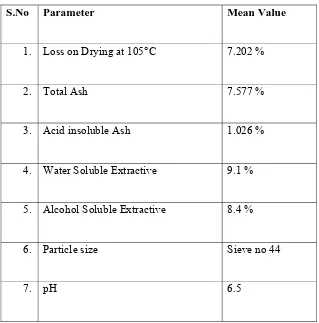
4.2.2. Physico-chemical analysis: 4.2.2.1 Ash and acid insoluble ash:
To the ash add 1:5 Hcl: Distilled water 15 ml boil, cooled and then filtered using whatman filter paper (No.41) repeat 3 to 4 times till the yellow colour disappear or colourless, then remove the filter paper and add to the filter to the original dish and keep
it in the muffle furnace at 600o C and take constant weight and calculate the acid
insoluble ash value.
Weight of acid insoluble residue x 100 Acid insoluble ash (%)=
Weight of the sample
Acid insoluble residue = Acid insoluble ash value – Empty weight of the dish
Loss on drying:
3gm of the drug is heated in a hot oven at 1050 c to constant weight. The % of
weight was calculated.
Potential of hydrogen (ph):
The pH scale is logarithmic and runs from 0.0 to 14.0 with 7.0 being neutral. Readings less than 7.0 indicate acidic solutions, while higher readings indicate alkaline or base solutions.
Above mentioned Quantitative analysis results are showed in the Table :1
4.2.2.2 TLC estimation of Ilavu ver chooranam:
Thin-layer chromatography is a technique in which a solute undergoes distribution between two phases, stationary phase acting through adsorption and a mobile phase in the form of a liquid. The adsorbent is a relatively thin, uniform layer of dry finely powdered material applied to a glass, plastic or metal sheet or plate. Glass plates are most commonly used.
Identification can be effected by observation of spots of identical Rf value and about equal magnitude obtained, respectively, with an unknown and a reference sample chromatographed on the same plate. A visual comparison of the size and intensity of the spots usually serves for semi-quantitative estimation.
Solvent system:
Toluene : Ethyl acetate (4:1.5).
TLC plate:
Aluminium plate precoated with silica gel 60F254 of 0.2 mm thickness (Merck).
Developing chamber:
Camag’s twin trough chamber.
Visualizing reagent:
Vanillin-sulphuric acid reagent.
Extract Preparation:
4 g of the chooranam was soaked overnight in chloroform. Boiled on a water bath for 10 mins, filtered and concentrated to 10 ml.
Procedure:
4.2.2.3 Fourier transform infrared spectroscopy (Ftir):
4.2.2.4 scanning electron microscope (sem):
The Scanning Electron Microscope (SEM) is a microscope that was electrons rather than light to form an image. There are many advantages to using the SEM instead of a light microscope.
Resolution : 1.2 nm gold particle separation on a carbon substrate
Magnification : From a min of 12 x to greater than 1, 00,000 X
The SEM has a large depth of field, which allows a large amount of the sample to be in focus at one time. The SEM also produces images of high resolution, which means that closely spaced features can be examined at a high magnification. Preparation of the samples is relatively easy since most SEM one require the sample to be conductive.
The combination of higher magnification, larger depth of focus, greater resolution, and easy of sample observation marks the SEM one of the most heavily used instruments in research areas today.
4000.0 3600 3200 2800 2400 2000 1800 1600 1400 1200 1000 800 600 400.0 0.0
5 10 15 20 25 30 35 40 45 50 55 60 65 70 75 80 85 90 95 100.0
cm-1 %T
3408.9 2930.8
2344.9 2138.3
1734.3
1627.8 1508.2
1430.0 1376.2
1321.0 1249.9
1159.8
1053.4
4.2.3. Qualitative phytochemical analysis:
S.No Experiment Observation Inference
1 Test for Alkaloids (Dragendorff’s Test) Few
mg of extract in separate test tube was warmed with 2% Sulphuric acid for 2 minutes. And it was filtered in separate test tube and few drops of Dragendorff’s reagent were added
. The presence of orange red precipitates indicates .
the presence of alkaloids.
2 Test for Flavonoids (Shinoda test)
Substance is dissolved in alcohol, added with magnesium bits and concentrated hydrochloric acid. On heating over a water bath
the appearance of majenta colour shows
presence of flavonoids.
3 Triterpenoids (Noller’s Test)
To fewmg of extract, add tin
and thionyl chloride and heat in water bath
Purple colour indicates presence of
tritepenoids.
4 Test for steroids:An
ethonolic extract of plant sample 2ml is mixed with 2
ml H2SO4 and 0.5 gm Acetic
anhydride
The solution turns in to blue to green colour
Presence of Steroids
5 Test for Phenol
Substance in water is added with 5 % alcoholic ferric chloride
.Dark blue or green colour shows
6 Test for Tannin
Substance is shaken with water and added with lead acetate solution
White precipitate shows presence of
tannin.
7 Test for Saponins
To few mg of extract distilled
water is added and shaken well
The formation of foam indicates
presence of saponin.
4.2.4. Chemical analysis :
Proximate Chemical Analysis of a Drug Methodology For Chemcial Analysis Preparation of Extract :
Add 5 gm of the sample to 50ml of distilled water. Boil the solution for 20 minutes, cool and then filter. Use the Extract for the following tests.
S.No Experiment Observation Inference
1. Test for reducing Sugar :
To 5ml of Benedicts qualitative reagent, add 10 drops of extract, then boil for two minutes
Green / Yellow / Red PPT
Presence of Reducing Sugar
2. Test for Starch :
To 5 ml of extract add 2ml of acetic acid and then add few drops of N/50 Iodine Solution.
Blue Colour Presence of
Starch
3. Test for Proteins :
To 2 ml of extract, add 2ml of 5% Sodium Hydroxide mix and add 2 drops of Copper Sulphate Solution.
Violet or Purple Colour
Presence of Proteins
4. Test for amino Acid :
Place 2 drops of extract on a filter paper and allow to dry well. Then spray 1% ninhydrin over the same and allow to dry.
Violet Colour Presence of
5. Test for Albumin :
To 2 ml of extract, add 2ml of Esboch’s reagent.
Yellow PPT Presence of
Albumin
6. Test for Phosphate :
To 2ml of extract, add 2ml of ammonium Molybdate solution and 2ml of concentrated Nitric Acid.
Yellow PPT Presence of
Phosphate
7. Test for Sulphate :
To 2 ml of extract add 2ml of 4% ammonium oxalate solution.
White PPT Presence of
Sulphate
8. Test for Chloride :
Add 2ml of extract to dilute nitric acid till the effervescence ceases. Then add 2 ml of Silver Nitrate Solution.
Cloudy White PPT
Presence of Chloride
9. Test for Iron :
To 2ml of extract, add 2ml of ammonium thio cynate solution and add 2ml of concentrated Nitric Acid.
Red Colour Presence of Iron
10. Test for Calcium :
To 2 ml of extract, add 2 ml of 4% ammonium Oxalate Solution.
White PPT Presence of
Calcium
11. Test for Sodium :
Make a paste with 2 pinches of the sample with Hcl and Introduce it into the blue flame.
Yellow Flame Presence of
Sodium
12. Test for Potassium :
Add a pinch of the sample to 2 ml of Sodium Nitrate Solution. Then add 2ml of Cobal Nitrate in 20% acetic acid.
Yellow PPT Presence of
S.No Experiment Observation Inference
13. Test for Zinc :
To 2ml of extract, add few drops of Sodium Hydroxide.
White PPT Presence of Zinc
14. Test for Magnesium :
To 2ml of extract, add few drops of Sodium Hydroxide Solution
White PPT Presence of
Magnesium
15. Test for Alkaloids :
a. To 2ml of extract, add 2ml
of Potassium Iodide Solution
b. To 2ml of extract add 2ml of
Picric Acid.
c. To 2 ml of extract add 2ml
of Phosphotunqstic Acid.
Red Colour
Yellow Colour
White PPT
Presence of Alkaloids
Presence of Alkaloids
Presence of Alkaloids
16. Test for Tannic Acid :
To 2ml of extract add 2 ml of Ferric Chloride Solution
Black PPT Presence of
Tannic Acid
4.3 PHARMACOLOGICAL STUDY:
Antimicrobial activity of ilavu ver chooranam Introduction:
Further, various bacteria have developed resistance to certain antibiotics, and thus, other forms of bactericidal agents are required. India is a land of rich biodiversity. The total number of lower and higher plants in India is about 45,000 species. The plants are potential source of medicines since ancient times. According to World Health Organization, 80% of the populations in the world depend on traditional medical practitioners for their medicinal needs. Many formulations of plants and their products such as medicines are said in the form of hymns in the Vedas. Yet a scientific study of plant to determine their anti-microbial material is comparatively new.
Numerous surveys on antimicrobial medicinal plants had been made in United
States and in many countries throughout the world. Since ancient times, plants have been model source of medicines as they are a reservoir of chemical agents with therapeutic properties. The general population is increasingly using herbal medicines as dietary supplements to relieve and treat many different human disorders. Herbs and spices are an important part of the human diet. They have been used for thousands of year to enhance the flavour, colour and aroma of food. In addition to boosting flavour, herbs and spices are also known for their preservative and medicinal value, which forms one of the oldest sciences. Yet it is only in recent years that modern science has started paying attention to the properties of spices.
Medicinal and spice plants are renewable raw materials. Their production is an alternative to the overproduction of traditional crops in agriculture. They also have an increasing economic importance. For thousands of years, plants have been used as a rich source of bioactive compounds. Plants are continually attacked by a multitude of pathogens and have adapted natural defense mechanisms which protect them from developing disease.
Materials and methods:
Antimicrobial assay-Isolation and maintenance of cultures
Esherichia coli and Bacil