DISSERTATION
SUBMITTED FOR
M.D.IN PATHOLOGY
THE TAMILNADU DR.M.G.R. MEDICAL UNIVERSITY
DEPARTMENT OF PATHOLOGY
PSG INSTITUTE OF MEDICAL SCIENCE AND RESEARCH
PEELAMEDU, COIMBATORE – 641 004
TAMILNADU, INDIA
CERTIFICATE
This is to certify that the dissertation work entitled “ Comparative histologic and immunohistochemical classification of ampullary carcinoma on endoscopic biopsy and resection specimens” submitted by Dr. Usha Mary Abraham is the work done by her during the period of study in the department of Pathology, PSGIMS & R from June 2009 to April 2012. This work was done under the guidance of Dr.V.Nirmala, Professor and Dr. Suma. B. Pillai, Associate Professor, Department of Pathology, PSGIMS & R.
Dr. V. Nirmala, Dr.Suma.B. Pillai,
Professor, Associate Professor,
Department of Pathology, Department of Pathology,
PSGIMS & R, PSGIMS & R,
Coimbatore-641 004. Coimbatore-641 004.
Dr. Alamelu Jayaraman, Dr.S. Ramalingam,
Professor & Head of the Department, Principal,
Department of Pathology, PSGIMS & R,
ACKNOWLEDGEMENT
It gives me great joy and happiness to express my heartfelt gratitude to
my most respected teachers and guides Dr.V.Nirmala, Professor and Dr.Suma
B.Pillai Associate Professor, Department of Pathology, PSG IMS & R for their
invaluable help.
I would like to thank Dr.Alamelu Jayaraman, Professor and HOD of the
Department of pathology, PSG IMS & R for her support and encouragement.
I express my humble thanks to all faculty members and colleagues of the
Department of Pathology, PSG IMS & R especially Dr.Subramaniam
Ramkumar for his help with the illustrations.
I am grateful to the technical staff of Histopathology section especially
CONTENTS
PAGE NO
1.INTRODUCTION 1
2.AIMS AND OBJECTIVES 3
3.MATERIALS AND METHODS 4
4.REVIEW OF LITERATURE 14
5.RESULTS 33
6.DISCUSSION 47
7.SUMMARY AND CONCLUSIONS 52
8.APPENDIX 1
9.BIBLIOGRAPHY
1
INTRODUCTION
Junctions of two different types of epithelia can give rise to tumors
which may show features of either of the colliding epithelia or a mixture or
intermediate of both. One such junction is the ampulla of Vater, the
luminal opening of common channel formed by the common bile duct and the
main pancreatic duct of Wirsung within the wall of the duodenum. Here the
pancreatic and bile duct epithelia meet the duodenal mucosa at the
ampulla(1).
Tumors involving the ampulla could be arising primarily in the
ampulla or extending from the adjacent sites like duodenum, bile duct and
pancreas. Tumors arising at any of these sites can present as an ampullary/
periampullary growth. When a neoplasm is centered primarily in the
ampulla with or without periampullary mucosal involvement, it is considered
a primary ampullary carcinoma (2). These tumors generally have a better prognosis compared to duodenal and pancreaticobiliary neoplasms
secondarily involving the ampulla ( 3,4).
Primary ampullary carcinomas arise from the ampulla and present as
exophytic or ulcerated lesions. Based on the microscopic appearance these are
2
between the two primary types has prognostic implications, as the
pancreatiobiliary type ampullary carcinomas are said to have a poorer prognosis
compared to the intestinal type (5,6.) Morphological analysis alone may not suffice in this context. The role of immunohistochemistry has therefore been
explored by various groups of workers. The cytokeratin expression pattern-
CK7 Negative / CK20 Positive in the intestinal type and CK7 Positive /
CK20 Negative in pancreaticobiliary phenotype was found to correlate with
morphology in some of these studies.(7, 8) However studies by other workers contradicted these observations .(9,10) The purpose of the present study was to determine the role of morphology and Cytokeratin profile in accurate
3 AIMS & OBJECTIVES
1. To correlate the morphology of ampullary carcinomas with cytokeratin
immunoprofile.
2. To determine whether the histopathological & immunohistochemical
subgroups correlate with clinical, radiological and known prognostic factors.
4
MATERIALS & METHODS
Seventy two cases of ampullary carcinoma were received in the
department of pathology, PSGIMS&R over a period of 8 years, from January
2003 to October 2011. Of these, 25 were pancreaticoduodenectomy
specimens and 47, endoscopic biopsy specimens. After screening the relevant
paraffin sections and blocks, thirty cases were selected out of 72, using the
following criteria.
1. Histopathological features were unequivocally those of a pure, invasive
adenocarcinoma.
2. The neoplasm involved primarily the ampulla of vater, with or without
periampullary extension, as judged by gross examination of the resection
specimens and by the endoscopic appearance recorded in the case of small
biopsies. Those with an exclusively periampullary pattern of growth were not
included in the study.
3. Availability of adequate tissue in the paraffin blocks, for further study.
Hematoxylin and Eosin stained paraffin sections of the selected cases were
5
A proforma including points of patient identification, relevant
clinical date and details of pathological findings (appendix 1) was prepared
and filled in.
Clinical data included the mode of presentation and endoscopic /per
operative findings, obtained from case files and pathology request forms.
Access to case files in the medical records section was obtained through
permission from concerned authorities.
Pathological examination required recording of gross, microscopic and
immunohistochemical findings.
A.GROSS EXAMINATION:
a. Resection specimens: The specimens available were examined and
findings recorded. In cases where the specimen had already been discarded, the
required details were obtained from the gross description in the histopathology
report. Location and pattern of growth i.e ampullary or mixed ampullary/
periampullary , size of the lesion, gross morphology i.e ulcerative, polypoid or
stenosing, involvement of head of pancreas, duodenal wall, common bile
6
b. Endoscopic biopsy samples: Whatever information regarding the
pattern of growth, size and gross morphology, that could be obtained from
the endoscopic observation, were collected from pathology request forms or
patient files and recorded.
A.MICROSCOPIC EXAMINATION:
Architecture of the neoplasm i.e tubular, papillary, mixed etc, cell
morphology including cell height and nuclear atypia and nature of the stroma
were the salient microscopic features included. An attempt was made to
classify each of the tumors as intestinal, pancreaticobiliary or mixed based on
their appearance in the routine hematoxylin and eosin stained sections.
B. IMMUNOHISTOCHEMISTRY:
Immunostaining for CK 7 and CK20 was done on sections from all the 30
selected blocks using the technique as described below.( 11)
TECHNIQUE:
Two cases of colonic carcinoma and one case of cholangiocarcinoma were
chosen as controls for CK 7 & CK 20 respectively. 4μ thick sections from the
chosen paraffin blocks were made. They were mounted onto the glass slides
and were stained with routine hematoxylin and eosin stain. These slides were
7
thickness and taken on to a Poly-L-Lysine coated slide. Same procedure was
carried out on the control sections too.
Immunohistochemistry for the detection of CK 7 & CK 20
expression was done using the super sensitive polymer – HRP detection system
along with appropriate controls. 3, 3’diaminobenzidine tetra hydrochloride
(DAB) was used as the chromogen. The procedure followed is described below.
Method: The supersensitive polymer – HRP detection system
Antibody: CK7 & CK 20 (Biogenex)
Principle: Antigens in tissues and cells was detected by a two stage
process: the binding of the primary antibody to specific epitopes and the
subsequent detection of this binding by a colorimetric reaction using a substrate
chromogen.
The method is based on the utility of compact dextran polymer
to which multiple molecules of the enzyme (Horse radish peroxidase) are
attached to each linker secondary antibody (specific for the unconjugated
primary antibody). The primary antibody (CK7 & CK 20) for the antigen to be
localised is first employed. This is followed by the addition of dextran polymer
8
react with different antigenic sites on the primary antibody, thereby increasing
the signal amplification with the use of a suitable chromogen 3,
3’diaminobenzidine tetra hydrochloride (DAB).
Primary antibody Secondary antibody
--- Polymerase enzyme
--- Secondary antibody
--- Primary antibody ---- Dextran polymer
9
ANTIGEN RETRIEVAL: Various methods are used to expose the
antigenic sites (epitopes) that may be unexposed (masked) during routine
processing due to formation of cross linking by the action of formalin. The
methods to unmask the epitopes include digestion with a variety of proteolytic
enzymes, microwave heating, and lastly exposure to the combined effects of
heat and pressure in a stainless steel pressure cooker as is said to be more
uniform. It can recover almost full antigenicity. In this study, antigen retrieval
was carried out using pressure cooker for 10 min, using Citrate Buffer at a pH
of 6.0.
REAGENTS:
1. Citrate buffer at pH of 6.0. It was prepared by dissolving Tri sodium
citrate (2.94g) in 1000ml of distilled water and 5ml of 1 N Hcl.
2. 3% H2O2 in distilled water to block endogenous peroxidase activity.
3. Phosphate buffered saline (PBS) with a molarity of 0.01M and the pH
value of 7.6. It was prepared by dissolving the following substances in 1000 ml
of distilled water.
Na2HPO4 Dibasic sodium phosphate, anhydrate 17.5g
10
NaCl Sodium chloride 17.0g
4. Blocking reagent- contained casein in PBS with 15mM sodium azide.
This was used to blocks non specific protein binding.
5. Primary antibodies against CK 7 & CK 20 mouse monoclonal antibodies
supplied in liquid form. (Biogenex).
6. Poly HRP reagent- anti-mouse and anti-rabbit IgG complex linked to
Horse radish peroxidase enzyme.
7. DAB (3, 3’Diamino Benzidine tetra hydrochloride) - Chromogen.
It offers great sensitivity as an HRP calorimetric chromogen and provides
insoluble permanent coarse brown precipitate.
8. Harris hematoxylin as counter stain.
9. DPX (Distrene dibutyl phthalate Xylene) - Mountant.
PROCEDURE:
1. Sections were cut at 4μ thickness and taken on a egg albumin coated
slide.
11
3. Sections were cut at 5 micron thickness and taken on a Poly-L-Lysine
coated slide.
4. Immunohistochemical Staining with CK 7 & CK 20 antigen was done as
follows:
a. Slides were dewaxed and dehydrated in graded alcohol.
b. Heat induced antigen retrieval in citrate buffer at pH 6.0 using
pressure cooker for 10 minutes
c. Washed in PBS buffer at pH 7.6 for 5 minutes twice
d. Slides were immersed in 0.3% H2O2 solution for 20 minutes to block
endogenous peroxidase activity.
e. Washed in PBS buffer thrice, each for 5 minutes
f. Slides incubated with blocking solution for 10 minutes to block
non-specific protein binding.
g. Washed in PBS buffer thrice.
h. Slides were incubated with CK 7 & CK 20 primary antibody for 1 Hr on
12
i. Enhancer was applied to the sections for 30 minutes to enhance the
signal intensity.
j. Washed in PBS buffer thrice.
k. Slides were incubated with polymer Horse radish peroxidase reagent.
l. Washed in PBS buffer thrice.
m. Diamino Benzidine (DAB) is applied for 7-8 minutes.
n. Washed in PBS buffer thrice.
o. Sections counter stained with Harris hematoxylin for 1 minute.
p. Washed in tap water.
q. Sections cleared in Xylene and mounted with DPX mountant.
The sections were assessed using the light microscope, under low
power and high power objectives. Tumors cells were scored positive if there
was brown cytoplasmic staining or membranous accentuation in the
neoplastic cells. Scoring is done in the well stained area with no interference by
nonspecific staining background.
13
Immunoreactivity was evaluated as follows:
The extent of staining was graded from 0-3 based on the percentage of
cells expressing the marker as shown below:
(0) No cells expressing the marker.
(1) < 25% of neoplastic cells expressing the marker.
(2) 25– 50% of neoplastic cells expressing the marker.
(3) > 50% of neoplastic cells expressing the marker.
The intensity of staining was also assessed, though in a subjective manner
and graded from 1 + to 3 +. This was done by two individuals independently
14
REVIEW OF LITERATURE
The word ampulla means a flask like dilatation of a tubular
structure. The ampulla of Vater is the confluence of the distal common bile
duct and the main pancreatic duct in the second portion of the duodenum. In
some individuals, the ampulla is a termination of the common bile duct only, as
the pancreatic duct enters the duodenum separately next to the ampulla. The
ampulla measures 1.5cm in length or lesser, traverses the duodenal wall and
open into the duodenal lumen through the major duodenal papilla (papilla of
Vater). It is approximately 0.5cm in diameter with mucosal reduplications
(valves of Santorrini) and is lined by pancreatobiliary type ductal epithelium
with occasional Goblet cells. There is a subtle transition to the intestinal type of
epithelium at the duodenal surface of the ampulla. Endocrine and paneth cells
are scattered in the non intestinal ampullary mucosa. The lamina propria has
occasional lymphocytes, plasma cells and mast cells.(1,3,12,13).
The lymphatics drain into the anterior and posterior
pancreaticoduodenal lymph nodes and to the superior and inferior lymph nodes
15
Ampullary carcinomas constitute 5% of gastrointestinal carcinomas
seen in people aged more than 60 years usually. Clinical symptoms and signs
include jaundice, itching, loss of appetite, loss of weight, pale colored
stools and others signs of biliary obstruction(14).
Ampullary carcinomas though usually small at the time of diagnosis
present with early obstructive symptoms thereby resulting in early detection.
Most of these tumors present as a small mass projecting into the duodenal
lumen or as periductal thickening or bulging of the papilla. Intrampullary
tumors extending into duodenal mucosa can present as an exophytic or
ulcerative growth.(12,15)
Spread of these tumors may be invasive or noninvasive. Noninvasive
spread includes spread intramucosally to the duodenum as well as to the
proximal areas of common bile duct and pancreatic duct. The invasive tumors
spread through the musculature into the adjacent duodenal and/or pancreas.
The lymph nodes involved are the peripancreatic group. Distant metastasis is
usually to the liver, also the peritoneum, lungs and pleura. Vascular and
16
Pathologic staging is difficult due to the complexity of the anatomy of the
ampulla of Vater. Ampullary carcinomas have been staged by the AJCC
Cancer staging manual6th edition.( 12)
Based on the primary tumor(T) , there are seven stages such as Tx
wherein the tumor cannot be assessed, T0 when there is no evidence of the
primary tumor, Tis for carcinoma in situ, T1 when the tumor is limited to
ampulla of Vater, T2 when the tumor invades duodenal wall, T3 when the
tumor invades pancreas and T4 when the tumor invades peripancreatic soft
tissues or other adjacent organs or structures.
The problem with this staging is that most invasive carcinomas
located at the ampulla of Vater would have invaded the duodenal mucosa by
default. So it is difficult to designate a tumor as belonging to stage T1 or T2
based on this criterion. Also , there are rare pancreatic acinar lobules in the
wall of the ampulla and therefore invasion of these acini is not considered T3.
Based on the regional lymph node involvement, there are three
stages, Nx when the regional lymph nodes cannot be assessed, N0 when there
are no regional lymph node metastasis and N1 with regional lymph node
17
Based on distant metastasis there are three stages such as Mx when
distant metastasis cannot be assessed, M0 when there are no distant metastasis
and M1 when there is distant metastasis.
Pancreaticoduodenectomy is the treatment of choice and the mainstay of
therapy. (3,4,18)
The ampullary carcinomas are different from those arising elsewhere in
the small intestine in that the ampullary epithelium shows features of both
duodenal epithelium and the associated ducts. Accordingly ampullary
carcinomas are divided into intestinal and pancreaticobiliary types. Mixed
tumors are composed of both types of epithelium. Other less common or rare
types are mucinous, signet ring cell, papillary, hepatoid, clear cell,
adenosquamous, squamous and medullary carcinomas.(12)
The mucinous adenocarcinomas are the third in frequency of ampullary
carcinomas. They are characterized by abundant extracellular mucin and are
also referred to as colloid carcinomas.
Signet ring cell adenocarcinomas show a diffuse pattern of individual
18
Invasive papillary carcinomas may mimic in situ neoplasia because of its
exophytic growth, formation of papillae , branching architecture and
relatively smooth contours. The stroma is more desmoplastic in this type.
The poorly differentiated adenocarcinomas are made up of poorly
differentiated cells growing in sheets.
The medullary carcinomas have pushing borders and show a syncitial
growth. Lymphoplasmacytic infiltration of the stroma is also a feature. They
are characterized by microsatellite instability and an association with colonic
adenocarcinoma. An exceedingly small percentage is constituted by
neuroendocrine carcinomas. They occur at a relatively higher proportion in the
ampulla. Sarcomatoid carcinomas are very rare.
Intestinal types of ampullary carcinomas consist of elongated, tubular
units lined by tall columnar to cuboidal cells with oval nuclei located in
the more basal aspects of the cytoplasm and often contain mucin.
Pseudostratification of nuclei, solid nests and cribriform areas are also features
described in this subtype. The neoplasm bears a resemblance to intestinal
19
The pancreatobiliary carcinomas have simple or branching glands
lined by cuboidal to low columnar epithelium in a single layer. They do not
exhibit nuclear pseudostratification and the nuclei are rounded with marked
pleomorphism. There often is a marked desmoplastic reaction in the stroma in
these tumors.(4,5,11,21) The intestinal type carcinomas have a better prognosis when compared to pancreaticobiliary type .(3,4,22,23)
MORPHOLOGY AND IMMUNOHISTOCHEMISTRY CORRELATION
Previous studies have indicated that immunostaining for cytokeratins
may help characterize intestinal and pancreatobiliary type of ampullary
carcinoma, due to differential expression of cytokeratins, especially CK 7 &
CK 20. Cytokeratins are proteins of keratin-containing intermediate filaments
found in the intracytoplasmic cytoskeleton of epithelial tissue.(24) Normal hepatobiliary and pancreatic epithelium express cytokeratin7, the small
intestinal mucosa expresses cytokeratin 7and Cytokeratin 20 and the colonic
epithelium is predominantly cytokeratin 20 positive. (25,26) Gastric mucosa has CK7 and CK 20 variably expressed in different parts & mucosal
20
Jennie et al studied the expression of cytokeratins 7 and 20 in carcinomas
of the extrahepatic biliary tract, pancreas and gall bladder. 53 carcinomas
were included in the study, of which 8 were of extrahepatic biliary duct,
7 ampullary carcinomas, 11 carcinomas of the gall bladder and 27
carcinomas of the pancreas. The study showed that most tumors of the
pancreas and bile duct were CK 7 +ve/ CK 20 –ve tumors. This same
pattern of CK 7/20 expresion was also seen in carcinomas of the ampulla,
pancreas and majority of gall bladder and bile duct carcinomas. The results
failed to support usefulness of CK immunohistochemistry in differentiating
pancreatobiliary from intestinal type ampullary carcinoma.(10)
Jong Sun Choi et al also studied the expression of cytokeratins 7 and 20
in periampullary carcinomas. 61 resected specimens were included in the
study, of which 20 were pancreatic duct adenocarcinomas, 13 distal bile duct
carcinomas, 10 duodenal adenocarcinomas and 18 ampulla of Vater
adenocarcinomas. > 5% tumor cells taking up diffuse staining was
considered positive reactivity. The pancreatic ductal adenocarcinomas and
the distal bile duct adenocarcinomas were CK 7 +ve/ CK 20 –ve. The
duodenal adenocarcinomas and carcinomas of the ampulla were either CK7
21
CK 20 positivity in less differentiated carcinomas of the ampulla of Vater. It
was suggested that CK 7/ CK 20 immunophenotype especially CK 20
expression correlated to some extent with the site of origin of periampullary
carcinomas and tumor grade.(28)
Cytokeratins 7, 20 and 17 reactivity were studied in pancreatic and
ampulla of Vater adenocarcinomas by Goldstein and Bassi. 64
pancreaticobiliary adenocarcinomas were included in their study. The
adenocarcinomas were divided into 3 groups based on the tissue which
was infiltrated by the tumor. There groups were ampulla only, pancreas only
and ampulla and pancreas. Immunohistochemical staining for cytokeratins 7, 20
and 17 was performed on these groups. The intensity of the stained cells
were quantified as weak reactivity, moderate reactivity and strong reactivity. In
the ampulla only group, cytokeratins 7 was strongly reactive in all and
cytokeratin 20 was positive in 43% of the cases. In the ampulla and pancreas
group, strong Cytokeratin 7 reactivity was observed in 91% of the tumors .
23% adenocarcinomas had strong cytokeratin20 reactivity. CK7 +ve/ CK20 –ve
pattern was the most common which constituted 55% to 75% of the neoplasms.
22
could not confirm a useful role for CK profile in the subtyping of these
tumors.(9)
Zhou et al were the first to show agreement between the histological
classification and immunohistochemical characterization based on cytokeratins.
A comparative histologic/ immunohistochemical classification and follow up of
carcinoma of ampulla of Vater was done by these authors.. The histologic
classification was done according to the histologic criteria published by
Albores-Saavedra et al29. They found 24 pancreatobiliary, 15 intestinal and 16 other types of carcinomas in a total of 55 cases. They found a high
specificity for CK7 for pancreatobiliary type carcinomas and CK20 for
intestinal type carcinomas. Most of the tumors correlated by histology and
immunohistochemistry. 21 of 24 (87.5%) carcinomas of histologically
pancreatobiliary type were of immunohistochemically CK7+ve / CK20-ve. 9
of 15 (60%) intestinal type carcinomas showed the intestinal CK
pattern(CK20). 16 carcinomas which were classified as the ‘other’ type were
immunohistochemically divided into 8 pancreatobiliary and 4 intestinal type
carcinomas. 4 remained as ‘other’. The histologic classification correlated
23
The expression of cytokeratins 7 and 20 in ampullary carcinomas
was studied by Le Pessot et al with an attempt to correlate the immunostaining
profile in those tumors. 54 cases of adenocarcinomas were studied
retrospectively. The details included were the macroscopic and histological
details and immunostaining for cytokeratin 7 and 20. In their study, 26% of
the tumors were intestinal, 65% were pancreatobiliary and 9% were of the
mixed type. There was a strong correlation between the histological subtype
and the CK7/CK20 immunostaining profile.(8)
Cytokeratin 7 & 20 expression were studied for differentiating the
tumor subtypes by Roh YH et al. According to these authors, performing
immunohistochemical staining was helpful to differentiate between the two
types of ampullary carcinomas.(31)
In an attempt to understand the accuracy with which
immunohistochemical markers differentiate between pancreatobiliary and
intestinal type adenocarcinomas in the pancreatic head, Westgaard et al did a
study on 114 resected adenocarcinomas of pancreatobiliary and intestinal types,
of which 67 were pancreatobiliary and 47 intestinal. The histopathological
features of these tumors were recorded and immunostaining for CK 7 and CK20
24
CK7/20 pattern. 58 pancreatobiliary & 30 intestinal tumors were correctly
identified by IHC evaluation indicating a moderate agreement between
morphology and immunohistochemistry.(32)
MORPHOLOGY-MUCIN TYPE CORRELATION
Mucins are a family of high molecular weight, heavily glycosylated proteins
(glycoconjugates) produced by epithelial tissues . Different types of mucins are
produced in different parts of the gastrointestinal tract, biliary tract and
pancreas. At the ampulla exists a combination of sulphated acid mucins
from pancreatic duct, sialomucins from common bile duct and duodenal
mucosa and neutral mucins from Brunner’s glands.(33) Mucins have been subtyped based on the structure of the core protein into MUC1, MUC2 etc. It
has been found that Mucin 1 is expressed in the pancreatobiliary
adenocarcinomas and Mucin 2 in the intestinal type.(34,35) Monoclonal antibodies have been developed to these phenotypes. Attempts have been made
to correlate ampullary carcinoma subtypes with mucin immunohistochemistry.
Kawabata et al conducted a study on surgically resected specimens
from 43 patients with ampullary carcinoma. The tumors were classified based
on morphology as intestinal and pancreatobiliary type carcinomas. Tumors with
25
the predominant morphological component. CK 20 and mucin1 expression was
studied on these tumors. They found that CK20 had high sensitivity for
intestinal type carcinoma (100%) and MUC1 had high sensitivity (94%) for
pancreatobiliary type carcinoma.(34) Sessa et al studied 53 resected ampullary adenocarcinomas and analyzed them for MUC1, MUC5 AC, MUC 6, MUC2
using immunohistochemical techniques. The expression of MUC1 and MUC5
AC appeared to be peculiar features of pancreatobiliary adenocarcinoma and a
strong production of MUC2 was associated with an intestinal histology.(33)
Other studies have also shown that MUC 2 expression is seen in
intestinal type adenocarcinomas and that MUC 1 is associated with
pancreatobiliary adenocarcinomas.(34,35)
TUMOR TYPE-SPREAD/PROGNOSIS STUDIES
The clinicopathological findings in the two histologic types of
carcinoma of the ampulla of Vater was investigated by Kimura et al.(6) They classified ampullary carcinomas into two types; the intestinal type that
resembles the tubular adenocarcinomas of the stomach or colon and the
26
scant fibrous cores. They did this study on 53 resected specimens of
carcinomas of the ampulla of Vater. The pancreatobiliary and intestinal
histologic types were found in 38 & 13 cases respectively. Both types of
epithelia was found in 11 cases, but they were classified according to the
predominant histologic type that was present. Undifferentiated carcinoma was
found in 2 cases. They also found that the incidence of lymph node metastasis
was higher in cases of pancreatobiliary type than in the intestinal type and
that histological infiltration of pancreatic parenchyma was also more
frequent in the pancreatobiliary type thereby denoting a worse prognosis for this
type of ampullary carcinoma.
Howe et al in their series had 70% of the ampullary cancers with an
intestinal morphology which showed a trend toward improved survival as
against the pancreatobiliary histologic subtypes ( median survival of 59.6
versus 22.5 months)(5).
In the study by Le Pessot et al, follow up details of the ampullary
carcinomas were documented in addition to the morphology and
immunohistochemical staining profile. The five year survival rate was 100% for
the intestinal subtype and 40% for the pancreatobiliary type ampullary
27
morphological subtype correlated well and that the intestinal subtype had a
favorable prognosis.(8)
Roh et al in their series of 34 patients with ampullary carcinoma found
that long term survival post resection was significantly greater in intestinal
type tumors as against the pancreatobiliary type. These authors had also found
statistically effective correlation between morphological subtype and CK7/
CK20 expression.(30)
In a study of 157 patients with ampullary carcinoma correlating tumor
histology and survival Jonathan et al found that lymphovascular invasion,
perineural invasion, stage and pancreatobiliary subtype predicted survival.
Pancreatobiliary type of ampullary carcinoma had worse survival.(9)
In the study by Westgaard(19), histological type of differentiation, tumor origin, pT stage, maximum tumor diameter, resection status, nodal
involvement, perineural infiltration, vascular involvement and degree of
differentiation were the factors taken into account for the histopathological
assessment of the resected specimens. All the tumors were assigned to one
of the two histological types. The pancreatobiliary tumors were associated with
resection margin involvement, perineural involvement, areas with poor
28
greater than 25mm and had a poor prognosis . They concluded that histological
type significantly discriminates between prognostically poor pancreatobiliary
and prognostically good intestinal types of pancreatic head adenocarcinomas.
Georgescu et al explored the relevance of histopathological typing of
periampullary tumors for survival in 38 patients. They assessed not only the
Histopathological features but also tumor stage , size, degree of
differentiation and lymph node involvement. 60.5% of cases were intestinal
and 39.5% of the cases were pancreatobiliary in type. The median overall
survival was found to be significantly higher in patients with well differentiated
intestinal type in T1-T2 stage without lymph node involvement. These authors
concluded that intestinal type of periampullary carcinoma has a longer survival
but lymph node involvement and degree of differentiation remained significant
prognostic factors associated with high mortality(36).
Kawabata et al in addition to establishing correlation between tumor
morphology, CK20 expression and MUC1 expression , also observed a better
prognosis in intestinal type ampullary carcinoma based on pT stage and node
metastasis. These authors found that intestinal type carcinoma is slow to
progress and carries a lower risk of nodal metastasis when compared to the
29
The above mentioned studies have been based on correlation between
the histomorphologic tumor type and prognosis. Very few studies have been
based on molecular characteristics of these carcinomas. One such study is done
by Gheza F et al (37). They studied the differential gene expression between pancreatoduodenal adenocarcinomas and ampullary carcinomas. Among the
different genes expressed, it was found that Hepatocyte nuclear factor 4 alpha
(HNF4α) was over expressed in ampullary carcinomas(7.61 fold greater)
compared to pancreatoduodenal adenocarcinoma. It was also seen that
HNF4α expression correlated with its histological subtype, grading , CDX 2
positivity, MUC1 negativity , which are all features of ampullary carcinomas.
Multivariate analysis also revealed that HNF4α negativity and lymph node
positivity are independent negative predictors of survival. Thereby HNF 4α
protein expression is an independent predictor of favorable prognosis in
carcinomas of the ampulla of Vater.
Sudo T et al (38) attempted to identify the prognostic factors in patients undergoing pancreatoduodenectomy with lymphadenectomy for ampullary
carcinoma. The records of 46 patients with ampullary carcinoma who underwent
pancreatoduodenectomy were reviewed. Their overall survival rate was 64%.
30
perineural invasion were significant predictors of poor prognosis. No attempt was
made to correlate tumor type with prognosis.
In a review of 36 patients with ampullary carcinoma who underwent
pancreatoduodenectomy, Yamaguchi et al (39) , studied the prognostic factors. They pointed out that pre operative serum Carcinoembryonic antigen levels,
lymphatic permeation, perineural invasion were significant parameters influencing
survival post-surgery. Of these , histologic invasion of the venous space was
found to be an independent prognostic factor .
A study was done on the pathological factors influencing survival of
carcinoma of the ampulla of Vater by Mori et al(40). 24 patients post pancreatoduodenectomy were examined pathohistologically and the prognostic
factors were evaluated. Five year survival at stage 1 was 100%, stage 2 was
64.2% and stage 3 was 15%. Only one patient with stage 4 survived for more than
5 years. Localization within the ampulla and lymph node metastasis were
significant prognostic indicators. Other factors like shape of the tumor, invasion
into veins and lymphatic vessels in the primary lesion and type of local invasion
were also indicative of influencing survival. Tumor size, histological type and
31
Iacono et al (41) attempted to evaluate the prognostic significance of different clinico-pathological and molecular factors, and to compare survival after
standard and extended pancreaticoduodenectomy in ampulla of Vater
adenocarcinoma. They have studied 5-10 year survival in 59 patients who
underwent pancreatoduodenectomy for ampulla of Vater adenocarcinoma.
Surgery alone proved to be curative in patients with only microsatellite
instability. But it was inadequate in patients showing chromosome 17p allelic
loss. It was suggested that these patients might benefit from adjuvant therapy.
Prognostic molecular factors in ampullary adenocarcinoma could be of
significant importance, thus necessitating the need to identify these factors. The
possible prognostic significance of cyclooxygenase-2 (COX-2) after surgical
resection was thereby studied by Santini.D et al (42). COX-2 is a rate limiting enzyme which is involved in the conversion of arachidonic acid to H2
prostoglandin. COX 1 and COX 2 gene have been identified, of which COX 1 is
constitutively expressed in many tissues and is involved in many functions
such as cytoprotection of the stomach, vasodilation in the kidney and the
production of thromboxane by the platelets. COX 2 which has been shown to
be involved in carcinogenesis is induced by inflammation or by mitogens,
32
histologic types of pancreatic carcinomas and so inhibition of COX 2 could be
effective in countering the development of human pancreatic carcinomas. This
study examines the possible prognostic significance of COX 2 expression in
radically resected ampullary cancer patients. These authors for the first time in
the literature , have reported a statistically significant association between high
COX 2 expression and poor clinical outcome and can be considered an
independent prognostic indicator in ampullary carcinomas. The mechanisms by
which over expression of COX 2 is associated with poor prognosis of carcinoma
of the ampulla of Vater have not been elucidated. COX 2 expression has
been linked to tumor invasion, thereby proving to be a poor prognostic factor
33 RESULTS
Twenty one out of the thirty cases studied showed correlation between
morphology and CK7/CK20 staining pattern. Of these two were intestinal in
type, fourteen were pancreaticobiliary type carcinomas and five showed a mixed
intestinal pancreaticobiliary morphology as well as immunoprofile (table 1).
MORPHOLOGICAL TYPE IMMUNE PROFILE NO. OF CASES
Intestinal (I) CK7(-)/CK20(+) 2
Pancreaticobiliary (PB) CK7(+)/CK20(-) 14
Mixed CK7(+)/CK20(+) 5
TOTAL 21
Table 1: Morphologicaland Immunohistochemical
correlationin thesubtypes ofampullary carcinoma.
The remaining nine cases showed immunoprofile at variance with morphology.
Thus, three cases interpreted as ‘intestinal’ on morphological grounds turned out
to be CK7 positive /CK20 Negative (PB type cytokeratin profile) and two were
CK7 positive /CK20 positive (“ mixed”) cytokeratin profile, another three cases
with pancreaticobiliary morphology were CK7 positive /CK20 positive
[image:38.595.81.540.262.423.2]34
(unclassified), respectively. One case had mixed morphological features but
showed cytokeratin profile confirming to pancreaticobiliary type carcinoma
(CK7 positive /CK20 negative) (table 2).
MORPHOLOGY CYTOKERATIN PROFILE NO. OF CASES
Intestinal CK7+ / CK20- (PB) 3
Intestinal CK7+ / CK20+ (M) 2
Pancreaticobiliary CK7+ / CK20+ (M) 1
Pancreaticobiliary CK7+ / CK20- (I) 1
Pancreaticobiliary CK7- / CK20- (-) 1
Mixed CK7+ / CK20- (PB) 1
TOTAL 9
Table 2: shows thenine caseslackingmorphology
[image:39.595.65.519.199.556.2]35 AGE AND SEX:
The age of the patient in the present study ranged from 38 to 74 yrs, the overall
average being 56yrs. The average age for intestinal type carcinoma was 58yrs,
for pancreaticobiliary type 57yrs and for the mixed type 56yrs.
Of the twenty one cases with morphological and immunohistochemical
correlation, sixteen were men and five women. No difference in the incidence of
tumor subtypes between the sexes was observed, pancreaticobiliary type being
the comment in both (M-12/16 and W- 4/5).
CLINICAL PRESENTATION:
All the patients presented with obstructive jaundice. Some complained of loss of
weight and abdominal pain and a few presented with recurrent pancreatitis.
RADIOLOGICAL FINDINGS AND TUMOR TYPE:
Radiological findings were obtained from the hospital records in ten cases. Of
these only six had correlation between morphology and cytokeratin pattern. All
but one (5 cases) showed dilatation of biliary duct system. One case showed a
mass lesion. All the five cases with dilated ducts on radiological examination
were of pancreaticobiliary subtype and the one case recorded as “mass lesion”
36
ENDOSCOPIC FINDINGS AND TUMOR TYPE:
Endoscopic findings were available in nineteen cases, of which sixteen had
morphological-cytokeratin correlation. The lesions had been described as
ulcer/ulceroproliferative growth (3 cases), mass /polyp/ampullary prominence
(12 cases) and stricture (1 case).
8/10 of the pancreaticobiliary subtype presented as mass lesion/ampullary
prominence and one each as ulcer and stricture. Mixed tumors were five in
number, of which two were ulcerative and three were mass lesions. The sole
intestinal subtype in this group of sixteen also presented as mass lesions. In short
there was considerably overlap in the endoscopic findings among the three
subtypes (Bar diagram-1).
37
LYMPH NODE METASTASIS AND TUMOR TYPES
A total of fifteen Whipple's resection specimens were included in the study, of
which nine showed metastatic deposits in the lymph nodes( Pie diagram 1). Of
these, only seven had morphology correlating with cytokeratin profile, enabling
the tumors to be subtyped.
Pie chart 1- Lymph node metastasis in 9/15 Whipple’s specimens.
Four out of seven cases with lymph node metastasis were of pancreaticobiliary
38
lymph node involvement . The numbers of this subtype however was too small
(2/15) to arrive at any definite conclusion.
Pie chart 2- Distribution of pancreatobiliary & mixed
subtypes among 7 tumors with metastasis which had
39
The two cases with no morphological immunohistochemical correlation, which
also showed tumor in lymph nodes, were of pancreaticobiliary (morphology
intestinal) and mixed (morphological pancreaticobiliary) by cytokeratin profile .
Of the six cases without lymph node metastasis, three had morphological and
immunohistochemical correlation. There was one each of intestinal,
pancreaticobiliary and mixed subtypes in these three. Of the remaining three,
two were mixed and one was intestinal by immunoprofile.
Regardless of morphological subtypes, it was observed that 9/9
tumors(100%) with lymph node metastasis expressed CK7 while only 4 out of
these 9 expressed CK20 (44%).( Bar diagram 2).
Of the six cases without metastasis 5(83%) expressed CK20 while only 4
40
Bar diagram 2: CK 7 & CK 20 expression in ampullary
carcinomaswithlymph nodemetastasis.
PATHOLOGICAL FINDINGS:
GROSS MORPHOLOGY:
Fifteen of the thirty selected cases were resection specimens. Eleven of these fell within the twenty one cases with histomorphological immunohistochemical correlation.
41
Of the six tumors that were < 2.0cm across four were of pancreaticobiliary type (4/6-66%). Of the five tumors measuring > 2.0cm across, three were of mixed or intestinal type (3/5 60%) (Bar diagram 3) . Smaller tumors were predominantly pancreatobiliary, while the larger ones were predominantly mixed and intestinal.
Bar diagram 3: Correlationbetweentumorsizeand subtype.
HISTOLOGICAL FEATURES AND CYTOKERATIN PROFILE
Morphological analysis of the hematoxylin and eosin stained sections
included assessment of architecture, cell height, nuclear grade and stromal
desmoplasia. Based on these features all the thirty neoplasms had been
tentatively grouped as pancreaticobiliary (PB), mixed (M) and intestinal (I)
types. Now an attempt was made to correlate each of these histological features
42 ARCHITECTURE:
The main architectural patterns observed were tubular (T), tubular and
papillary (TP) and cribriform with solid areas(C.S) in some. Twenty six out of
the thirty (26/30) cases expressed CK7. Seven out of these twenty six expressed
a tubular pattern (figure 1), eleven of twenty six expresed T-TP pattern and eight
of twenty six had C.S pattern.
Eleven out of the thirty cases expressed CK20 (11/30). All the eleven had
tubular- papillary (figure 6), cribriform or solid architecture (100%). None
showed an exclusively tubular pattern.
ARCHITECTURE CK7/CK20 NO TOTAL NO
Tubular
CK7(+)/CK20(-) 7
8 CK7(-)/CK20(-) 1
Tubular-papillary
CK7(+)/CK20(-) 8
14 CK7(+)/CK20(+) 3
CK7(-)/CK20(+) 3
Cribriform- solid
CK7(+)/CK20(+) 5
8 CK7(+)/CK20(-) 3
TOTAL 30
[image:47.595.106.485.386.618.2]43
Pure tubular pattern is seen only in CK7positive and CK 20 negative tumors.
CK20 positive tumors show either mixed tubular-papillary or cribriform- solid
architecture.
CELL HEIGHT AND CK EXPRESSION
Twelve of the neoplasms assessed consisted of cuboidal and /or low
columnar cells. Eleven of these showed CK7 +/CK20 - immunoprofile and one
was CK7 -/ CK20-.
Eighteen tumors had tall columnar cells either exclusively or in
combination with cuboidal/low columnar cells types (figures 11&12). Seven of
these were CK7 (+)/CK20 (-), eight were CK20 (+)/ CK7 (+) (figures 13&14)
and three were CK7 (-)/ CK 20 (+) (figures 9&10).
While CK7 expression showed no significant correlation with cell height all the
eleven CK20+ cases had a considerable proportion of tall columnar cells.
NUCLEAR GRADE AND CYTOKERATIN PROFILE.
Ten out of ten cases with +++ nuclear grade(figure 2) show CK7+ expression.
Nine of eleven cases with CK20 expression are of low to medium nuclear grade
44
CK PROFILE
NUCLEAR GRADE
+ ++ +++
CK7(+)/20(-) 4 6 8
CK7(-)/20(-) 0 1 0
CK7(+)/20(+) 1 5 2
CK7(-)/20(+) 2 1 0
TOTAL- 20 TOTAL 10
Table4: Nucleargrade and CK profile.
.
DESMOPLASIA AND CYTOKERATIN PROFILE.
CK7 expression was present in 11/12(92%) tumors with 2 to 3+ desmoplasia
(figures 3&4) (92%) and CK 20, in 5/12 (42%) (figure 5).
In tumors with 0-1+ desmoplasia (figure 8), 83% expressed CK7 (15/18) and
[image:49.595.82.518.103.485.2]45
CYTOKERATIN
PROFILE
DESMOPLASIA
+ ++ +++ -
CK7(+)/20(-) 7 5 2 4
CK7(-)/20(-) 1 0 0 0
CK7(+)/20(+) 3 3 1 1
CK7(-)/20(+) 1 1 0 1
TOTAL 12 9 3 6 30
46
SUMMARY OF HISTOLOGICAL FEATURES AND CYTOKERATIN
EXPRESSION 42% 58% 19% 42% 39% 19% 38% 31% 12% 27% 42% 31%
CK 7+ 26/30
CUBOIDAL-LOW … 100% 27% 55% 18% 18% 36% 36% 9% 55% 45%
CK20+ 11/30
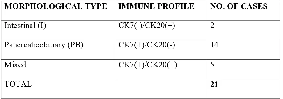
FIGURE 1. PANCREATOBILIARY TYPE CARCINOMA WITH TUBULAR PATTERN(HEMATOXYLIN-EOSIN ,40X)
[image:52.595.121.475.403.670.2]FIGURE 4. PANCREATOBILIAY TYPE CARCINOMA WITH CK 7 POSITIVITY (IMMUNOHISTOCHEMISTRY,400X)
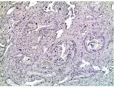
[image:54.595.117.481.421.702.2]FIGURE 6. INTESTINAL TYPE CARCINOMA WITH TUBULOPAPILLARY PATTERN
(HEMATOXYLIN-EOSIN, 100X)
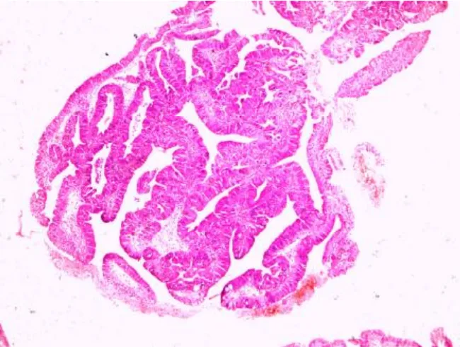
[image:55.595.126.472.410.674.2]FIGURE 9. INTESTINAL TYPE CARCINOMA WITH CK 20 POSITIVITY(IMMUNOHISTOCHEMISTRY,100X)
[image:57.595.128.469.389.647.2]FIGURE 11. TALL COLUMNAR CELLS IN MIXED TYPE CARCINOMA(HEMATOXYLIN-EOSIN ,400X)
[image:58.595.129.467.440.697.2]FIGURE 13. CK 7POSITIVITY IN MIXED TYPE CARCINOMA (IMMUNOHISTOCHEMISTRY,100X)
[image:59.595.130.467.443.698.2]47
DISCUSSION
Carcinomas of the Ampulla of Vater may arise in the ampulloduodenal part
lined by intestinal mucosa or in the deeper part lined by pancreatic or bile
duct mucosa. Intestinal and Pancreatobiliary represent the main histological
types of ampullary carcinoma. Morphologically the two types resemble their
colonic and pancreatic counterparts (43). Moreover intestinal type ampullary
carcinomas often possess immunohistochemical marker profile similar to that
of colonic carcinomas and the pancreatobiliary type ampullary carcinomas
show an immunoprofile resembling pancreatic carcinomas (44). Immunomarkers
most useful in this context are cytokeratin 20, cytokeratin 7 and Mucin 2. Thus
the intestinal type tumors express “CK 20+, CK7-, MUC 2+” marker profile,
while pancreatobiliary type carcinomas express “CK 7+, CK 20-, MUC 2-“
profile.
The present study is an attempt to correlate the morphology of
ampullary adenocarcinomas with their immunoprofile. The markers used were
CK7 and CK 20, both simple cytokeratins with restricted tissue and neoplastic
distribution. Cytokeratins are intermediate filaments found in the epithelial cells
of all types and are therefore specific markers for an epithelial cell lineage.
48
frequently organ and tissue specific. The subtypes of cytokeratins expressed
depend, in addition to the type of epithelium , on the stage of differentiation
and development(45). Applications of cytokeratin 7 and cytokeratin 20 in
combination include study of Barret’s mucosa,and assessment of metastatic
carcinomas with unknown primary, in addition to typing of ampullary
carcinomas.
Cytokeratins 7 and 20 expression could also have a bearing on the
prognosis of carcinomas irrespective of subtype. At least in one series, on
colorectal carcinomas , cytokeratin 20 expession was found to be more in low
grade tumors and cytokeratin 7 expression more in tumors with lymph node
metastasis (45). Other studies have however contradicted these observations (26).
In the present study , 70% of ampullary carcinomas (21/30) showed
correlation between morphology and CK7/CK20 profile. Some of the previous
studies have also yielded similar results (7,8). In the series of Zhou et al overall
correlation was present in 76% of cases and pancreatobiliary group alone
showed a higher percentage of correlation between morphology and
cytokeratin profile.
Clinical , radiological and endoscopic data showed considerable overlap
49
pancreatobiliary and mixed subtypes, but not in intestinal type of ampullary
carcinoma. However only two cases out of fifteen Whipple’s resection
specimens were of intestinal type and therefore no definite conclusion could be
drawn. But the cytokeratin 7 and cytokeratin 20 expression taken independently
did show that 100% of metastatic tumors were CK 7 (9/9) and only 44%
(4/9) were CK 20+. Among the cases without metastasis in our series 83%
(5/6) expressed CK 20 and only 63% (4/6) expressed CK 7.
This is in agreement with earlier observations of more cytokeratin 7
expression in colorectal adenocarcinomas with lymph node metastasis i.e
cytokeratin expression rather than histological type correlated with
metastasis (46).
We have in this study, made an attempt to correlate individual
histological features like architecture , predominant cell type, nuclear grade
and degree of desmoplasia separately, with CK 7 expression and CK 20
expression, also taken separately. It was found that tumors with an exclusively
50
With regard to cell height, CK 20 expression was found to have a
strong correlation with tall columnar cells in the sense that all CK 20 +
tumors had a tall columnar cell component.
Distribution of the different nuclear grades did not differ between
cytokeratin 7+ and CK 20+ tumors. But , 100 % of high nuclear grade (+++)
neoplasms expressed cytokeratin 7 and 20 % expressed cytokeratin 20. While
this would suggest a correlation between CK 7 expression and high grade,
one has to bear in mind that majority of tumors in the present series were
either pancreatobiliary or mixed in type, and were CK 7+.
Observations on desmoplasia also followed a similar pattern and
the results should be viewed against the fact that majority of the tumors were
anyway cytokeratin 7+.
Finally, the significance of our findings can be derived from the
fact that accurate histologic typing of ampullary carcinomas will provide
predictive information influencing staging and planning of operative
procedures. It will also provide a solid basis to analyze pathogenetic
mechanisms of these tumors by more advanced methods such as molecular
studies. Above all it raises the fundamental question whether ampullary
51
is to be clubbed under other intestinal and pancreatic carcinomas. The number of
cases studied is small. The three subtypes were not represented equally. In
spite of these drawbacks the following conclusions can be drawn from the
52
SUMMARY & CONCLUSIONS
1) Tumor morphology of ampullary adenocarcinomas correlates to an extent with
cytokeratin immunoprofile, and morphological- immunohistochemical
subtyping is possible in 70% of cases.
2) Frequency of lymph node metastasis is more in CK 7+ tumors as against CK
20+ tumors.
3) Among the microscopic features, an exclusively tubular architecture is
associated with cytokeratin 7 expression.Tall columnar cell component is
unequivocally related to CK 20 expression.
4) It may further be concluded that correlation between some of the individual
microscopic features like architecture and cell height with independent
cytokeratin7 and cytokeratin 20 expression is greater than that between
intestinal / pancreatobiliary morphological types and combined cytokeratin 7/
PROFORMA
SERIAL NO: BIOPSY NO:
NAME: OP/IP NO:
AGE/SEX: CLINICAL DETAILS: RADIOLOGICAL FINDINGS: ENDOSCOPIC FINDINGS: PATHOLOGY: GROSS:
A. TYPE OF SPECIMEN: SURGICAL/ENDOSCOPIC
B. LOCATION:
INTRAAMPULLARY/PERIAMPULLARY
DUODENUM
PANCREAS
CBD
UNCERTAIN
C. SIZE (GREATEST DIMENSION)
MICROSCOPY:
B. ARCHITECTURE: TUBULAR/PAPILLARY/CRIBRIFORM/MIXED
C. CELL TYPE: TALL COLUMNAR/LOW COLUMNAR/CUBOIDAL
D. MULTILAYERING: +/-
E. NUCLEAR ATYPIA: +/++/+++
F. STROMA:
G. ADJACENT DUODENAL MUCOSA:
H. ADJACENT DUCT MUCOSA:
I. ADJACENT PANCREATIC DUCT MUCOSA:
EXTENT OF INVOLVEMENT: GROSS MICROSCOPY
AMPULLA
DUODENUM
PANCREAS
CBD
PANCREATIC DUCT
LYMPH NODE
MARGINS DYSPLASIA/INVASIVECA
DIAGNOSIS:
IMMUNOHISTOCHEMISTRY
BIBLIOGRAPHY
1. Frierson H F. The Gross Anatomy and Histology of the Gallbladder,
Extrahepatic Bile Ducts, Vaterian System, and Minor Papilla. Am J Surg
Pathol 13(2); 146-162, 1989.
2. Rosai J. Pancreas and ampullary region. In: Rosai and Ackerman’s Surgical
Pathology. Ninth edition. Elsevier, St. Louis, Missouri: 2004.1092-95.
3. Cecilia M. Preiser F, Noffsinger A E, Stemmerman G E, Lantz P E, Isaacson P
G. Epithelial tumors of the small intestine. In: GI Pathology An Atlas and Text.
Third edition. Philadelphia, Baltimore, New York, London, Beunos Aires,
Hong Kong, Sydney, Tokyo : Wolters Kluwer , Lippincott Williams & Wilkins;
2008.482-85.
4. Yantiss R K, Antonioli D A. Polyps of the small intestine. In: Goldblum R R,
Odze R R. Surgical Pathology of the GI tract, Liver, Biliary tract, and Pancreas.
Second edition. Philadelphia,PA:Elsevier Inc; 2009. 465-6, 586-9.
5. Howe JR, Klimstra DS, Moccia RD, Conlon KC, Brennan MF. Factors predictive
6. Kimura W, Futakawa N, Yamagata S, Wada Y, Kuroda A, Muto T, Esaki Y.
Different clinicopathologic findings in two histologic types of carcinoma of
papilla of Vater. Jpn J Cancer Res. 1994 Feb;85(2):161-6.
7. Zhou H, Schaefer N, Wolff M, Fischer HP. Carcinoma of the ampulla of Vater:
comparative histologic/immunohistochemical classification and follow-up. Am
J Surg Pathol. 2004 Jul;28(7):875-82.
8. Le Pessot F, Ranty ML, Hellot MF, Lemoine F, Ténière P, Testart J, Métayer J.
Cytokeratins 7 and 20 immunohistochemistry in ampullary carcinomas. Ann
Pathol. 2004 Sep;24(4):312-8.
9. Goldstein N S and Bassi D. Cytokeratins 7, 17, and 20 reactivity in Pancreatic
and Ampulla of Vater Adenocarcinomas. Am J Clin Pathol 2001; 115:695-702.
10. Duval J V, Savas L, Banner B F. Expression of cytokeratins 7 and 20 in
carcinomas of the Extrahepatic Biliary Tract, Pancreas, and Gallbladder. Arch
Pathol Lab Med- Vol 124, August 2000.
11. Jackson P and Blythe D. Immunohistochemical techniques. In :
John.D.Bancroft, Marilyn Gamble. Theory and Practice of Histological
12. Adsay N V. Gall Bladder, Extrahepatic Biliary Tree, and Ampulla. In: Mills S
E, CarterD, Greenson J K, Reuter V E, Stoler M H. Sternberg’s Diagnostic
Surgical Pathology. Fifth edition.Philadelphia: Lippincott Williams & Wilkins;
2010.1636-43.
13. Schirmacher P and Büchler M W. Ampullary adenocarcinoma – differentiation
matters. BMC Cancer 2008; 8:251.
14. Chan M, and Adler D G. Ampullary cancer: review and clinical update.
Commun Oncol 2010;7:61–66.
15. Kim JH, Kim MJ, Chung JJ, Lee WJ, Yoro HS, Lee JT.Differential Diagnosis
of Periampullary Carcinomas at MR Imaging. Radiographics. 2002
Nov-Dec;22(6):1335-52.
16. Westgaard A, Tafjord S, Farstad I N et al. Resectable adenocarcinomas in the
pancreatic head: the retroperitoneal resection margin is an independent
prognostic factor. BMC Cancer 2008; 8:5
17. Stiff M G, Alabraba E, Tan YM et al. Assessment of survival advantage in
ampullary carcinoma in relation to tumour biology and morphology. Eur J Surg
18. Michelassi F, Erroi F, Dawson P J et al. Experience with 647 consecutive tumors
of the duodenum, ampulla, head of the pancreas, and distal common bile duct.
Ann Surg. 1989 October; 210(4): 544–556.
19. Westgaard A, Tafjord S, Farstad I N et al. Pancreatobiliary versus intestinal
histologic type of differentiation is an independent prognostic factor in resected
periampullary adenocarcinoma. BMC Cancer 2008, 8:170.
20. Carter JT, Grenert JP, Rubenstein L, Stewart L, Way LW. Tumors of the ampulla
of vater: histopathologic classification and predictors of survival. J Am Coll
Surg. 2008 Aug;207(2):210-8.
21. Goldstein NS and Silverman J.F. Immunohistochemistry of the gastrointestinal
tract, Pancreas, Bile ducts, Gall bladder and Liver. In: Diagnostic
Immunohistochemistry. Philadelphia, Pennsylvania: Churchill Livingstone;
2002. 333-64.
22. Chen ZM, Wang HL. Alteration of cytokeratin 7 and cytokeratin 20 expression
profile is uniquely associated with tumorigenesis of primary adenocarcinoma
of the small intestine. Am J Surg Pathol. 2004 Oct;28(10):1352-9.
23. Tot T. Cytokeratins 20 and 7 as biomarkers: usefulness in discriminating
24. Moll R, Löwe A, Laufer J, Franke WW. Cytokeratin 20 in human carcinomas. A
new histodiagnostic marker detected by monoclonal antibodies. Am J Pathol.
1992 Feb;140(2):427-47.
25. Jovanovic I, Tzardi M, Mouzas I A et al. Changing pattern of cytokeratin 7 and
20 expression from normal epithelium to intestinal metaplasia of the gastric
mucosa and gastroesophageal junction. Histol Histopathol (2002) 17: 445-454.
26. Choi JS, Kim NR, Ahn GH, Park CK. Expression of Cytokeratin 7 and 20 in
Periampullary Carcinomas. Korean J Pathol. 2000 Jan;34(1):34-38.
27. Saavedra A J, Schwartz AM, Batich K, Henson DE. Cancers of the ampulla of
vater: demographics, morphology, and survival based on 5,625 cases from the
SEER program. J Surg Oncol. 2009 Dec 1;100(7):598-605.
28. Roh YH, Kim YH, Lee HW et al. The clinicopathologic and immunohistochemical
characteristics of ampulla of Vater carcinoma: the intestinal type is associated
with a better prognosis. Hepatogastroenterology. 2007 Sep;54(78):1641-4
29. Westgaard A, Schjølberg A R,Cvancarova M, Eide T J, Clausen O P F,and
Gladhaug I P.. Differentiation markers in pancreatic head adenocarcinomas:
MUC1 and MUC4 expression indicates poor prognosis in pancreatobiliary
30. Stefan H; Alain P C. Ampullary cancer. Current Opinion in Gastroenterology:
2010 May; 26 (3):280–285.
31. Chu P G, Schwartz R E, Lau S K, Yen Y, Weiss L M. Immunohistochemical
staining in the diagnosis of pancreatobiliary and ampulla of Vater
adenocarcinoma. Application of CDX2, CK 17, MUC1 and MUC 2. Am J Surg
Pathol 2005;29: 359-67.
32. Kawabata Y, Tanaka T, Nishisaka T, Inao T, Nishi T, Yano S. Cytokeratin 20
(CK20) and apomucin 1 (MUC1) expression in ampullary carcinoma:
Correlation with tumor progression and prognosis. Diagn Pathol. 2010 Nov
25;5:75.
33. Sessa F, Furlan D, Zampatti C, Carnevali I, Franzi F, Capella C. Prognostic
factors for ampullary adenocarcinomas: tumor stage, tumor histology, tumor
location, immunohistochemistry and microsatellite instability. Virchows Arch.
2007 Sep;451(3):649-57.
34. Paulsen FP, Varoga D, Paulsen AR, Corfield A, Tsokos M. Prognostic value of
mucins in the classification of ampullary carcinomas. Hum Pathol. 2006 Feb;