ACUTE AND CHRONIC UNPREDICTABLE STRESS (CUS)
INDUCED MODELS IN SD RATS
Dissertation submitted to
The Tamil Nadu Dr. M.G.R. Medical University, Chennai-32.
In partial fulfillment for the award of the degree of
MASTER OF PHARMACY
IN
PHARMACOLOGY
Submitted by
Reg.No: 26113093
Under the Guidance of
Dr. R. SHANMUGA SUNDARAM, M.Pharm., Ph.D.,
APRIL – 2010-2011
DEPARTMENT OF PHARMACOLOGY J.K.K. NATTRAJA COLLEGE OF PHARMACY
Komarapalayam – 638183. Tamil Nadu.
Certificates
This is to certify that the work embodied in this dissertation entitled
“Evaluation of Potential Central Protective Role of Ethanol Extract of
Pedalium murex Linn. in Acute and Chronic Unpredictable Stress (CUS)
Induced Models in SD Rats”,, submitted to “The Tamil Nadu Dr. M.G.R. Medical
University”, Chennai, in partial fulfillment to the requirement for the award of Degree of Master of Pharmacy in Pharmacology, is a bonafide work carried out
by Miss. SUDHA.M [Reg.No.26113093]during the academic year 2012-2013,
under the guidance and supervision of Dr. R. SHANMUGA SUNDARAM, M.Pharm.,Ph.D., Vice Principal, Department of Pharmacology, J.K.K. Nattraja
College of Pharmacy, Komarapalayam.
Place: Komarapalayam Dr. R. Sambathkumar, M.Pharm., Ph.D.,
Date: Principal & Professor
Department of Pharmaceutics, J.K.K. Nattraja College of Pharmacy,
This is to certify that the work embodied in this dissertation entitled
“Evaluation of Potential Central Protective Role of Ethanol Extract of
Pedalium murex Linn. in Acute and Chronic Unpredictable Stress (CUS)
Induced Models in SD Rats”, submitted to “The Tamil Nadu Dr. M.G.R. Medical
University”, Chennai, in partial fulfillment to the requirement for the award of Degree of Master of Pharmacy in Pharmacology, is a bonafide work carried out
by Miss. SUDHA .M [Reg.No.26113093], during the academic year 2012-2013,
under my guidance and direct supervision in the department of Pharmacology, J.K.K. Nattraja College of Pharmacy, Komarapalayam.
Place: Komarapalayam Dr. R. Shanmuga Sundaram, M.Pharm., Ph.D.,
Date: Vice Principal and Professor Department of Pharmacology, J.K.K. Nattraja College of Pharmacy.
This is to certify that the work embodied in this dissertation entitled
“Evaluation of Potential Central Protective Role of Ethanol Extract of
Pedalium murex Linn. in Acute and Chronic Unpredictable Stress (CUS)
Induced Models in SD Rats”, submitted to “The Tamil Nadu Dr. M.G.R. Medical
University”, Chennai, in partial fulfillment to the requirement for the award of Degree of Master of Pharmacy in Pharmacology, is a bonafide work carried out
by Miss. SUDHA.M [Reg.No.26113093], during the academic year 2012-2013,
under my guidance and direct supervision in the Department of Pharmacology, J.K.K. Nattraja College of Pharmacy, Komarapalayam.
Internal Examiner External Examiner
I hereby declare that the dissertation entitled “Evaluation of Potential Central Protective Role of Ethanol Extract of Pedalium murex Linn. in Acute
and Chronic Unpredictable Stress (CUS) Induced Models in SD Rats”, has been
carried out under the guidance and supervision of Dr. R. Shanmuga Sundaram, M.Pharm., Ph.D., Vice Principal, Department of Pharmacology, J.K.K. Nattraja
College of Pharmacy, Komarapalayam, in partial fulfillment of the requirements for the award of degree of Master of Pharmacy in Pharmacology during the academic
year 2012-2013.
I further declare that, this work is original and this dissertation has not been submitted previously for the award of any other degree, diploma associateship and fellowship or any other similar title.
Place: Komarapalayam SUDHA.M
Date: Reg.No: 26113093
Department of Pharmacology, J.K.K. Nattraja College of Pharmacy.
We, the Undersigned Chairman/Members of the Animal Ethical Committee, functioning in JKK Nattraja College of Pharmacy have studied the proposed research Subject/Project of M. Sudha titled “Evaluation of potential central protective role of ethanol extract of Pedalium murex Linn. In acute and chronic unpredictable stress
(CUS) induced model in SD Rats” applying for permission for animal usage and
hereby give the certificate of clearance of approval by this Ethical Committee.
Signature of the Chairman/ Members of the
Animal Ethical Committee
Name of the Institution:
Station : Date : Seal :
g
`ç
gxtv{
Wxw
TÄ
`ç uxÄ
g v{xÜá
Wxw|vtàx
TÄÅ|z{
xÄÉäxw y
tÇw
àxw àÉ
z{àç?
w ytÅ
w
YÜ|
É
Å|Äç?
Y |xÇwá
I am proud to dedicate my deep sense of gratitude to the founder, (Late) Thiru J.K.K. Nattaraja Chettiar, providing us the historical institution to study.
My sincere thanks and respectful regards to our reverent Chairperson
Smt. N. Sendamaraai, B.Com., Managing Director Mr. S. Omm Sharravana,
B.Com., LLB., and Executive Director Mr. S. Omm Singarravel, B.E., M.S.,
J.K.K. Nattraja Educational Institutions, Komarapalayam for their blessings, encouragement and support at all times.
It is most pleasant duty to thank our beloved Principal Dr. R.SambathKumar, M.Pharm., Ph.D., J.K.K.Nattraja College of Pharmacy,
Komarapalayam for ensuring all the facilities were made available to me for the smooth running of this project.
I express my whole hearted thanks to my guide Dr. R. Shanmuga Sundaram, M.Pharm., Ph.D., Vice Principal and Professor, Department of
Pharmacology, for suggesting solution to problems faced by me and providing indispensable guidance, tremendous encouragement at each and every step of this dissertation work. Without his critical advice and deep-rooted knowledge, this work would not have been a reality.
My sincere thanks to Mr. V. Rajesh, M.Pharm., Ph.D., Head of the
Department, Department of Pharmacology, Mr. C. Sridharan, M.Pharm.,
Lecturer, Department of Pharmacology, Mr. S. Venkatesh, M.Pharm., Lecturer,
Dr. N. Mahadevan, M.Pharm., Ph.D., Professor and Head, Department of
Pharmacognosy and Mr. P. Balasubramaniam, M.Pharm., Lecturer, Department
of Pharmacognosy for their valuable suggestions during my project work.
My sincere thanks to Dr. M. Vijayabaskaran, M.Pharm., Ph.D., Assistant
Professor and head Department of Pharmaceutical chemistry, Mr. S.V. Arunachalam, M.Pharm., Lecturer, Department of Pharmaceutical chemistry, Mrs. S. Gomathi, M.Pharm., Lecturer, Department of Pharmaceutical chemistry
and Mrs. B. Vasuki, M.Pharm., Lecturer, Department of Pharmaceutical
chemistry, for their valuable suggestions and inspiration.
My sincere thanks to N. Venkateswaramurthy, M.Pharm., Assistant
Professor and Head, Department of Pharmacy Practice. Mrs. K. Krishna Veni, M.Pharm., Lecturer, Department of Pharmacy Practice, Mrs. Christy John, M.Pharm., Lecturer, Department of Pharmacy Practice and Dr. K. Sattanathan, M.Pharm., Ph.D., Lecturer Department of pharmacy practice, for their help during
my project.
My sincere thanks to Dr. V. Sekar, M.Pharm., Ph.D., Professor and Head,
Department of Analysis, Mr. M. Senthilraja, M.Pharm., Assistant Professor, and Mr. S. Jayaseelan, M.Pharm., Assistant Professor, Department of Pharmaceutical
Analysis for their valuable suggestions.
My sincere thanks to Mrs. S. Bhama, M.Pharm., Assistant Professor, Dr. S.K. Senthilkumar, M.Pharm., Ph.D., Assistant Professor, Mr. R.
Lecturer, Department of Pharmaceutics and Mr. Kamalakannan M.Pharm.,
Lecturer, Department of pharmaceutics for their valuable help during my project.
I greatly acknowledge the help rendered by Mrs. K. Rani, Office
Superintendent, Miss. Prabha, Mrs. V. Gandhimathi, M.A., M.L.I.S., Librarian,
and Mrs.S. Jayakala, B.A., B.L.I.S., Asst. Librarian for their co-operation.
My special thanks to all the Technical and Non Technical Staff Members
of the institute for their precious assistance and help. I am extremely thankful to
Mr. Ariharasivakumar M.Pharm., Dept. of Pharmacology, KMCH College of
Pharmacy, Coimbatore.
Last, but nevertheless, I am thankful to my lovable parents and all my friends for their co-operation, encouragement and help extended to me throughout my project work.
M. SUDHA
INDEX
S.
No.
CONTENTS
Page
No.
1. INTRODUCTION 1-49
2. REVIEW OF LITERATURE 50-62
3. AIM AND OBJECTIVE 63-65
4. PLAN OF WORK 66-68
5. MATERIALS AND METHODS 69-91
6. RESULTS AND DISCUSSION 92-128
7. SUMMARY AND CONCLUSION 129-130
Dept. of Pharmacology 78 J.K.K.Nattraja College of Pharmacy
1.STRESS
Stress is a common experience of daily life and all organisms have developed mechanisms to cope with it. Sustained stress can have numerous pathophysiological effects such as activation of neuro-endocrine [limbic-hypothalamic-pituitary adrenal system] (Bonfiglio et al., 2011) and hormonal (corticosterone release) functions (Fuchs & Flugge, 1998). Sustained and persistent stressful conditions can precipitate anxiety and affective disorders such as depression, which further leads to the excessive production of free radicals and oxidative burden (Maes et al., 2011).
Stressful events can activate the Hypothalamo-Pituitary-Adrenal (HPA) axis
(Kvetnansky et al., 2002) and increase the release of Corticotrophin Releasing
Hormone (CRH) from the hypothalamic paraventricular nucleus, causing the secretion of Adrenocorticotropin (ACTH) from anterior pituitary, which in turn stimulates the secretion of glucocorticoids from the adrenal cortex (Pacak et al.,
1993; Venihaki et al., 1997). Glucocorticoids possess broad spectrum of actions
affecting expression and regulation of genes throughout the body readying the organism for changes in energy and metabolism required for coping (Akil &
Morano, 1995; Levine, 2005). Stress has been postulated to be involved in the
etiopathogenesis of a variety of disease state including hypertension, coronary heart disease (Roy et al., 2001), gastric ulcers (Yadin & Thomas, 1996), diabetes
(Fitzpatrick et al., 1992), immuno-suppression (Purret, 2001), mental depression,
memory loss (Gareri et al., 2000), and host of other diseases. The resultant disturbances may vary depending upon type, intensity, and the duration of a particular stressor and the strain\sex differentiation of the subjects (Kioukia-Fougia
Dept. of Pharmacology 79 J.K.K.Nattraja College of Pharmacy
frequently to evaluate the anti-stress activity of compounds of both natural and synthetic origin.
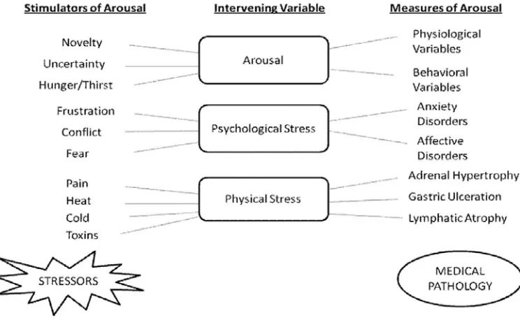
In an organism, diverse stressors activate a wide spectrum of interacting hormonal and neuronal systems resulting in behavioral (anxiety disorders, decrease in food intake, decrease in sexual behavior, and loss of cognitive function) and physiological responses [activation of pituitary adrenal axis and release of glucocorticoids into the blood stream] (Henry & Stephens, 1977). These stressors are stimulators of arousal and lead to autonomic (changes in body temperature and tachycardia) and behavioral changes; however, when arousal increases to stress‐like levels, it results in psychiatric and physical disorders (Hennessy et al., 1979) [Figure 1]. Different animal models have been developed for chronic stress induced neurological disorders such as the olfactory bulbectomy model, and the chronic unpredictable stress model. These animal models are used to screen various new chemical entities and to develop a better understanding of the underlying molecular pathway in chronic stress pathology. Stress responses are variable and there are individual differences both physiologically and behaviorally in how an organism perceives a perturbation and in the resulting adaptational/maladaptational processes (Weiner,
1992).
Stress and Stressors
Dept. of Pharmacology 80 J.K.K.Nattraja College of Pharmacy
stress can be any threat, either real or perceived, to the well being of an organism and it can be of two types.
[image:19.612.137.509.236.470.2]Kumar, et al.: Stress
Figure 1: Relationship between arousal, psychological stress, physical stress and
pathology
Dept. of Pharmacology 81 J.K.K.Nattraja College of Pharmacy
stress‐induced degenerative conditions (Sahin & Gumuşlu, 2007). The brain tissue is made up of large amounts of polyunsaturated fatty acid, thus making it vulnerable to free radical attacks (Gutteridge, 1995).
Consequences of Stress
Normal development and preservation of life and species depend on a normally functioning stress system. Maladaptive neuroendocrine responses, i.e., dysregulation of the stress system, may lead to disturbances in growth and development, and cause psychiatric, endocrine/metabolic, and/or autoimmune diseases or vulnerability to such diseases.
Stress and anxiety
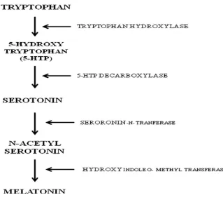
According to previous reports stress induces anxiety‐like behavior in both humans and animals (Liezmann et al., 2011). In response to stress, there is an increase in CRF levels. The CRF level decreases when the stressor is no longer present. Lee, et al. reported that chronic stress increases the length and volume of expression of CRF in areas of the brain associated with fear and emotion, including the amygdale (Lee
et al., 2008) [Figure 2]. Such chronic stress changes the body’s response, and the
Dept. of Pharmacology 82 J.K.K.Nattraja College of Pharmacy
[image:21.612.157.476.73.358.2]
Figure 2: De novo synthesis pathway of melatonin.
Dept. of Pharmacology 83 J.K.K.Nattraja College of Pharmacy
This results in oxidative burden, which has been implicated in stress as well as in the pathogenesis of several disease states. Since brain tissues consist of a high content of polyunsaturated fatty acids and one of the important consequences of oxidative stress is peroxidation of membrane lipids, this reaction produces marked damage to the structure and function of cell membranes in these tissues (Jain et al., 1991). Therefore, lipid peroxidation was supposed as the major biochemical alteration and consequence of oxidant‐induced cell injury. Thus, the important consequences of stress could be attributed to stress‐induced lipid peroxidation.
Stress and immunological changes
Stress has been associated with impaired immune function and increased susceptibility to infectious diseases (Connor & Leonard, 1998). It is now believed that the nervous, endocrine, and immune systems are so intimately connected that they should be regarded as a single network rather than as three separate systems
(Connor & Leonard, 1998). It is widely accepted that psychological stress and
psychiatric illness can compromise immune function (Leonard, 1995) and soluble mediators released by immune cells can affect the central nervous system, thus producing alterations in behavior. Exposure to stressful life events such as academic examinations and divorce was reported to cause impairments in various aspects of cellular immune function (Bartrop et al., 1977). There are also reports of immune activation, (Bartrop et al., 1977) in addition to immunosuppression in both the depressed and subjects exposed to stressful life events.
Dept. of Pharmacology 84 J.K.K.Nattraja College of Pharmacy
However, none of the models available is able to entirely reproduce stress response. Some models reproduce physical stress and associated neuroendocrine changes
(Kvetnansky & Mikulai, 1970), whereas others better reproduce the psychological
stress and associated behavioral changes (Marcelo et al., 2007). Acute models do not reproduce the neuroendocrine dysfunction whereas a chronic model might be able to do so. Therefore, a correct model should be used to evaluate specific aspects of the stress response. Each model has inherent limitations including lack of stability, lack of predictability of tissue damage, and lack of adjustability. And hence a literature survey of more than 35 years (1970-2007) was conducted based on the description of the models, potential utilization of the models, and value of the models for testing of new medical interventions for the management of stress. The purpose of this review was to assess different models of stress.
Different Types of Stress (Based on the Time Period of Application of Stressor)
Acute Stress (Single Application of
Stressor)
Chronic Stress
(Application of Stressor for a Long Period of Time)
Chronic Variable Stress (Repeated Application of different types of stressors
Dept. of Pharmacology 85 J.K.K.Nattraja College of Pharmacy
ANIMAL MODELS COMMONLY USED
• Physical stress models
Animal models of stress that use physical stress can be subdivided into:
¾ Temperature fluctuation induced stress:
1. Immersion in cold water with no escape
2. Cold environment isolation
o Immobilization induced stress
o Electric foot shock induced stress
o Forced swimming induced stress
• Psychological stress models
Animal models of stress that use psychological stress can be subdivided into:
¾ Neonatal isolation induced stress
¾ Predatory stress
¾ Day-night light change induced stress
¾ Noise induced stress.
• Chronic unpredictable stress
Dept. of Pharmacology 86 J.K.K.Nattraja College of Pharmacy
stressor. It involves the use of both physical and psychological stress models in a random way.
PHYSICAL STRESS MODELS
Temperature fluctuation induced stress
Acute change in temperature leads to stressful conditions by activation of temperature regulatory centre in the hypothalamus and subsequently HPA axis. It leads to acute release of adrenocortical hormones in the blood stream responsible for acute stressful response (Sapolsky et al., 1986). A sharp decrease in temperature using either cold water or freezer has been used frequently to induce acute stress.
1. Immersion in Cold Water (ICW)
In this method, the rats are placed individually in a tank of cold water (depth = 15.5 cm; temperature = 15-20°C) where they either swim or remain in an upright position, keeping their heads above water level (Retana-Marquez et al., 2003; Iwona
et al., 2003; Fernandez-Landiera, 2004; Yun et al., 2003). This situation lasts for 15
Dept. of Pharmacology 87 J.K.K.Nattraja College of Pharmacy
2. Cold environment isolation
In this method, rats are individually kept in a freezer with a temperature maintained at 4°C. The rats are kept for 15 minutes once for acute stress and for 7-10 days to develop chronic stress (Kvetnansky et al., 1971). This sharp fall in temperature leads to a sharp increase in the level of adrenocorticoids as explained above culminating in the development of stress response (Kvetnansky et al., 2002; Staratakis & Chrousos, 1995). Unlike the ICW model, rats are prevented from drowning in cold water hence it is relatively safe model however it also suffers from same drawback of development of resistance/adaptation on chronic exposure.
Immobilization induced stress
Immobilization has been used extensively as a stressor for the study of stress-related biological, biochemical and physiological responses in animals (Kvetnansky &
Mikulai, 1970; Kasuga et al., 1999; Marty et al., 1997). Immobilization can be
produced in two different ways. Animal can be either kept immobilized in a semi cylindrical acrylic tube (4.5 cm diameter and 12 cm long) with proper holes in it for air to pass (Das et al., 2000). Another way is to keep the animal with its limbs stretched on a board and its limbs are immobilized with adhesive tape. Movement of head is restricted by keeping the head in a metal loop coiled around the neck. The rats are kept immobilized in either of the above two ways for 150 minutes once to produce acute stress and for 7-10 days to produce chronic stress (Dronjak &
Gavrilovic, 2006). The major advantage of using immobilization as a model of stress
Dept. of Pharmacology 88 J.K.K.Nattraja College of Pharmacy
Electric foot shock induced stress:
Electric foot shock (EFS) of mild intensity has also been used as a stressor. Rodents are very susceptible even to mild shock and exhibit rapid stress response. Researchers have used electric foot shock of varying degree to produce stressful conditions and hence to evaluate adaptogenic activity of various compounds. Stress by electric foot shock is given by placing the rats individually in a chamber with an electrified floor. Rats receive unavoidable electric foot shocks with an intensity of 3 mA, 200 ms of duration and a frequency of 1 per second over a 5-min period. For acute stress response, the rats are exposed once and sacrificed after 15 minutes of stress. Chronic stress is also produced by repeating the same treatment for 7-10 days and rats are sacrificed 1 h after the last stress session (Retana-Marquez et al., 2003). Some researchers have modified the method in which rats are subjected to inescapable electric foot shock for 60 minutes (0.15 mA shock, on a variable interval schedule with a mean inter shock interval of 60 seconds) (Taysse et al., 2005). The biggest advantage of this model is that it effectively produces high degree of stress in the animal. The major disadvantage of this model is the hazard of electric shock causing death of the animal and special caution that is required to perform this methodology.
Forced swimming induced stress
Dept. of Pharmacology 89 J.K.K.Nattraja College of Pharmacy
filled to a height of 20 cm with 15 cm of space above the head of the rat) for a single session of 2 h duration for acute stress, or for one 2 h session a day for five consecutive days for chronic stress (Ferry et al., 1991). Some authors have used forced swimming in warm (20°C) water for 3 minutes with the total session lasting for 1 h (Kitchen & Pinker, 1990). Although forced swimming induced stress is a highly safe model, adaptation to chronic swimming induced stress has been reported and inter-strain differences between rats to forced swimming behavior have also been documented (Armario et al., 1995).
PSYCOLOGICAL STRESS MODELS
Neonatal isolation stress
Dept. of Pharmacology 90 J.K.K.Nattraja College of Pharmacy
animal colony facility. Containers are placed 20-30 cm apart. After 1 h period the litters are placed back with their dams in home cage (Kosten et al., 2000; Kosten et
al., 2004). This isolation procedure continues up to 8 days and hence it is used to
induce chronic stress only. Neonatal isolation stress model has be used extensively to demonstrate the effect of early lifetime stress on vulnerability to addiction
(Kosten et al., 2005a), and response to psychostimulants by impairment of
hippocampal-dependent context induced fear in adult male rats.
Predatory stress
Dept. of Pharmacology 91 J.K.K.Nattraja College of Pharmacy
1993) was used consists of a PVC box (30x20x20 cm) covered with Plexiglas and subdivided into six equal square exploratory units, which are all interconnected by small entries. It could be divided in half lengthwise by closing three temporary partitions. Approximately 20 h before cat exposure, each subject is placed in one half of the apparatus with the temporary partitions in place, in order to be familiarized with it. The floor of this half was covered with fresh sawdust and the animal is given unlimited access to food and water. On the test day, mice of each strain are randomly allocated to the following four groups.
(a) Naive clay: animals are exposed to both familiar and novel compartments by removal of the temporary partitions. The novel compartment contains three modeling odor-free clay pellets.
(b) Naive feces: animals are exposed to both familiar and novel compartments. The novel compartment contains three cat feces pellets.
(c) Exposed clay: subjects are removed from the free-exploration box and confronted individually with a cat during a 5 min. session. The cat cage consists of a PVC box (82x56x62 cm) subdivided into two compartments, one containing the cat, the other the mouse. Separation consists of a transparent PVC wall with holes allowing the cat to reach the other side with its paws. The mouse is then put back in the free-exploration apparatus and is exposed 1 h later to both familiar and novel compartments. The novel compartment contains three modeling odor-free clay pellets.
Dept. of Pharmacology 92 J.K.K.Nattraja College of Pharmacy
is observed under red light for 5 min via a closed circuit TV camera by an observer located in an adjacent room.
The following parameters are recorded:
(a) Time spent in the novel compartment; (b) total unit entries and (c) total number of rearings.
The results are expressed as mean percentage of time spent in the novel compartment, mean total number of novel unit changes, and mean total number of rearings. Marmosets (Callithrixpenicillata) have also been employed for induction of predatory stress in a test battery known as Marmoset Predator Confrontation Test [MPCT] (Cilia & Piper, 1997). This model compares the behavioral response of experienced versus naïve adult black tufted-ear marmosets in confrontation with a taxidermized wild-cat predator stimulus. After four initial 20-min cage habituation sessions, each subject is submitted to two randomly-assigned 20-min predator confrontation sessions. Confrontation with the predator induces significant behavioral changes; i.e., proximic avoidance and tsik-tsik alarm call. Anti-stress drug administration, concomitant to predator exposure, reverses the behavioral changes observed (Barros et al., 2004). Predator induced stress is an established model to induce short term acute stress response but its major disadvantage is development of habituation to predator exposure hence the use of this model for inducing stress is justified for developing only acute stress.
Day-night light change induced stress
Dept. of Pharmacology 93 J.K.K.Nattraja College of Pharmacy
subjected to abrupt changes in day-night light pattern, exhibit acute stress response
(Kosten et al., 2005b). Changes in circadian rhythms are regulated by pineal gland
through the secretion of melatonin (Nicholsonet al., 1985). Melatonin is released from the pineal gland in response to dark or dim light where as its functional antagonist serotonin is secreted in response to bright light. It is this serotonin-melatonin cycle that is responsible for regulation of sleep-awake state of the body
(Bermudez et al., 1983; Hamm et al., 1983). To induce stress, cages of rat or mice
are kept under bright light from 19:00 h over night (in the dark phase) and cages are kept in dark room with no light from 12:00 h in the light phase for 180 minutes for 7-10 days (Marcelo et al., 2007). This method is suitable for inducing short term stress response. Generation of stress can be evaluated by measuring the biochemical parameters associated with chronic stress response (Rai et al., 2003). The major disadvantage of this model is that it can be effectively used to generate short term stress response as on repeated exposure to this type of stressor, the animal adapts to the changed day-night light pattern. This major drawback can be minimized by using this model as a part of chronic unpredictable stress protocol.
Noise induced stress
Dept. of Pharmacology 94 J.K.K.Nattraja College of Pharmacy
Noise stress has a depletory effect on free radical scavenging enzymes in the brain leading to moderate to severe oxidative stress (Samson et al., 2005) which can be a potential basis for hearing loss (Fechter, 2005). Noise stress in laboratory rats can be produced by loudspeakers (15 W), driven by a white noise generator (0-26 kHz), installed 30 cm above the cage. Thus a noise level can be set at 100 dB or above uniformly throughout the cage and can be monitored by a sound level meter. Each animal to be treated is exposed to noise stress for 4 h/day for 15 days. Control group rats are also kept in the above described cage during the corresponding period of time, without noise stimulation to avoid the influence of handling stress on evaluation of effects due to noise exposure (Ravindran et al., 2005; Manikandan &
Devi, 2005). The effect of noise stress exposure can be determined by estimating the
brain biogenic amine level.
CHRONIC VARIABLE (UNPREDICTABLE) STRESS MODELS
Dept. of Pharmacology 95 J.K.K.Nattraja College of Pharmacy
adaptations of HPA axis depend on type, duration and severity of the stress regime
(Blanchard et al., 1998; Gadek-Michalska & Bugajski, 2003). To prevent the
Dept. of Pharmacology 96 J.K.K.Nattraja College of Pharmacy
the development of effective and long-term stress response. Thus CUS models are nowadays the preferred models for generation of a stress response.
Stress has been postulated to be involved in the etiopathogenesis of a variety of disease state including hypertension, coronary heart disease (Roy et al., 2001), gastric ulcers (Yadin & Thomas, 1996), diabetes (Fitzpatrick et al., 1992), immunosuppression (Purret, 2001), mental depression, memory loss (Gareri et al., 2000), and host of other diseases.
NEURODEGENERATIVE DISEASE
Neurodegenerative diseases comprise a wide range of diseases that share the common characteristic of progressive loss of structure or function of neurons and glial cells in the brain and spinal cord. Many neurodegenerative diseases are a result of neuronal loss, although glial cells are also involved (Glass et al., 2010).
Neurodegenerative diseases present a chronic and slowly progressive process. Neurons in neurodegenerative diseases are affected by neuronal dysfunction at the level of synaptic transmission, synaptic contacts, and axonal and dendritic degeneration. In different neurodegenerative diseases, neurite degeneration and cell loss of neurons are present within specific neurotransmitter populations. In addition, numbers of functional neurons in neurogenic regions, and adult neurogenesis are altered or decreased. Adult neurogenesis increases after several acute pathologic stimuli, including stroke, seizure and acute trauma (Arvidsson et al., 2002; Rice et
al., 2003; Parent, 2007). Brain regions differ in their vulnerability to aging; some
Dept. of Pharmacology 97 J.K.K.Nattraja College of Pharmacy
zone (SVZ) ⁄ olfactory bulb (OB) (Braak et al., 2003). The generation and cell death of newly generated cells have critical roles in brain development and maintenance in the embryonic and adult brain, and alterations in these processes are seen in neurodegenerative diseases.
DIFFERENT NEURODEGENERATIVE DISORDERS:
Although neuronal degeneration predominantly affects or starts with specific neuronal populations [including DAergic neurons in PD, striatal medium spiny neurons in HD, motor neurons in amyotrophic lateral sclerosis, and cortical and hippocampal neurons in Alzheimer’s disease (AD)], there are many similarities between different neurodegenerative disorders. These include atypical protein assemblies and oligomerization as well as induced cell death. At a late disease stage, protein aggregation is no longer restricted to specific brain regions.
Interestingly, in Parkinson’s disease (PD) and Huntington’s disease (HD), the specific alterations in neurogenic areas such as the DG and SVZ ⁄ OB system parallel the early or premotor symptoms that are seen in the early stages of neurodegenerative disease, such as depression, anxiety or olfactory dysfunction
(Simuni & Sethi, 2008). Therefore, it is intriguing that the mechanisms of
neurodegenerative diseases are closely linked to brain plasticity. Brain plasticity in the adult, originally conceived of as changes at the level of synaptic transmission, synaptic contacts and gene expression (reviewed in Buonomano & Merzenich, 1998), became a more complicated concept.
Dept. of Pharmacology 98 J.K.K.Nattraja College of Pharmacy
brain plasticity in the embryonic brain, specifically as membrane proteins and when concentrated in synapses. These proteins commonly show high conservation between species and are located close to membranes or are involved in microtubule transport. α-synuclein is a protein that is physiologically enriched in presynaptic termini (Abeliovich et al., 2000). Initially shown to be upregulated in a discrete population of presynaptic terminals of the song bird brain during a period of acquisition-related synaptic rearrangement (George et al., 1995), α-synuclein can interact with tubulin (Alim et al., 2002). In addition, it is involved in dopamine (DA) synthesis, metabolism and release, and slight changes in concentration can have vast effects on neurotransmitter release (Nemani et al., 2010).
NEURODEGENERATION IN DIFFERENT DISORDERS:
In non-pathological aging, cognitive impairment would seem easiest to attribute to the effects of oxidative stress, inflammatory reactions and changes in the cerebral microvasculature (Riddle et al., 2003). However, the aging brain in the absence of dementia is also affected to varying degrees by the neuropathological features. Many neurodegenerative diseases occur as a result of neurodegenerative processes including
• Parkinson’s disease (PD)
• Alzheimer’s disease (AD)
• Huntington’s disease (HD)
• Amyotrophic lateral sclerosis (ALS) and
Dept. of Pharmacology 99 J.K.K.Nattraja College of Pharmacy
Alzheimer's disease (AD)
Alzheimer’s disease (AD) was first described by Alois Alzheimer more than a century ago in Germany, and it constitutes one of the most common causes of senile dementia.AD refers to a clinical syndrome that occurs in the elderly and is severe enough to interfere with social and occupational activities. At least two clinical abnormalities are essential for diagnosis of the disease, namely, memory loss in an alert person and impairment of one or more of the following functions: language, attention, perception, judgment or problem solving (Forstl & Kurz, 1999).
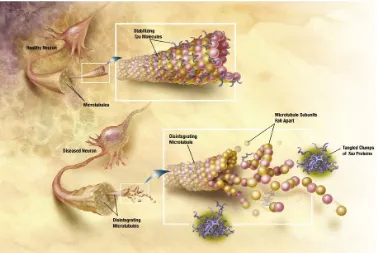
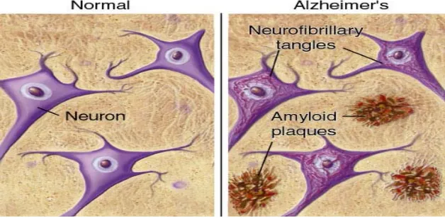
The pathology of AD includes neuronal and synaptic loss, neurofibrillary tangles due to hyperphosphorylated tau proteins and deposition of amyloid-β (Ab) protein in senile plaques in the basal forebrain cholinergic neurons as well as in the cortex, hippocampus and amygdala (Hardy & Selkoe, 2002). Ab is the product of proteolysis of amyloid precursor protein (APP) by b- and c-secretase enzymess.
[image:38.612.133.511.439.692.2]
Dept. of Pharmacology 100 J.K.K.Nattraja College of Pharmacy
[image:39.612.163.477.77.231.2] [image:39.612.129.543.317.501.2]Fig.4 "Tangles" of a protein called "tau" occur in Alzheimer's patients' brains-causing neurons to lose their function and increasing memory loss. Illustration: National Institute on Aging/U.S. National Institutes of Health.
[β amyloid] [Normal Neuron] [Amyloid Plague]
Fig.5 Enzymes act on the APP (amyloid precursor protein) and cut it into fragments.
The beta-amyloid fragment is crucial in the formation of senile plaques in AD.
Dept. of Pharmacology 101 J.K.K.Nattraja College of Pharmacy
in both transgenic mice and humans, the presence of oligomers in the brains of transgenic mice, the toxicity of Ab dimer and trimer measured by long-term potentiation, and lack of a good correlation between plaque amount and AD (at least in the early phase of the disease) (Walsh et al., 2002; Walsh & Selkoe, 2007). Patient deficits include olfactory deficits, memory impairment, cognitive and functional decline, and death. These symptoms can be partly related to regions and functions of adult neurogenesis.
Parkinson’s disease (PD)
Parkinson disease (PD) is the second most common neurodegenerative disease, affecting about 1% of the population over 65 years of age. PD is characterized clinically by resting tremor, rigidity and bradykinesia, resulting from the progressive and selective loss of dopamine (DA) neurons in the substantianigra (SN) pars compacta, and histopathologically by the eosinophilic proteinaceous intracytoplasmic inclusion known as Lewy bodies (LBs) in surviving dopaminergic cells (Forno, 1996). The etiopathogenesis of PD is probably multifactorial, including both environmental and genetic factors (Di Monte, 2003; Hardy et al., 2003). Asmentioned above, α-synuclein (α-syn) is the principal component of LBs
(Spillantini et al., 1997). This was seen in molecular genetic investigations that
Dept. of Pharmacology 102 J.K.K.Nattraja College of Pharmacy
It was suggested that α-syn aggregation is the key event that triggers neuronal damage and death (Forloni et al., 2000; Volles & Lansbury, 2003). In this context, α-syn mutations would speed up the protein aggregation (Conway et al., 2000). It is likely that the α-syn concentration is crucial to switch from a physiological to a pathological condition, and only when a threshold concentration is reached do the deleterious effects of aggregation become evident. Misregulation in the homeostasis of α-syn (caused by mitochondrial complex I inhibition, environmental toxins, oxidative stress or proteasome impairment) would be sufficient to trigger α-syn chemical modifications and aggregation in sporadic PD too (Sherer et al., 2003;
Norris et al., 2003).
Dept. of Pharmacology 103 J.K.K.Nattraja College of Pharmacy
monogenetic forms of PD show a decreased gray matter volume in the hippocampal region (Reetz et al., 2010).
Chaperone-like activity of α-synuclein:
Chaperones are proteins that prevent irreversible protein aggregation and facilitate the correct folding of non-native proteins through regulated binding and release in vivo (Slavotinek & Biesecker, 2003). α–Syn (α–Synuclein) has been suggested to function as a chaperone protein in vivo because, besides lipids (Eliezer et al., 2001;
Li et al., 2001; Davidson et al., 1998), it appears capable of interacting with a
variety of ligands and cellular proteins (Ostrerova et al., 1999; Xu et al., 2002), thus modifying their activities. It has recently been reported that the amino-terminal portion of α–syn shares 40% a homology with molecular chaperone 14-3-3
(Ostrerova et al., 1999), suggesting that the two proteins could sub serve the same
function. The molecular chaperone 14-3-3 is particularly abundant in brain, where it comprises ~1% of total soluble proteins (Boston et al., 1982). Chaperone 14-3-3 accumulates in LBs, participates in neuronal development and cell growth control
(Fu & Masters, 2000), and prevents apoptosis by antagonizing BAD, a proapoptotic
Dept. of Pharmacology 104 J.K.K.Nattraja College of Pharmacy
Dept. of Pharmacology 105 J.K.K.Nattraja College of Pharmacy
Figure 6. α-Syn aggregation and toxic effects in dopaminergic neurons. A
hypothetical scheme depicts various pathways that, leading to aggregation of natively unfolded α-syn, oxidative stress, or mitochondrial impairment, cause cell death. For further details, refer to text. DA-dopamine; DOPA- dihydroxyphenylalanine; LBs-Lewy bodies; MAO-monoamine oxidase; ROS- reactive oxygen species; TH-tyrosine hydroxylase; THP-phosphorylated tyrosine hydroxylase; Tyr-tyrosine; UPS-ubiquitin proteasome system.
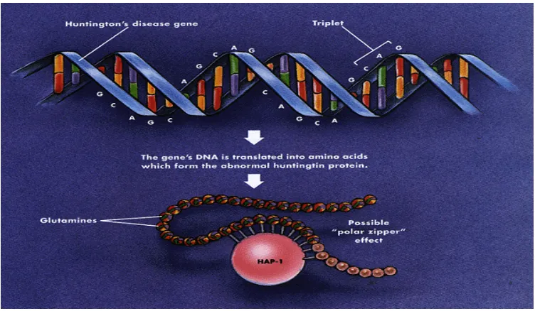
Huntington’s disease (HD)
Hungtington’s disease (HD) is an autosomal dominant neurodegenerative disorder characterized by motor dysfunction, cognitive impairment and psychosis (Sharp et
al., 1995). The disease is caused by an IT15 gene mutation on chromosome 4 (Sharp
et al., 1995). This mutation produces a CAG / polyglutamate repeat expansion in the
Dept. of Pharmacology 106 J.K.K.Nattraja College of Pharmacy
[image:45.612.133.509.134.353.2]mutation produces a toxic huntington protein with a distinct conformation that enables it to stick to both itself and normal huntington (Catteneo et al., 2002).
Figure 7. This conformation allows mutant huntington fragments to clump in
aggregates and simultaneously inhibit the normal protein’s proper function
(Catteneo et al., 2002). Despite these opposing hypotheses regarding the exact role
of CAG expansion, one idea is accepted across the board: mutant huntington forms inclusions in cell nuclei depending on the length of its CAG repeat, and longer repeats correlate with an increased presence of huntington inclusions (Senut et al., 2000). Moreover, these nuclear inclusions are associated with premature neuronal cell death, especially in the striatum and globuspallidus (Hickey & Chesselet, 2003).
Amyotrophic lateral sclerosis (ALS)
Dept. of Pharmacology 107 J.K.K.Nattraja College of Pharmacy
membrane protein (VAMP)-Associated Protein B (hVAPB) causes a range of dominantly inherited motor neuron diseases including ALS8 (Nishimura et al.,
2004; Chen et al., 2010). VAP family proteins are characterized by an N-terminal
major sperm protein (MSP) domain, a coiled-coil (CC) motif and a transmembrane (TM)-spanning region. They are implicated in several biological processes, including regulation of lipid transport, endoplasmic reticulum (ER) morphology and membrane trafficking (Lev et al., 2008). Drosophila Vap-33-1 (DVAP) hereafter, regulates synaptic structure, synaptic microtubule (MT) stability and the composition of postsynaptic glutamate receptors (Pennetta et al., 2002; Chai et al., 2008). MSP domains in DVAP are cleaved and secreted into the extracellular space where they bind Ephrin receptors (Tsuda et al., 2008). MSPs also bind postsynaptic Roundabout and Lar-like receptors to control muscle mitochondria morphology, localization and function (Han et al., 2012). Transgenic expression of the disease-linked alleles (DVAP-P58S and DVAP-T48I) in the larval motor system recapitulates major hallmarks of the human disease, including aggregate formation, locomotion defects and chaperone upregulation (Chen et al., 2010; Chai et al.,
2008; Ratnaparkhi et al., 2008). Several studies have also implicated the ALS
mutant allele in abnormal unfolded protein response (UPR) (Chen et al., 2010; Kanekura et al., 2006; Langou et al., 2010; Suzuki et al., 2009; Gkogkas et al.,
2008) and in the disruption of the anterograde axonal transport of mitochondria
(Mo´rotz et al., 2012). However, it is unclear how these diverse VAP functions are
Dept. of Pharmacology 108 J.K.K.Nattraja College of Pharmacy
lipids that localize to the membrane–cytoplasm interface and function by binding various effector proteins. The inositol group can be reversibly phosphorylated at the 3′, 4′ and 5′ positions to generate seven possible phosphoinositide derivatives, each with a specific intracellular dynamic distribution (Di Paolo & De Camilli, 2006). Interestingly, SAC3 (also known as FIG4), another member of the Sac phosphatase family, is mutated in familial and sporadic cases of ALS (Chow et al., 2009). Inactivation of SAC3 in mice also results in extensive degeneration and neuronal vacuolization in the brain, most relevantly in the motor cortex (Chow et al., 2007). We identified Sac1 and DVAP as binding partners and show that DVAP is required to maintain normal levels of PtdIns4P. Loss of either Sac1 or DVAP function disrupts axonal transport, MT stability, synaptic growth and the localization of a number of postsynaptic markers. We also show that the disease causing mutation (DVAP-P58S) induces neurodegeneration and displays synaptic phenotypes similar to those of either Sac1 or DVAP loss-of-function, including an increase in PtdIns4P levels. Importantly, reducing PtdIns4P levels rescues the neurodegeneration associated with DVAP-P58S and suppresses the synaptic phenotypes associated with DVAP-P58S and DVAP loss-of-function alleles.
Multiple sclerosis (MS)
Multiple sclerosis is a chronic idiopathic demyelinating and neurodegenerative disease of the central nervous system. As such, both the onset and exacerbation of MS are thought to be influenced by multiple factors, including infectious agents, genetic composition and environment (Hauser et al, 2006).
Dept. of Pharmacology 109 J.K.K.Nattraja College of Pharmacy
DEFINITION:
MS is a chronic disease of the CNS, characterized by discrete areas of demyelination and axon injury associated with inflammatory activity as shown in figure 8. A key defining feature of MS is that lesions are disseminated in both space and time, i.e., they occur at more than one site and develop on more than one occasion. Additional information on the pathology of MS is provided below. Clinically, MS symptoms emerge Sbetween the ages of 20 and 40 years in approximately 70% of patients
(Weinshenker et al., 1989; Confavreux et al., 1980) although changes visible on
MRI are much more common than clinical activity and may well precede the latter
[image:48.612.128.526.352.620.2](O’Riordan et al., 1998; Sailer et al., 1999; Brex et al., 2002).
Dept. of Pharmacology 110 J.K.K.Nattraja College of Pharmacy
SYMPTOMS AND SIGNS:
Because MS lesions can occur in many different parts of the CNS, they can cause a wide variety of symptoms and signs. An exhaustive list of clinical findings seen in MS clinics at the Universities of British Columbia and Western Ontario, Canada
(Paty & Ebers, 1997) together with estimates of the frequencies of each finding at
onset and at any time. According to this list, initial neurologic symptoms and signs seen in 10% or more of patients include fatigue (20%, probably more common than this in many populations), optic neuritis (16%), internuclear ophthalmoplegia (17%), nystagmus (20%), vertigo (4–14%), gait disturbances (18%), sensory loss (30–50%, most commonly in the legs and implicating the posterior columns), increased deep tendon reflexes (20%), weakness in the legs (10%), spasticity (10%) and bladder disturbance (3– 10%). Symptoms and signs seen in 50% or more of patients at any time include cognitive changes (70%), euphoria (10–60%), depression (25–54%), fatigue (80%, probably nearer 90% in many populations), optic neuritis (65%), optic atrophy (77%), retinal nerve fiber loss (80%), nystagmus (85%), vertigo (5–50%), dysarthria (50%), limb ataxia (50%), ataxia of the gait and trunk (50–80%), sensory loss (90%, again, most commonly in the legs and implicating the posterior columns), increased deep tendon reflexes (90%), weakness in the legs (90%), spasticity (90%), extensor or flexor spasms (50%), cramps (50%), amyotrophy (50%), bladder disturbance (80%), and sexual disturbance (50% in women, 75% in men).
COMMON MOTIFS IN NEURO DEGENERATION
Dept. of Pharmacology 111 J.K.K.Nattraja College of Pharmacy
increased life expectancy and changing population demographics (i.e., the aging of baby boomers), neurodegenerative dementias and neurodegenerative movement disorders are becoming more common (Brookmeyer et al., 1998; Samii et al., 2004). As our population ages, an improved understanding of these diseases will be vital to developing more effective therapies and combating the staggering personal, social, and economic costs of these diseases (Ernst et al., 1997). Unifying theories of pathogenesis in neurodegenerative disease provide an avenue for developing therapeutic strategies with broad applicability for disease prevention and an opportunity for decreasing morbidity and mortality from these disorders in the elderly population (Forman et al., 2004). Converging lines of investigation have revealed a potential single common pathogenic mechanism underlying many diverse neurodegenerative disorders.
COMMON NEUROPATHOLOGICAL HALLMARKS:
Dept. of Pharmacology 112 J.K.K.Nattraja College of Pharmacy
between the increased production of both the reactive oxygen species (ROS) and the reactive nitrogen species (RNS) and the cellular antioxidant defense systems (Valko
et al., 2007). At low levels, ROS function as signaling intermediates for the
modulation of cellular activities but, at higher concentrations, they contribute to neuronal membrane damage.
ROS is a collective term, which includes not only the oxygen radicals (O2 •−, and
•OH) but also some non-radical derivatives of oxygen. These include hydrogen peroxide (H2O2), hypochlorous acid (HOCl) and ozone (O3) (Bandhopadhyay et al.,
1999).
Over about 100 disorders like rheumatoid arthritis, hemorrhagic shock, cardiovascular disorders, cystic fibrosis, metabolic disorders, neurodegenerative diseases, gastrointestinal ulcerogenesis and AIDS have been reported as ROS mediated. Some specific examples of ROS mediated diseases include Alzheimer’s disease, Parkinson’s disease, Atherosclerosis, Cancer, Down’s syndrome and ischemic reperfusion injury in different tissues including heart, liver, brain, kidney and gastro intestinal tract. The role played by ROS in stress induced gastric ulcer and inflammatory bowel diseases have been well established, as well as their involvement in the process of ageing. The role of radicals in various diseases is dealt in detail.
Aging biology
Dept. of Pharmacology 113 J.K.K.Nattraja College of Pharmacy
decrease of their activities. Telomere shortening with decrease or interruption of cell proliferation, which can mean an unbalance between cells lost and reposition rates, culmination with organ and system failure and death of the organism. Cell damages provoked by free radicals are common during aging; once in this life step cells produce less concentrations of antioxidant enzymes like superoxide dismutase (SOD), catalase (CAT), etc. Gene activation with inevitable aging-related physiological changes (Harman, 1998; Ferrari, 2001). In healthy human centenarians, although both plasmatic and red blood cell-SOD were decreased (in concentration with the increasing levels since <60 years until 99 years), the remarkable increase of plasmatic Vitamins A and E contribute the first indication that these vitamins are favorable to longevity (Mecocci et al., 2000).
Inflammation
An inflammatory response implicates macrophages and neutrophils, which secrete a number of mediators (eicosinoids, oxidants, cytokine and lytic enzymes) responsible for initiation, progression and persistence of acute or chronic state of inflammation
(Lefkowitz et al., 1999). NO along with superoxide (O2 •−) and the products of their
interaction initiates a wide range of toxic oxidative reactions causing tissue injury
(Hogg, 1998). Likewise, the neutrophils too produce oxidants and release granular
anti-
Dept. of Pharmacology 114 J.K.K.Nattraja College of Pharmacy
inflammatory agents in carrageenan-induced rat paw edema method as SOD. These may be due to the removal of O2 •− by SOD, so preventing O2 •− dependent
formation of a factor chemotactic for nutrophils (Miller et al., 1992).
REACTIVE OXYGEN SPECIES
There have been several reports on the role of ROS / RNS in neurodegenerative diseases. Parkinson’s disease usually appears in the middle to old age often as a rhythmic tremor in a foot or hand especially when the limb is at rest. Comparison of the brains of Parkinson’s disease with that of the neurologically normal brains shows several parameters consistent with increased oxidative stress and defective mitochondrial function. Damaged mitochondria may generate more ROS than usual and ROS / RNS (includingO2 •−, •OH, ONOO−) can inactivate complex I. Hence it
is possible that oxidative stress and mitochondrial defects form a vicious cycle
(Halliwell & Gutteridge, 1999).
The ROS mainly involved in neurodegeneration are the superoxide anion (O2 −), the
hydrogen peroxide (H2O2), and the hydroxyl radical (HO•). RNS, such as nitric
oxide (NO), can react with O2 to produce peroxynitrite (ONOO−), a powerful
Dept. of Pharmacology 115 J.K.K.Nattraja College of Pharmacy
Antioxidant defense
It is evident through the reactions of oxygen, that it is toxic; still only the aerobes survive its presence, primarily because they have evolved an inbuilt antioxidant defense. Antioxidant defenses comprise:
• Agents that catalytically remove free radicals and other reactive species like SOD, CAT, peroxidase and thio specific antioxidants.
• Proteins that minimize the availability of peroxidase such as iron ions, copper ions and haem.
• Proteins that protect biomolecules against oxidative damage example heat shock proteins.
• Low molecular mass agents that scavenge ROS and RNS, example GSH, ascorbic acid, tocopherol. The antioxidants may be defined as “any substance, when present at low concentrations compared with that of an oxidizable substrate that significantly delays or prevents oxidations of that substrate”. The term oxidizable substrate includes every type of molecule found in vivo. Antioxidant defense include the antioxidant enzymes like SOD, CAT, GSH-px, etc, low molecular agents and dietary antioxidants (Halliwell & Gutteridge, 1999).
Dept. of Pharmacology 116 J.K.K.Nattraja College of Pharmacy
lower in patients with Alzheimer’s disease and that these levels are associated with the degree of cognitive impairment (Riviere et al., 1998).A prospective study of 633 patients aged 65 years and older found that high-dose supplementation with vitamin C decreased the risk of developing Alzheimer’s disease (Morris et al., 1998).None of the 23 high-dose vitamin C users in this study developed Alzheimer’s when, statistically speaking, 3.3 would have been expected to develop the disease. A case-control study found that beta carotene levels are lower in Alzheimer’s disease patients than in healthy controls (Zaman et al., 1992).A study of 38 Alzheimer’s patients and 42 healthy control subjects found that beta carotene levels are lower in patients than in healthy individuals (Jimenez-Jimenez et al., 1999).Other antioxidants such as selenium, glutamine, taurine, coenzyme Q10, pantethine, and magnesium would likely be beneficial to Alzheimer’s patients but have not yet been thoroughly studied. Magnesium, for instance, is a particularly good agent to study because of its demonstrated to block the absorption of aluminum in the intestines as well as across the blood-brain barrier.
CURRENT SCENARIO
Dept. of Pharmacology 117 J.K.K.Nattraja College of Pharmacy
impairment, seizure disorders, head injury, Parkinsonism can be severely functionally debilitating in nature (Commenges et al., 2000).
A recent study (Andlin-Sobocki et al., 2005) has evaluated the total cost of brain diseases per year, including direct and indirect costs, in 28 countries in Europe at about 386 billion Euros for the year 2004. This represented 35% of the total burden of diseases affecting about 27% of the 465 million people who are suffering brain diseases. If mental disorders are excluded from the calculation the total cost of neurological diseases including dementia could be about 146 billion Euros per year and the total specific cost of the neurodegenerative diseases could be as much as 72 billion Euros (Table 1). These diseases are found in about 5% of the total number of patients suffering brain diseases. They are characterized by more or less selective neuronal degenerations inducing neurological syndromes, and affect both sensory-motor areas and cognitive functions.
Table 1.Number of cases and cost per case and per year of neurodegenerative
diseases in 28 selected European countries (2004)*
Number of cases Cost per case/year Total cost/year
Parkinson’s disease
Dementia
Multiple sclerosis
1 160 000
4 890 000
380 000
7 500 Euros
11 000 Euros
24 000 Euros
8.70 billion Euros
53.80 billion Euros
9.12 billion Euros
* from Andlin-Sobocki et al. (2005)
Dept. of Pharmacology 118 J.K.K.Nattraja College of Pharmacy
This disease rarely occurs before the age of 50, and men are at higher risk than women. In Europe, PD affected 1.2 million people in 2010, resulting in costs per patient of EUR 5,626 for direct health care and EUR 4,417 for non-medical care. In 30 European countries, the total cost of all care for patients with PD in 2010 was EUR 13.9 billion (de Lau & Breteler, 2006).
According to a recent estimation, it is possible that almost 80% of individuals with dementia suffer from AD (Jellinger & Attems, 2010). AD is a severe progressive neurodegenerative brain disorder that affects approximately 5% of the population older than 65 years (Shah et al., 2008). According to the US Centers for Disease Control and Prevention (2003), the number of people in the world who are over the age of 65 will increase to around 1 billion by 2030. It has also been projected that by 2050 the number of dementia cases will reach around 14 million in Europe (Mura et
al., 2010) and 13.2 million in the United States (Hebert et al., 2001). Furthermore, it
Dept. of Pharmacology 119 J.K.K.Nattraja College of Pharmacy
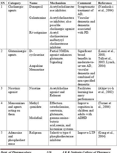
CURRENT THERAPEUTIC APPROACHES IN NEURODEGENERATION
[image:58.612.125.536.204.742.2]Drugs to improve memory generally work by altering the balance of particular chemicals (neurotransmitters) in the brain that are involved in the initial
Table 2. List of cognition enhancing drugs acting at neurotransmitter level
SN Category Name Mechanism Comment Reference
1 Cholinergic agents Donepezil Galantamine Rivastigmine Acetylocholinester ase inhibitor. Acetylcholinestera se inhibitor; also possible cholinergic agonist Acetyl cholinesterase andbutyryl cholinesterase inhibitor. Symptomatic treatment of AD, Vascular dementia and dementia associated with PD. (Narahash-i et al., 2004)
2 Glutaminergic
agents D-cycloserine
Ampakine Memantine Partial NMDA agonist enhances glutamate. Signaling Significant broad benefits in moderate-to-severe AD, vascular dementia and combined of non-specified dementia
(Lanni et al., 2008; Tullyet al., 2003; Lynch, 2004) 3 Nicotinic agonist Nicotine Acetylcholine agonist and Releaser Facilitates learning/me mory performance (Ahijevyc-h et al., 2002)
4 Monoamines and agents acting on them Methyl -penidate Modafinil Effectson catecholamine, serotonin, glutamate, gamma amino-butyric acid,orexin, and histamine systems Improve cognition in children and adults with ADHD (Turner et al., 2004) 5 Adenosine and phosphodiest erase
Rolipram Selective type-4 phosphodiesterase inhibitor
Dept. of Pharmacology 120 J.K.K.Nattraja College of Pharmacy
learning of a memory or its subsequent reinforcement. Some of them along with their mechanism are listed in table 2. Some acts by selective enhancement of cerebral blood flow and metabolism, including enhanced glucose uptake, which may protect against the effects of hypoxia and ischemia. Reports from literature reveal that some medications currently available to patients with memory disorders mayalso increase performances in healthy people. Drugs designed for psychiatric disorders can also be used to enhance certain mental functions. However, the long-term effects of these drugs are unknown. Drugs which act as cognition enhancer increase synaptic plasticity by, regulating release of neurotransmitter from the pre-synaptic terminal and increasing sensitivity and specificity of receptors and ion channels in the membranes of synapse to neurotransmitter signaling. Some of the agents also modulate the process at transcriptional and translational level.
HERBAL MEDICINES
Dept. of Pharmacology 121 J.K.K.Nattraja College of Pharmacy
potential cognitive enhancement activity are listed in table 3, (Howes & Houghton,
2003; Kennedy et al., 2003).
The past decade has also witnessed an intense interest in herbal medicines in which phytochemical constituents can have long-term health promoting or medicinal qualities. In contrast, many medicinal plants exert specific medicinal actions without serving a nutritional role in the human diet and may be used in response to specific health problems over short- or long-term intervals. Phytochemicals present in vegetables and fruits are believed to reduce the risk of several major diseases including cardiovascular diseases, cancers as well as neurodegenerative disorders. Therefore people who consume higher vegetables and fruits may be at reduced risk for some of diseases caused by neuronal dysfunction (Selvam, 2008; Lobo et al.,
[image:60.612.134.502.437.708.2]2010).
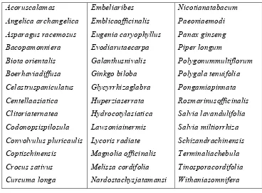
Table 3. Some putative cognitive enhancing plants
Dept. of Pharmacology 122 J.K.K.Nattraja College of Pharmacy
Herbal medicine has long been used to treat neural symptoms. Although the precise mechanisms of action of herbal drugs have yet to be determined, some of them have been shown to exert anti-inflammatory and/or antioxidant effects in a variety of peripheral systems. Now, as increasing evidence indicates that neuroglia-derived chronic inflammatory responses play a pathological role in the central nervous system, anti-inflammatory herbal medicine and its constituents are being proved to be a potent neuroprotector against various brain pathologies. Structural diversity of medicinal herbs makes them a valuable source of novel lead compounds against therapeutic targets that are newly discovered by genomics, proteomics, and high-throughput screening. This review will highlight the importance of phytochemicals on neuroprotective function and other related disorders, in particular their mechanism of action and therapeutic potential (Pueyo & Calvo, 2009).
Phytochemicals in neuroprotection
Dept. of Pharmacology 123 J.K.K.Nattraja College of Pharmacy
effects of various phytochemicals are associated with reduced levels of oxidative stress. For example, resveratrol, quercetin and catechins reduced oxidative stress and protected cultured hippocampal neurons against nitric oxide-mediated cell death
(Larson, 1988). Some of the neuroprotective herbs with their major bioactive
compound and mode of action were shown in table 4. Hundreds of articles have been published reporting neuroprotective effects of compounds in natural products, including α-tocopherol, lycopene, resveratrol, ginkgo biloba and ginsenosides (Ikeda
et al., 2003).
Flavonoids
[image:63.612.130.517.134.569.2]
Dept. of Pharmacology 124 J.K.K.Nattraja College of Pharmacy
Table 4: Nootropic herbs with their active constituents' that help in
neuroprotection
Dept. of Pharmacology 125 J.K.K.Nattraja College of Pharmacy
brain barrier (BBB), which controls entry of xenobiotics into the brain (Ehrnhoefer
et al., 2006). Flavanones such as hesperetin, naringenin and their in vivo metabolites,
along with some dietary anthocyanins, cyanidin-3-rutinoside and pelargonidin-3-glucoside, have been shown to traverse the BBB in relevant in vitro and in situ models (Youdim et al., 2004; Youdim et al., 2002). Anthocyanins can possibly cross the monolayer in blood-brain barrier models in vitro.
Flavonoids and tannins are phenolic compounds that are a major group of compounds act as primary antioxidants or free radical scavengers (Polterait, 1997).
THE ROLE OF PLANT FLAVONOIDS IN NEURODEGENERATION
There has been a recent explosion of interest by research scientists in the flavonoid compounds, with a multitude of medically useful properties having been demonstrated in experimental, as well as, clinical studies of flavonoids. For instance, flavonoids have been shown to act as powerful free radical scavengers for a multitude of free radical species, even the powerful peroxynitrite radical (Jovanovic
et al., 1998). In addition, several flavonoids have shown powerful metal-chelating
Dept. of Pharmacology 126 J.K.K.Nattraja College of Pharmacy
1991). Finally, some of the flavonoids have the unique ability to inhibit certain enzymes, such as the COX-2 enzyme (Kim et al., 1998).
FLAVONOIDS AS FREE RADICAL SCAVENGERS
The flavonoid compounds have two properties that make them especially useful as antioxidants. First, many are powerful, primary free radical scavengers against a wide variety of radicals, including singlet oxygen, superoxide, peroxyl, hydroxyl, and the peroxynitrite radicals (Saija et al., 1995). Second, several are known to be very effective metal chelators (Duthie et al., 1997). Most flavonoids are present in plants as glycosides. In the intestines, this moiety is cleaved off, leaving the aglycone form of the flavonoids (Griffiths, 1982). It is the aglycone form that is thought to have the highest antioxidant activity in biological systems. There is experimental evidence that hydrogen peroxide accumulation occurs during the process of catecholamine catabolism, making it especially important in PD (Li et al., 1995). Recent evidence also indicates that H2O2 plays an important role in the
toxicity of Alzheimer’s plaques. As we have seen, iron accumulation within neurons is characteristic of ageing of the nervous system, but is especially high in the case of neurodegeneration. A multitude of phytochemicals have specific properties that make them especially useful in combating neurodegeneration, and a list of nutrients that stimulate energy generation, primarily through the mitochondrial system.
D
S
Dept. of Phar
cientific cla Kingd Divisi Class Subcl Order Famil Genu Speci Binom rmacology assification dom ion lass r ly us es
mial name
12
2. PLA
27 J
ANT PRO
: Plan : Mag : Mag : Lam : Car