A COMPARATIVE STUDY OF OPEN
SURGICAL DRAINAGE AND EARLY
PERCUTANEOUS NEEDLE
ASPIRATION OF LIVER ABSCESS.
Dissertation Submitted to
THE TAMIL NADU DR. M.G.R. MEDICAL UNIVERSITY in partial fulfillment of the regulations
for the award of the degree of
M. S. General Surgery
Branch – I
Chengalpattu Medical College
The Tamilnadu Dr. M. G. R. Medical University
Tamilnadu, India.
CERTIFICATE
Certified that this dissertation entitled “A COMPARATIVE STUDY OF
OPEN SURGICAL DRAINAGE AND EARLY PERCUTANEOUS
NEEDLE ASPIRATION OF LIVER ABSCESS” is a bonafide work
done by Dr. C. R. SIDDARTHA, post graduate student of the Department
of General Surgery, Chengalpattu Medical College, during the academic
year 2010 – 2013. This work has not previously formed the basis for the
award of any degree.
Prof. Dr. G. Raja Billy Graham, M. S. Dr. R. Kannan, M. S.,
Professor and Head, Associate Professor,
Department of General Surgery, Department of General Surgery, Chengalpattu Medical College. Chengalpattu Medical College.
Dr. Thenmozhi Valli, M. D.
Dean,
DECLARATION
I, Dr. C. R. SIDDARTHA, solemnly declare that dissertation titled,
“A COMPARATIVE STUDY OF OPEN SURGICAL DRAINAGE
AND EARLY PERCUTANEOUS NEEDLE ASPIRATION OF
LIVER ABSCESS” is a bonafide work done by me at Chengalpattu
Medical College during 2010-2013 under the guidance and supervision of
Prof. Dr. G. RAJA BILLY GRAHAM , Professor and Head, Department
of General Surgery, Chengalpattu Medical College.
The dissertation is submitted to The Tamilnadu Dr. M.G.R. Medical
University, in partial fulfilment of requirement for the award of
M. S. GENERAL SURGERY.
Place: Chengalpattu. (Dr C. R. SIDDARTHA)
ACKNOWLEDGEMENT
I thank my Dean, Dr. Thenmozhi Valli, M. D., for permitting me to utilize
the facilities provided at the Hospital and to do my Dissertation.
I thank my Professor and Head of Department of General Surgery,
Dr. G. Raja Billy Graham. M. S., for his constant guidance and support in
completing this endeavour.
I extend my sincere thanks to Dr. R. Kannan. M. S., my unit chief for his
never ending support and guidance during the study.
I thank Dr. Santha seelan. M. S., Dr. V. Mohanraj. M. S., Professors of
General Surgery for their valuable guidance.
I express my deepest gratitude to my Assistant professors,
Dr. P. Sankarlingam, Dr. Shanmugavelayutham, Dr. Raja Mahendran, Dr.
Mathusoothanan, and Dr. Raja. Without their constant support, guidance
and motivation this venture of mine wouldn’t have been possible.
I am grateful to the Assistant professors of Department of Radiology,
Department of Microbiology and Department of Anaesthesiology, for their
utmost help in working up and also in therapeutic management of the
My most sincere thanks to my fellow post graduates, Dr. Vijayabhasker,
Dr. Hariharan, for their constant help in managing the patients.
I thank the members of the nursing staff, laboratory technicians, for their
assistance. Without which the undertaking of mine wouldn’t have been
possible.
CONTENTS
Si. No. Page No.
1. Introduction...1
2. Review of literature...2
3. Objectives...42
4. Material and methods...43
5. Observation and results...48
6. Discussion...71
7. Conclusion...80
8. Bibliography...81
9. Annexure. a. Proforma...89
Introduction
Liver Abscess noted in the writings of Hippocrates, was considered to be
dreadful disease. It was uniformly fatal until a study by Ochsner and
Debakey in 1938. They suggested an aggressive surgical approach to the
liver abscess which reduced the mortality rates considerably. [1] Advances
in radiological imaging which can diagnose the presence of liver abscess
and its accurate anatomical localisation, combined with advances in
pharmacotherapeutics and critical care have shifted the treatment modality
of choice from open surgical drainage to minimally invasive procedures.
With many therapeutic options now available, the effective treatment of
liver abscess is still controversial. Do the minimally invasive procedures
provide effective drainage of the abscess with reduction of mortality when
compared with open surgical drainage? This study aims at comparing the
two methods of abscess drainage, open surgical and percutaneous
Review of literature
History
Liver abscess has been known since the time of Hippocrates (460 - 377
BC). In one of his aphorisms he mentions “when abscess of the liver is
treated by the cautery or incision, if the pus which is discharged is 'pure
and white', the patient recovers, but if it resembles the lees of the oil as it
flows, they die” .[2]
Around 300 BC, Alexander the Great, after his campaign reaching the
Indus region, where amoebiasis is endemic, felt sick and died on his return
in Gedrosia desert. It is believed that is death was probably due to amoebic
liver abscess. [3]
In 1818, George Ballingall, an army surgeon at the Madras establishment
had made a note of an officer who got accidentally shot, resulted in
draining of liver abscess and obtaining a complete cure. [4]
Napoleon Bonaparte, the French emperor in 18th century was exiled to a
tropical island St. Helena after his defeat at the Battle of Waterloo. He had
contracted a disease which was common on the island. Antommarchi, a
physician appointed by Napoleon’s family described the features of
Napoleon’s last illness. At the emperor's post-mortem, he had noticed “a
curve of stomach, communicating with the liver.” It is believed that
Napoleon had died of amoebic liver abscess of the left lobe which had
ruptured into stomach. [3]
In 1887, Robert Koch investigating cholera in Egypt and India had come
across 2 cases of dysentery complicated by liver abscess. He identified
Entamoeba histolytica in the capillary walls, adjacent to the abscess and
found them similar to the ones in stool. [3]
Sir Charles Morehead, Professor of Medicine at Grants Medical College,
Bombay in 1948, had described the first case of hepatic abscess in India. [3]
In 1891, Councilmann and Lafleur in their monograph coined the term
“Amoebic Abscess of the liver” and described the occurrence of liver
abscess as a complication of intestinal amoebiasis even in patients who did
not have the symptoms of latter disease. [5]
Sir William Osler, in his 1892 edition of “The Principles and Practice of
Medicine” described the major cause of liver abscess as “suppuration in the
territory of portal vessels”. He also stated that “results from dysentery and
other ulcerative affections of the bowels, appendicitis, occasionally typhoid
fever, in rectal affections, and in abscesses in the pelvis.” He termed the
process of portal bacteraemia with abscess formations as “suppurative
Ochsner and DeBakey in 1938 described 137 cases of amoebic abscess and
47 cases of pyogenic abscess. They also described the aggressive surgical
management to decrease mortality which was a uniformly fatal disease
until then. [1] Mcfadzean et al in 1953 first described the successful
treatment of pyogenic liver abscess by percutaneous aspiration. [7] Whereas
in 2005, a study by Tan et al showed open surgical drainage maybe a better
modality compared to percutaneous drainage for the treatment of liver
abscess. [8] Numerous studies have been published and yet effective
treatment of liver abscess remains controversial.
Epidemiology
Liver abscess is a rare occurrence whose aetiology, diagnosis and treatment
have changed over time. Traditionally, it has been classified into pyogenic
abscess – caused by various bacteria, and Amoebic – caused by Entamoeba
histolytica. Recent trends show increase in the incidence of fungal and
mycobacterial abscess, probably due to increase in patients with AIDS and
other forms of immunosuppression. [9]
Liver abscesses are the most common type of visceral abscess; in a report
of 540 cases of intra-abdominal abscesses, pyogenic liver abscesses
accounted for 48 percent of visceral abscesses and 13 percent of
intra-abdominal abscesses. [10] The annual incidence of liver abscess has been
[11]
Risk factors include diabetes, hepatobiliary or pancreatic disease, and
post liver transplantation. A primary invasive liver abscess syndrome due
to Klebsiella pneumoniae has been described in East Asia indicating
interplay between host and geographical factors. [12] With the effective
treatment of conditions like appendicitis and other acute colonic diseases,
there has been a shift in the aetiology and age distribution of patients
presenting with hepatic abscess. Numerous studies have reported biliary
tract disease to be the most frequent underlying lesion associated with
pyogenic liver abscess, with a peak incidence in the seventh and eight
decades. [11]
Worldwide, amoebiasis is the third most common parasitic cause of death.
[13]
Amoebic liver abscess is 7 to 10 times more common among adult men,
despite equal gender distribution of intestinal amoebiasis. It is
predominantly seen in the fourth and fifth decades of life. The reasons for
these observations are not fully understood; suggested mechanisms include
hormonal effects and a potential role of alcoholic hepatocellular damage in
creating a nidus for portal seeding. Immigrants and travellers from endemic
regions, people of low socioeconomic status, mentally retarded individuals
Pyogenic Liver Abscess
Aetiology
Liver is constantly exposed to the infective organisms from the
gastrointestinal tract. Kupffer cells act as a filter for the clearance of micro
organisms. Abscesses occur when normal hepatic clearance mechanism fail
or the system is overwhelmed.
The potential cause of liver abscess is classified as [14]:
1. Biliary.
2. Portal vein.
3. Haematogenous.
4. Traumatic.
5. Direct extension.
6. Cryptogenic.
30 – 40% of all pyogenic abscesses are due to diseases of the biliary
system. 40% of these abscesses occur due to underlying malignancy.
Obstruction of biliary tree, intrahepatic biliary stone, associated stricture,
manipulation of biliary tract like cholangiography, percutaneous hepatic
stents, endoscopic stent placement and biliary enteric anastomosis
Intestinal pathology is responsible for 20% of pyogenic liver abscess.
Gastrointestinal perforation or bacterial translocation cause transient
bacteraemia and resultant spread via the portal vein to the liver.
Diverticulitis, perforated colonic carcinoma and other intra-abdominal
abscess predispose to pylephlebitis and cause liver abscess. [14]
Gangrenous cholecystitis, perforated ulcers and subphrenic abscesses cause
liver abscess by contiguous spread. Trauma to liver causes parenchymal
necrosis and clot, which creates an ideal milieu for the seeding and
proliferation of microorganisms and subsequent abscess formation.
Haematogenous spread can occur from distant infection from heart, lungs,
kidneys, bones, ears and teeth through the hepatic artery and cause about
12% of pyogenic liver abscesses. Cryptogenic abscesses, those of unknown
aetiology occur in 10 – 45% of patients who have co morbidities like
diabetes, immunosuppression. [14]
Pathology
The number, size, location of liver abscess is determined by the source.
Portal, cryptogenic, and traumatic abscesses are solitary, large whereas
biliary and arterial abscesses are small and multiple. [14]
Huang et al reported that liver abscess involved the right lobe in 63% of
predilection for the right hepatic lobe can be attributed to anatomic
considerations. [14]
The right hepatic lobe receives blood from both the superior
mesenteric and portal veins, whereas the left hepatic lobe receives
inferior mesenteric and splenic drainage.
The right lobe contains a denser network of biliary canaliculi and,
overall, accounts for more hepatic mass.
Studies have suggested that a streaming effect in the portal
circulation is causative.
Bacteriology
Abscess cultures are positive for growth in 80 – 90% of cases, but blood
cultures yield growth in 50 – 60%. E. coli, klebsiella sp, enterococci,
pseudomonas are commonly isolated aerobic bacteria. [15] Bacteroides and
fusobacterium are the commonly isolated anaerobes. [16]
The organisms commonly isolated from the abscess cavity and their
Category of organisms % of patients
Gram negative aerobes 50 – 70
Eschericihia coli 35 – 45
Klebsiella 18
Proteus 10
Enterobacter 15
Serratia Rare
Morganella Rare
Acinetobacter Rare
Gram positive aerobes 55 Streptococcal species 20
Enterococcus 10
β – Streptococci 5
α - Streptococci 5
Staphylococcal sp 15
Anaerobes 40 – 50
Bacteroides 24
Fusobacterium 10
Peptostreptococcus 10
Clostridium 5
Actinomyces Rare
Fungal 26
Clinical features
The clinical presentation of liver abscess is usually sub-acute and
nonspecific, requiring high index of suspicion for diagnosis. The classic
presentation triad of fever with chills, right upper quadrant pain and
general malaise is rarely present. [15] The most common presenting
symptom is fever with chills. The abdominal pain is constant dull aching,
localised to the right upper quadrant. Associated diarrhoea is rarely present.
Abscesses of the left lobe present with symptoms in the epigastric region.
Associated features like malaise and weight loss are present when patients
present late. Jaundice occurs when there is compression of the biliary tree.
Pleuritic pain or pain at the tip of right shoulder occurs due to irritation of
the right hemi-diaphragm. [14]
The following table shows the common signs and symptoms of
presentation of patients with pyogenic liver abscess and their frequency of
Symptom Percentage of pyogenic abscesses
Fever 83
Weight loss 60
Pain 55
Nausea and vomiting 50
Malaise 50
Chills 37
Anorexia 34
Cough or pleurisy 30
Pruritis 17
Diarrhoea 12
Signs
Right upper quadrant tenderness 52
Hepatomegaly 40
Jaundice 31
Right upper quadrant mass 25
Ascites 25
Complications
Complications of liver pyogenic liver abscess can be classified into
1. Local complications include:
Rupture [17]:
o Into the peritoneal cavity resulting in peritonitis.
o Into the pleural cavity causing empyema and hepatopleural
fistula.
o Into the pericardial cavity from an abscess of the left lobe.
o Into adjacent organ and fistulise into stomach, colon, small
bowel, or kidney.
o Subphrenic space resulting in subphrenic space.
o Into biliary tract resulting in haemobilia.
Liver Failure.
Acute pancreatitis.
2. Vascular complications include:
Hepatic vein thrombosis.
Portal vein thrombosis.
Hepatic artery pseudo – aneurysm due to erosion of abscess
3. Systemic septic complications are common in diabetic patients who
are infected with Klebsiella pneumonia through haematogenous
dissemination. [18]
Endophthalmitis or uveitis,
Pulmonary abscess,
Brain abscess and/or purulent meningitis,
Bacteriuria and/or prostate abscess,
Osteomyelitis and/or pyogenic arthritis, and
Psoas abscess.
Amoebic liver abscess
Aetiology
Amoebic liver abscess is caused by Entamoeba histolytica. After fecal oral
transmission, the infective quadrinucleate cyst passes through the stomach.
The trophozoite is released after the pancreatic enzymes digest the outer
cyst wall. Usually, the trophozoite multiplies in the intestine with no
invasion resulting in amoebic dysentery alone or asymptomatic carrier. In a
few patients, the trophozoite invades the intestine and travels along the
Life cycle of Entamoeba histolytica
Pathology
Entamoeba histolytica as implied by its name lyses through the tissues
through a complex set of interactions, cell adherence, cell activation and
subsequent release of enzymes results in cell necrosis. Hepatic amoebic
liquefactive necrosis of the liver forming a cavity of blood and liquefied
liver tissue. The hepatic necrosis continues until it reaches the Glisson's
capsule. As the capsule is resistant to hydrolysis, the amoebic liver abscess
tends to abut the liver capsule and the cavity is crisscrossed by portal
triads. [19]
Early on, the infection of liver results in amoebic hepatitis which progress
to from multiple small abscess which later coalesce to form a single large
hepatic abscess. The abscess itself contains acellular fluid which is
red/brown and yellow in colour and similar to “anchovy paste” in
consistency. Trophozoites are absent in the abscess but reside in the
necrotic tissue surrounding the abscess. [20]
Clinical presentation
Majority of amoebic liver abscess occur in the young adult males. The
presentation may be acute, with fever and right upper quadrant pain, or sub
acute, with weight loss. History of alcohol abuse is common.
The following table shows the common signs and symptoms of
presentation of patients with pyogenic liver abscess and their frequency of
Symptom Percentage of Amoebic liver abscesses
Pain 90
Fever 87
Nausea and vomiting 85
Anorexia 50
Weight loss 45
Malaise 25
Diarrhoea 25
Cough or pleurisy 25
Pruritis <1
Signs
Hepatomegaly 85
Right upper quadrant tenderness 84
Pleural effusion or rub 40
Right upper quadrant mass 12
Ascites 10
Complications
Secondary bacterial infection of the abscess can occur.
Pleural manifestations may include development of a sympathetic
serous effusion.
Rupture is the most common complication of amoebic liver abscess.
Rupture may occur into peritoneum, pleural cavity or pericardium,
the latter being common with left lobe abscess. Rupture may also
occur into the stomach and colon. Abscess rupture into the pleural
space results in an amoebic empyema; rupture into the lung can lead
to consolidation, abscess formation, or a hepatobronchial fistula
resulting in a spontaneous cure. [21]
Metastatic amoebic abscess can occur in brain, lung, skin and
genitourinary tract, presumably due to haematogenous spread from
the intestinal infection. [21]
Compression syndrome: A posterior located Amoebic liver abscess
of the right lobe may manifest as hepatic outflow obstruction or
inferior vena cava obstruction. Clinical features include bilateral
pedal oedema, ascites and visible veins on anterior and posterior
abdominal wall. These features disappear after aspiration of the
Differential Diagnosis [19]
1. Right subphrenic abscess.
Recent abdominal surgery, inflammatory or perforating conditions of
the intra abdominal viscera or abdominal trauma may suggest the
diagnosis of subphrenic abscess; a lung and liver scan may help in
differentiating it from the liver abscess.
2. Malignancy of liver.
This may be a primary liver malignancy or secondary metastatic
deposits in the liver. A hard nodular and markedly enlarged liver
both with and without jaundice suggests malignancy.
3. Acute cholecystitis.
Sudden onset of severe pain in right upper quadrant, nausea,
vomiting fever and minimal icterus should suggest the diagnosis of
acute cholecystitis. Murphy’s sign will be positive.
4. Hydatid cyst of liver.
Patients with hydatid cyst usually present with a mass in the upper
abdomen. Cardinal features of liver abscess like fever, pain and
tenderness are absent. Hydatid thrill can be elicited.
5. Alcoholic hepatitis.
This condition occurs in chronic alcoholics. Jaundice is more
6. Viral Hepatitis.
Although liver abscess and viral hepatitis are both common in under
developed countries, differentiating the two is not difficult.
Complete loss of appetite, nausea, vomiting and absence of pain all
favour the viral hepatitis.
7. Congestive liver of congestive cardiac failure or constrictive
pericarditis.
Oedema, breathlessness and hepatomegaly occurring occasionally in
liver abscess may be mistaken for congestive cardiac failure.
Absence of fever, severe pain, diffuse hepatomegaly and presence of
engorged neck veins along with signs in the chest suggest
involvement of heart.
8. Acute Abdomen
Occasionally “Reflex ileus” in association with persistent vomiting,
constipation and distension provides a picture which is difficult to
distinguish from an acute intestinal obstruction.
Investigations
Haematological investigations:
Almost all patients with liver abscess have a haemoglobin level <12g% and
a haematocrit level < 36%. There will be associated leukocytosis >10,000
Liver function tests
Elevation of bilirubin > 2 g/dl is common with pyogenic liver abscess. In
an acute liver abscess, serum alkaline phosphatase will be normal and
alanine transferase will be elevated and a reverse occurs in a chronic liver
abscess. Hypoalbuminemia (Albumin <3g/dl) is present in about 90 % of
the patients. [14]
Stool examination
The role of wet mount stool examination is limited as less than 30-40% of
patients with amoebic liver abscess have concomitant intestinal
amoebiasis. Microscopically similar but non-pathogenic Entamoeba dispar
contributes for the high false positive rate. [24]
Examination of the stool is done by a combination of wet mount,
iodine-stained concentrates, and trichrome-iodine-stained preparations. Haematophagous
trophozoites of E histolytica require at least 3 fresh specimens as the
trophozoites are very sensitive and may be excreted intermittently. Cysts
must be differentiated morphologically from nonpathogenic Entamoeba
hartmanni, Entamoeba coli, and Endolimax nana. Nonpathogenic E dispar
cannot be differentiated morphologically and require fecal antigen
ELISA targeting the galactose inhibitable adherence protein of E.
histolytica helps to differentiate between the two amoebae. Stool antigen
detection facilitates early diagnosis before an antibody response. The
drawbacks are the requirement for fresh, unpreserved stool specimens and
the lack of intestinal amoebiasis in as many as 60% of patients with
amoebic liver abscess. [13]
The PCR stool test is highly sensitive for detection of E histolytica and for
distinguishing nonpathogenic amoebas. However, this test is expensive and
less sensitive than ELISA for stool antigen. Real-time (rapid) PCR is being
developed and is a sensitive test for detection of E histolytica. [25]
Serology
Indirect haemagglutination tests targeting the antibodies against E
histolytica were used in the past. These tests remain positive after infection
for many years and interpretation is difficult in patients from endemic
areas.
These tests have been replaced by enzyme immunoassay (EIA) which is
rapid and inexpensive. The EIA test detects antibodies specific for E
histolytica in approximately. [26]
95% of patients with extraintestinal amoebiasis,
10% of persons who are asymptomatic cyst passers.
The EIA serology reverts to negative in 6 – 12 months following
eradication of infection. Even in highly endemic areas, fewer than 10% of
patients who are asymptomatic have positive amoebic serology findings.[26]
Initial test results may be negative in as many as 10% of patients with
amoebic liver abscess, but repeat serology testing within a week will
usually be positive.
Entamoeba histolytica galactose lectin antigen is detectable by
Enzyme-linked immunosorbent assay (ELISA) in 75% of patients with amoebic
liver abscess [26]. Initial antigen seropositivity is 96% with a reversal rate
of 82% after 1 week of treatment with metronidazole. This test is useful for
patients who present acutely, before an antibody response occurs. The
serum sample is to be obtained before initiation of treatment, as medical
therapy leads to rapid antigen loss. This test can be used for rapid diagnosis
in highly endemic areas, where serology can be misleading.
Rapid testing for antigen and antibodies are being currently developed and
Blood culture
Blood culture reveals growth in only about 50 % of cases. But there has
been a poor correlation between the organism isolated from the abscess
itself and the blood culture due to polymicrobial nature of the disease.
Diagnostic Aspiration
Under CT scan or ultrasound guidance, needle aspiration of cavity material
can be performed.
Needle aspiration enables rapid recovery of material for microbiologic and
pathologic evaluation. When the diagnosis is in doubt, it helps to identify
whether the abscess is amoebic or pyogenic. [26]
Needle aspiration of the abscess can be performed with the initial
diagnostic procedure.
Radiological investigations
Roentgenography
Chest X ray is abnormal in about 50% of cases. [19] Features include
1. Elevated right hemi diaphragm.
2. Pleural effusion.
3. Atelectasis.
Abdominal X ray may reveal
1. Hepatomegaly.
2. Gas within the abscess cavity due to
a. Infection by gas forming organisms.
b. Secondary infection of amoebic abscess.
c. Prior percutaneous intervention.
d. Rupture into a hollow viscous.
3. Air in the portal vein if associated pylephlebitis is present. [14]
4. Aerobilia may be present if cholangitis is present. [27]
Absence of the findings however doesn’t rule out liver abscess.
Ultrasonography
Ease of access and low cost has made ultrasonography a boon in evaluation
and treatment of live abscess. USG can identify lesions as small as 2cm.
The typical lesion is a hypo echoic round lesion with an echogenic wall,
acoustic enhancement and internal echoes. Ultrasonography allows for
close evaluation of the biliary tree and simultaneous aspiration of the
cavity. The major benefits of this technique are its portability and
diagnostic utility in patients who are too critical to undergo prolonged
The drawbacks of USG include. [19]
1. Doesn’t visualize the dome of liver.
2. Multiple micro abscesses may not be seen.
3. Fatty infiltration increase echogenicity of liver and decrease the
sensitivity of the study.
4. Operator dependence affects its overall sensitivity.
CT scan [14]
Computed tomography scanning has the advantage of being able to detect
intrahepatic lesions as small as 0.5 cm and also image the entire abdomen.
A classical lesion of liver abscess will be a hypo dense cystic lesion with
thick segmental wall enhancement and surrounding low density oedema. A
central large lesion can be surrounded by adjacent “daughter” abscess
representing coalescing of multiple small abscess – Cluster sign, is
suggestive of bacterial cause. [28]
MRI
Magnetic resonance imaging can better characterize hepatic lesions
compared to CT. MRI can distinguish liver abscess from other cystic and
necrotic lesions. [9] High cost, length of study and lack of easy access for
drainage procedure has limited the usefulness in management of liver
Other imaging
Technetium – 99m liver scanning is useful test for differentiating an
amoebic liver abscess from a pyogenic abscess; however, it is not a
first-line test. [26]
Amoebic liver abscesses do not contain leukocytes; they appear as cold
lesions on hepatic nuclear scanning, with a typical hot halo or a rim of
radioactivity surrounding the abscess. [26]
In contrast, pyogenic liver abscesses contain leukocytes and, therefore,
typically appear as hot lesions on nuclear scanning.
Gallium scanning is helpful in differentiating pyogenic abscess but requires
delayed images, which makes the test less helpful. Gallium and
Technetium 99m scans have largely been abandoned in their role of
investigating and treatment of liver abscess due to advances in CT and
USG. [9]
Treatment
Pyogenic liver abscess
An untreated hepatic abscess is nearly uniformly fatal due to the
complications that follow. Management of pyogenic liver abscess involves
appropriate antibiotic therapy and drainage of abscess, followed by
Appropriate antibiotic therapy involves the identification of the organism
by blood culture or culture of the pus from the abscess. Treatment is
initiated with broad spectrum antibiotics covering gram positive and
negative organisms and anaerobic bacteria. A prolonged course of
antibiotics lasting 4 – 6 weeks is required as there is a perception of
presence of avascularity in the abscess. [14]
The differences in clinical presentation of pyogenic and amoebic liver
abscess are given in the table below. [19]
Clinical features Amoebic liver
abscess
Pyogenic liver abscess
Age (years) 20 – 40 >50 Male : Female >10:1 1.5:1 Solitary vs. Multiple Solitary 80% Solitary > 50 %
Location Usually Right lobe
Usually right lobe
Travel to endemic area Yes No Diabetes and other forms of
Immunosuppression No Yes
Alcohol use Common Common
Jaundice Uncommon Common
Drainage procedures
Most liver abscesses require some form of drainage, whether it is closed
aspiration, percutaneous drainage or surgical.
Closed aspiration
Giorgio and colleagues reported a series of 115 patients who underwent
percutaneous aspiration and reported a 98.3 % success rate with no
mortality or morbidity. [29] Percutaneous aspiration is usually undertaken
under USG or CT guidance. At present, the indications for closed needle
aspiration include [14]:
1. Lack of improvement with subsidence of symptom and signs in
48-72 hours after medical line of treatment.
2. Abscess greater than 5 cm in diameter. [19].
Percutaneous Aspiration is not recommended in patients with [14]
1. Multiple large abscesses.
2. A known intra- abdominal source that requires surgery.
3. An abscess of unknown aetiology.
4. Abscess that would require transpleural drainage.
Complications
1. Haemorrhage: External bleeding from punctured site is usually of
short duration and is only about 5 to 10ml. Inter or intraperitoneal
haemorrhage can occur from perforation of portal vein, hepatic veins
and aberrant or intercostal arteries. Intrahepatic haematoma can
occur but usually is asymptomatic. Rupture of intrahepatic
haematoma into a bile duct may lead to haemobilia.
2. Puncture of gall bladder is usually followed by biliary peritonitis and
surgical intervention may be warranted.
3. Pleurisy, perihepatitis and pneumothorax can occur. [29]
4. Death due to vagal shock can occur very rarely.
Percutaneous aspiration is less invasive, less expensive; however
recurrence rates and the requirement for surgical intervention may be
greater who undergo aspiration alone.
Catheter drainage
In 1998, a randomised control trial conducted by Rajak et al compared
percutaneous aspiration to catheter drainage. [30] They reported a 60%
success with percutaneous aspiration versus a 100% success with catheter
drainage. The indications and complications are similar to percutaneous
Advantages
1. It can be used as therapeutic and diagnostic procedure.
2. Relatively safe economic and informative.
3. Follow up of the resolution of the abscess cavity can be ascertained
by studies like air cavitogram or by use of dyes. [31]
Disadvantages
1. Catheter blockade can occur and require re – insertion.
2. Can’t be used when there is evidence of threatening rupture.
Open surgical drainage
Surgical drainage had been an accepted treatment modality since 1938. [1]
Initially abscesses were drained extraperitoneally to avoid contamination of
the peritoneum. [9] With the advent of antibiotics and improvements in post
operative care, transperitoneal exploration is a safe surgical approach. The
transperitoneal approach has the advantage of the ability to [14]:
1. Treat the inciting pathology in the remainder of abdomen or pelvis.
2. Gain access and exposure of entire liver for evaluation and
treatment.
3. Access the biliary tree for cholangiography and bile duct
Surgical drainage is currently reserved for patients that [11]
1. Ruptured liver abscess.
2. Have failed non – operative therapy.
3. Those needing surgical treatment of the underlying source.
4. Multiple macro abscesses.
5. Concomitant ascites.
For high posterior lesions, a posterior transpleural approach is advocated.
This allows easier access to the abscess, but the advantage to identify
multiple lesions or a concurrent intra-abdominal pathology is lost.
Laparoscopic surgical drainage [32, 33]
Laparoscopic drainage is an attractive alternative for patients requiring
open surgical drainage. Laparoscopic surgery has the advantages of both
open surgery and the minimal invasiveness of percutaneous drainage. The
advantages of laparoscopic surgery in terms of reduced analgesia
requirements, reduced morbidity, faster postoperative recovery and shorter
hospital stay compared to laparotomy are well documented. Laparoscopic
localization of liver abscess may be more difficult than at open surgery due
to lack of tactile feedback. However, aspiration with a long endoscopic or
spinal needle may aid localization. Laparoscopic ultrasonography is useful
There is no single best treatment modality for pyogenic liver abscess.
Therapy has to be individualised to each patients to achieve the best
possible outcome.
Factors associated with poor outcome in Pyogenic liver abscess [14]:
Age >70 years.
Diabetes mellitus.
Associated malignancy.
Biliary aetiology.
Multiple abscesses.
Septicaemia.
Polymicrobial bacteraemia.
WBC count >20,000 cells/mm3.
Increasing bilirubin.
Increasing SGOT.
Albumin <2 g/dL.
Aerobic Abscess.
Follow up
Patients will require prolonged parenteral antimicrobial therapy that may
continue after discharge. Radiologic evaluation to document progress of
therapy after discharge is required.
Amoebic liver abscess
Majority of the amoebic abscess can be treated with medical management.
Drainage procedures, regardless of the approach, are reserved for patients
with questionable diagnosis or when complications ensue.
Anti amoebic drugs
Classification [34]
1) Tissue amoebicides.
a) Intestinal and extraintestinal amoebiasis:
i) Nitroimidazoles: Metronidazole, Tinidazole, Secnidazole,
Ornidazole, Satranidazole.
ii) Alkaloids: emetine, Dehydroemetine.
b) For extraintestinal amoebiasis only: Chloroquine.
2) Luminal amoebicides:
a) Amide: Diloxanide furoate.
b) 8 – Hydroxyquinolones: Quinidochlor, Iodoquinol.
Metronidazole: Prototype Nitroimidazole compound. It has broad spectrum
cidal activity against protozoa and anaerobic bacteria. Achieves high
concentration in liver with small amounts of drug, absorbs rapidly, rapidly
excreted without cumulative effect. The current recommendation for liver
abscess is 750 mg TDS for 10 days. In serious cases of liver abscess 1g
may be infused IV followed by 0.5g 12th hourly till oral therapy is
instituted. For mild intestinal disease 400mg TDS for 7 days is
recommended. Adverse effects include Headache (10%), dizziness, nausea,
anorexia, Heavy coating of tongue, brownish urine, metallic taste,
ataxia(<1%), seizures, paraesthesias, disulfiram like reaction with
alcohol.[34]
Tinidazole: A congener of Metronidazole with slower metabolism resulting
in less frequent dosing. 2g OD for 3 days or 0.6g BD for 5 – 10 days. The
incidence of adverse effects is lower.
Chloroquine: useful in hepatic amoebiasis only as it is completely absorbed
in the upper intestine without achieving concentration in the intestinal wall.
Amoebae do not develop resistance to chloroquine but duration of
treatment is longer and relapses are more frequent. Dose for amoebic liver
abscess is 600mg base for 2 days followed by 300mg base for next 2 – 3
Other adverse effects include G.I. disturbances, headache, visual
disturbances and Pruritis.
Diloxanide furoate: is a highly effective luminal amoebicide which directly
kills the trophozoites responsible for cyst production. Effective in cyst
passers and therefore used for treatment of asymptomatic carriers, family
members and intimate contacts of patients diagnosed with liver abscess.
Dosage is 500 mg TDS for 10 days. Adverse effects include flatulence,
nausea, vomiting, pruritis and urticaria.
Therapeutic protocol [9]
Metronidazole is the current drug of choice. It is administered as a single
drug (750mg TDS) after diagnosis, with concomitant correction of
hypoprothrombinemia, hypoproteinemia and anemia. If improvement
occurs within 48 – 72 hours, metronidazole is continued till 14 days. A
luminal agent such as Diloxanide furoate (500 mg PO, TDS. x 10 days)
must be administered following metronidazole therapy for the eradication
of intestinal infection as a part of the complete treatment. In patients who
do not respond, Emetine or Dehydroemetine is added.
Freeman et al in their study of 36 patients with amoebic liver abscess
and accelerates resolution particularly in patients with large abscess
cavities. [35]
Therapeutic aspiration of amoebic liver abscess is considered when there
are [36]:
1. High risk of abscess rupture, as defined by cavity size greater than 5
cm;
2. Left lobe liver abscess, which is associated with higher mortality and
frequency of peritoneal leak or rupture into the pericardium;
3. Failure to observe a clinical response to medical therapy within 5-7
days; and
4. A difficulty in differentiating from a pyogenic liver abscess.
5. Age older than 55 years.
In endemic areas as many as 50% of patients may require aspiration, as
many present late and have multiple abscesses.
Routine needle aspiration offers only minimal benefit over medical care
alone. Needle aspiration is avoided for uncomplicated amoebic liver
abscess unless one of the above indications exists. Prompt medical care
decreases the need for aspiration.
Catheter drainage is considered when large volume of abscess needs to be
compared to needle aspiration, but the average time for clinical
improvement, mean hospital stay were similar in the two groups.[30]
Generally, surgical drainage is not necessary and should be avoided;
however, open surgical drainage is considered when the abscess is
inaccessible to needle drainage or a response to therapy has not
occurred.[14]
Evidence of pulmonary, peritoneal or pericardial extension is a dreaded
complication and an indication for drainage of the liver abscess with an
intercostal tube or catheter into a closed-circuit collection system. Failure
to adequately control the abscess by these means or when there are
increasing signs or features of peritonitis or fistulisation into a hollow
viscus or secondary infection with septicaemia constitutes an indication for
laparotomy. [9, 14, 19]
Factors associated with poor outcome in Amoebic liver abscess. [14].
Serum bilirubin >3.5 mg/dL.
Encephalopathy.
Albumin <2 g/dL.
Follow up
After clinical cure, patients are usually asymptomatic and ultrasonographic
follow up demonstrates evidence of persistent hypoechoic lesion. The
mean time for the disappearance of the sonographic abnormality is 6 - 9
months. The patterns of resolution seen on sonographic follow up
include[37]:
Type I: where complete disappearance of the cavity occurs within 3
months,
Type II: a rapid reduction till 25% of the original cavity size and then a
delayed resolution. Clinical resolution does not correlate with radiologic
resolution; the result of the therapeutic intervention is monitored by clinical
criteria rather than USG.
Fungal liver abscess.
It is an unusual cause of liver abscess whose recent incidence is on the rise.
Aetiology.
The common causative organisms causing liver abscess include:
Candida species like C. albicans, C. glabrata.
Cryptococcus sp.
Predisposing factors.
Fungal liver abscess is more common in patients in
Immunocompromised states.
Diabetes mellitus.
Hematologic malignancies like lymphoma and leukemia.
Few cases are reported in immune competent patients also. [38]
Investigations
Ultrasonography and CT reveal multiple abscesses which are suggestive of
fungal nature in the predisposed patients.
Fungal culture and microscopy of the aspirate are diagnostic.
Treatment
Amphotericin B and Fluconazole are the drugs of choice.
Percutaneous aspiration can be done in cases where conservative
management fails.
Open surgical drainage is reserved for resistant cases.
FDG PET helps in the evaluation of treatment response in fungal liver
Tubercular Liver abscess.
Tubercular liver abscesses are extremely rare. It was first described by
Bestowe in 1858.
Etiology
Tubercular liver abscess is most commonly due to haematogeneous spread
from co existing pulmonary or gastro intestinal tuberculosis. Formation of
tubercular liver abscess in patients with miliary tuberculosis is also
explained. [40]
Clinical features.
The clinical features are non specific which include fever, abdominal pain,
nausea and anorexia.
Jaundice is rarely seen.
Hepatomegaly with smooth enlargement and right hypochondriac
tenderness are the most common signs. [41]
Investigations
USG and CT scan reveal either granulomatous tubercle, calcification
depending on the activity of the disease. ‘Honey comb’ pattern of
Confirmation of the diagnosis is made by demonstrating AFB in the pus
aspirate by microscopy or culture.
PCR is a useful investigation in diagnosis of tubercular liver abscess and
helps to differentiate M. tuberculosis from other mycobacterium.
Treatment
Antitubercular therapy using Isoniazid, Rifampicin, Pyrizinamide and
Ethambutol for a period of 1 year can be considered in all patients.
Percutaneous aspiration is a useful adjunct to antitubercular therapy.
Objectives
1. To study the etiology, clinical manifestations of liver abscesses.
2. To know the clinical response in patients with liver abscess,
undergoing open surgical drainage and percutaneous needle
Materials and Methods
Patients admitted at Chengalpattu Medical College and Government
hospital during October 2010 to October 2012 and diagnosed with liver
abscess were included into the study.
Inclusion criteria.
1. Patients aged between 18 and 65 years admitted with
a. A diagnosis of liver abscess with USG/ CT with size > 5cm in
diameter.
Exclusion criteria.
1. Patients aged above 65 and below 18 years.
2. Patients with liver abscess with size <5cm in diameter.
3. Patients with liver abscess ruptured into peritoneal, pleural or
pericardial cavity.
4. Patients with liver abscess and coexisting ascites.
5. Patients who do not consent for the study.
Patient data collection
A detailed history was collected from the patients who were selected for
the study. The investigations were done and recorded as in the proforma.
Metronidazole 800 mg tds received as the initial treatment. Patients were
divided into two groups and consent was taken regarding the procedure
being undertaken.
Group I underwent USG guided percutaneous needle aspiration.
Group II received open surgical drainage.
Patients’ coagulation profile was normalised before any intervention.
Percutaneous needle aspiration.
The patient was kept NPO for six hours.
0.6 mg Atropine was injected to counteract vagal attack.
Aspiration was undertaken in an operation theatre with precautions
for asepsis.
The site and depth of abscess was assessed and marked using USG
guidance prior to the procedure.
The patient was put in semi-recumbent position, leaning against a
backrest.
The selected area for aspiration was infiltrated with 5% Xylocaine
after surgical draping.
Percutaneous aspiration was done with 18 gauge large bore needle.
Aspiration was done using 10 ml syringe and continued until no
The aspiration site was sealed with tincture Benzoin. An adhesive
plaster covering the right lower chest was applied.
The aspirated pus was sent for culture and sensitivity or microscopy
accordingly.
Adequate analgesia was provided for the patient.
Patient was observed for twenty four hours with monitoring of the
temperature, pulse, respiratory rate and abdominal girth to identify
any signs of haemorrhage or peritonitis.
Patients were started orally the next day as tolerated.
Open surgical drainage.
Patients were made to fast for at least six hours and abscess localised
by USG and marked.
General or epidural anaesthesia was administered.
With patient in supine position and meticulous surgical toileting of
the abdomen was done. The abdomen was opened with a midline
vertical incision.
The liver was isolated from the rest of the peritoneal cavity by
packing surgical mops. The abscess site was confirmed by needle
A hepatotomy was made with an electrocautery to open the abscess
cavity. The abscess is completely drained out with suction. A
thorough wash was given with saline.
Care was taken to not break inter - running biliary radicles within the
abscess cavity. A thorough examination of the intra – abdominal
contents was made to rule out other intra – abdominal pathology.
Large bore tube drain was placed and brought out through a separate
stab incision.
Abdomen was closed in layers after confirming the instrument and
mopp count.
Patient was monitored regularly with watch on the temperature,
pulse, respiration in the post operative period.
Oral feeds were started as tolerated by the patient. Drains were
removed accordingly after confirming that there was no bile leak.
Post procedure follow up
Patients were examined daily for clinical improvement. Improvement of
pain, fever, anorexia, and hepatomegaly were considered as a criterion for
successful treatment. Complications arising from either of the procedures
were noted and appropriate measures were undertaken.
Ultrasonography was done as indicated. Relapses were noted and repeat
Cure was defined as improvement clinically with subsidence of fever, and
local signs, symptoms, decrease in WBC count and follow-up
ultrasonography showed reduction in size < 3 cm in diameter and no
evidence of relapses. Mean Hospital Stay was recorded.
Patients were followed up for a minimum period of 3 months for recurrent
attacks or development of complications and to monitor the efficacy of the
PHOTOGRAPHS
X ray showing an Air – fluid level in the liver.
Percutaneous Aspiration of Pyogenic Liver Abscess being undertaken.
Observations and Results
Patient data was collected using the proforma enclosed in the Annexure
and tabulated using Microsoft Excel 2007.
Tables and graphs were generated using the Microsoft Excel 2007.
All statistical analysis was done using Winstats (v1.11) software. Standard
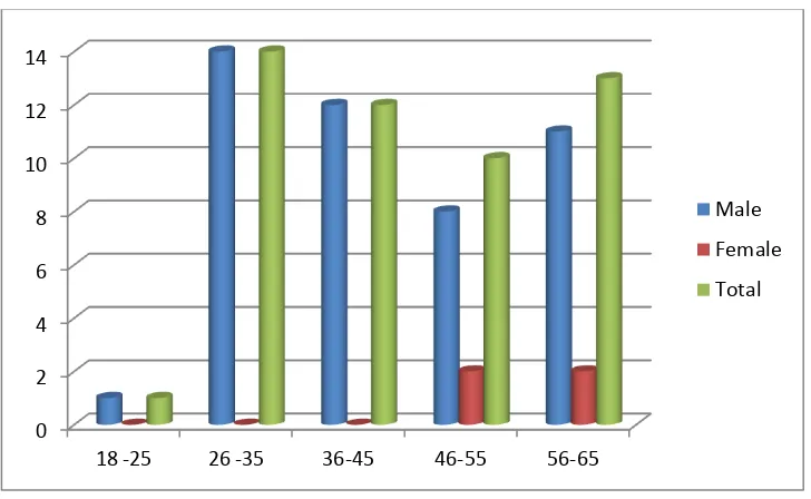
Table 1: Table showing age and sex wise distribution of the studied
population.
Age Male Female Total Percentage
18 -25 1 0 1 2
26 -35 14 0 14 28
36-45 12 0 12 24
46-55 8 2 10 20
56-65 11 2 13 26
Total 46 4 50 100
The studied population was between the age group of 18 to 65 years. The
majority of the patients were in the age group of 26 - 35 years. The mean
age of the patients was 44.8 years. Majority of the studied population were
males (92 %) with females constituting 8 %.
0 2 4 6 8 10 12 14
18 -25 26 -35 36-45 46-55 56-65
Male
Female
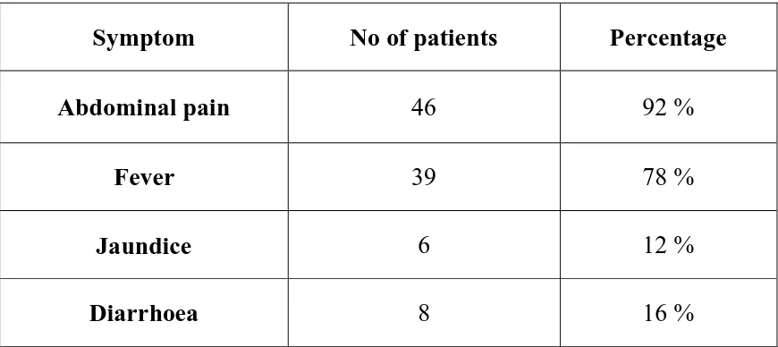
[image:59.595.100.464.397.621.2]Table 2: Table showing the symptoms associated with liver abscess in
the studied population.
Symptom No of patients Percentage
Abdominal pain 46 92 %
Fever 39 78 %
Jaundice 6 12 %
Diarrhoea 8 16 %
Abdominal pain was the most common symptom (92 %) present in patients
with liver abscess. Fever was present in 78 % of the patients.
0 5 10 15 20 25 30 35 40 45 50
Table 3: Table showing the most common signs associated in patients
with liver abscess.
Signs Number of patients Percentage
Tachycardia >100/min 22 44
Hepatomegaly 23 46
Rt intercostal tenderness 30 60
Right lower intercostal tenderness was the most consistent sign and was
present in 60 % of the patients while tachycardia (>100/min) was present in
44 % and hepatomegaly was present in 46 % of the patients.
0 5 10 15 20 25 30
Table 4: Table showing the signs and symptoms of liver abscess,
according to its aetiology.
Signs and Symptoms Pyogenic Amoebic
Abdominal Pain 97.22 % 78.57 %
Fever 77.78 % 78.57 %
Diarrhoea 11.11 % 28.57 %
Jaundice 11.11 % 14.28 %
Tachycardia >100/min 38.89 % 57.14 %
Hepatomegaly 41.67 % 57.14 %
Rt intercostal
tenderness 58.33 % 64.28 %
Abdominal pain was the most consistent complaint in both pyogenic and
amoebic liver abscess. Right lower intercostal tenderness was the most
consistent sign in both pyogenic and amoebic liver abscess.
0 20 40 60 80
100 Pyogenic
[image:62.595.93.527.148.438.2]Table 5: Table showing the number of patients with deranged
investigations in the studied population.
Investigations Total Percentage
Hb <9 16 32
TC >11000 38 76
Urea >40 20 40
Creatinine >1.4 2 4
76 % of the patients had a total count > 11,000. 40 % of patients had
urea > 40mg/dl and 32 % had haemoglobin < 9 g/dl.
0 5 10 15 20 25 30 35 40
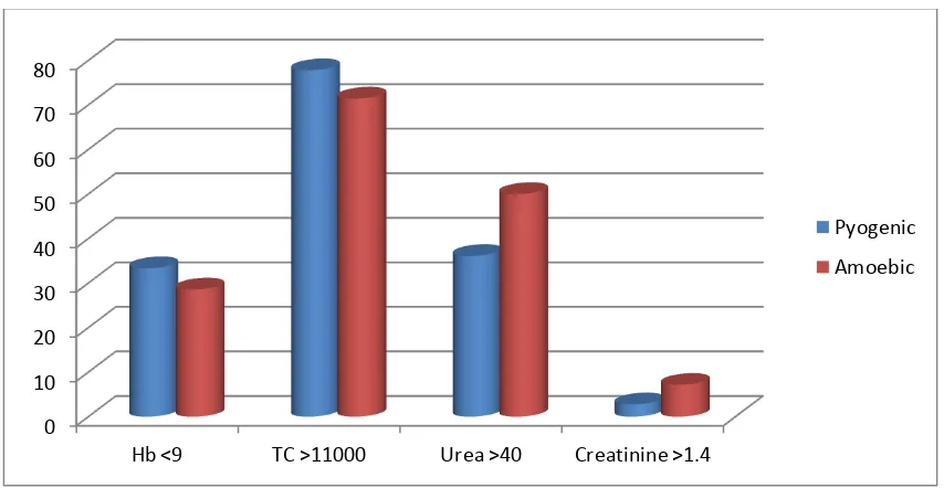
[image:63.595.101.540.384.601.2]Table 6: Table showing the number of patients with deranged
investigations in the studied population based on the type of liver
abscess.
Investigations Pyogenic Amoebic
Hb <9 33.33 % 28.57 %
TC >11000 77.78 % 71.43 %
Urea >40 36.11 % 50 %
Creatinine >1.4 2.78 % 7.14 %
Total count > 11,000 cells/mm3 was seen in 77.8 % and 71.4 % of
pyogenic and amoebic liver abscess respectively. Elevation of urea was
more common in amoebic liver abscess compared to pyogenic liver
abscess.
0 10 20 30 40 50 60 70 80
Hb <9 TC >11000 Urea >40 Creatinine >1.4
Pyogenic
[image:64.595.99.526.378.599.2]Table 7: Table showing the number of patients with abnormal liver
function test parameters.
Investigations Total Percentage
T Bilirubin >3 6 12 %
AST >40 42 84 %
ALT >40 46 92 %
SAP >115 17 34 %
Albumin <3g/dl 16 32 %
PT >20 s 3 6 %
Most of the patients had elevation of AST and ALT levels. 32 % of
patients had Albumin < 3 g/dl. 34 % of patients had elevation of Serum
Alkaline phosphatase.
0 5 10 15 20 25 30 35 40 45 50
T Bilirubin >3
[image:65.595.101.534.385.615.2]Table 8: Table showing the number of patients with abnormal liver
function test parameters based on the type of liver abscess.
Investigations Pyogenic Amoebic
T Bilirubin >3 13.89 % 7.14 %
AST >40 77.78 % 100 %
ALT >40 91.67 % 92.86 %
SAP >115 30.56 % 42.86 %
ALB <3 27.78 % 42.86 %
PT >20s 5.56 % 7.14 %
All patients with amoebic abscess had elevation of AST. Elevation of AST
and ALT were the most common parameter to be elevated in both amoebic
and pyogenic liver abscesses.
0 10 20 30 40 50 60 70 80 90 100
T Bilirubin >3
AST >40 ALT >40 SAP >115 ALB <3 PT >20s
Pyogenic
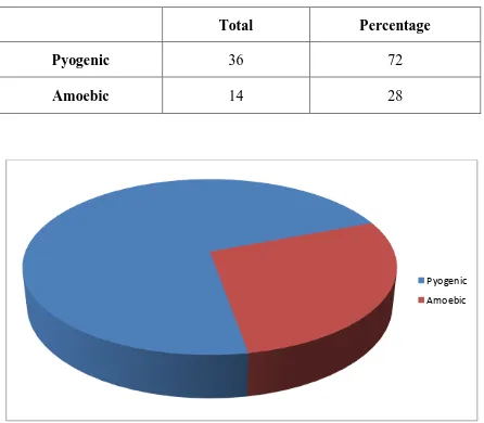
[image:66.595.100.533.401.627.2]Table 9: Table showing the distribution of patients based on the type
of liver abscess.
Total Percentage
Pyogenic 36 72
Amoebic 14 28
72 % of the studied population had pyogenic abscess while the remaining
28 % had amoebic abscess.
Pyogenic
[image:67.595.95.541.140.530.2]Table 10: Table showing the distribution of patients in the studied
population based on the lobe of involvement.
No. of Patients Percentage
Right lobe 43 86
Left lobe 4 8
Bilateral 3 6
Total 50 100
Involvement of the right lobe was seen predominantly in 86% of cases.
Isolated left lobe involvement was seen in 8 % of cases and 6 % had
involvement of both lobes of liver.
Right lobe
Left lobe
[image:68.595.98.533.176.599.2]Table 11: Table showing the distribution of patients in the studied
population based on the lobe of involvement and the type of abscess.
Pyogenic Amoebic
Right 83.33 % 92.86 %
Left 8.33 % 7.14 %
Bilobar 8.33 % 0
Predominant right lobe involvement was seen in both pyogenic and
amoebic abscess. Simultaneous involvement of both lobes was seen in
pyogenic liver abscess only.
0 10 20 30 40 50 60 70 80 90 100
Right Left Bilobar
Pyogenic
[image:69.595.101.518.318.577.2]Table 12: Table showing the occurrence of liver abscess as a solitary or
multiple lesions.
Total Percentage
Solitary 45 90
Multiple 5 10
Total 50 100
90% of the liver abscess occurred as a solitary lesion and the remaining 10
% had multiple lesions.
Solitary
Table 13: Table showing the occurrence of liver abscess as solitary or
multiple lesions based on the aetiology.
Solitary Multiple
Pyogenic 86.11 % 13.89 %
Amoebic 100 % 0
All of amoebic abscess in the studied population had a solitary lesion. All
the multiple lesions were of pyogenic origin.
0 10 20 30 40 50 60 70 80 90 100
Solitary Multiple
Pyogenic
Table 14: Table showing the distribution of patients in the studied
population based on the aetiology of pyogenic abscess.
Aetiology Number Percentage
Biliary 9 25
Sepsis 1 2.8%
Cryptogenic 26 72.2%
Total 36 100
72.2 % of the pyogenic abscess was of cryptogenic origin. Biliary origin of
abscess was present in 25 % of cases.
Biliary
Sepsis
[image:72.595.97.530.171.578.2]Table 15: Table showing the bacteria isolated from blood culture in
patients with liver abscess.
Bacteria Number Percentage
Klebsiella 10 47.6
E coli 6 28.6
Enterococci 5 23.8
Total 21 100
Blood culture was positive in 58.3 % of pyogenic abscess. Klebsiella was
grown in 47.6 % of cases. E. coli was grown in 28.6 % of cases.
Enterococci were isolated in 23.8 % of cases.
0 1 2 3 4 5 6 7 8 9 10
[image:73.595.99.532.260.591.2]Table 16: Table showing the bacteria isolated from the culture of pus
from patients with pyogenic liver abscess.
Bacteria Number Percentage
Klebsiella 10 52.6
E coli 9 47.4
Total 19 100
Pus culture yielded growth in 52.8 % of pyogenic abscess. Klebsiella was
the predominant organism, and was present in 52.6 % of cultures. E coli
constituted 47.4 % of the cultures.
8.4 8.6 8.8 9 9.2 9.4 9.6 9.8 10
Table 17: Table showing the average size and volume of the abscess
cavity based on the type of liver abscess.
Diameter Volume
Pyogenic 8.4 310
Amoebic 11.2 736
Amoebic abscesses were larger with mean diameter of 11.2 cm to pyogenic
abscesses with mean diameter 8.4 cm. The average volume of amoebic
abscesses was 736 ml compared to 310 ml of pyogenic liver abscess.
0 2 4 6 8 10 12
Pyogenic Amoebic
Diameter
0 100 200 300 400 500 600 700 800
Pyogenic Amoebic
[image:75.595.98.538.141.659.2]Table 18: Table showing the number of aspirations required in
successful treatment of liver abscess.
Number Percentage
Single 21 84
Twice 4 16
Percutaneous aspiration was successful in all the patients employed where
84 % were treated with a single aspiration and 16 % required aspiration
twice.
Single
[image:76.595.97.466.217.505.2]Table 19: Table showing the common complications encountered in
patients who underwent open surgical drainage of liver abscess.
Complications Number Percentage
Wound sepsis 12 48 %
Atelectasis 6 24 %
Bile leak 2 8 %
Total 19
76 % of the patients who underwent open surgical drainage developed
complications. Wound sepsis (superficial surgical site infection) was
encountered in 48 % patients who underwent open surgical drainage. Right
basal atelectasis was seen in 24 % of cases and 8 % cases had postoperative
bile leak.
0 2 4 6 8 10 12
[image:77.595.95.540.186.530.2]Table 20: Table showing the mean values of the investigations done in
patients with pyogenic and amoebic liver abscess.
Investigations Pyogenic liver
abscess S.D
Amoebic liver
abscess S.D
Hemoglobin 9.5 0.69 9.5 0.93
White Blood Cell
count 11.7 x 10
3
1483 12.3 x 10³ 1793
Urea 38.6 8.73 37 8.7
Creatinine 0.9 0.18 0.9 0.2
Total Bilirubin 2 0.8 2.23 0.68
SAP 103.4 26.58 119.1 34.6
ALT 49.8 12.72 89.1 23.72
AST 58.2 13.20 89.7 15.94
Albumin 3.3 0.35 3.1 0.34
Prothrombin time 15.4 2.73 15.8 2.42
Patients with amoebic liver abscess had a marginal increase in mean values
of WBC count compared to pyogenic liver abscess. The mean values of
serum bilirubin, serum alkaline phosphatase, AST and ALT were higher in
Table 21: Table showing the average size of liver abscess following
aspiration and at follow up.
Size of Abscess Percentage
At Aspiration 9.68 cm 100
At Discharge 4.76 cm 49.2
At 1 month of follow up 3.26 cm 33.7
At 3 months of follow up 2.15 cm 22.2
There was a 50 % reduction in the size of abscess following needle
aspiration at discharge. Most of the patients had a residual abscess cavity at
3 months of follow up.
0 1 2 3 4 5 6 7 8 9 10
At aspiration At discharge After 1 month After 3 months
[image:79.595.101.524.325.574.2]