JOURNAL OF VIROLOGY,Apr.1994,p.2230-2238 Vol. 68,No. 4 0022-538X/94/$04.00+0
Copyright C) 1994,American SocietyforMicrobiology
Identification
of Three Feline Immunodeficiency Virus (FIV)
env
Gene Subtypes and Comparison of the FIV and
Human
Immunodeficiency Virus
Type 1
Evolutionary
Patterns
DONALD L.
SODORA,l
EUGENEG. SHPAER,1 BARBARA E. KITCHELL,2 STEVEN W.DOW,3 EDWARD A.HOOVER,3 ANDJAMES I.MULLINS'*
Departmentof Microbiologyand
Immunology'
andDepartmentof ComparativeMedicine,2 Stanford University School of Medicine, Stanford, California, and DepartmentofPathology, Colorado State University, FortCollins, Colorado3Received27September1993/Accepted 17December 1993
Felineimmunodeficiencyvirus (FlI)is alentivirusassociatedwith AIDS-like illnesses in cats. Assuch, FIV appears to be a feline analogofhumanimmunodeficiencyvirus(HIV).A hallmark ofHIVinfection is thelarge degree of viral genetic diversity that can develop within an infected individual and the even greater and continually increasinglevel ofdiversityamong virus isolates fromdifferent individuals. Our goal in this study was to determine patterns of FIV genetic diversity by focusing on a 684-nucleotide region encompassing variable regions V3, V4, and V5of theFIVenvgeneinorder toestablishparallels anddistinctions betweenFIV and HIVtype 1 (HIV-1). Ourdata demonstratethat, likeHIV-1, FIVcanbeseparatedinto distinctenvelope sequence subtypes (three are described here). Similar to that found for HIV-1, the pairwise sequence divergencewithinanFIVsubtyperanged from 2.5to15.0%,whereas that betweensubtypes rangedfrom 17.8 to26.2%. However,thehigh number of synonymousnucleotidechanges among FIVV3 to V5 envsequences may also include a significant numberof backmutations and suggeststhattheevolutionarydistancesamong FIV subtypes areunderestimated. Although onlyafewsubtypeB viruses were available forexamination,thepattern ofdiversity betweentheFIVAandBsubtypeswasfound to be
significantly
distinct;subtypeB sequenceshad proportionally fewer mutations that changed aminoacids,compared with silentchanges, suggestinga more advanced stateofadaptationtothe host. Nosimilar distinctionwasevident forHIV-1 subtypes.Thediversity
ofFIVgenomeswithin individualinfected cats wasfound to be ashigh as3.7% yet twofold lower than that within HIV-1-infected peopleover acomparableregion ofthe env gene.Despite thesedifferences, significant parallels between patterns of FIV evolution and HIV-1 evolution exist, indicating that a wide array of potentially divergentviruschallenges needtobe consideredinFIVvaccine andpathogenesis studies.
Thestudy of lentiviral diversity hasgenerally focusedonthe envelope (env) gene, the most variable structural gene (32). The Env protein of human immunodeficiency virus type 1 (HIV-1) is an important antigenic target of the virus and encodes theprincipal neutralizingdeterminant(12, 26).HIV-1 sequences can be separated intophylogenetically distinct en-velope sequence subtypes(32). To date, seven HIV-1 subtypes have been identified: subtypes A and D are found predomi-nately in central Africa; subtype B is common in the United Statesand western Europe;subtype C iscommoninsouthern Africa, East Africa, and India; subtype E is common in Thailand and has been found in the Central African Republic; subtype F has been found in Brazil and Romania; and the newest subtype, 0, was found in Cameroon (7, 24, 29, 32). Becauseof thepotential need to generate geographically and temporally specificvaccine formulations, investigations to de-termine thediversity and representation of the various HIV-1 subtypesthroughout the world are under way.
The relationship between viral diversity and disease progres-sion and the impact of diversity on vaccine strategies can be addressed in a lentiviral animal model system. Feline immu-nodeficiency virus (FIV) was first isolated in 1986 from a cat with symptoms of an immunodeficiency-like syndrome (40) andisemerging as a useful model for understanding
immuno-*Correspondingauthor. Mailing address: Department of
Microbi-ology and Immunology, Stanford University School of Medicine, Stanford,CA94305-5402.Phone: (415) 723-0668. Fax: (415) 725-0678. Electronic mail address:jmullins@jmullins.stanford.edu.
deficiencydisease. LikeHIV-1,FIV caninduceimmunological abnormalities, includingadecline inCD4+Tcells(1, 14, 34), inversion of the CD4+/CD8+ T-cell ratio (1, 34), and de-creased lymphocyteproliferative response tomitogens (2, 48, 53). FIVinfection ofdomesticcatshasbeen found worldwide (16, 17), and related viruses have been isolated from wild members of the family Felidae (i.e., panthers, lions, and bobcats) (3, 36).
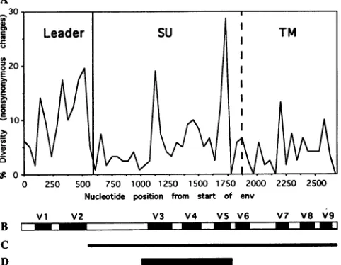
The primary translation product of the FIV env gene is processedthrough proteolyticcleavage into the surfaceprotein (approximately 433 amino acids) and the transmembrane protein (approximately 246 amino acids) (54). Pancino and colleagues have defined nine variableregions throughout the FIV Envprotein(38).The firsttwooccurin the leaderregion, which is also the firstcodingexonof the rev gene(41),and are not present in the matureEnvprotein because of proteolytic processing(54). VariableregionsV3 throughV6occurin the surface protein, and variable regionsV7through V9 occur in thetransmembrane protein.
The workpresented here buildsonprevious studies of FIV diversity within the domesticcatpopulation(25, 42, 43). Prior to this study, Rigbyandcolleagues (43) investigated FIV env gene diversity and found that the available sequences were nearly equidistant by genetic divergence (with the exception of oneisolate from Japan[27]).We evaluated 12 additional FIV sequences obtained in North America and show that theycan be divided into three distinctsubtypes(designatedA, B,andC) by the same criteria used to group HIV-1 strains, with the Japanese isolateservingasthe prototypeof whatwerefer to as 2230
on November 9, 2019 by guest
http://jvi.asm.org/
TABLE 1. FIVstrains evaluated in this study
No.of cells/pl
Subtype and strain" Origin Clinical symptom(s)"
CD4 CD8
A
USCAlemy()A Fremont, Calif. ND" ND wt loss,01
USCAhnkyOOA ElCerrito, Calif. ND ND wt loss,01, diarrhea
USCAtt__(A Oakland, Calif. 108 907 Wtloss, behavior
USCAzepyOOA San Francisco area, Calif. 392 241 None
USCAsam_(OA Albany,Calif. 789 252 Behavior
B
USOKlgrl(OB Tulsa,Okla. 675 114 None
USILbrnyOOB Chicago,Ill. 40 148 None
USMOglwdOOB Kansas City, Mo. 602 345 None
USMAsboyOOB Salem, Mass. 999 1,518 None
USTXmtexOOB Arlington, Tex. 682 1,981 None
C
CAEBCpbarOOC Vancouver, British Columbia, Canada 190 519 None
CABCpadyOOC Abbotsford, British Columbia, Canada 77 50 wtloss, lethargy
"The first two capital letters indicate the country of origin; the next two designate the location within the country (state or province); the four lowercaselcttersor
numbers refer to the name of the cat, virus strain, orisolate; the two numbers represent a specific viral isolate or clone (00 represents the consensussequence);and the last letter represents the subtype assigned to the virus. Forshort names, an underline is inserted so that thecorrectspacing ismaintained. This nomenclature evolved from the HIV-1nomenclature currently being developed (19).
"O,opportunisticinfections;behavior, abnormal motor behavior potentially indicative of neurological damage. ND, not determined.
subtype B. Parallels and distinctions between the patterns of FIV diversity and HIV-1 diversitywere alsoestablished.
MATERIALSAND METHODS
Viral DNA isolation.Whole-blood sampleswereobtainedin heparinized tubes, and peripheral blood mononuclear cells were separated by Ficoll-Hypaque density centrifugation. For two samples (USCAlemyOOA and USCAtt_OOA), DNA was extracted from lymph nodes at necropsy. High-molecular-weight DNA was purified by proteinase K digestion and phenol-chloroform extraction (44).
PCRamplification. PCRswereperformedin a mixture(100
p.l
total) containing10 mMTris(pH 8.3), 50mMKCI, 1.7mM MgCl, 1% dimethyl sulfoxide, 200 FM deoxynucleoside triphosphates,2.5 Uof Taq polymerase (Perkin-Elmer Cetus), 10 pmol of each amplimer, and a dilution of cellular DNA corresponding toone FIVprovirus (31). Tworoundsof PCR (94°C for 45 s, 55°C for 1 min, and 72°C for 2 min 10 s; 30 cycles) in aPerkin-Elmerthermocyclerwere usedtogenerate a 2,048-nucleotide fragment encompassing the surface and transmembrane portionsof theFIVenvgene. The first-round amplimers were Fenv23 (5'GCGCAAGTAGTGTGGAG ACT) (corresponding to bp 6788 to 6807 of the JapanTM2 genome [27])and Fenv22(5'GCTTCATCAYTCCTCCTCTT)
(correspondingtobp8836to8817of theJapanTM2genome). The second-round amplimers were a combination of either
Fenv27 (5
'GACTGGAATTCGCGCAAGTAGTGTGGAGA
CTTCCCCCTTTA)
(corresponding
tobp6788 to6817 ofthe JapanTM2 genome; with viruses from subtypes B and C) or Fenv3 1 (5'GACTGGAAT1CGCTCAGGTAGTATGGAGA
CTTCCACCAYT)
(corresponding tobp 6784 to 6812 ofthe Petaluma FIV-14 genome; withviruses from subtype A) and Fenv24 (5'GACTGGGATCCTCATCATTCCTCCTCT11TT11 CAGA) (corresponding tobp 8836to 8810of theJapanTM2
genome). Although no cloningwas performed in the experi-ments outlined here, restriction enzyme sites
(underlined)
were includedtofacilitatecloningofthefull-lengthenvgenes.
DNAsequencing. The 12 newamplifiedFIV env genes were sequenced directly. In order to ensure that each sequence originated from only one FIV proviral genome template, dilutions of the infected peripheral blood mononuclear cell DNA with which less than half of the PCR amplifications resulted in a positive signalwere used. The existence of only one FIV genome was further verified by a heteroduplex mobilityassayin whichthe presence oftwo ormore divergent sequences, having a single insertion or deletion, or of point mutations resulting in a mismatch of greater than about 2% would be detected through the formation of slowly migrating heteroduplexes in acrylamide gels (7). Amplified DNA frag-ments werepurified byagarose gel electrophoretic separation andSpin-X centrifuge filtration (Costar). Each fragment was sequenced by using an Applied Biosystems automated se-quencerand adye-deoxy terminator procedurespecified by the manufacturer.
Sequence analyses. Overlapping sequences werejoined by using the Gel program from the Intelligenetics suite (5). Nucleotide divergence (distance) for pairs of sequences was estimated by using the maximum likelihood method and the DNADISTprogram from thePHYLIPsoftwarepackage(10). Onthe basis of theoutputfromDNADIST,phylogenetictrees wereconstructed by using the FITCHprogram,andbranching orderreliabilitywasevaluatedby bootstrapanalysis (by using the DNABOOT program [10]).
KA,
thefrequency
ofamino acid replacement substitutions perreplacement site, andKs,
thefrequency of substitutionspersilentsite,weremeasuredby usingthe method and programdeveloped byLi (21, 22).
Viral sequences. Thenewlydescribed FIV strains evaluated in this studyoriginated from naturally infected North Ameri-can petcatsandare describedinTable 1. GenBankaccession numbersfortheV3toV5FIVenvgenesequencesareU02392 through U02422.Other FIV sequences used inthisstudyare as
follows (GenBank accession numbers and
geographic
origins
are in parentheses): CA.Petaluma clone FIV-14
(M25381)
(Petaluma, Calif. [35, 52]); CA.PPR
(M36968) (San
Diego,
Calif. [42]); CA.Dixon (L00608) (northern California
[55]);
on November 9, 2019 by guest
http://jvi.asm.org/
2232 SODORA ET AL.
JapanTM2 (M59418) (Tokyo, Japan [27]); SwissZl (X57002) and SwissZ2 (X57001) (Zurich, Switzerland [30]); DutchKl (M73964) and DutchK32 (M73965) (Amsterdam,the Nether-lands [49]); DutchUtr (X60725) (Utrecht, the Netherlands [54]); FranceWo (L06312) (France [28]); ScotUK2 (X69494) (Perth, Scotland [43]); EngUK8 (X69496) (Portsmouth, En-gland [43]); WalesUK14 (X69497) (Colwyn Bay, Wales[43]); Dutch4 (X69498) and Dutch6 (X69499) (Amsterdam, the Netherlands [43]); and ItalyMl (X69500), ItalyM3 (X69502), and ItalyM4 (X69503) (Pisa, Italy [43]).
Foreach HIV-1 subtype,thefollowing sequenceswereused (GenBankorreferencenumbers are inparentheses): subtype A,U455(M62320), Z321 (M15896), SF170 (M66535), 1UG06 (M98503), KIG93 (L07082), and D687 (32); subtype B, ALA1 (M38430), BRVA (M21098), SC (M17450), JH32 (M21138), CDC42 (M13137), OYI (M26727), SF2 (K02007), JFL (M31451),RF(M17451), SF162 (M65024), JRCSF (M38429), LAI (K02013), BAL2 (M68894), MN (M17449), NY5NEW (M38431), ADA (M60472), WMJ2 (M12507), 537-lpre and 1058-lpre (6), and HAN (32); subtype C, NOF (L07426), ZAM20 (L03707), D1044 (L07651), D747 (L07653), D757 (07654), D760 (L07655), and IND744, IND766, and IND868 (32); subtype D,ELI(K03454), NDK (M27323), JY1 (J03653), MAL(K03456), UG23 (M98504),Z2Z6(M22639), Uganda-9 (13), and U44342 (32); and subtype E, TN235 (L03698), TN239 (L03699), TN241 (L03700), TN242 (L03701), TN2432 (L03703), and TN244 (L03704). Intrapatient diversity of HIV-1 was determined with 13 sequences from patient MA (M79342toM79354) (20), 15 sequencesfrompatientBU (6), 6sequencesfrompatientPE(6), and 5 sequences frompatient JO (6).
RESULTS
The viruses introduced in this study originated from adult domestic catsfrom the United States and southwestern Can-ada(Table 1). Data obtained with uninfected adultcatssuggest that in general, T-cell counts below 200 cells per ,ul are abnormally low (15). According to this criterion, 4 of the 12 cats examined had abnormally low CD4+ T-cell levels at the time of sampling. Some of the cats displayed symptoms of immunodeficiency disease (weight loss and opportunistic in-fections), and two (USCAlemyOOA and
USCAtt-OOA)
had died because of complications ofimmunodeficiency. Clinical symptomswereevident infive cats, includingtwoof the three catswith the lowest CD4+ T-cell numbers (Table 1).Variableregions withinFIV envgenes.Figure1Adepicts the amino acid variation across the FIV env open reading frame determined for the nine full-length env genes currently avail-able. Variable regions defined by Pancino and colleagues (38) are indicated (Fig. iB). To obtain env genes directly from feline infected-cell DNA,anested PCR protocol (31) was used toamplify singleFIVproviral templates from peripheral blood mononuclear cells or lymph node cells. The entire coding sequence of the mature Envprotein was amplified (Fig. 1C). Thenucleotide sequence of the 684-nucleotide region encom-passing themostvariable region in the env gene, V3 through V5,wasthen determined (Fig. 1D).
To assessthe variability of the env gene, the 18 sequences available from GenBank were included along with a total of 16 sequencesfrom the subtypes described in Table 1 (Fig. 2). As expected, ourresultsdemonstrated a high degree of diversity within the predicted 228-amino-acid region (Fig. 2). This region contains 13 N-linked glycosylation sites, the majority of which were not conserved in all viruses. V5 was the only segmentof theenvgenewherelength variations were detected.
A
B
250 500 750 1000 1250 1500 1750 2000 2250 2500 Nucleotide position from start of env
Vi V2 V3 V4 VS V6 V7 V8 V9
3mK:3mK3
C D
FIG. 1. Variableregionsof the FIV Envprotein. (A) Variability plot generated from the nucleotide sequence alignment of nine full-length FIVenvgenes(each from adifferentcat) obtained from GenBank. The sequences used to generate the plot were CA.Peta-luma, CA.Dixon, CA.PPR, FranceWo, SwissZl, SwissZ2, DutchKl, DutchUtr, and JapanTM2. Points on the graph were generated by usingawindowof 45 nucleotides, and onlynonsynonymouschanges (those which resulted in anamino acidchange) were included. (B) Variable regions of the FIV env gene identified by Pancino and colleagues(38).(C) Region ofthe FIVenvgenePCRamplified in this study.(D) Region of theenvgenesequencedandcharacterizedin this study.
Inaddition, therewere 13cysteineresidues(Fig. 2)conserved in all sequences except ItalyMl, in which threewereabsent, and ItalyM3, in which one was absent (Fig. 2). All env gene segments examined contained intact open reading frames, except the CABCpbarO7C sequence, which hadone termina-tion codon.
Phylogenetic groupings. An unrooted phylogenetic tree generated for all 34 sequences from V3toV5 is shown inFig. 3. Thisanalysisreveals thatFIV env genes fall within one of threeclusters,heredesignatedasenvelope sequencesubtypes A, B, and C. TheAsubtype consists of the original isolate from Petaluma plus all other viruses from California and several Europeancountries. The only isolate highly divergent from the isolate from Petaluma inprevious studies was the TM2 isolate fromTokyo, Japan(27).TM2isnowviewed as the prototype of what we refer to as subtype B, which includes sequences from the central andeasternUnited States. Both viruses from subtype Cwerefrom southwestern Canada.
In assessing the reliability of the branching order of the phylogenetictreeshown, the data were subjected to bootstrap analysis(9).Thebootstrap method recreates the phylogenetic treemultiple times, each time after swapping some characters between sequences, and records the number of times each group of sequences is placed together on the same branch. Branching is considered significant if it occurs in at least 95% of thebootstrap estimates (9). Results from bootstrap analysis of the major branch points are presented in Fig. 3. These analyses demonstrate that sequences within each of the three subtypes aremonophyletic (they cluster together) in a signifi-cantproportion of the analyses (99 to 100 of 100).
The diversity of the V3-to-V5 region of env between FIVs from the same subtype rangedfrom 2.5 to 15% and did not J. VIROL.
on November 9, 2019 by guest
http://jvi.asm.org/
[image:3.612.318.559.79.266.2]SvissZl...R...R.L...L.E...K...H.S..
IialyM3 ...D...I....R..GTD....P.H...L.V...K...TS...P...D..
ItalyM4...I....R..RTE...P...L.V....H...K...T....T...PP...D..
SWi3sZ2...A....R..STD...L..T.L.T...RPK...E..-.2....N...K....S..
FranceWo ...N....R..HTD-0.0...I...E.V...T... .Y.T..
ScotUK2 ....SD...I.R...R.OTD...T.L.T....K.K...E.RV....T...SD. EnqUJK ...I.R...R.STD...L.L...E.A.V...T.. Wale3UK14...D...R....R. .STD...K.... S...E...s..S. Italy4l G. ....HI.R...R.STV...L.L.0.. ...G.E...E..V..PG.T. .T....I.---RK.E.E...T..
USCAlemyOlA...L...OTD...L...K.. .V....E. V..K...Q.D..
USCAemyO2A...L...OTD...L.V...V.N...V..K...O.D..
USCAhnkyllA...I...RTE...L....R...E...V..K...L...T..
USCAhnky12A...I...RTE...L.T...R.G...E.V...K....R...L...T..
USCAtt__09A...S...I...OTO...R.T....R.G...E..V...R..T..
USCAtt__10A...S...R.Q....TD...R.T....R.G.E...E...V... ...T.. USCAzepyOlA...I.. .T....KTD...L.T....R.E...E.. V..K...T..
CA.Dixon ...I...lTD...I...E.R...E.. ...K...T..
USCAsa,._01A...Q...KTD...RV ...R.K...E. .V... ...M...T..
Dutch4 ...I....R..KTD...H....R...E... V... ..E...V...M.SA. CA.PPR...I....R..STN. ...Y...T.I.T....R.K...E...H... ...RF.T..
Dutchl ...K....O..5..TD..Q.0...I....R.K...R. .V...0...I...H.N..
DutchK32 ...K....O.S..QTD..Q...I....R...ER..V...0...I...
DutehUtr I...0.... N...RTD....0...I...G...ER..V...R...I...IR.NA.
Dutch6 I.R...R.QTD. ...0...H....I.T...R...VNER.VV...R...KI...R..NA.
JapanTM2 ..L.K.S.... ...V..DT....N.T.Q.H...S...I.T...E...S.. .YD.Q ...R.S.. USOK1qrlD2B .L.K.S...A...A.N...N ...K...K.I....R.K...E...YD.Q....Q....YS.. USILbrnyO3B .L.K.S.... ...V...A....N.T.Q.H...S..I.T...E...R...S..YD.0Q...R.S..
USMo0g1d03B .L.K.S.R...R...A....N.T.Q.Y...I....RH.D... ..I....R...S..SD...Y....T.TGT
USMAsboyO3BA.L...A.QTD...OT.. .0...I.N.A...E...S..YD...Y...N..G.
USTXmtexO3B .L.K...K....R..KTD. .Q.0...I...R.K...E...R...S..YD.0...Y..T.RR.TGT
CABCpbarO3C ..L.K.D.0...A...N.T.0...R.T... R.0.1....,... KY.K.N...AS.F.DI...D..T.PT.R.T..
CAECpbarO7C ..L.K..D...A...Q.N.T.0...T.R.T....R.W ...L.... AS.F.DI...D..APT.R.T..
CABCpadyO2C ..L.KN.D...A.R...R.K.D.Q.Q...N.I....R.K...ER...S...D.V...RR.TD.
4 4 ~~~~5/344 30/34 31/34 429/34
457 570
CA.Petaluma RFRIRCRWNVGSNTSLIDTGT SAPDTYNMNSOGFMVDIHNKAEYIGWCSLPSGMCCNSSS-TMCSR
SWisaZ ... ...D...V..S.T..K...K...
ItalyM3 .... ...D...T...V...T ..K..H..RRTR.--..I..K... ItalyM ... ...D...S...S.T...V..R...K...R.TR.--..I..TK...
Swis.sZ2 ...DK.D...K..N.L...A.R...I..V....T...PT...TS.SSGN....GDK. FranceWo 0... ...D...E..N..R...A.R...KT...K..PT...TN.GT.--IR...R.Q. ScotUK2 ...D...DPN...A.R...T...T.TO... TD---N....KRO.
EnqUKS ... .E.D...E.N...A.R...T....0...PT...TN.---V....K.0.
Wale3UKl4 ...D...E..N.DHO...A...L...T...PT...GTTT---NQ...GDH.
Italyt4l ...GD...E. .N...A.R...T...K.T...T TH---V...R.K.0. USCAlemyOlA...E.N.A...PNI...V...T...PT...TDDA----N...
USCA1emyO2A ..K....E.N.A...K.PHI...A...T...PT...TDGT----EK...
USCAhnkyllA...N.A...N...A...T...PT...TNNPT---G...0.
USCAhnkyl2A...N.A..0..D.N...A...T...PT...TNNLD---SR....0.
UJSCAtt__09A...K.K.A...E.N.... H.8....A...T...PT...RTNPDGT--A....KQ. USCAtt_10A...K.K.A...E..N... ...A...T...PT...GT.GI..--...KQ.
USCAzepyOlA...N.A...RNI...A...T...PT...N.NDS----N.0....O CA.Dixon ...N.A...PD...N....A.N...I...T...PT...K...N.DDTR---G....RTQ.
USCAsam_01A...E.D.A...E..N.T ...A.E...T...PT....K...VKDNFT---K..E....0. Dutch4 ...E...E..N....K...A...S...T...OE...GTE.NNS--N....R.Q.
CA.PPR .0...D...KNLN...A...T...K..ON...GT.ND----N....EDK.
Dutchl ...E.N.N...K..N.L...A.R... 0. ...T... M...TN....K....DT.N`NHT--I..E .EEK. DutchK32 ...E.N.N.K...K.N ...A.R...0 ...T....N....M..TN...K.O-.T.KN`YT--I..E..EEK. DutchUtr ...E.D.N.E...E.N...A....0..D...T...M...TE...DT.NN`NT--R..K..KEN. Dutch6 ...E.N.N....T.KN...A.R....D..II...T... M...TN ...P----D.0 .E..AD.
JapanTM42 .K... ..E.N.I...TNPN.T...KA.T...D ....VE...V...T....LL...KG...GTDNSEK-..-...K.O.
USOK1qrlO2B....K..E.N.I...TNPN.TR...KA.T.O...S....IE..VV...T...KG...GTDNSG---P..T..K.Q.
USILbrnyO3B....K..E.N.I...TNPN.T...KA.T...S....IE...VV...T...KG...GTNNN----N....K.Q. USMoglwdO3B...E.N.A...TNPN.T...KASIL....0....E... V...T...KG...GTNDN--- E..KAO.
USt4AsboyO3B....K..E.K.I...TNPN.TR...KA.T...SS.N..IE...VV...T...KE...T.HIA---P..E..Q.N. USTXmtexO3B....K..E.N.A...TDPN.T.N..N.LKA.T...S....IE...V....T...KE...KK....TK.E.IG---. .T. ..DTA. CABCpbarO3C... KDKNI...N. D.VTL . E. TR.K....KD...K.KDANAT---E. K. AED.
CABCpbarO7C... KDltGI...N.D.AKTL.... . TR.K... KD..E.K.K ATEYV---G..K..AED. CABCpadyO2C....K. KDKNIT. TAKTL... .. T. K .K... TE....K. SK.ET---D. K. .AKD.
33/34 25/34 2/34 33/34 32/34 34/34 34/34 8/34
_________________
4
4
44~~~~~~~~~~~~~~~29/34
4
[ ~~
~~V4
1 -Vs
FIG. 2. Deduced amino acid sequences ofFIV Env overtheregion sequenced, beginning342amino acids from thestartof the leader. Each hatched boxindicates the location ofanN-linkedglycosylation site,and the numbersunder each box indicate the ratio of the number of sites presenttothe total number of sequences examined.Arrowsindicate thepositionsofcysteine residues,andarrowswith linesthroughthemindicate cysteineresiduesnotconserved in all sequences(absentfrom eitherItalyMIaloneorbothItalyMIandItalyM3). Openboxes indicatethe positions of V3throughV5asdefinedbyPancino andcolleagues(38). Twenty-fivesequences(from21cats)fromsubtypeAaregroupedabove six sequences (from 6cats)fromsubtype Bandthree sequences (from2cats) fromsubtypeC.
2233
on November 9, 2019 by guest
http://jvi.asm.org/
[image:4.612.87.541.53.668.2]2234 SODORA ET AL.
FIG. 3. Phylogenetic analysis of FIV env gene
includes 18 viruses for each of which a684-nucleoti4
through V5)wasavailable from GenBank and 16 vira
thisstudy,including those from each of the subtypes di 1.Thediversity within infectedcatsisrepresentedby divergent sequences obtained from each cat
USCAhnkyOOA,USCAlemyOOA, andCABCpbarOOC obtained from the same cat by Siebelink and coll presented (DutchK) (49). Only horizontal lines ar
assessing divergence; the distanceonthetreewhichcc
divergence isindicated. To evaluate the consistency neticgroupings, the dataweresubjectedtobootstrap number at each branch point indicates the numb
sequencestothe right of thebranch of thetreewere
bootstraprepetitions.
overlapwith viral diversity betweendifferent s
ranged from 17.8 to 26.2% (Table 2). This diversity is comparable to the diversity obsen between HIV-1subtypes (see Fig. 6).
FIVenvgenevariation within infected anim;
assess the diversity of the FIV quasispecies
multiple viruses were obtained from four c
Thetwo mostdivergentsequencesfrom eachca
in Fig. 2 and 3 (USCAlemyOlA and
TABLE 2. Diversityof FIVenvgene segmen
differentcats
Avg.% diversity of subtype (ra Subtype
Subtype A SubtypeB
A 9.8(2.5-15)
[image:5.612.60.298.71.365.2]B 21.3(18.7-24.7) 11.1(3.3-14.5) C 22.9(20.3-26.2) 20.7(17.8-22.6)
TABLE 3. Diversity of FIV env gene segments within the samecat
Avg.%
Strain No.ofsequences diversity (range)
USCAlemyOOA 6 1.1(0.0-2.1)
USCAhnkyOOA 4 1.3(0.9-1.9)
USCAtt_OOA 6 1.8(0.2-2.8)
CABCpbarOOC 7 2.5(1.2-3.7)
EuropeandUSA
A(California)
USCAhnkyllA and USCAhnkyl2A, USCAtt_09A and
USCAtt-1OA,
and CABCpbarO3C andCABCpbarO7C). env genes obtained from the same cat were usually more closely related than those fromdifferent animals.Inall, fourtoseven different env genes were obtained from each cat, and the diversity ranged from 0 to 3.7% and averaged 1.1 to 2.5% (Table 3). IntracatdiversityofFIV wasapproximatelyhalfof thatobserved in HIV-1-infected subjects (see Fig. 6).Relative numbers of amino acid and silent changes. The phylogenetic analysis described above evaluated nucleotide divergence, whichincludes both amino acidand silentchanges. The
Ks
and KA have been examined previously in order to B Japan and USA understand the selective forces which affect lentiviral evolution (notCalifornia) (4,43),including that of FIV(43). In thisstudy,wecompared theKS
and KA values obtained for pairs of FIV env gene sequences from different cats (the 684-nucleotide region in-CBritishColumbia,cluding
V3toV5)
with thoseobtainedforacomparable region
Canada (V3 toV5)
of HIV-1 envgene sequences(Fig.
4),
each asafunction of nucleotide divergence.
KS
values were high(ap-desequence(V3
proaching
1)
forpairs
ofFIVsequences when thenucleotideLe
sequencesfromdivergence
exceeded
15%
(Fig. 4A), suggesting
that the
rate
of
escribedinTable back mutation was partially counterbalancing forward muta-two of themost tions. Thus, at about 15% overall nucleotide divergence and (USCAtt_OOA, above
(between
env sequence subtypes), DNA distances are ). Twosequences likely to be underestimated. In contrast, theKs
values for leagues are also HIV-1 sequence pairs were lower; the majority did not exceed re meaningful in 0.5 and thehighest was 0.75 (Fig. 4A). If one assumes that the )rrespondsto5% reverse transcriptase error rates for these two viruses are lof thephyloge-
similar, then this result implies that FIV has been in cataraofsims
(9That
populations
longer
than HIV-1 has been in humans and has er oftvmes
that therefore accumulatedrelatively
more silent mutations. Incontrast,the KA valuesobtainedfor FIV sequences werequite similartothose obtained forHIV-1sequences. However, when thenucleotidedivergenceexceeded15%, the values for HIV-1 sequences were generally higher, indicating somewhat more ubtypes,
whIch
replacement substitutions per replacement site for HIV-1 than;level of FIV for FIV.
ved within and Ininvestigating the evolutionary pressures to which any pair of sequences has beensubjected, the values of both
Ks
(which als. Inorderto generally reflects the length of time the two sequences have,awithin
a cat, been evolving apart) and KA (amino acid replacement sites)vats
(Table3).
are important. In this study, we examined these values as a it arepresented ratio(KA/KS)
inorder to analyze thedifferent FIV and HIV-1JSCAlemyO2A,
subtypes (Fig. 5).KA/KS
ratios of greater than 1 reflect some positiveselection for aminoacid change (4, 43). In comparison, theKA/KSratio ofbeta-globin sequences is 0.27, which reflects tsbetween thehigh degree
of amino acid conservation observed for most eucaryoticgenes (22). Inexamining the KA/KS ratios for the FIV subtypes,wefound that only seven of the sequence pairs nge): hadaKA/KSratio of>1.0, each from acomparison of closely SubtypeC relatedviruses(<7.5% divergence) (Fig. 5A).
Therefore, the majorityof sequence pairs studied exhibited no clear evidence for positive selection for amino acid change. Rigby and col-10.5 (9.9-11)leagues
described evidence forpositive
selection forchange
in FIV env when the region analyzed was restricted to theJ. VIROL.
on November 9, 2019 by guest
http://jvi.asm.org/
[image:5.612.314.555.88.160.2] [image:5.612.57.298.658.724.2]A
0o9
08 0.7 0.6
Ye
0.5-04
0.3-0.2
-0.1. 0*
0.3
B
0.25-
0.2-<
0.15-0.1
-0.05
A 4
3.5-3
2.5-
2-1
.5-
0.5-A\ c)
Il
0.5 -1
0.5-~
30
%Nucleotide Divergence
FIG. 4. Silent and amino acid substitutions as functions of total
nucleotide divergence. Only viralsequencesobtainedfrom
epidemio-logically unrelated FIV- and HIV-1-infected subjects (within and betweensubgroups butnotintrasubject)wereused for these analyses. KSandKAwere estimatedby the method described by Liet al.(21, 22). TheKs(A) and KA (B) values relativetotheamountof nucleotide divergence for bothFIV(Ol) and HIV-1 (.)arepresented.
variable regions, with V4 having the highest proportion of changesat amino acidreplacement sites (43).
The range ofKA/KS ratios appeared tovary between FIV
subtypes (Fig. SA). When these data were subjected to the Mann-Whitney nonparametric test and a normal
approxima-tion, theKA/Ksratioswerefoundtobe significantly lower (P
<0.001)forsubtypeBthan forsubtypeA. Thisresultsuggests
that the evolutionary pressures that B subtype viruses were
subjectedto differ fromthose affectingAsubtypeviruses. No subtype-specificpatternfor theparallel analysisof HIV-1 was
noted (Fig. SB).
DISCUSSION
FIV sequences previously described were predominately
from what we can now define as FIV envelope sequence
subtype A,with theexception of theJapanTM2 (27) andthe Maryland MDisolates(36) (FIVMDwasnotexamined in this studysinceonlythepolsequenceisavailable).Thedistribution ofFIVsubtypesexhibitedsomegeographic clustering. Subtype
Awasfound in California andEurope,whereassubtypeBwas
found in Japan and the central and eastern United States. Subtype Cwasdefined bytwo sequences found in
southwest-ern Canada. Geographic clustering of sequences was also evident within subtypeA (e.g., Dutch viruses and California viruses) (Fig. 3),while results obtainedwithin subtype Bare moredifficulttointerpret.Forexample,thesimilaritybetween
10*
- I 15- *
5 10 15 20 25 30
%Nucleotide Divergence
FIG. 5. KA/Ksratios and total nucleotide divergence of FIV (A) andHIV-1(B)envgenesegments.Only viralsequencesobtained from
epidemiologically unrelated FIV- and HIV-1-infected subjects (within
and betweensubgroups butnotintracat)wereused for theseanalyses.
The KA/Ksvalues relative tothe amountof nucleotide diversity for each pair of sequences within subtypes, as well as for between
subtypes,arepresented.
JapanTM2 and USILbrnyO3B from Chicago, Ill., is quite striking,considering theirgeographic distance. Weknow that this result was not due to contamination since neither the JapanTM2isolatenoritsclonehas everbeen in our
laborato-ries.
Theseresultsobtained forFIVcanbecompared with those
for the simian immunodeficiency virus SIVAGM, a primate
lentiviruswhichcanalsobedivided intophylogenetic subtypes
(18, 23). SIVAGM subtypes coincide with different African
green monkey species in geographically separate areas of
Africa.SIVAGM subtypesexist within cleargeographic bound-aries, whereas this does not appear to be the case for FIV
subtypes, suggesting an earlier entry into and/or the clearly greater mobilityof the host (feline) species.
Majorinterest in FIV derives from itsimportanceas amodel
forAIDS in humansaswellasitsimportanceas adomesticcat
pathogen.Wethereforesoughttodetermineparallelsbetween theenvdiversity of FIV and that of HIV-1. The regionofthe HIV-1envgenechosenforcomparisonincluded theregionsof thesurfaceprotein codingsequence fromV3 through V5; the regionis similar insize, location, and relative levelofdiversity
tothe FIVenvgenefragmentanalyzed.A total of 47 different HIV-1envsequences obtained fromGenBank and represent-ingsequence subtypes A, B, C, D,and Ewereevaluated (Fig. 6). Parallels in FIV diversity and HIV-1 diversity can be
observedamongvirusesobtainedfrom differentsubjectsof the
samesubtype (2.5to 15% for FIV and 2to 19.5%for HIV-1) FIV Subgroup A
0 SubgroupB A SubgroupC x BetweenSubgroups
. u;
C i' a U S-
~~~~~~x
5 10 I . .20
5 10 15 20 2
H IV-1
oSubgroup
A
Subgroup B
A SubgroupC o SubgroupD
v v SubgroupE
x BetweenSubgroups
0
V
VV A~~
A A r
y
~~~~~W4
on November 9, 2019 by guest
http://jvi.asm.org/
[image:6.612.331.558.75.369.2] [image:6.612.62.305.78.370.2]2236 SODORA ET AL.
O:r 5 10o 1 5 20 25
a)
O 80 B HIV-1
0)
50
30
0 5 10 15 20 25
% Nucleotide divergence
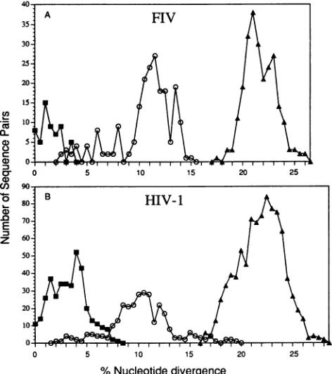
FIG. 6. FIV (A) and HIV-1 (B) diversity within and between
infected individuals. Each symbol indicates the number of sequence pairs at a givenlevelof nucleotide divergence. Data within a subject (U) and for subjects infected with viruses from the same (0) or different (A) subtypes are presented.
Or different subtypes (17.8 to26.2%o for FIV and 15.5 to 28% for HIV-1) (compare Fig. 6A and B). The observation that parallels in FIV diversity and HIV-1 diversity exist increases the perceived usefulness of FIV as a lentiviral model system in vaccine development and pathogenesis studies confounded by viral diversity.
Lentiviral diversity within an infected subject was also studied. Multiple viruses representing the pool of variants from four HI V-i-infected people (between 5 and 17 sequences per person) (20, 45) were compared with the pool from four FIV-infected cats (4 to 7 sequences per cat) (Fig. 6). The diversity of sequences of FIV from a cat was approximately half of that observed with sequences of HIV-1 from a patient (mean, 3.5%; range, 0 to 8%). The relatively lower level of FIV diversity within a cat was in concordance with a smaller data set obtained from another investigation (49). However, the level of HIV-1 diversity in humans is in largepart a function of the length of time the individual has been infected (39), and the length of time these cats have been infected is unknown.
The analysis ofKSvalues (proportion of the potential silent changes that have occurred) and KA values (proportion of the potential amino acid replacement changes that haveoccurred) provides information as to the selective forces which affect genetic evolution (21, 22). Previous studies have found evi-dence for positive selection for amino acid change (KA > KS) within lentiviral env genes (4, 33, 43, 46, 51). Previous studies examined the selective forces of FIV diversity by considering the variable and constant regions separately and established that a selection for amino acid change exists within some variable regions of FIV env genes, particularly V3 (37) and V4 (43). This selection is likely due to viral mutations resulting in
escape from immune
recognition. Indeed,
biochemical andimmunological
studies have since verified that the variable regionsofEnvaretargetsof the feline immune system(37, 50).
The
KA/Ks
ratioanalysis performed by Rigby
etal. utilized the FIV env gene sequencesavailable,
whichwerepredominately
of the Asubtype
(37, 43). They
foundevenstrongerselection for amino acid change within closely related sequences (termedsubgroups
[43])
of what we now refer to as the A subtype,butthey
wereunabletoevaluate differences between subtypes becauseonly
one member of the Bsubtype
was available foranalysis
(JapanTM2).
Theadditionalenvgene sequences
presented
here enableus to study theevolutionary
differences that exist between FIV subtypes. Bsubtype
viruses were found to havesignificantly
lower KA/KS ratios
(P
=<0.001), suggesting
apredominant
negativeorpurifyingselection rather thana
positive
selection for change. In aprevious study, Shpaer
and Mullins(46)
examined the selection for amino acid
replacement
or for silentchangeswithinprimate
lentiviruses which exhibit diversepathogenic phenotypes
in theirrespective
hosts(8, 11).
TheKA/Ks
ratios for the Asubtype
ofFIV resembled those for HIV-2 and were about two times lower than those for the pathogenic HIV-1,while theKA/KS
ratios for Bsubtype
FIV sequences weresignificantly
lower and were similar to those for therelatively
nonpathogenic SIVAGM
(46, 47).
Positive selection results from immune pressure exertedby
the infected host(4),
andtherefore,
our datapredict
arelatively
low immune responseandareducedpathogenicity
incatsinfected bysubtype
B FIVcompared
with that in cats infectedby
subtype
A. Itisthereforeinteresting
that in this limitedstudy,
none of the animals from which
subtype
B viruses were obtained hadevidence ofdisease, although
someofthem had low CD4+ cell numbers.Furthermore,
thesehypotheses
cannow be tested
through
experimental analysis
of theimmuno-genic
andpathogenic properties
of FIVs from different sub-types.KA/Ks ratioswerealsodetermined for five HIV-1
subtypes
(Fig.
5B).
We observed that eachsubtype
displayed
abroad range of KA/Ksratios,
with some sequencepairs
of agiven
subtype
exhibiting positive
selection for amino acidchange
and others not(ranges:
HIV-1subtype
A,0.64 to1.92;
subtype B,
0.23to3.7;
subtype C,
0.43to1.64;
subtype D,
0.55to1.5;
andsubtype
E, 0.49 to2.44). Therefore,
in contrast to results for FIV, nosubtype-specific
patternswere noted forHIV-1.The
finding
that FIV exists worldwide in the domestic catpopulation, the
relatively high
rate ofseroprevalence
among domestic cats,andthehigh
Ks
valuesobserved forFIVsuggest that FIV has been prevalent in catpopulations
longer than HIV-1 has been inhumans. These data support thehypothesis
that FIV and its host are more
adapted
to coexist than are HIV-1 and humans. Nonetheless,the breadth ofdiversity
that exists between FIV sequences suggests that a wide array ofchallenge
strainsareavailable forstringent
vaccineprotection
studies and for the
analysis
oflentiviralpathogenesis.
ACKNOWLEDGMENTS
WethankC. Mathieson-DuBard for excellent assistance insample preparation and T-cell enumeration; J. Brojatsch, J. Arthos, and E. Delwart for helpful discussion and comments; M. Bachman for
comments on the manuscript; B. Korber and G. Myers for their assistance in developing the FIV nomenclature; and W.-H. Li for providingthe computer program for theKAandKsanalyses.
This work was supported by a postdoctoral fellowship from the Giannini Foundation,Bank ofAmerica, toD.L.S., byPublic Health Service grantA133773 toE.A.H. andJ.I.M.,andbyagrantfrom the Morris Animal FoundationtoE.A.H.
J. VIROL.
on November 9, 2019 by guest
http://jvi.asm.org/
[image:7.612.60.296.80.346.2]REFERENCES
1. Ackley, C. D., J. K. Yamamoto, N. Levy, N. C. Pedersen, and M. D. Cooper. 1990. Immunologic abnormalities inpathogen-free cats experimentally infected with feline immunodeficiency virus. J. Virol. 64:5652-5655.
2. Barlough, J. E., C. D. Ackley, J. W. George, N. Levy, R. Acevedo, P. F. Moore, B. A. Rideout, M. D. Cooper, and N. C. Pedersen. 1991. Acquired immune dysfunction in cats with experimentally induced feline immunodeficiency virus infection: comparison of short-term and long-term infections. J.Acquired ImmuneDefic. Syndr. 4:219-227.
3. Barr, M. C., P. P. Calle, M. E. Roelke, and F. W. Scott. 1989. Feline immunodeficiencyvirusinfection innondomestic felids.J. ZooWildl. Med. 20:265-272.
4. Brown, A. L., and P. Monaghan.1988. Evolution of the structural proteins of human immunodeficiencyvirus: selective constraints onnucleotide substitution.AIDS Res. Hum. Retroviruses 4:399-407.
5. Clayton, J., and L. Kadef. 1982. Gel,a DNAsequencing project management system.NucleicAcidsRes. 10:305-321.
6. Delwart, E. L., J. Goudsmit, H. Sheppard, and J. Mullins. Unpublished data.
7. Delwart, E. L., E. G. Shpaer,F. E.McCutchan,J. Louwagie, M. Grez,H. Rubsamen-Waigmann, andJ. I. Mullins. 1993.Genetic relationships determined byaheteroduplex mobilityassay: analy-sis ofHIVenvgenes.Science262:1257-1261.
8. Doolittle,R. F.1989.Immunodeficiencyviruses: the simian-human connection. Nature (London)339:338-339.
9. Felsenstein, J. 1985. Confidence limits on phylogenies: an ap-proach usingthe bootstrap.Evolution 39:783-791.
10. Felsenstein, J. 1989. PHYLIP-phylogeny inference package. Cladistics 5:164-166.
11. Fultz, P. N., H. M.McClure, D.C.Anderson,R. B. Swenson,R. Anand, and A. Srinivasan. 1986. Isolation of a T-lymphotropic retrovirusfromnaturally infectedsootymangabey monkeys (Cer-cocebus atys).Proc. Natl. Acad.Sci. USA83:5286-5290. 12. Goudsmit, J., C. L.Kuiken,and P. L. Nara. 1989. Linearversus
conformational variationofV3 neutralization domains ofHIV-1 during experimental and natural infection. AIDS 3(Suppl. 1): s119-s123.
13. Hahn, B.(University of Alabama). 1992. Personalcommunication. 14. Hoffmann-Fezer, G., J.Thum,C.Ackley,M.Herbold, J. Mysliwi-etz, S. Thefeld, K. Hartmann, and W. Kraft. 1992. Decline in CD4+ cellnumbers in catswithnaturallyacquiredfeline immu-nodeficiencyvirusinfection.J. Virol. 66:1484-1488.
15. Hoover,E. A.Unpublisheddata.
16. Hosie, M. J., C. Robertson, and 0. Jarrett. 1989. Prevalence of felineleukaemia virusandantibodiestofelineimmunodeficiency virusincatsin theUnitedKingdom. Vet. Rec. 125:293-297. 17. Ishida,T.,A.Taniguchi,T.Kanai,Y.Kataoka,K.Aimi,K.Kariya,
T. Washizu, and I. Tomoda. 1990. Retrospective serosurvey for felineimmunodeficiencyvirusinfection inJapanesecats.Nippon
Juigaku Zasshi 52:453-454.
18. Johnson,P.R.,A.Fomsgaard, J. Allan,M.Gravell,W. T.London, R.A.Olmsted,and V. M. Hirsch. 1990.Simianimmunodeficiency viruses from African green monkeys display unusual genetic diversity.J.Virol.64:1086-1092.
19. Korber, B., and G. Myers (Los Alamos National Laboratory). 1993. Personalcommunication.
20. Kusumi, K., B. Conway, S. Cunningham, A. Berson, C. Evans,
A.K. N.Iversen,D.Colvin,M. V.Gallo,S.Coutre,E.G.Shpaer, D. V. Faulkner, A. deRonde, S. Volkman, C. Williams, M. S. Hirsch, anidJ.I. Mullins. 1992. Human immunodeficiencyvirus type 1 envelope gene structure and diversity in vivo and after cocultivationinvitro.J.Virol.66:875-885.
21. Li,W.-H. 1993.Unbiased estimation of theratesofsynonymous andnonsynonymoussubstitution.J.Mol. Evol.36:96-99. 22. Li, W.-H., C.-I. Wu, and C.-C. Luo. 1985. A new method for
estimating synonymous and nonsynonymous rates of nucleotide substitutionconsideringthe relative likelihood ofnucleotide and codonchanges. Mol. Biol.Evol.2:150-174.
23. Li, Y.,Y. M. Naidu, M. D. Daniel,and R. C.Desrosiers. 1989. Extensive genetic variability of simian immunodeficiency virus
fromAfricangreenmonkeys.J. Virol.63:1800-1802.
24. Louwagie, J., F. E. McCutchan,M. Peeters, T. P. Brennan, E. Sanders-Buell,G. A.Eddy, G.vanderGroen,K.Fransen,G.-M. Gershy-Damet,R. Deleys, and D. S. Burke. 1993. Phylogenetic analysisofgag genes from seventy internationalHIV-1 isolates providesevidence formultiplegenotypes. AIDS7:769-780. 25. Maki, N.,T.Miyazawa,M.Fukasawa,A.Hasegawa,M.Hayami,
K. Miki, and T. Mikami. 1992. Molecular characterization and heterogeneity of feline immunodeficiency virus isolates. Arch. Virol.123:29-45.
26. Matsushita, S.,M.Robert-Guroff, J.Rusche,A.Koito,T.Hattori, H.Hoshino, K. Javaherian,K.Takatsuki, and S.Putney. 1988. Characterization ofahumanimmunodeficiencyvirusneutralizing monoclonalantibodyandmappingof theneutralizing epitope.J. Virol. 62:2107-2114.
27. Miyazawa, T.,M.Fukasawa,A.Hasegawa,N.Maki,K.Ikuta,E. Takahashi,M.Hayami,andT.Mikami. 1991.Molecularcloning ofanovelisolateof felineimmunodeficiencyvirusbiologicallyand genetically different from the original U.S. isolate. J. Virol. 65:1572-1577.
28. Moraillon, A.,F.Barre-Sinoussi,A.Parodi,R.Moraillon,andC. Dauguet. 1992. In vitro properties and experimentalpathogenic effect of three strains of feline immunodeficiencyviruses (FIV) isolated fromcatswith terminaldisease.Vet.Microbiol. 31:41-54. 29. Morgado, M. G., E. C. Sabino, E. G. Shpaer, V. Bongertz, L. Brigido, M. D.C. Guimaraes,E. A. Castilho,B. Galvao-Castro, J.L.Mullins, R M.Hendry, and A.Mayer.V3region polymor-phisms in HIV-1 from Brazil: prevalence of subtype B strains divergent from the North American/European prototype and detection ofsubtype F.Submittedforpublication.
30. Morikawa, S., H. Lutz, A. Aubert, and D. H. Bishop. 1991. Identification of conserved and variable regions in the envelope glycoprotein sequences of two feline immunodeficiency viruses isolated inZurich,Switzerland.Virus Res. 21:53-63.
31. Mullis,K.B.,and F. A. Faloona.1987. Specific synthesisofDNA in vitro via apolymerase-catalyzed chain reaction. Methods En-zymol. 155:335-350.
32. Myers, G., B. Korber,S. Wain-Hobson, R. F. Smith,and G. N. Pavlakis. 1993. Human retroviruses and AIDS. Los Alamos NationalLaboratory,LosAlamos,N.Mex.
33. Myers,G.,K. Maclnnes,and B.Korber. 1992.Theemergenceof simian/human immunodeficiencyviruses. AIDS Res. Hum. Ret-roviruses 8:373-386.
34. Novotney,C.,R. V.English,J.Housman,M.G.Davidson,M. P. Nasisse, C. R.Jeng, W. C. Davis, and M. B.Tompkins. 1990. Lymphocyte population changes in cats naturally infectedwith feline immunodeficiencyvirus. AIDS 4:1213-1218.
35. Olmsted,R.A., A. K.Barnes,J.K.Yamamoto,V. M.Hirsch,R.H.
Purcell, and P. R Johnson. 1989. Molecular cloning of feline immunodeficiencyvirus.Proc.Natl. Acad. Sci. USA86:2448-2452. 36. Olmsted, R. A., R. Langley, M. E. Roelke, R. M. Goeken, D. Adger-Johnson,J.P.Goff,J.P.Albert,C.Packer,M. K. Lauren-son, T. M. Caro, L. Scheepers, D. E. Wildt, M. Bush, J. S. Martenson, and S.J. O'Brien. 1992. Worldwide prevalence of lentivirus infection in wild feline species: epidemiologic and
phylogeneticaspects.J.Virol.66:6008-6018.
37. Pancino, G., C. Chappey, W. Saurin, and P. Sonigo. 1993. B epitopesandselectionpressures in feline
immunodeficiency
virus envelope glycoproteins.J. Virol. 67:664-672.38. Pancino, G., I. Fossati, C.Chappey, S. Castelot, B. Hurtrel, A. Moraillon, D. Klatzmann, and P. Sonigo. 1993. Structure and variations of feline immunodeficiency virus
envelope
glycopro-teins.Virology192:659-662.
39. Pang, S.,Y. Shlesinger, E. S. Daar,T. Moudgil, D.D. Ho, and I. S. Y. Chen. 1992.Rapidgenerationof sequencevariation
during
primaryHIV-1 infection. AIDS6:453-460.
40. Pedersen, N. C., E.W. Ho, M. L. Brown,andJ. K.Yamamoto. 1987.Isolation ofaT-lymphotropicvirusfromdomesticcatswith
animmunodeficiency-like
syndrome.
Science235:790-793. 41. Phillips,T.R,C.Lamont,D. A.M.Konings,B.L.Shacklett,C.A.Hamson,P. A.Luciw,andJ.H. Elder.1992.Identification of the Revtransactivationand
Rev-responsive
elementsoffelineimmu-nodeficiencyvirus.J.Virol. 66:5464-5471.
on November 9, 2019 by guest
http://jvi.asm.org/
2238 SODORA ET AL.
42. Phillips,T. R., R.L. Talbott,C.Lamont,S. Muir,K. Lovelace, and
J. H. Elder. 1990. Comparison oftwohost cell rangevariants of
feline immunodeficiency virus. J. Virol. 64:4605-4613.
43. Rigby,M. A.,E. C. Holmes,M. Pistello, A. Mackay, A.L.Brown, and J. C. Neil. 1993. Evolution of structural proteins of feline immunodeficiency virus: molecular epidemiology and evidence of selection for change. J. Gen. Virol. 74:425-436.
44. Sambrook, J., E. F. Fritsch, and T. Maniatis. 1989. Molecular cloning: alaboratory manual, 2nd ed. Cold Spring Harbor
Labo-ratoryPress, Cold Spring Harbor,N.Y.
45. Shpaer, E. G., E.D.Delwart,A. K. N. Iversen, M. V.Gallo, R.Oh,
A. Berson, J. Brojatsch, M. S. Hirsch, B. D. Walker, and J. I.
Mullins. Selection, geneconversion and successive evolution of
theHIV-1envelopegeneinvivo.Submitted for publication.
46. Shpaer, E. G., and J.I.Mullins. 1993. Rates of amino acid change in the envelope protein correlate with thepathogenicity of primate lentiviruses.J.Mol. Evol. 37:57-65.
47. Shpaer, E. G.,and J.I.Mullins.Unpublished data.
48. Siebelink,K.H. J., I.H.Chu, G. F.Rimmelzwaan, K.Weijer, R.
vanHerwijnen,P. Knell, H. F.Egberink, M. L.Bosch, and A. D. Osterhaus. 1990. Feline immunodeficiency virus (FIV) infection in the cat as a model for HIV infection in man: FIV-induced
impairment of immune function. AIDS Res. Hum. Retroviruses 6:1373-1378.
49. Siebelink, K. H. J., I.-H. Chu, G. F. Rimmelzwaan, K. Weijer,
A. D. M. E. Osterhaus, and M. L. Bosch. 1992. Isolation and
partial characterization of infectious molecular clones of feline immunodeficiency virus obtained directly from bonemarrowDNA
of anaturally infected cat. J. Virol. 66:1091-1097.
50. Siebelink, K. H. J., G. F. Rimmelzwaan, M. L. Bosch, R. H. Meloen, and A. D. M. E. Osterhaus. 1993. A singleamino acid substitution inhypervariable region5of the envelope protein of feline immunodeficiency virus allows escape from virus neutraliza-tion. J. Virol.67:2202-2208.
51. Simmonds, P., P. Balfe, C. A. Ludlam, J. 0. Bishop, and A. J. L. Brown. 1990. Analysis of sequence diversity in hypervariable regions of the externalglycoproteinof human immunodeficiency virus type 1. J.Virol.64:5840-5850.
52. Talbott, R. L., E. E. Sparger, K. M. Lovelace,W.M.Fitch, N. C. Pedersen, P. A. Luciw, and J. H.Elder.1989. Nucleotide sequence andgenomicorganization of feline immunodeficiency virus. Proc. Natl. Acad. Sci. USA 86:5743-5747.
53. Torten, M., M.Franchini, J. E.Barlough, J. W. George, E. Mozes, H.Lutz, and N. C.Pedersen. 1991.Progressive immune dysfunc-tion in catsexperimentallyinfected with feline immunodeficiency virus. J. Virol. 65:2225-2230.
54. Verschoor, E. J., E. G. Hulskotte, J. Ederveen, M. J. Koolen, M. C. Horzinek, and P. J. Rottier. 1993.Post-translationalprocessingof the feline immunodeficiency virus envelope precursor protein. Virology 193:433-438.
55. Yamamoto, J. K., T. Hohdatsu, R. A. Olmsted, R. Pu, H. Louie, H. A.Zochlinski, V. Acevedo, H. M. Johnson, G. A. Soulds, and M. B. Gardner. 1993. Experimental vaccine protection against homologous and heterologous strains of felineimmunodeficiency virus. J. Virol. 67:601-605.
J VlIROL.