JOURNAL OFVIROLOGY, June 1996, p. 4131–4135 Vol. 70, No. 6 0022-538X/96/$04.0010
Copyrightq1996, American Society for Microbiology
Processing in the Pestivirus E2-NS2 Region: Identification
of Proteins p7 and E2p7
KNUT ELBERS,1† NORBERT TAUTZ,2PAUL BECHER,2DIETER STOLL,3TILLMANN RU¨ MENAPF,2
ANDHEINZ-JU¨ RGEN THIEL2*
Federal Research Center for Virus Diseases of Animals, D-72076 Tu¨bingen,1Institut fu¨r Virologie, Fachbereich
Veterina¨rmedizin, Justus-Liebig-Universita¨t, D-35392 Giessen,2and NMI, D-72762 Reutlingen,3Germany
Received 14 August 1995/Accepted 21 February 1996
The pestivirus genome encodes a single polyprotein which is subject to co- and posttranslational processing by cellular and viral proteases. The map positions of all virus-encoded proteins are known with the exception of a hypothetical peptide (p?) which interlinks the glycoprotein E2 and the nonstructural protein NS2-3 approximately between amino acid positions 1060 and 1130. Expression studies with recombinant vaccinia viruses bearing a set of C-terminally truncated E2-p?-NS2-encoding sequences derived from a bovine viral diarrhea virus (BVDV) strain led to the identification of a minor fraction of E2 which had an increased molecular mass due to a C-terminal extension. This larger form of E2 (E2p7) was specifically recognized by an antiserum raised against the amino acid sequence from 1065 to 1125. In addition, the antibodies revealed a BVDV-encoded 7-kDa protein (p7) in infected cells. By radiosequencing it was determined that Val-1067 was the N-terminal amino acid of in vitro-synthesized p7. Analyses of BVDV and classical swine fever virus virions suggest that neither p7 nor E2p7 is a major structural constituent.
Pestiviruses are classified as a separate genus within the family Flaviviridae, which also includes flaviviruses and the hepatitis C virus group (22). Currently three pestivirus species are recognized, namely, bovine viral diarrhea virus (BVDV), classical swine fever virus (CSFV), and border disease virus (BDV) of sheep. The genomes of pestiviruses are positive-stranded RNAs, usually of about 12,300 nucleotides, which encode polyproteins of about 4,000 amino acids (3, 4, 9). En-tire or partial genomic sequences of numerous BVDV, CSFV, and BDV isolates have been determined (1, 2, 4, 7, 9, 11, 12), and their comparison demonstrated a high degree of sequence conservation among pestiviruses.
The virions of pestiviruses consist, together with the RNA, of four structural proteins, the nucleocapsid C protein and the envelope glycoproteins Erns, E1, and E2 (20). Currently 11
pestiviral proteins have been identified as products of polypro-tein processing which occurs co- and posttranslationally be-cause of viral and host cell proteases. In the hypothetical polyprotein, the proteins are arranged in the order Npro/C/
Erns/E1/E2/NS2/NS3/NS4A/NS4B/NS5A/NS5B (3, 5, 15, 16,
19) (these are designations proposed by theFlaviviridaestudy group of the International Committee on the Taxonomy of Viruses). The existence of an additional protein (p?) located between E2 and NS2 was recently hypothesized (14). The speculation arose because of a gap in the organization of the polyprotein which was left between the C terminus of CSFV E2 (14) and the estimated N terminus of the NS2 of BVDV NADL (5). Recently, a hydrophobic p7 protein of hepatitis C virus was demonstrated to be either part of unprocessed E2p7NS2, E2p7, or an individual protein of 7 kDa (8, 10).
BVDV E2 has two different C termini.During glycoprotein maturation, a thus-far-uncharacterized cleavage occurs at the C terminus of the E2 glycoprotein. For the positions of the C
terminus of E2 and the N terminus of nonstructural NS2-3, only rough estimates were available. We wished to map the C-terminal border of E2 and to identify the putative processing product(s) between E2 and NS2. A set of recombinant vaccinia viruses was generated which express Npro, C, Erns, E1, E2,
and serially decreasing portions of p? and NS2-3 of BVDV CP7. The resulting vaccinia virus recombinants, among them Vac1–1140 and Vac1–1116, were named according to the
ex-pressed portions of the BVDV CP7 polyprotein. Analysis of the apparent molecular masses of the proteins expressed by the vaccinia virus recombinants revealed that the
predomi-* Corresponding author. Mailing address: Institut fu¨r Virologie, Fachbereich Veterina¨rmedizin, Justus-Liebig-Universita¨t, Frankfurter Strasse 107, D-35392 Giessen, Germany. Fax: (641) 702 86609.
[image:1.612.351.515.467.635.2]† Present address: Boehringer Ingelheim Vetmedica GmbH, 55216 Ingelheim, Germany.
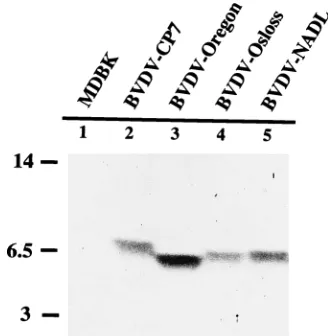
FIG. 1. Immunoblot detection of a 6- to 7-kDa polypeptide with antibodies generated against amino acids 1065 to 1125 of BVDV CP7; the respective BVDV cDNA fragment was expressed as a fusion protein with the N-terminal part of the MS2 polymerase, which is under the transcriptional control of the heat-inducible
lambda pL promoter in plasmid pEX34 (17). pEX p7 was expressed inE. coli
C600 carrying the repressor plasmid pc1857, and 200mg of the purified protein
was used for each immunization of laboratory rabbits. Lysates of cells infected with BVDV strains CP7 (6), Oregon (from Intervet, Netherlands), Osloss (12), and NADL (4) were subjected to SDS-PAGE (12% polyacrylamide) in a Tris/ Tricine buffer system, transferred to nitrocellulose membranes, and probed with anti-p7 antibodies.
4131
on November 9, 2019 by guest
http://jvi.asm.org/
increased molecular mass (58 kDa), termed E2*, could only be observed in extracts from cells infected with BVDV or Vac1–1140; it was absent from those infected with Vac1–1116
(data not shown). To exclude the possibility that a variable degree of glycosylation accounted for the formation of E2*, immunoprecipitated material was treated withN -glycopep-tidase F to fully remove the carbohydrate side chains. Pre-cipitates from Vac1–1116-infected cells yielded a single
41-kDa protein, while deglycosylation of precipitates from Vac1–1140- as well as BVDV-infected cells resulted in the
formation of two bands with apparent molecular masses of 41 and 46 kDa (data not shown). The results suggest that the protein backbone of E2* is extended compared with that of E2 and that the additional amino acids are located at the C terminus.
Demonstration of p7.The bulk of the E2 glycoprotein is in the 53-kDa form, while E2* accounts for only a minor fraction. The generation of E2 probably results from proteolytic pro-cessing which does not occur with E2*. To identify the putative cleavage product, antibodies were raised against amino acids 1065 to 1125 of the BVDV CP7 polyprotein (see Fig. 6B). A cDNA fragment encoding this peptide was fused to a fragment of the MS2 polymerase gene, and the fusion protein was ex-pressed inEscherichia coli; the purified fusion protein served for immunization of two rabbits. The antibody response was poor, probably because of the hydrophobic nature of the pep-tide. Seven booster injections were needed to obtain an anti-serum, which is referred to as anti-p7.
Extracts of cells infected with different BVDV strains (NADL, Oregon, Osloss, and CP7) were tested for their reac-tivity with the anti-p7 antibodies (Fig. 1). All BVDV isolates reacted with the antiserum, while the apparent molecular masses of the proteins differed slightly among the tested strains. Analysis of the set of vaccinia virus-BVDV recombi-nants with the anti-p7 antibodies resulted in detection of p7 in cells infected with Vac1–1140 but not in cells infected with
FIG. 2. Anti-p7 antibodies identify E2* as unprocessed E2-p7. A total of 1.5
3106
MDBK cells were infected at a multiplicity of infection of 0.1 for 24 h. At 7 h postinfection, the growth medium was replaced with Dulbecco’s modified
Eagle’s medium without methionine, and 500mCi of [35
S]methionine was added
30 min later. CVI (13106
) cells were infected with recombinant vaccinia viruses
at a multiplicity of infection of 5 and labeled with 500mCi of [35
S]methionine for 16 h beginning at 4 h postinfection. Extracts from cells infected with BVDV CP7, Vac1–1140, or Vac1–1116were incubated with anti-BVDV E2 (a-E2) (a mixture of
monoclonal antibodies provided by E. Weiland, Tu¨bingen) and anti-p7 (a-p7)
antibodies. Afterwards, the extracts were incubated with formaldehyde-fixed
Staphylococcus aureuscells. Precipitates were subjected to SDS-PAGE. Gels
were treated with En3
[image:2.612.85.270.37.322.2]Hance (NEN) for fluorography. The positions of molecular mass markers (sizes are in kilodaltons) are shown on the left.
FIG. 3. p7 does not copurify with BVDV virions. Virus from the culture medium of BVDV Oregon-infected MDBK cells was precipitated with polyethylene glycol 8000 and further concentrated by ultracentrifugation. Pelleted virions (‘‘0’’) were layered on top of a 10 to 60% glycerol–TNE gradient and centrifuged for 1 h at 30,000 rpm in an SW55 rotor. A total of 30 fractions were collected from the bottom and analyzed by immunoblotting with anti-p7 and anti-C antibodies (20). The nitrocellulose membrane was blocked with phosphate-buffered saline (PBS) containing 2% bovine serum albumin and 0.05% Tween 20 for 1 h. The anti-p7 antibodies were used at a dilution of 1:500, and the anti-C antibodies were used at a dilution of 1:5,000. The choice of the blocking substance proved to be critical since p7 could not be detected after the filter was immersed in 2.5% skim milk–PBS. For revelation of a secondary signal, antibodies conjugated to alkaline phosphatase or peroxidase were used as recommended by the suppliers. A light-emitting detection system (ECL; Amersham) was used in combination with peroxidase-labeled antibodies according to the manufacturer’s instructions.
4132
on November 9, 2019 by guest
http://jvi.asm.org/
Vac1–1116(Fig. 2). The anti-p7 antibodies specifically reacted
with E2* present in cells infected with BVDV, Vac1–1212, or
Vac1–1140, which identifies E2* as the fusion protein E2p7 (Fig.
2).
p7 is not a major structural component of the virion.The anti-p7 antibodies enabled us to address whether p7 and/or E2p7 is an integral part of the virion. Pestiviruses remain cell associated, and virions in the culture medium are sparse. In addition, pestiviruses are difficult to purify by their physical properties since their density and sedimentation constants are similar to those of various vesicular components of the host cell (13). Sufficient amounts of cytopathogenic BVDV can be har-vested after virus-induced disintegration of the monolayer. Un-fortunately, enrichment of virions by differential centrifugation yielded virus preparations that were heavily contaminated with cellular debris and nonstructural proteins such as NS3. By using linear glycerol gradients, we found that zonal-rate cen-trifugation gave a sufficient degree of virus purification to at least address whether a protein copurifies with virus particles. For this purpose, virus from 1 liter of culture medium was concentrated and applied to a linear 10 to 60% glycerol–Tris-buffered sodium chloride-EDTA solution (TNE) gradient. Thirty fractions were collected and analyzed by immunoblotting (Fig. 3). Intact virus particles peaked in fractions 14 to 17, as deter-mined by immunoelectron microscopy (data not shown). The blots were analyzed with anti-p7 and anti-CSFV capsid protein antibodies (20). The latter were used as an indicator of the position of virions along the gradient; in pilot experiments it was determined that in gradients, infectivity colocalizes accu-rately with the capsid protein (data not shown). p7 present in the starting material (Fig. 3) was detected after centrifugation at the bottom of the tube in the pellet, which mostly consisted of cellular debris. Even long exposures of the blots did not indicate the presence of p7 in fractions 13 to 20. In accordance with earlier analyses of the molecular compositions of pestivi-ruses (20), we reason that p7 is at least not a major constituent of the virion.
The question of whether E2p7 represents a structural pro-tein could only be answered indirectly. Western blot (immu-noblot) analyses with BVDV virions and anti-p7 antibodies gave no evidence for the presence of E2p7 in viral particles. This result, however, should be validated by the use of anti-bodies which detect E2 in a Western blot. Unfortunately, none of the anti-BVDV E2 antibodies available to us is reactive after sodium dodecyl sulfate-polyacrylamide gel electrophoresis (SDS-PAGE). Also, the amounts of metabolically labeled an-tigens from virions were insufficient for immunoprecipitation analysis. We therefore turned to the CSFV system, the system in which the desired antibodies have been prepared. Analo-gous to the situation described for BVDV, two forms of E2 could be detected in CSFV-infected cells which vary in their protein backbones by about the size of the p7 peptide. Since CSFV grows to reasonable titers without an apparent cyto-pathic effect, we considered the pelleted virions clean enough to assess if they also contain different forms of E2. A compar-ison of the molecular mass of E2 from infected cells with that of E2 from virions required complete deglycosylation because the molecular masses contributed by carbohydrate chains in the two proteins are significantly different (20). E2 from in-fected cells clearly comprises two different protein backbones, while E2 from virions is exclusively in the smaller form (Fig. 4).
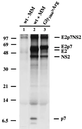
Generation of p7 requires microsomal membranes.To de-fine the biosynthesis of p7 and E2p7 in more detail, processing was studied by in vitro translation. A cDNA served as the template for mRNA transcription in which the sequences en-coding E2p7NS2 (BVDV amino acids 693 to 1588) were
placed downstream from an SP6 promoter. For efficient trans-location, a preprolactin cDNA encoding amino acids 1 to 53 was fused to the 59end of the E2 gene. RNA transcribed from the respective plasmid was used to direct in vitro translation in a rabbit reticulocyte lysate (Fig. 5). Addition of microsomal membranes allowed the generation of the individual products E2, E2p7, p7, and NS2, while in the absence of the membranes, only the unglycosylated precursor preprolactin-E2p7NS2 ap-peared. Thus, cleavage between E2 and p7 as well as between
FIG. 4. E2p7 is not detectable in CSFV virions. Pelleted viruses and extract
from CSFV-infected (inf.) pig lymphoma cells (38A1D) were treated with
[image:3.612.346.522.335.610.2]N-glycopeptidase F and analyzed by immunoblotting with monoclonal antibody A18 (anti-E2) (21). E2p7 is present in CSFV-infected cells but not in pelleted virions.
FIG. 5. Generation of p7 depends on the presence of microsomal mem-branes. The plasmid used for the generation of SP6 transcripts was obtained by cloning a cDNA fragment encoding amino acids 693 to 1588 of BVDV CP7
(restricted withKpnI andBamHI and treated with Klenow fragment) into the
SmaI site of pRN654 (derived from pRN653 [18]), downstream of and in frame with the coding sequence for amino acids 1 to 53 of preprolactin. Translation of
the synthetic mRNA encoding preprolactin1–53-E2p7NS2 was carried out in
reticulocyte lysate in the presence of [35S]methionine with (1MM) or without
(2MM) canine microsomal membranes. While only minor amounts of E2p7 can be detected with wild-type (wt) E2p7NS2, mutagenesis of Gly-1066 to Arg abolished the cleavage which generates E2 and p7 (lane 3). The uncleaved precursor, E2p7NS2, and the resulting processing products are indicated at the right margin. The positions of molecular mass markers (sizes are in kilodaltons) are on the left.
VOL. 70, 1996 NOTES 4133
on November 9, 2019 by guest
http://jvi.asm.org/
p7 and NS2 apparently depends on translocation and is facil-itated by microsome-associated proteases, presumably signa-lase. The basic requirements for signalase cleavage are fulfilled in the sequence MASG/V-1067 together with the preceding hydrophobic amino acids (Fig. 6B). Further support for signa-lase cleavage was obtained by substitution of Arg at the as-sumed P1 position (Gly-1066). While cleavage between E2p7 and NS2 was unaffected, the generation of E2 and p7 could no longer be detected (Fig. 5, lane 3).
The N terminus of p7 is Val-1067.Further characterization of p7 biosynthesis was expected from N-terminal sequencing of p7. For this procedure, [35S]methionine-labeled p7 was
synthe-sized by in vitro translation, purified by SDS-PAGE, and elec-troblotted onto polyvinylidene difluoride membranes (14, 15). The release of radioactivity in the course of the cyclic Edman degradation peaked in cycles 10 and 11, which, according to the positions of the methionine residues, results in Val-1067
being the N terminus of p7 (Fig. 6A). Labeling with a single amino acid was found to be sufficient for radiosequencing because two consecutive methionines are unique within the in vitro-translated protein. p7 sequences of different pestiviruses, including BVDV and CSFV, were aligned (Fig. 6B). It is likely that the p7 proteins of all these viruses share the same N terminus, especially since the signalase cleavage site is pre-served.
p7 forms the junction between the structural and the non-structural genes in pestiviruses, and the same is probably true for hepatitis C virus. The nature of p7 and E2p7 and the peculiar processing of the E2p7NS2 region are well conserved among pestiviruses and hepatitis C virus, which is indicative of a common function for these proteins. The spatial proximity of E2 and p7 in the polyprotein and the formation of an E2p7 fusion protein suggest a role in glycoprotein maturation and/or virus morphogenesis. Pestiviruses are believed to mature at
FIG. 6. (A) N-terminal sequencing of p7. In vitro-synthesized, [35S]methionine-labeled p7 was purified by SDS-PAGE, transferred to a polyvinylidene difluoride
membrane, and subjected to peptide sequencing. The graph depicts the distribution of radioactivity released during automated Edman degradation after subtraction of background radiation. At the top of the panel, the amino acid sequence deduced from the BVDV genome is aligned with the appearance of two consecutive methionines, which are unique within the sequence of the in vitro-translated protein. According to this alignment, Val-1067 forms the N terminus of p7. (B) Alignment of the p7 peptide sequences of BVDV CP7 with other pestiviruses. With an N terminus at Val-1067 and a C terminus tentatively located around amino acid 1135, p7 consists of about 70 mostly hydrophobic amino acids. The sequences N terminal to p7 conform to the requirements for signalase (21,23 rule), and the alignment makes cleavage at the analogous position likely for all pestiviruses. The difference in numbering results from the additional amino acids of the BVDV isolates compared with the CSFV and BDV polyproteins. The overall structure, shown as a Hopps and Wood diagram, is remarkably conserved among the listed strains. The dashed line at the top depicts the amino acid sequence which was fused to the MS2 polymerase and subsequently served for immunization of rabbits.
on November 9, 2019 by guest
http://jvi.asm.org/
[image:4.612.62.555.67.455.2]internal membranes, presumably the endoplasmic reticulum, but distinct steps of pestivirus morphogenesis have not been revealed. Assembly of a number of enveloped viruses requires specific interactions between the membrane-associated glyco-protein(s) and the capsid glyco-protein(s) in the cytosol. In general, this is facilitated by the cytoplasmic domain of a glycoprotein which is anchored in the membrane by a transmembrane se-quence. A cytoplasmic domain could not be identified for any of the E proteins of pestiviruses. Interestingly, the pestivirus p7 protein contains the charged sequence 1100-REENTKK-1106 (RDEPIKK in CSFV), which probably faces the cytosol (Fig. 6B). It is conceivable that this charged stretch interacts with the capsid protein and initiates the budding process without necessarily being enriched in the virion.
We thank Silke Esslinger and Petra Wulle for excellent technical assistance.
This work was supported by the Bundesministerium fu¨r Forschung und Technologie and Intervet International BV (project 0319028A).
REFERENCES
1.Becher, P., M. Ko¨nig, D. Paton, and H.-J. Thiel.1995. Further
character-ization of border disease virus isolates: evidence for the presence of more
than three species within the genus pestivirus. Virology209:200–206.
2.Becher, P., A. D. Shannon, N. Tautz, and H.-J. Thiel.1994. Molecular
characterization of border disease virus, a pestivirus from sheep. Virology 198:542–551.
3.Collett, M. S., R. Larson, S. Belzer, and E. Retzel.1988. Proteins encoded by
bovine viral diarrhea virus: the genome organization of a pestivirus. Virology 165:200–208.
4.Collett, M. S., R. Larson, C. Gold, D. Strick, D. K. Anderson, and A. F.
Purchio.1988. Molecular cloning and nucleotide sequence of the pestivirus
bovine viral diarrhea virus. Virology165:191–199.
5.Collett, M. S., M. A. Wiskerchen, E. Welniak, and S. K. Belzer.1991. Bovine
viral diarrhea virus genomic organization. Arch. Virol.3:19–27.
6.Corapi, W. V., R. O. Donis, and E. J. Dubovi.1988. Monoclonal antibody
analyses of cytopathic and noncytopathic viruses from fatal bovine viral
diarrhea virus infections. J. Virol.62:2823–2827.
7.Deng, R., and K. V. Brock.1992. Molecular cloning and nucleotide sequence
of a pestivirus genome, noncytopathogenic bovine viral diarrhea virus strain
SD-1. Virology191:867–879.
8.Lin, C., B. D. Lindenbach, B. M. Pra´gai, D. W. McCourt, and C. M. Rice.
1994. Processing in the hepatitis C virus E2-NS2 region: identification of p7 and two distinct E2-specific products with different C termini. J. Virol. 68:5063–5073.
9. Meyers, G., T. Ru¨menapf, and H.-J. Thiel.1989. Molecular cloning and
nucleotide sequence of the genome of hog cholera virus. Virology171:555–
567.
10. Mizushima, H., M. Hijikata, S.-I. Asabe, M. Hirota, K. Kimura, and K.
Shimotohno.1994. Two hepatitis C virus glycoprotein E2 products with
different C termini. J. Virol.68:6215–6222.
11. Moormann, R. J. M., P. A. M. Warmerdam, B. Van der Meer, W. M. M.
Schaaper, G. Wensvoort, and M. M. Hulst.1990. Molecular cloning and
nucleotide sequence of hog cholera virus strain Brescia and mapping of the
genomic region encoding envelope glycoprotein E1. Virology177:184–198.
12. Renard, A., D. Dino, and J. Martial.1987. Vaccines and diagnostics derived
from bovine diarrhea virus. European patent application number 86870095.6. Publication number 02.08672.
13. Ru¨menapf, T., G. Meyers, R. Stark, and H.-J. Thiel.1991. Molecular
char-acterization of hog cholera virus. Arch. Virol. Suppl.3:7–18.
14. Ru¨menapf, T., G. Unger, J. H. Strauss, and H.-J. Thiel.1993. Processing of
the envelope glycoproteins of pestiviruses. J. Virol.67:3288–3294.
15. Stark, R., G. Meyers, T. Ru¨menapf, and H.-J. Thiel.1993. Processing of
pestivirus polyprotein: cleavage site between autoprotease and nucleocapsid
protein of classical swine fever virus. J. Virol.67:7088–7095.
16. Stark, R., T. Ru¨menapf, G. Meyers, and H.-J. Thiel.1990. Genomic
local-ization of hog cholera virus glycoproteins. Virology174:286–289.
17. Strebel, K., E. Beck, K. Strohmaier, and H. Schaller.1986. Characterization
of foot-and-mouth disease virus gene products with antisera against
bacte-rially synthesized fusion proteins. J. Virol.57:983–991.
18. Tautz, N., G. Meyers, and H.-J. Thiel.1993. Processing of poly-ubiquitin in
the polyprotein of an RNA virus. Virology197:74–85.
19. Thiel, H.-J., P. G. W. Plagemann, and V. Moennig.1996. Pestiviruses, p.
1059–1073.InB. N. Fields, D. M. Knipe, H. P. Howley, et al. (ed.), Virology,
3rd ed. Lippincott-Raven Publishers, Philadelphia.
20. Thiel, H.-J., R. Stark, E. Weiland, T. Ru¨menapf, and G. Meyers.1991. Hog
cholera virus: molecular composition of virions from a pestivirus. J. Virol. 65:4705–4712.
21. Weiland, E., R. Stark, B. Haas, T. Ru¨menapf, G. Meyers, and H.-J. Thiel.
1990. Pestivirus glycoprotein which induces neutralizing antibodies forms
part of a disulfide-linked heterodimer. J. Virol.64:3563–3569.
22. Wengler, G., D. W. Bradley, M. S. Collett, F. X. Heinz, R. W. Schlesinger,
and J. H. Strauss.1995. Flaviviridae, p. 415–427.InF. A. Murphy, C. M.
Fauquet, D. H. L. Bishop, S. A. Ghabrial, A. W. Jarvis, G. P. Martelli, M. A. Mayo, and M. D. Summers (ed.), Virus taxonomy. Sixth report on the International Committee on Taxonomy of Viruses. Springer-Verlag, New York.
VOL. 70, 1996 NOTES 4135

![FIG. 6. (A) N-terminal sequencing of p7. In vitro-synthesized, [35S]methionine-labeled p7 was purified by SDS-PAGE, transferred to a polyvinylidene difluoridemembrane, and subjected to peptide sequencing](https://thumb-us.123doks.com/thumbv2/123dok_us/1278580.80391/4.612.62.555.67.455/sequencing-synthesized-methionine-transferred-polyvinylidene-diuoridemembrane-subjected-sequencing.webp)
