MORPHOEA – A CLINICAL STUDY
Dissertation Submitted to
THE TAMIL NADU
DR. M.G.R. MEDICAL UNIVERSITY
in partial fulfillment of the regulations
for the award of the degree of
M.D. (Dermatology, Venereology and Leprology)
BRANCH – XII A
MADRAS MEDICAL COLLEGE
THE TAMIL NADU DR. M.G.R. MEDICAL UNIVERSITY CHENNAI, INDIA.
CERTIFICATE
Certified that this dissertation entitled “MORPHOEA – A CLINICAL STUDY” is a bonafide work done by Dr. B.VIJAYALAKSHMI, Post graduate student of the Department of Dermatology and Leprology and Institute of
Venereology, Madras Medical College, Chennai- 3, during the academic year 2004 –
2007. This work has not previously formed the basis for the award of any degree or
diploma.
Prof. Dr. B. PARVEEN, M.D., D.D., Professor and Head of the Department, Department of Dermatology and Leprology, Madras Medical College,
Chennai- 3.
Prof. Dr .KALAVATHI PONNIRAIVAN, B.Sc., M.D., DEAN,
DECLARATION
I, DR.B.VIJAYALAKSHMI, solemnly declare that dissertation titled, “MORPHOEA – A CLINICAL STUDY” is a bonafide work done by me at Madras Medical College during 2004-2007 under the guidance and supervision of
Prof. Dr. B. PARVEEN, M.D.,D.D., Professor and Head, Department of Dermatology, Madras Medical College, Chennai - 600 003.
The dissertation is submitted to The Tamilnadu, Dr.M.G.R.
Medical University, towards partial fulfillment of requirement for the
award of M.D. Degree in Dermatology, Venereology and Leprology (BRANCH – XII A).
Place: Chennai.
Date:
SPECIAL ACKNOWLEDGEMENT
My sincere thanks to
Prof. Dr .KALAVATHI PONNIRAIVAN, B.Sc., M.D., DEAN,
Madras Medical College
for allowing me to do this
I am gratefully indebted to Prof. Dr. B. Parveen M.D., D.D., Professor and Head, Department of Dermatology and Leprology for her invaluable guidance,
motivation and help through out the study. I would like to express my sincere and
heartfelt gratitude to Prof. Dr. V.S. Dorairaj, M.D., D.V., Director in charge, Institute of Venereology.
I wish to thank Dr.N.Gomathy, M.D.,D.D., former Professor, Department of Dermatology and Dr.N.Usman, M.D.,D.V.,Ph.D., former Director, Institute of Venereology for their constant support and motivation.
I am very grateful to Dr.S.Jayakumar M.D., D.D., Additional Professor, Department of Dermatology for his invaluable guidance and help.
I sincerely thank Dr.C.Janaki, M.D., D.D., Reader of Dermatology (Mycology) for her priceless support.
I express my earnest gratefulness to Dr.D.Prabavathy M.D., D.D., Professor and Head of Department of Occupational Dermatology and Contact Dermatitis for
her constant motivation and guidance. I thank
Dr.V.Somasundaram M.D., D.D., Additional Professor, Department of Occupational Dermatology and Contact Dermatitis for his benevolent help and
support.
I express my sincere gratitude to Dr.K.Rathinavelu M.D., D.D., Professor of Leprosy and Dr.R.Arunadevi M.D., D.D., Lecturer / Registrar, Department of Dermatology for their support.
I thank Dr. A. Hameedullah M.D., D.D., Dr. S. Kumaravelu M.D., D.D., Dr. J. Manjula M.D., D.N.B., (Derm) and Dr. Aftab Jameela Wahab M.D., D.D.,
Assistant Professors, Department of Occupational Dermatology and Contact
Dermatitis for their support and help.
My sincere thanks to Dr.S.Mohan M.D, D.V. Former Registrar,
Dr.V.Thirunavukkarasu M.D., D.V., Dr.K.Venkateswaran M.D., D.V., Dr.P.Elangovan M.D., D.V., Dr.D.Ramachandra Reddy M.D., D.V., Dr.S.Thilagavathy M.D., D.V., Dr.P.Mohan M.D., D.V., Dr.S.Arunkumar M.D., D.V., and Dr.S.Kalaivani M.D., D.V., Assistant Professors, Institute of Venereology for their help and suggestions.
I am also thankful to Dr.K.Manoharan M.D., D.D., and Dr. V. Sampath M.D., D.D., for their continuing guidance and support.
I duly acknowledge the paramedical staff and my colleagues for their help and
favour.
Last but not least I am profoundly grateful to all patients for their cooperation
CONTENTS
Sl.No Title Page No.
1 INTRODUCTION 1
2 REVIEW OF LITERATURE 2
3 AIM OF THE STUDY 29
4 MATERIALS AND METHODS 30
5 OBSERVATIONS AND RESULTS 31
6 DISCUSSION 49
7 CONCLUSION 54
BIBLIOGRAPHY
PROFORMA
INTRODUCTION
Morphoea is a benign disorder characterized by localized thickening of
the skin with no known aetiological factors. Although the skin disorder
resembles systemic scleroderma there are no internal organ involvement.
Morphoea is more commonly found in females and it has a characteristic
morphology which differs in early and late stage. There are varying clinical
types. All have characteristic distribution across the age groups. Morphoea may
cause a lot of cosmetic problems which is very distressing to the patients.
Sometimes it may cause growth retardations, deformity, contractures and
disfigurement. There have been rare occurrence of pain, oedema, arthritis,
colicky abdominal pain, visual disturbances and neurological problems in
patients of morphoea. There are also interesting serological associations in
morphoea.
The interesting fact about morphoea is that it resolves on its own even
without treatment. So far no conclusive evidence regarding its etiopathogenisis
has been proposed in spite of the advancements in the field of medicine and
technology. This disease, with such peculiar manifestations has kindled the
interest of dermatologist from time immemorial. Several eminent
REVIEW OF LITRATURE
Morphoea is localized scleroderma caused by vascular sclerosis
resulting in increased collagen deposition in the dermis and sometimes in
deeper structures also1.
The word morphoea is derived from Greek mythological character
“Morph” who could change his body at will2. Erasmus Wilson is first credited
for using the word to describe this lesion which he considered to be areas of
vestiges of true leprosy3. Later Morphoea came to be used synonymously with
localized scleroderma.
Morphoea has varied clinical presentations. There are also several
classifications of the clinical types. The latest classification proposed by
Peterson et al is as follows4.
PLAQUE TYPE OR CIRCUMSCRIBED TYPE
MORPHOEA EN PLAQUE
KELOIDAL
BULLOUS
NODULAR
GUTTATE /LICHEN SCLEROSUS ET ATROPHICUS
LINEAR TYPE
LINEAR CLASSICAL
EN COUP DE SABRE
PARRY ROMBERG SYNDROME
DEEP MORPHOEA
SUBCUTANEOUS MORPHOEA
MORPHOEA PROFUNDUS
EOSINOPHILIC FASCIITIS
PANSCLEROTIC MORPHOEA
GENERALIZED MORPHOEA
EPIDEMIOLOGY OF MORPHOEA
Incidence of morphoea is more in females.5 The sex ratio is 3:15. Whites
are more susceptible.5 Age group in which it is more common is 20 to 40 years,
but it has been reported in a 1year old child and a 70 year old patient also.
Morphoea is rare in children. Incidence below 10 years is 1.5% and below 20
years is 7.2%.5 There are exceptions to this pattern. Linear morphoea has an
incidence of 20% before the age of 10 years and 75% before the age 20 years
.The plaque type of morphoea occurs much later in life. Only 10 % occur
before 10 years and 75 % occurs between 20 and 40 years. Generalized
The onset of morphoea is slow and insidious. The duration of morphoea
is very variable, ranging from 3 to 25 years. The average is 3 to 5 years.
morphoea is a self limiting disease. Therefore no conclusive studies on
treatment are available.5
Morphoea may be associated with various other abnormalities like
atrophy of fat, muscles, periostium and bones.6 Vascular anomalies of the brain,
mesentery, and kidney under lying the lesion have also been reported7.
Most serious complications include the disturbances in bone growth
under lying the lesion. Linear morphoea on limbs can give rise to limb length
discrepancy and subsequent alteration in gait and height. Claw hand, hammer
toes and fixed contracture of joints may also occur8.
In a study conducted in sixty eight patients with linear morphoea 47% of
them had spina bifida occulta in the radiological examination of the spine5.
Sacralization of lumbar vertebra, presence of six lumbar segments,
prolongation of transverse arches, scoliosis, rudimentary ribs, atrophic clavicle,
shortened ulna, torticolis, kyphosis and contracted pelvis were some of the
other bony anomalies reported.1 Melorheostosis was seen along bones under
lying linear morphoea.43 In a rare study Prasad et al showed morphoea was
associated with bone cyst.10
In a study conducted by Christianson on 235 patients showed that 44%
of them reported with arthralgia and pain lateralized to site of lesion on the
children had arthritis at a site distant to the lesion on the affected side before
the occurrence of lesion.6
Raynaud’s phenomenon was reported in 8 out of 191 patients by
Christianson’s study.5 An interesting feature that has been reported is unilateral
Raynaud’s on side of the lesion.11 Emotional instability, anxiety,
psychoneurosis, and seizures were also reported in the same study.5 The same
study also showed 31 patients had migraine and 21 patients had colicky
abdominal pain and infact 20 of these patients under went appendicectomy
with no relief.
Vascular and pigmented nevi, Becker’s melanosis and café au lait
macules have been reported in few patients of morphoea.5 Other autoimmune
conditions like alopecia areata, vitiligo135, icthyosis, pigmentation, nail
dystrophy, hirsutism, carpal tunnel syndrome, absent pectoralis major, biliary
cirrhosis and nephrotic syndrome, were some of the other features reported.9
Rarely bullous pemphigoid and subcorneal pustulosis have also been reported
in few cases of morphoea.12
A rare occurrence of entrapment neuropathy due to constriction by
sclerosing process in subcutaneous tissue and associated hypertrichosis on
lesions has been reported in a patient of linear morphoea.13.
Blood investigations revealed eosinophilia in both active and quiescent
lesions.14.
Elevated immunoglobulins IgG and IgM have been demonstrated by a
IgG and IgM. The same study also showed elevated ANA in most of patients
with linear morphoea and also in children. He also showed that incidence of
rheumatoid factor was more in children.6. Winkleman’s study of 77 patients,
showed that direct immunofluorence was positive in 35.8% of the cases.15
VARIOUS TYPES MORPHOEA
MORPHOEA EN PLAQUE:
This the most common type of morphoea encountered. The skin is
usually smooth, shiny, thickened, and faintly purplish or mauve coloured, or
rarely waxy.16 The border is lilac coloured, well defined and circumscribed and
the maximum diameter of the plaque may reach 30cm. Older lesions loose their
original colour and become ivory coloured. The surface may sometimes be
nodular. The hair is usually absent and there is decreased sweating.17There may
also be loss of sensation due to the thickening of skin. The shape of the plaques
may be circular, oval or irregular. There may be associated
hypertrichosis.13They may be single or multiple or bilateral but are usually
asymmetrical. The usual sites of occurrence of these lesions are the face, limbs,
trunk and genitalia. They are less commonly found on axilla, perineum and
nipples.17 Sometimes vesicles, bullae, telengectasia and haemorrhages may also
be seen on the lesion.18
The onset is usually insidious and sometimes rapid with associated
oedema and erythema.17 Sometimes pigmentation may precede the appearance
colicky abdominal pain may be associated with lesions occurring on the face
and abdomen respectively..5
KELOIDAL MORPHOEA:
This is a rare variant of morphoea. This resembles a keloid and it shows
greater depth of inflammation, but limited to dermis.19 There is increased
fibrosis and collagen deposits. This type of morphoea is mostly seen associated
with systemic scleroderma.
NODULAR MORPHOEA
These can occur along with plaque type of morphoea. The surface is
extremely nodular.20.
BULLOUS TYPE
Blisters may be seen in morphoea. This may be due to lymphatic
dilatations or vascular sclerosis causing obstructions. Eosinophils capable of
releasing major basic proteins causes subepidermal lysis and produces bullous
lesons.22. Similar condition may also be seen in Lichen sclerosis et atrophicus.
GUTTATE MORPHOEA (LICHEN SCLEROSUS ET ATROPHICUS/WHITE SPOT DISEASE)
Guttate means rain drops. Since these lesions are also small like rain
drops, chalky white and size varying from 1 to 10 mm these are called guttate
morphoea.It is a very superficial type of morphoea .These lesions also have a
neck, shoulder and extremities. Multiple lesions can be seen on the same
person.28
As per the classification of Peterson4, Lichen sclerosus et atrophicus is
also considered to be type of guttate morphoea. Bizzero in his study also
considers it to be a type of guttate morphoea.23 Certain authors like K.G.
Bergstorm consider it to be different because of early development of
epidermal atrophy and follicular dellings and variations in the histopathology.
LICHEN SCLEROSUS ET ATROPHICUS:
The lesions are porcelain white and have follicular dellings ( widened
pilosebacious orifices) on them. They have specific HLApredispsition.24, 25
Female related HLA includes :
MHC I - HLA -B40, HLA -A29, HLA -B44.
MHC II - DQ7, DQ8, DQ9.
Males related HLA includes :
- DR11, DR 12, DQ7
Skin is atrophied and numerous lesions are seen on trunk, limbs and
genitalia.23. Number of autoimmune disease are associated with Lichen
sclerosis et atrophicus.26. Borrelial infection have been implicated in
development of these lesions.90
These lesions may also follow the Blasckho’s line. Lichenification may
telengectasia, purpura, or bulla may be seen on the lesion. In later stages, they
may become wrinkled and depressed. Scarring alopecia may occur on the
scalp.28 Involvement of superior oblique muscle of the eye may give rise to
diplopia.29.
Anogenital lesion in women and men may occur at 45 to 60 years of
age. Rarely female children may also have anogenital lesions. Hormonal
influence may cause of such lesions.86 Constant friction on these surfaces may
give rise to raw areas which may later heal to produce flat and glistening
lesions. Patients usually complain of soreness and dyspareunia. A late
complication may be resorption of labia, vulva, and clitoris leading to
narrowing of vaginal introitus. Normal pregnancy and delivery have occurred
despite this problem.30The male counterpart of this disease is called balanitis
xerotica obliterans23 which presents with recurrent balanitis phimosis and
painful erection.
ATRPHODERMA OF PIERINI AND PASINI
This is a primary abortive form of morphoea in which indurations fails
to occur.31. It is an uncommon superficial form of localised morphoea in which
there is an oval, hyper pigmented or slate grey atrophic plaque on the trunk.
The plaques may be multiple and the centre is usually depressed and the border
is described as typically being cliff-drop like.32. Some authors consider it to be
an optical effect because of the difference in colour.33 It occurs much earlier in
life around 10 to 20 years of age and has a protracted course. It also lacks the
typical violacious border. It may coexist with other types of morphoea,
Another peculiar feature of this morphoea is that, atrophy occurs much before
sclerosis.36 Borrelia burgdorferi has also been implicated in the development of
this lesion..90
LINEAR MORPHOEA (SYN. CLASSICAL LINEAR MORPHOEA)
Localised scleroderma occurs in linear bands in this condition. They are
usually unilateral or rarely bilateral. The extremities are usually affected and
lower limbs are much more commonly affected. These linear lesions may also
occur on anterior aspect of thorax, thighs and buttocks. Homolateral lesions
involving one arm and same side leg have also been reported. Rarely one half
of the body may be affected.37.
These lesions usually lack the usual violacious ring but they may be
seen in the advancing boarders. They may also follow the lines of Blaschko.38.
These distributions along lines of Blaschko shows that the tendency to develop
scleroderma is predetermined during embryogenesis with formation of clones
of vulnerable cells which on appropriate trigger may transform to develop full
blown lesion.39.
Patients usually complain of pain, arthralgia or oedema before the onset
of the lesions.5The underlying bones and muscles may be involved in 20% of
cases leading on to severe growth disturbance and limb length discrepancy.
When these lesions cross a joint it can give rise to contractures.40 On
radiological examination, the underlying involved bone may have a
characteristic picture of wax flowing out of the candle due to cortical
considered to be developmental anomaly. A sclerotome147 supplied by a spinal
nerve may be affected in this condition This is a painful condition seen mainly
in children and presenting with asymmetrical contractures and thickening of the
overlying skin.42 Any surgical correction may further worsen the condition.
Rarely linear scleroderma may be associated with overlying
hypertrichosis.44. Ulcerated dystrophic calcinosis45 and nodular morphoea46
may also be seen on linear morphoea. Sometimes bands47 of lesions occur
circumferentially around limbs, fingers or breasts, resembling an ainhum.48,50,
The tissues distal to these bands may be oedematous and depigmented.49
Fibrosis may spare areas with in a linear morphoea producing skip lesion.
Linear morphoea of lower limbs is more commonly associated with spina
bifida occulta.5
FRONTOPARIETAL MORPHOEA: (SYN. EN COUP DE SABRE)
En coup de sabre in French means a strike with a sword. Atrophy of skin
is usually present and the skin over the affected area is usually contracted and
firm in the early stages. Later an ivory coloured sclerotic plaque develops and
there may be rarely overlying telengectasia and hyperpigmentation of border.
Later a depressed groove appears on the frontoparietal region, extending on to
the scalp. In the scalp there is alopecia which in some cases may be preceded
by bleaching of hair.54The groove may extend further down on to the cheeks,
nose, upper lip and gums. In severe case it may involve the chin and neck also.
It may affect the gingiva and jaws also leading to alteration in arrangement,
spacing and direction of teeth.52 Tongue may be atrophied or grooved. There
Rarely frontoparietal lesions may be bilateral, trilinear or may follow
Blaschko’s lines.53 There may be associated morphoea lesions on other sites of
the body or there may be total atrophy of one side of the body either
homolateral or contralateral or rarely both sides.56,57 The underlying bone can
also get affected. There may be EEG abnormalities showing dysarrythmias
under the affected site.28 Variety of ocular lesions like enophthalmous, lid
ptosis, extroccular muscle weakness, iris atrophy , heterochromia iris , atrophy
of fundus ,loss of eye lashes (along the line of involvement ) and eye lid
oedema may be the presenting feature.58,59 Rarely ossification may occur in
later stages.57
PARRY ROMBERG SYNDROME
Hemiatrophy of Parry Romberg is similar to frontoparietal morphoea but
there is usually no cutaneous sclerosis. It usually begins as hyperpigmentation
on the face. The skin is less bound down to the deeper structures and it is
usually seen along the distribution of the trigeminal nerve.56.
DEEP MORPHOEA
MORPHOEA PROFUNDUS
Whittaker et al described this morphoea, in which all layers of the skin
are involved.61They appear as solitary fibrotic plaque on shoulders, back, neck,
or paraspinal areas. The skin is invariably hyperpigmented .Osteoma cutis
develops very often in these lesions.62 The skin is bound down and borders are
usually ill defined. They have a cobble stone or pseudocellulite appearance.
groups) may be evident.28. In subcutaneous morphoea, the subcutaneous plane
alone is involved and there no signs of inflammation on the skin. Flexions
contractures are more common in this type.21
DISABLING PANSCLEROTIC MORPHOEA OF CHILDREN
This a rare forms of morphoea. Dermis, fat, fascia, muscles and even
bones are sclerosed before the age of 14 years. It may develop from linear
morphoea or de novo. Trunk, extremities, scalp, and face may be involved .The
finger, soles and toes are exceptionally spared.63 The patient may walk on tip
toes due to contractures of Achilles tendon. The onset is usually acute and there
may be associated arthralgia and stiffness. The intense pain may be due to
involvement of cutaneous nerves.64 Oesophageal, pulmonary and periodontal
changes may be seen. Flexion contractures, osteoporosis and bone changes are
some of the common complications seen.
Electromyogram is usually abnormal. Histopathological changes in
muscle may be seen .Creatinine phosphokinase level is normal. Serological
investigation shows elevate ESR, eosinophilia and hypergammaglobulinemia.
Treatment with PUVA and cyclosporine have been useful. With out treatment
this condition is progressive and occasionally fatal.
GENERALIZED MORPHOEA (SYN. GENERALIZED SCLERODERMA)
In this condition there is generalized sclerosis of the skin. There is no
systemic disturbance. It occurs between 30 to 40 years of age.65A rare
more commonly affected more than men. The sex ratio is 3:1.5 On set is usually
insidious. Individual plaques look like localized morphoea. It usually begins in
the trunk. The hands may resemble the tumid phase of systemic scleroderma.
Spindling of fingers may be seen. Flexion contractures have also been
reported5. Rarely the whole body from head to foot may be involved.66 There
may be dyspnoea when chest wall is involved. Respiratory failure has been
reported due to inter costal muscles involvement. The face may be shiny and
brown, and expressionless due to indurations of the under lying skin and there
may be restricted mouth opening. Raynaud’s phenomenon have also been
reported in few cases.5 Whitlows following trauma have been reported.67Non
pitting oedema may precede the development of morphoea. Bullous, keloidal or
nodular type may coexist with generalized morphoea. Acral myofibromas may
occur in few cases.67 Overall hyperpigmentation may be the presenting
symptom.5The skin lesions of generalised morphoea may be superimposed by
keratosis or calcinosis.65
Contracture of joints, thinning of limb and soreness may be the
presenting complaints in acute phase of the disease. Joint pains were reported
in 50% of cases5. Rheumatoid arthritis, polymyositis and sick sinus syndrome
may coexist along with this condition.68 Sometimes intractable pain, infection
and atrophy of the limbs may occur in this condition. Squamous cell
carcinomas have occurred in long standing case of morphoea.1
Generalised morphoea may also coexist with lichen sclerosus et
atrophicus.133 Subcutaneous involvement of lesions occurring in generalised
subcutaneous involvement in generalised morphoea may occur with systemic
eosinophilia.114
EOSINOPHILIC FASCIITIS: (SYN. SCHULMAN’S SYNDROME)
Although it is a distinct entity some authors consider it to be a variant of
morphoea.69. It forms one end of spectrum of linear morphoea and the other end
of systemic scleroderma. It may be associated with other connective tissue
disorders. It occurs both in children and adults. Males are more commonly
affected. There is acute pain before the onset of the lesions, which is followed
by swelling and tenderness of the distal parts of the limbs and later there is
induration.70There is limitations of movement of feet and hands. Occasionally
the lesions can occur on face or abdomen and there may be superficial
blistering or haemorrhage on them. Trauma or strenuous exercise is considered
to precipitate this condition. There is no Raynaud’s phenomenon, internal
organ involvement or history of previous infections in this condition.
Eosinophilic fasciitis like syndrome have also been reported after
ingestion of L-Tryptophan.71 In addition to the usual feature of muscle
weakness, muscle enzymes are also elevated. Enhanced type I procollagen gene
expression in the skin has been demonstrated.72 In another similar condition
called eosinophilia-myalgia syndrome, there may be dyspnoea, oedema,
arthralgia and rashes in addition to the above features.73. This condition is also
Histopathological examination of eosinophilic fasciitis may be similar to
morphoea. There may be raised ANA titre, hypergammaglobulinemia and
eosinophilia.75This condition also resolves on its own.72.
ASSOCIATIONS OF MORPHOEA
Although morphoea is a localised condition with out any systemic
involvement, there are many interesting associations with morphoea.
Arthralgia,5unilateral Raynaud’s11 migraine and colicky abdominal pain5 have
been reported. Spina bifida was found in some of the cases of linear
morphoea.5,96,101 Sacralization of lumbar vertebra, presence of six lumbar
vertebrae, prolongation of transverse arches, kypohosis, scoliosis, lumbar
intervertebral disc prolapse, rudimentary clavicle, torticolis, atrophic clavicle,
absent pectoralis muscle, contracted pelvis, shortened ulna and deformities of
feet and toes were other bony defects described.5 Warty vascular pigmented
naevi, usually on the same side of morphoea, lichen planus146 café au lait spots,
alopecia areata, vitiligo,135 generalised icthyosis, pigmentation, dystrophy of
nails and hirsutism have also been reported in few patients.1 Children may be
intellectually precocious.5 Variants of morphoea occurring later in life may also
be associated with tissue calcification. Hair loss and beaking of nose were also
seen in few studies.9 Bullous pemphigoid, subcorneal pustulosis and primary
biliary cirrhosis have also been reported as associations.12 Localised morphoea
and Lichen sclerosus et atrophicus have occurred in same patients.136
No systemic changes were found in morphoea except for oesophageal
changes in 27% of cases.5 systemic lupus erythematosis107, progressive
nephritis, elastosis perforans serpiginosa, discoid lupus erythematosis,
rheumatoid arthritis146 and mixed connective tissue disorder were other
autoimmune disorders seen in morphoea.108 Silicon implants into breast for
augmentation may also cause morphoea.104 Morphoea like features have also
followed carcinoid syndrome.105 There is no relation between internal
malignancy and morphoea. Squamous cell carcinoma1 have occurred in long
standing cases morphoea.
ETIOPATHOGENISIS
Pathogenesis of morphoea is still a mystery. Various studies have been
conducted and numerous theories have been put forward to explain the
evolution of morphoea. The basic fact is that there are increased fibroblasts,
and increased production of collagen.
There is always a triggering factor for production of sclerosis. A
vascular injury may be the primary insult promoting migration of mononuclear
cells across the damaged endothelial cells. The endothelial cells promote
increased production of platelet derived growth factors and their receptors.
There is increased production of Transforming growth factors-β
(TGF-β), which may increase connective tissues growth factor76 (CTGF) gene
expressions and promote fibrosis. There are also increased production of
interleukin 2 (IL 2)77 receptors, IL6 receptors, CD4, CD8, CD28, CD30,
TNF-α78, soluble vascular cell adhesion molecules (VCAM-1), E selectin79 and
been reported in graft versus host disease and idiopathic thrombocytopenic
purpura.80
A possible pathogenic role of fibrillin –1 in localised sceleroderma has
been demonstrated by isolation fibrillin –1 antibody82,83 in the serum of
patients. The progenitor cell antigen CD3484 identified in a subpopulation of
dendritic cells are decreased in localised scleroderma, proving that a loss of
immunomodulations may result in pathogenesis of morphoea. In
melorheostosis of bone of, TGF-β is increased causing proliferation of
periosteal fibroblasts.77
Connective tissue growth factor (CTGF) has the ability to induce TGF-B
and ultra microscopy and RNA analysis shows that CTGF m RNA is increased
in lesional cells. TGF-β & CTGF form a loop that induce and sustain fibroblast
proliferation and extracellular matrix production.83 Antibody to U1RNP has
also been implicated in development of morphoea.81
ETIOLOGICAL THEORYS
Various theories and precipitating factors have been proposed for the
development of morphoea. Few of them are as follows.
GENETICS
There is increased incidence of morphoea in family members of
patients.85 Monozygotic twins have had morphoea. So far HLA predisposition
has not been elucidated. There are increased organ specific antibodies in
TRAUMA
Trauma preceding the development of lesions have been reported in
several studies.87 Trauma may trigger vascular sclerosis and lead on to
cutaneous sclerosis.88 Surgical trauma has caused morphoea following surgery
for atriovenous fistula and rhinophyma.89
VACCINATION
Vaccination for B.C.G,93 varicella zoster94 and tetanus90 have triggered
morphoea.
Injection
Injection vitamin K91 has produced lesions around the hip girdle. The
characteristic distribution is called gun-belt and holster sign.91 In several other
studies shows injections whose nature was unknown have also precipitated
morphoea probably due to trauma.
HORMONAL CAUSES
Pregnancy has precipitated morphoea in many females.86 Extract of
pratharmone injections in rats have produced sclerodermatous changes and
calcium deposits in skin and decalcification of bones.93 Lichen sclerosis et
atrophicans in prepubertal girls have resolved after puberty. Improvement of
lesions in menopausal women after hormonal therapy further supports this
METABOLIC CAUSES
Phenylketoneuria has been implicated in development of morphoea.92
Low phenylalanine diet has resulted in resolutions of these lesions. Diabetes
mellitus has also produced scleroderma like changes.
INFECTION
Several infections have been implicated in development of morphoea.
Important among them is Borrelia burgdorferi.94 Polymerised chain reactions
have detected DNA of these bacteria in lesional skin. Generalized morphoea
has followed measels.95
DRUGS
Several drugs have been implicated in development of morphoea. They
include penicillamine98, bromocriptine99, hydroxytryptophan109, carbidopa104,
pentazocine,96 docetaxal100 and bleomycin.97 Isolated limb perfusion with
melphalan104 have caused morphoea in few patients on limbs103. Among this
penicillamine have been used as a therapy for morphoea and has induced
morphoea in few patients. There has been spontaneous improvement after
stopping these drugs. Valproic acid used in treatment of seizure has also
triggered morphoea.104
RADIOTHERAPY
Morphoea at the sites of radiotherapy have been reported. Breast cancer
treated with chest wall irradiation has later on lead to development morphoea in
MISCELLANEOUS CAUSES
Exposure to epoxy resins, vinyl chloride, organic solvents and pesticides
have produced scleroderma like changes.
NEURAL THEORY
Linear morphoea of lower limbs have been associated spina bifida
occulta.5 In a study conducted by Rubin et al 17.3% to 33% patients had
spinabifida occulta in lumbosacral spine.101 This defect could have damaged the
spinal cord, by trauma or adhesion of cauda equina to the defect may give rise
to trophic changes of lower limbs resembling the changes seen on morphoea.
This view supports the theory that nerve damage is the important cause of
development of morphoea.102
Segmental morphoea or linear morphoea that usually follows the course
of peripheral nerves further supports this theory that injury of these nerves
could have triggered morphoea. The prolongation of sensory chronaxie in
systemic scleroderma further strengths this theory.105
IMMUNOLOGICAL THEORY
Other autoimmune conditions have also been associated with
morphoea.86This association suggests that morphoea could also be an
autoimmune disorder. Pernicious anaemia, hypothyroidism, thyrotoxicosis,
alopecia aereata, diabetes and vitiligo135 have been reported to occur in
antibodies108 have also been isolated in considerable cases of morphoea.
Morphoea like lesion have been reported in graft versus host reactions.80
Deposition of immunoreactive substances in lesional skin further
strengthens this theory. Winkle man et al has demonstrated positive direct
immunofluorence in 24 out of 77 patients( 35.8%) in his study.15
There are many studies that show morphoea has followed or coexisted
with other autoimmune diseases like systemic lupus erythematosus111,112 and
progressive systemic sclerosus.110
Presence of hypergammaglobulinemia6, positive rheumatoid factor6 and
L.E110 cells are additional supports for immunological theory.
INVESTIGATIONS
Anaemia is seen in several patients of morphoea.28. ESR is raised in
many patients with active morphoea.137 Eosinophilia (>400cells/cumm) is also
present in many of the patients.14 Antihistone antibodies 115 were positive in
25% of plaque type of morphoea and 32% of linear morphoea. Anti single
stranded antibodies to DNA116 are frequently found, in generalized morphoea
(75%), linear morphoea (53%) and plaque type (27.3%). Rheumatoid factor117
is especially raised in children with linear morphoea.6 Hereditary deficiency of
C2 has been demonstrated by Hulsman in study.118 Organ specific antibodies
were also found in relatives of morphoea.86 Idiopathic thrombocytopenia were
present in many patients, which improved with corticosteroid therapy were
showed both homogenous and speckled pattern. Procollagen peptide I and
III121, antibody to topoisomerase II28 and antibody to Zn/Cu super oxide
dismutase28havebeen recently reported to be raised in patients of morphoea.
Severe hypocomplimentemia, eosinophilia, raised ESR, and anti DNA
are useful in judgement of activity of morphoea.120 The progression to
systemic scleroderma is assessed by presence of Anti ku antibodies.123
ULTRASONOGRAM
Ultrasound scanning of the lesion with 20MH B mode pulse measures
the thickness of the morphoea122and is also useful in assessing the prognosis.
RADIOLOGICAL EXAMINATION
Radiological examination of skull and limbs shows underlying growth
arrests. A peculiar condition called melorheostosis41 of bones showing cortical
hyperostosis can be made out. Shortening of limbs, indurations of skull and
other bony abnormalities like sacralization of lumbar vertebra, contracted
pelvis, rib anomaly, atrophy of clavicle, spina bifida occulta, shortened ulna,
kypohosis, and scoliosis and bone cysts can be made out.1,5
MRI OF BRAIN
Frontoparietal morphoea may show cortical atrophy, ventricular
dilatation, calcification of leptomenninges or anomalies of intracranial
MRI IN EOSINOPHILIC FASCIITIS
This shows oedema of subcutaneous tissue and increased signal
intensity on T2- waited images and contrast enhancement of the facial planes.28.
ELECTROENCEPHALOGRAM
Dysarrythmias in EEG underlying the lesional skin have been
reported.24in frontoparietal morphoea.
DIRECT IMMUNOFLUORENCE
This shows IgM, IgG, andC3 deposits were seen around the blood
vessels of the sclerosed skin.15
HISTOPATHOLOGY
It is difficult to differentiate the different clinical types of morphoea by
histopathology but the difference in depth of involvement can be appreciated.
Severity can be graded according to the depth of involvement.
Based on the evolution of morphoea they can be graded as early,
intermediate and late.
EARLY STAGE
Biopsy of the specimen in early inflammatory stage especially from
violacious border shows dense lymphocytic inflammatory infiltrate in dermis
stages vascular changes are seen in subcutaneous tissue also. Endothelial
swelling are seen in the vessal walls.125
INTERMEDIATE STAGE
A much more pronounced inflammatory infiltrate is seen in dermis and
subcutaneous tissue extending upwards to surround the eccrine glands.
Trabeculae subdividing the subcutaneous fat are also thickened due to
deposition of newly formed collagen. Collagen is usually composed of fine,
wavy fibres which are arranged in bundles and stain faintly with
eosin-haematoxylin stain.124
LATE STAGE
Biopsy from the centre of the plaque or from an old plaque shows no
inflammatory infiltrate in dermis except for few in subcutaneous tissue .The
epidermis may be atrophied and there is loss of rete ridges. The collagen in
reticular dermis appears thickened, closely packed, hypocellular and stain more
deeply with eosin than normal skin. In papillary dermis the collagen is more
homogenous.
The eccrine glands are markedly atrophic and are conspicuous by
absence of adipocytes surrounding them and are surrounded by newly formed
collagen. There are very few blood vessels seen with in the sclerotic area. The
walls of vessels are also fibrotic and the lumen is narrowed. Special elastic
stains show thick elastic fibres lying parallel to the collagen strands. Similar
DEEP MORPHOEA
The fascia shows increased fibrotic process as seen in subcutaneous
tissue and muscle fibres appear vacuolated and separated from one another by
oedema and there is focal collection of inflammatory cells.125
BULLOUS MORPHOEA
This is a rare type of morphoea. There is a sub epidermal separation due
to oedema because of lymphatic obstruction.
ATROPHODERMA OF PASINI AND PEIRINI
Histopathological changes are minimal, mild and non-specific. There is
only minimal thickening of collagen bundles and scattered inflammatory
infiltrate. Older lesions show no inflammatory infiltrate but only thickened
collagen tightly packed in deeper layers of dermis. Indurated area show
homogenous, hyalinized, hypertrophic collagen. The lesions are usually present
in the back. The normal collagen in the back is usually thick. Therefore for
comparison sake either bit of normal skin from same side or from the opposite
side has to be included in biopsy.126
EIOSINOPHILIC FASCIITIS
A deep wedge shaped biopsy from a lesion shows thickened fascia
which appears homogenous and permeated by mononuclear cells. In some
cases infiltrate is predominantly eosinophils.28The under lying myofibrils in the
skeletal muscles shows degeneration and severe inflammation with a increased
The adipose tissue shows no significant changes except for fibrous septa
separating fat lobules which are thicker, paler, homogenous and hyalinized.
Sometimes the collagen in lower reticular dermis and subcutaneous tissue also
appears pale and later they may get horizontally oriented merging with the
fascia.127
LICHEN SCLEROSUS ET ATROPHICUS
Epidermis is atrophic with follicular hyperkeratosis. The basal layer of
cells shows hydropic degeneration.129The stratum malpighii also shows
atrophy. Rete ridges are completely absent in most of the lesions. Appearance
of keratotic plugging is associated with disappearance of appendageal
structures. Keratotic plugging is also absent in mucosal lesions.130 There is also
mild inflammatory infiltrates in the middle of the dermis. There is pronounced
oedema and homogenization of the collagen in upper dermis.131The
collagenous fibres also appear swollen, glassy and contain only few nuclei.
They stain poorly with eosin stain and elastic fibres are very sparsely
present.132
ELECTRON MICROSCOPY
Endothelium of blood vessel shows vacuolizasation. There is also
reduplication of the basement membrane. Mononuclear cells, pericytes and
fibroblasts show enlarged endoplasmic reticulum which confirms their
increased activity. Perivascular inflammatory infiltrate precedes the stage of
nm or less. The normal diameter is 70 to 140nm. The ratio of Type I & Type III
collagen is unaltered.133
HISTOCHEMICAL STUDY
There is excessive production of ground substance and fine collagen
fibres. There is increased amount of PAS positive diastase resistant material in
areas of homogenization of collagen. The glycosaminoglycons show
alteration in structure. Chemical analysis shows increased hexosamines and
hexoses bound to collagen fibres. Sugars attached to the collagen give the
homogenous appearance in eosin- haematoxylin stain.134.
TREATMENT
There is no specific treatment for morphoea since it resolves on its own.
Various topical, intralesional and systemic therapies have been tried. Some of
the topical application includes steroids,138calcipotriol138 and topical
photodynamic therapy.139
Intralesional steroids138 have also been tried. Some of the systemic
therapies tried includes PUVA,140 methotrexate,141 grisiofulvin,142 phenytoin,143
doxycyclin,138 etretinate,138 chloroquine,138 pencillamine,98 steroids144 vitamin
AIMS OF THE STUDY
1 To study the incidence of Morphoea in Government General Hospital,
Chennai during the period of two years between September 2004 and
September 2006
2. To study the incidence of various types of Morphoea.
3. To study the sex wise distribution.
4. To study the age wise distribution.
5. To study the commonest site of lesions.
6. To study the main presenting complaints.
7. To study the precipitating factors.
8. To study the relevant serological abnormalities.
9. To study the associated autoimmune disorders.
10. To study the other associated anomalies.
11. To correlate the Histopathological findings with various types of
morphoea.
MATERIALS AND METHODS
All the patients attending the outpatient department of Government
General Hospital Chennai during the period between September 2004 and
September 2006 were screened and patients with morphology suggestive of
morphoea were enrolled in the study. There were forty one patients who had
morphology similar to that of morphoea. A detailed history as given in the
proforma was elicited. Various presenting complaints like loss of sensation,
hair and sweating over the patch and other problems like contractures,
shortening, head ache, joint pains and seizures were also recorded. Biopsy was
done on all patients. All patients whose biopsy was similar to that of morphoea
was subjected to various other investigations. Total count, differential count,
haemoglobin, erythrocyte sedimentation rate, absolute eosiniphil count, blood
sugar, blood urea, urine albumin, urine sugar, liver function test, Antinuclear
antibody titer, Rheumatoid factor and ‘C’ reactive proteins were tested.
Radiological pictures of spine of patients with linear morphoea of the lower
limbs were taken. Relevant radiological examination of the limbs and skull
were also taken. Opinion of Neurologist and Ophthalmologist were obtained in
patients of frontoparietal morphoea. All the data were compiled and inference
OBSERVATIONS AND RESULTS
INCIDENCE
Of total 30,000 patients attending Skin O.P Government General
Hospital, Chennai, during the period between September 2004 and September
2006, total number of patients with morphoea was 41.
[image:38.612.143.498.389.550.2]Incidence of morphoea was 1per 1000 or 0.14%
TABLE - 1
Total no. of patients attending Skin O.P in Govt. Gen.
Hosp. (Sep 2004 to Sep 2006 )
30,OOO
Total no. of patients with morphoea 41
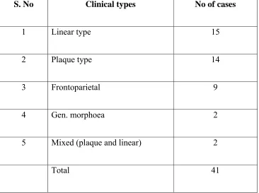
DIFFFERENT TYPES OF MORPHOEA AND THEIR INCIDENCE
Percentage of different types are as follows:
Linear – 36%
Plaque – 34%
Frontoparietal –20%
Generalized –5%
Mixed –5%
TABLE – 2
S. No Clinical types No of cases
1 Linear type 15
2 Plaque type 14
3 Frontoparietal 9
4 Gen. morphoea 2
5 Mixed (plaque and linear) 2
[image:39.612.138.504.362.635.2]AGE WISE DISTRIBUTION
Youngest person was 4 year old female child
Oldest person was 49 year old woman
Incidence of morphoea peaked between 10 to 25 years
Incidence below 10 years was 9%
Incidence below 20 years was 48%
[image:40.612.156.487.307.699.2]TABLE – 3
Age in years Females Males Total
0 - 4 0 1 1
5 - 9 3 0 3
10 - 14 6 2 8
15 - 19 7 1 8
20 - 24 3 5 8
25 - 29 0 1 1
30 - 34 2 0 2
35 - 39 3 0 3
40 - 44 0 1 1
45 - 50 4 2 6
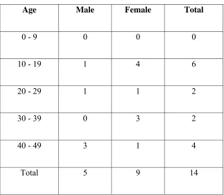
AGE WISE DISTRIBUTION OF PLAQUE TYPE
Incidence below 10 years was nil
Incidence between 20 to 50 years was 34%
TABLE - 4
Age Male Female Total
0 - 9 0 0 0
10 - 19 1 4 6
20 - 29 1 1 2
30 - 39 0 3 2
40 - 49 3 1 4
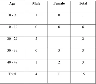
[image:41.612.163.486.283.564.2]AGE WISE DISTRIBUTION OF LINEAR TYPE
Incidence below 10 years was 2%
[image:42.612.164.486.286.565.2]Incidence below 20 years was 14%
TABLE - 5
Age Male Female Total
0 - 9 1 0 1
10 - 19 0 6 6
20 - 29 2 - 2
30 - 39 0 3 3
40 - 49 1 2 3
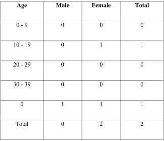
AGE WISE DISTRIBUTION OF GENERALIZED TYPE
Incidence between 11 and 50 years was 2%
TABLE - 6
Age Male Female Total
0 - 9 0 0 0
10 - 19 0 1 1
20 - 29 0 0 0
30 - 39 0 0 0
0 1 1 1
[image:43.612.162.486.248.527.2]AGE AND SEX WISE DISTRIBUTION FRONTOPARIETAL
Incidence in males and females are equal
Incidence between 10 to 30 years was 20%
TABLE - 7
Age Male Female Total
0 - 9 0 0 0
10 - 19 2 2 4
20 - 29 2 2 4
30 - 39 0 0 0
40 - 49 0 0 0
Total 4 4 8
[image:44.612.179.480.282.559.2]AGE AND SEX WISE DISTRIBUTION OF MIXED TYPE
Incidence of mixed type of morphoea was 5%
TABLE - 8
Age Female Male Total
0 - 10 2 0 2
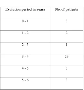
DURATION OF EVOLUTION OF MORPHOEA
[image:45.612.161.469.188.272.2]Average durations of evolution of morphoea was 2 to 3 years
TABLE – 9
Evolution period in years No. of patients
0 - 1 3
1 - 2 2
2 - 3 1
3 - 4 29
4 - 5 3
5 - 6 3
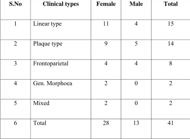
[image:45.612.174.454.401.700.2]SEX WISE DISTRIBUTION OF DIFFERENT TYPES OF
MORPHOEA
Incidence of morphoea was more in females
[image:46.612.120.493.307.581.2]The female to male ratio was 2 : 1
TABLE - 10
S.No Clinical types Female Male Total
1 Linear type 11 4 15
2 Plaque type 9 5 14
3 Frontoparietal 4 4 8
4 Gen. Morphoea 2 0 2
5 Mixed 2 0 2
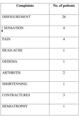
SYMPTOMS
[image:47.612.186.461.179.585.2]The most common presenting complaint was disfigurement - 63%
TABLE - 11
Complaints No. of patients
DISFIGUREMENT 26
SENSATION 4
PAIN 4
HEAD-ACHE 1
OEDEMA 1
ARTHRITIS 2
SHORTENNING 1
CONTRACTURES 3
ASSOCIATED CONDITIONS
[image:48.612.128.496.263.503.2]Associated disorders seen were as follows ;
TABLE - 12
Associated features No. of patients
MUCOSAL LICHEN PLANUS 1
VITILIGO 1
LICHEN SCLEROSUS ATROPHICUS 1
CAFÉ AU LAIT MACULES 1
NUMBER OF LESIONS SEEN IN MOST PATIENTS
The maximum number of lesion encountered in a patient – 6
[image:49.612.196.461.247.543.2]The number of patients with single lesions were 33 – (80% )
TABLE -13
No. of lesions No. of patients
1 LESION 33
2 “ 2
3 “ 2
4 “ 2
5 “ 0
6 “ 1
SITE COMMONLY INVOLVED
The most common site was lower limbs – 39%
Bilateral involvement of lower limbs was present in 2 patients
[image:50.612.177.434.270.583.2]The next most commonly affected area was head
TABLE - 14
FACE & SCALP 8
CHEST 2
ABDOMEN 6
BACK 1
UPPER LIMBS 4
LOWER LIMBS 16
EOSINOPHILIA
[image:51.612.143.512.262.502.2]Percentage of patients with eosinophilia was 66%
TABLE – 15
Eosinophils > 400/µl No. of Patients
TOTAL NO. OF PATIENTS
INVESTIGATED
41
TOTAL NO. OF PATIENTS WITH ↑
EOSINOPHILS
27
PERCENTAGE OF PATIENTS WITH ↑
EIOSINOPHILS
66%
PATIENTS WITH RAISED ESR
[image:52.612.127.511.164.347.2]Percentage of patients with raised ESR was 41%
TABLE – 16
(ESR > 30mm/hr) No. of Patients
TOTAL NO. OF PATIENTS WITH
INVESTIGATED
41
TOTAL NO. OF PATIENTS WITH ↑ ESR 17
PERCENTAGE OF PATIENTS WITH ↑ ESR 41%
HISTOPATHOLOGICAL EXAMINATION
Most of the patients showed feature of late morphoea
Percentage of patients with late morphoea was 80%
TABLE - 17
NO. OF PATIENTS WITH EARLY MORPHOEA 8
ANA POSITIVITY IN DIFFERENT TYPES OF MORPHOEA
( 1/10 ++,1/40 ++)
Percentage of patients with ANA positivity was 24%
[image:53.612.128.513.326.551.2]Percentage of ANA positivity in patients with linear morphoea was 14%
TABLE - 18
S.No. Different types of morphoea ANA positivity
1 LINEAR MORPHOEA 6
2 GEN. MORPHOEA 1
3 PLAQUE 2
5 FRONTOPARIETAL 1
TOTAL 10
RHEUMATOID FACTOR
Rheumatoid factor was positive in one patient – 24 units
C REACTIVE PROTEINS
C reactive proteins was positive in one patient – 320 ng/dl
ANA PATTERN
The most common ANA pattern seen was homogenous type
TABLE - 19
HOMOGENOUS PATTERN 9
SPECKELED PATTERN 1
PRECIPITATING FACTORS
Trauma, organophosphorus compounds and injections have precipitated
morphoea in following patients. Trauma (physical and iatrogenic factors) can
[image:55.612.167.473.211.316.2]precipitate morphoea.
TABLE - 20
TRAUMA 2
ORGANOPHOSPHORUS COMPOUNDS 2
INJECTIONS 2
RADIOLOGICAL ABNORMALITIES
Relevant radiological examination of patients with limb shortening,
frontoparietal and linear morphoea were undertaken. Radiological examination
of spine was carried out in all patients with linear morphoea.
The percentage of linear morphoea with spina bifida occulta was 6%.
TABLE - 21
TOTAL NO. OF PATIENTS WITH LINEAR MORPHOEA SCREENED
15
NO. OF PATIENTS WITH RADIOLOGICAL ABNORMALITY ( SPINA BIFIDA OCCULTA )
1
[image:55.612.137.511.542.696.2]DISCUSSION
In this study of 41 patients the incidence of morphoea was found to be 1
in 1,000. (Tab.1) Incidence of various clinical types of Morphoea are as
follows: (Tab. 2)
Plaque-35%
Linear-36%
Generalaized-5%
Mixed-5%
Frontoparietal-19%
A study by Christianson H.B. of 235 cases showed an incidence of 35%
of plaque type, and 49% of linear type and 19% of generalized morphoea.5
The youngest person in this study was a 4 year old female child and the
most aged person was a 49 year old woman. (Tab.3). In Christianson’s study
the youngest person was a 1 year old child and the oldest was a 76 year old
person. In this study, the maximum number of cases was between 10 to 25
years of age, whereas in Christianson’s study, the peak incidence was between
20 to 40 years. In this study the incidence of morphoea below 10 years was 9%
and below20 years was 48%. In Christianson’s study incidence below 10 years
In this study incidence of plaque type below 10 years was nil and
between 20 to 50 years was 34%. (Tab.4). In Christianson’s study incidence of
the same below 10 years was 10% and between 20 to 50 years was 75 %.
In this study incidence of linear type below 10 years was 2% and below
20 years was 7.2%. (Tab.5). Christianson’s study showed 10% incidence below
10 years and 75% below 20 years.
In this study incidence of generalized morphoea between 11 and 50 year
were 2% (Tab.6) whereas Christianson’s study showed 80 %.
Incidence of frontoparietal morphoea peaked between 10 and 30 years
of age. (Tab.7). Maximum incidence of mixed(linear and plaque) type of
morphoea was below 10 years of age. (Tab.8).
The average duration of evolution in this study was 2 to 3 years. (Tab.9).
In Christianson’s study, the average time was 3 to 5 years.
The incidence of morphoea in this study was more in females. The sex
ratio between females to males was 2:1. The sex ratio in Christianson’s study
was 3:1 male. (Tab.10).
The most common complaint of the patients was disfigurement. (63 %)
(Tab.11). In Christianson’s study 44% of patients presented with pain and
arthralgia whereas in this study pain in near by joints was the presenting
complaints in only two (5%) of the patients. One of them had hemiatrophy on
Four persons presented with loss of sensation. There was oedema preceding the
appearance of lesion in one patient. There was shortening of 1cm of the lower
limbs of one child due to soft tissue contracture. One patient with frontoparietal
morphoea complained of head ache and ophthalmologist diagnosed him of
having myopia and he was prescribed spectacles. Despite wearing spectacles he
continued to have head ache and after which he did not report for follow up.
Head ache may be an association of morphoea.146
Associated disorders seen in patients in this study includes mucosal
lichen planus, vitiligo, rheumatoid arthritis, café au lait macules and lichen
sclerosus et atrophicus. (Tab. 12). These associations have also been reported
in various other studies. Finklestein E, reported a case of vitiligo with
morphoea.135 Winkleman reported a case of lichen sclerosus with morphoea.136
Rheumatoid arthritis and lichen planus has also occurred frequently with
morphoea.146
The maximum number of lesions in one person was six. (Tab.13). Most
patients however had only single lesions. (80%)
The most commonly affected site was lower limbs. (39%) (Tab.14).
Two of the patients showed bilateral involvement of lower limbs. The next
common site was head and third most commonly affected area was the
abdomen and chest.
The serological investigations showed eosinophilia and raised ESR.
41% of patients. (Tab.16) A study conducted by Flagana showed similar
findings.137.
Histopathological examination of biopsy from patients showed features
of late morphoea in 33 patients (80%) and early morphoea in the rest (20%) of
the patients. (Tab. 17). In late morphoea there was no inflammatory infiltrates
and the epidermis was atrophied with loss of rete ridges. The collagen in the
dermis was thick, hypertrophied, homogenised, hyalinized and hypocellular.
There was also high uptake of eccrine glands. The glands were also atrophic
and adipocytes were absent. In early morphoea, inflammatory infiltrates was
seen extending up to the eccrine glands and also around perivascular spaces.
Endothelial swelling was also seen in blood vessals. Collagen bundles were
only slightly thickened.
ANA positivity was 24% in this study.(Tab-18). Signsen et al in his
study showed 40% positivity. He also showed that ANA positivity was more in
children and in patients with linear morphoea. In this study ANA positivity in
linear morphoea was 14%.
Rheumatoid factor was also positive in one patient of morphoea and
incidentally he was also having rheumatoid arthritis. He was also positive for C
reactive proteins.(320ng/dl). Singsen et al in his study has proved similar
association.
Among the 10 patients with ANA positivity, homogenous pattern was
There was history of trauma preceding lesions in 2 patients and there
was history of administration of injection(nature unknown) in 2 of patients
preceding the appearance of lesions. This shows that trauma could be
precipitating factor for development of morphoea as reported in
literature.87There were 2 farmers in this study with increased exposure to
organophosphorus compounds. (Tab. 20). No definite conclusions regarding
this factor could be drawn from the above history because of the small sample
size.
One child with linear morphoea showed spina bifida occulta on
radiological examination of the spine. (6%) (Tab.21) Christianson showed 47%
of association of spina bifida occulta with linear morphoea. Rubin et al also
CONCLUSION
* The incidence of Morphoea in Government General Hospital during the
period of September 2004 to September 2006 was 1 in 1000.
* The incidence of various types of Morphoea were as follows
PLAQUE TYPE 34%
LINEAR TYPE 36%
GENERALIZED TYPE 5%
MIXED TYPE 5%
FRONTOPARIETAL TYPE 20%
* The female to male sex ratio was 2:1.
* The maximum number of patients were in the age groups of 10 to
25years of age.
* Linear morphoea was more commonly seen in lower limbs.
* The main complaints of patients was disfigurement.
* One of the precipitating factors was found to be trauma.
* Serological investigations showed eosinophilia in 66% of patients.
* ANA was positive in 24% of patients.
* Homogenous pattern of ANA was most commonly seen.
* The associated autoimmune disorders seen were lichen planus, vitiligo
and rheumatoid arthritis.
* The other associated anomalies seen were spina bifida occulta, café au
lait macules, lichen sclerosus atrophicus.
* Histopathological study showed compatibility with late morphoea in
most of the patients.
1. Jablonska’S. The concept of scleroderma and its classification. In Jablonska Ed scleroderma and pseudo scleroderma. Warsaw polish medical publishers1975 (3-5)
2. The surgery of Henride Mandeville. Hand book of surgery. (769-770).
3. Curtis A.C. Jansen T.G. The prognosis, localized scleroderma, Arch dermatol Vol. 78, 1958, (749-755)
4. Peterson L.S, nelson A.M.S.U W.P, classification of Morphoea, Mayo clinic Proc 1995 Vol. (1068- 76)
5. Christianson H.B. Dorsey C.S. O Leary P.A. Kierland R.R. Localized scleroderma clinical study of 235 cases. Arch dermatol Vol. 74, 1956(6295-639).
6. Singsen B.H. scleroderma in childhood pediatric clinic of north America Vol. 33 No.5. Oct. 1986. (1119-113).
7. Rowel N.R. The collagen or connective tissue disease. In dermatology Vol. 11. Forth edition, EDs, Rook A. Wilkinson D.S. Ebling F.J.G.Champion R.H.Burton J.L.Blackwell scientific publication Oxford.1986-(1334-1343).
8. Eisner A2, vitto. J J. Bauer E A: scleroderma In: Dermatology in General Medicine. 3rd.ed. Fitzpatric. TB Eisner.
9. Falgana v. Localized scleroderma. Medical clinics of North America. Vol.73, No 5. September 1989.
10. Prasad PVS, Mathai R.Mary Jacobs: General. Linear morphea with bone cyst.Ind. J.D.V.L. Vol.56, No 4.1990. 308-309.
11. Bargava NC. Sings. Lin.scl. unilateral Ranaud’s phenomenon. IJDVL L.Vol.47.NO.3 1931 182—183.
12. Burnestein J. Medicer M. coexistence of localized Bullous pemphigoid, morphea, subcorneal pustules .Arch. Dermatol Novem. 1981 Vol.117. 725-727.
13. Pavithran. k. Linear morphea with hypertrichosis, entrapment neuropathy. IJDVL. Vol.47. NO: 1 .1981 45-48
Int.J.Dermatol. sept. 1988.vol.27. NO.7.487---490.
16. Peterson L.S nelson A.M., S.W.U.P et al The epidemiology of Morphoea ( localized scleroderma) in Olmsted Country 1960-1993, Rheumatology 1997-24 7380.
17. Rheumatology Mosby. Localised. Scleroderma idiopathic and environmental induced scleroderma variant .Johr Varga 1513---1516.
18. Grab J, sims. F. Scleroderma with Bullous lesions : report of acase and review of the literature .Dermatological 1959,119,341---59. 301
19. Pre - Wilson J, Pujol R M, Alejo.Metal Nodular keloidal scleroderma Int.J.Dermatol 1992; 31: 422—3.
20. Micalizzi, Purodi A Rebora A .Morphea with nodular lesions .Br.J.Dermatol 1994,131 298-301.
21. Frankel H. Ein. dermatologisch-neurologisches Genztall Nervenastz 1957,28,84. (Deep morphea).
22. Daoud, M,S.U,W.P.D, Leifornam K.M, Peniciceso C. Bullous morphea: clinical,pathological,and immunopathological evaluation of13 cases.J.Am Acaa distinct entity A M J Dermatopathol 1992: 16; 41.
23. Bizzero E. scleroderma Guttate, Lichen sclerois, Krausis penis Arch Dermatol Syphilol. 1943; 183, 493.
24. Farrel A.M, Marren. P.M, Wojnarowska F. Genital Lichen sclerosis associated with morphoea or systemic sclerosis: clinical and H.L.A charectoristical. Br. J. Dermatol 2000 143; 598-603.
25. Harrington C.F, Gulsthrope K, The association between lichen sclerosis et atrophicans and H.L.a B40. Bor J Dermatol 1981; 104 ;561-2.
26. Meyrick Thomas R.H., Ridley C.M, Black M.M. The association of lichen sclerosis et atrophicans and autoimmune related disease in males. Br. J. Dermatol 1983; 109. 661-4.
27. Azuridia R.M, Luzzi G.A.Bgren I et al.lichen sclerosis in adultman; a study of H.L.A association and susceptible to the autoimmune disease. Br. J. Dermatol 1999. 140; 29-83.
30. Ridley C.M. lichen sclerosis et atrophicans of the female genital tract. Arch Dermatol 1962. 85. 362-73.
34. Kencka. D, Blaszcyk. M, Jablonska. S. Atrophoderma Pasini- Pierni is primary atrophic abortive morphoea. Dermatology 1995. 190. 203-206.
35. Weines.M, in discussion on to Eshelman. O.m: Idiopathic Atrophoderma of Pierni and Pasini . Arch dermatol1965; 92 737.
36. Buechnu. S.A. Reyli. T. Atrophoderma of Pasini and Perini clinical and Histopathological findings and antibodies to Borrelia burgdorferi in 34 patients. J. Am Acad Dermatol 1994. 30. 44137][50] Bramley p, Forbes A.A. case of progressive hemiatrophy presenting with spontaneous fractures of the lower jaw. Br, J. 1960. I;1476-8.
37. Bramley p, Forbes A.A. case of progressive hemiatrophy presenting with spontaneous fractures of the lower jaw. Br, J. 1960. I;1476-8.
38. Husen C, Sakria A, Harms. M, Suareet J.H. Morphoea following Blaschko’s line. Br. J. Dermatol. 1996, 14, 594-5.
39. Mukhopadya Amiyakumar Linear scleroderma following Blaschko’s line I.J.D.V.L.2005 71 421-422.
40. Longacre, JI Wegner E.A. The surgical management of disabling contracture due to linear scleroderma. Plast Rconstruction surg. 1952 ; 9 367-80.
41. Soffa DJ, Sire DJ, Dodson JH. Melorhoessosis with linearscleroderma skin hanges. Radiology 1975 114; 577-9.
42. AlverezMjm, Lazano MA, Espada G. Barala H.A. Maldonado Cocco A. Linear scleroderma and melorhoestosis case presentation and literature review, clinical Rheumatology 1966, 15. 389- 393.
43. Wagers LT, Young Aw, Ryan SF< Linear melorhoestotic scleroderma. Br. J. Dermatol1072; 86 297- 301
44. Juhn BJ CHO YH, Lee Mh linear scleroderma associated with hyper trichosis in absence of melorhoestosis. Acta Dermatol Venereol 2000; 80. 62-3.
45. Verucken P, Stallenberg B Tas S. De Dobbleen, G. Huness, M. Ulcerared dystrophic, calcinosis cutis secondaryto Localized linear scleroderma. Int. J. clinic Prac 1998; 52; 593-4.