COMPARATIVE EVALUATION OF ANALGESIC EFFICACY OF CLONIDINE AS AN ADJUVANT TO BUPIVACAINE FOR
CAUDAL ANAESTHESIA IN CHILDREN
STUDY OF 60 CASES
DISSERTATION SUBMITTED FOR THE DEGREE OF
DOCTOR OF MEDICINE
BRANCH X (ANAESTHESIOLOGY)
APRIL -2011
THE TAMILNADU Dr. M.G.R MEDICAL UNIVERSITY
CHENNAI.
INSTITUTE OF ANAESTHESIOLOGY
ACKNOWLEDGEMENT
I am deeply indebted to Dr.S.P.MEENAKSHI SUNDARAM, MD.,DA., Director & Head of the Department of Anaesthesiology, Madurai Medical College, Madurai for the able guidance, inspiration and encouragement he rendered at every stage of this study.
I express my heartful gratitude to Dr.R.SHANMUGAM ,MD., Professor of Anaesthesiology, for his able guidance in doing this project.
My profound thanks to Dr.EDVIN JOE, M.D., Dean, Madurai Medical College and Dr.S.M.SIVAKUMAR, M.S.,Medical
Superintendent, Government Rajaji Hospital, Madurai for permitting me to conduct this study and to utilize the clinical materials of this hospital in the completion of my dissertation.
My sincere thanks to Dr. I.CHANDRASEKARAN M.D.,D.A., former Director, Institute of Anaesthesiology, for his encouragement and critical suggestions.
I sincerely thank my Assistant Professor Dr. S.SENTHIL KUMAR, M.D., D.A. for his valuable assistance and technical guidance in doing this study.
I am also thankful to my other Assistant Professors and my post graduate colleagues of the Institute of Anaesthesiology and Paediatric Surgery for their kind co-operation in doing this study.
CONTENTS
S.NO TITLE PAGE NO.
1 INTRODUCTION 01
2 HISTORY 03
3 AIM OF THE STUDY 04
4 ANATOMICAL CONSIDERATIONS 05
5 PHYSIOLOGICAL CONSIDERATIONS 09
6
PHARMACOLOGY
BUPIVACAINE 14
CLONIDINE 19
7 CAUDAL ANAESTHESIA 29
8 REVIEW OF LITERATURE 36
9 MATERIALS AND METHODS 44
10 OBSERVATIONS AND RESULTS 49
11 DISCUSSION 56
12 SUMMARY 60
13 CONCLUSION 61
BIBLIOGRAPHY PROFORMA
INTRODUCTION
“Pain following surgery is a universal phenomenon and it is not just slapping the patient on the face and telling him or her that ‘it is all over’. As for as the patient is concerned, it is just the beginning”(Berry, 1979)
Pain is often underestimated and undertreated, and its intensity varies among people. Inadequate knowledge regarding the necessity and adequate pain relief, combined with difficulty in assessment of pain has resulted in undertreatment of pain.
Pain is perhaps the most feared symptom of disease and man has tried his level best to discover methods to relieve pain. Effective analgesia reduces postoperative morbidity and mortality. Children need special care with regard to pain relief because of their lack of communicability.
The mode of pain relief should be such that it is technically less harmful, has minimal side effects, makes the surgeon comfortable, has extended post operative comfort and also cost effective. Parents should be satisfied when the child is given back to them at the end of surgery.
extended analgesia and to avoid the complications and side effects of general anaesthesia.
HISTORY
1885 – CORNING first used epidural analgesia
1901 – SICARD and CATHELIN described epidural injection through sacral hiatus.
1920 – ZWEIFEL was able to analyse 4200 caudal epidural blocks and recorded it in literature.
1921 – PAGES applied lumbar epidural analgesia in clinical surgery.
1933 – CAMBEL M.F first described sacral epidural block in children and infants.
1957 – synthesis of Bupivacaine.
1963- Bupivacaine was used in clinical practice.
1965 – MELZACK and WALL – propounded the Gate control theory of pain
1974 – KAY B used caudal block for postoperative pain relief in children
AIM OF THE STUDY
ANATOMICAL CONSIDERATIONS
EPIDURAL SPACE(Fig. 1)
The epidural space is a potential space within the bony cavity of the spinal cord and outside dural sac. It extends from the foramen magum to the coccyx. Within the cranium the endosteal and meningeal layers are united but below the foramen magnum, the two layers are separate, the outer becoming periosteal layer of the spinal cord, while the inner layer forms the spinal duramater. Between these two layers lies the epidural space. The spinal canal is triangular in cross section and the epidural space is widest in midline posteriorly in the lumbar region averaging about 5 to 6 mm in diameter. In the midthoracic region, the distance is somewhat less in the range of 3 to 5 mm in midline.
Boundaries of the epidural space:
Above: the foramen magnum, where the periosteal and spinal layers of the dura fuse together.
Below: the sacrococcygeal membrane
Behind: the anterior surface of vertebral lamina and ligamentum flavum
[image:9.612.160.442.206.469.2]Laterally: the pedicles of vertebra and intervertebral foramina
Fig. 1 EPIDURAL SPACE
ANATOMY OF SACRUM(Fig. 2)
It has concave anterior and convex posterior surfaces; the anterior surface bears four transverse lines (demarcating the boundaries between the fused bodies) which terminate on each side in four anterior sacral foramina. The anterior primary rami of the first four sacral spinal nerves, emerge from the anterior sacral foramina.
The posterior surface is convex and in midline runs a bony ridge, the median sacral crest with 3 or 4 rudimentary spinous process. The lamina of the 5th and sometimes the 4th sacral vertebra fail to fuse in the
midline. The deficiency thus formed is known as “sacral hiatus ”.The lateral margins of this space each bear a prominence – “sacral cornu” – which represents the remnant of the inferior articular process of the 5th sacral vertebra.
Sacral canal
It is a prismatic cavity running throughout the length of the bone and following its curves. Superiorly, it is triangular in its section and is continuous with the lumbar epidural space. Its lower extremity is the sacral hiatus, closed by posterior sacro-coccygeal membrane. Fibrous bands may be present in the canal and divide the epidural space into loculi which prevent the spread of solution and
these may account for occasional incomplete anaesthesia. The anterior wall of the sacral canal is formed by the fusion of the scaral vertebrae and posterior wall is by fusion of laminae.
Contents of the sacral canal
1.The dural sac extends and ends at the lower end of 2nd sacral vertebra on a line joining the posterior superior iliac spine
2.Sacral and coccygeal nerve roots with their dorsal root ganglia. 3.The filum terminale which is the continuation of piameter
4.Epidural plexus of veins formed by the lower end of vertebral veins. These vessels are numerous in anterior aspect than posteriorly and are valveless.
6.In infants the sacral canal is filled with fluid fat and areolar connective tissue, which allows easy spread of anaesthetic solutions.
7.In older children of 6 -7 years of age, the epidural fat becomes more densely packed, thus reducing this spread.
Sacral hiatus
This is a triangular opening present in the posterior wall resulting from failure of fusion of laminae of the 5th sacral vertebra and sometimes the 4th sacral vertebra. Its apex is at the level of the spine of 4th sacral vertebra. In some cases the apex is at the level of 3rd sacral spine, due to the absence of the 3rd and 4th laminae and occasionally the whole of the bony
posterior wall is deficient. When the laminae of the 5th sacral vertebra are almost fused in the midline, the hiatus may be very small with a diameter of as narrow as 2mm.
The hiatus is covered by the sacro-coccygeal membrane and pierced by the coccygeal nerves and the 5th sacral nerve. This membrane may be ossified in elderly subjects making the introduction of caudal needle difficult.
volume of the solution, force of injection, amount of leakage through the eight sacral foraminae and the connective tissue in the space.
The sacral canal may be wholly or partially obliterated by three anomalies. There may be a transverse fold in the posterior wall of the canal in conjunction with a forward projection of the corresponding segment of the sacral body, there may be a dorsal projection of a sacral vertebral body into the canal or there may be bony outgrowths which obliterate the hiatus. These anomalies may lead to incomplete caudal blockade or total failure.
PHYSIOLOGICAL CONSIDERATIONS
PAIN PATHWAYS
Pain receptors consist of peripheral plexus of unmyelinated nerves, activated by high-intensity stimuli which may be thermal, mechanical, electrical or chemical.
conducting(12-30m/sec). They conduct the sharp pain produced by pin prick or electrical stimulation as well as thermal stimuli and responsible for withdrawal reflex. A delta conducted pain is felt quickly and is well localised. ‘C’ fibres are very fine non-myelinated fibres which conduct at a very slow rate 2-3m/sec or less. Their threshold for stimulation is higher and is responsible for delayed and truly noxious burning or throbbing pain.
The activation of two different type of fibres(A delta &C) by noxious stimuli explain the double sensation for pain evoked in human by a single short noxious stimulus: rapid pricking pain(0.1sec, latency, first pain) carried by A delta fibres is followed approximately, 1 sec later by a burning pain (second pain) mediated by C fibres.
Peripheral sensory nerves have their cell bodies in the dorsal root ganglion and the central projection of A delta and C fibre neurones enter the dorsal horn in the lateral division of the dorsal root.
A delta fibres, give off axons which ascend in the contralateral anterior columns without synapsing with neurons from deeper layers. The majority of the pain fibres, however synapse in the substantia gelatinosa with intermediate neurons which send projections to deeper layers or with the dendrites of neurons whose cell bodies reside in deeper layers, principally in lamina V.
The central projection from cell bodies in lamina IV, V & VI with contribution from lamina I, cross midline in the anterior commissure to form the spinothalamic tract, which ends in thalamus, principally in the ventroposterior nucleus, sending a few fibres, enroute, to the periaqueductal grey matter. The ventroposterior nucleus of the thalamus projects to the post central gyrus, the sensory cortex (fig 4)
Pain stimuli can also pass via interneurons to cell bodies in the intermediate grey matter (laminae VII & VIII) whose central projections also ascend in the contralateral anterolateral columns, forming spinoreticular pathway.
While it appears that the thalamus is involved in the experience of pain, the post-central gyrus is necessary for its accurate localization and prefrontal cortex for unpleasant affective reactions to it.
INHIBITORY PATHWAYS:
stimulation of large A beta cutaneous afferents may inhibit pain transmission(Gate theory of Melzack and Wall)(Fig.5)
2. Inhibitory fibres, which descend in the dorsolateral white funiculus and whose cell bodies lie in the medullary raphe nuclei, may also inhibit pain transmission, presumably by an action of the inhibitory interneurons. Activity in these descending inhibitory fibres may be provoked by stimulation of the cell bodies in the medulla directly or of the periaqueductal grey matter.
3. Endorphins : long axons originating in the Hypothalamus release endorphins into the third ventricle, which is conveyed to the spinal cord through cerebrospinal fluid. This endorphin depresses pain conduction in Substantia gelatinosa.
4. Fig.5 GATE CONTROL THEORY OF PAIN 5.
7.
8. 9. 10.
PHYSIOLOGICAL CHANGES
certain cells, found principally in lamina IV respond only to a low intensity of stimulation such as light touch: These are termed low threshold or LT cells. They respond maximally to low- threshold stimuli and do not increase their firing rate with increased stimulus intensity. They are therefore incapable of conducting pain. Another type of cell, found principally in lamina V, responds over a wide range stimulus intensities, the so called
wide-dynamic range or WDR cells. Noxious stimuli can excite a variable response in WDR cells in lamina V. A third type of cell is responsive to stimuli only within the noxious range. Such cells are known as High threshold cells or HT cells and are found mainly in lamina I. A delta and C fibre stimulation in the periphery results in increased firing in HT and WDR cells in lamina I and Vwhich is conducted up in spinothalamic tract.
Surgery produces local tissue damage with consequent release of algesic substances like prostaglandins, histamine, serotonin, bradykinin, hydroxytryptamine, substance P and generation of noxious stimuli that are transduced by nociceptors and transmitted by A delta and C fibres to the neuraxis.
The majority of the opioid receptors in the dorsal horn are µ receptors although Delta and Kappa receptors are present.
Segmental reflex responses associated with surgery includes increased skeletal muscle tone and spasm with associated increase in O2 consumption and lacticacid production. Stimulation of sympathetic neuron cause tachycardia, increased stroke volume, cardiac work and myocardial oxygen consumption.
Suprasegmental reflex responses result in further increase in sympathetic tone and hypothalamic stimulation, metabolism and increased O2 consumption. Hence the most obvious motive for relieving postoperative pain is not only humanitarian but also contribute to more rapid and complete postoperative recovery after surgery.
APPLIED PHARMACOLOGY
Bupivacaine was synthesised in Sweden by Ekenstam and his colleagues in 1957. It was introduced into clinical practise by L J Telivuo in 1963.
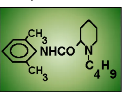
The structure of Bupivacaine is the same as Mepivacaine, with a Butyl group replacing a methyl group in the Piperidine ring. This increases lipid solubility and protein binding. High potency is associated with high lipid solubility.( fig.6)
PHYSICO CHEMICAL PROPERTIES
Molecular Weight : 288 (base) Pka at 25°C : 8.1
Percentage of protein binding : 95.6% Plasma protein binding : 2 µgm/ml
Partition coefficient :27.5 (n-Haptane pH7.4 buffer) Apporoximate anaesthetic duration: 175minutes.
Site of metabolism : liver
Safe dosage : 2 - 3 mg/kg
Toxicity :4-6 times more toxic than Lignocaine.
been reported to relieve the pain of labour. Sensory blockade with very minimal motor blockade is
Fig. 6 BUPIVACAINE
provided by 0.25% solution. Motor blockade is provided by 0.5% solution. Bupivacaine has a much more pronounced effect upon sensory nerves than motor nerves, and intense anaesthesia may often be obtained without any motor blockade. This is a special advantage in the treatment of pain such as post operative, post traumatic and labour pain.
Action on the cardio vascular system
Bupivacaine is relatively more cardiotoxic than Lignocaine. It is a powerful myocardial depressant and this is made worse by hypoxia, hypercarbia and by pregnancy. Ventricular arrhythmias including ventricular fibrillation are more lethal forms of Bupivacaine toxicity. Bupivacaine induced convulsions is probably due to its high lipid solubility. According to Moore et al (1979), an arterial plasma concentration of 5. 4 µgm/ml following an intravenous bolus of Bupivacaine resulted in convulsions. The threshold arterial plasma level for Bupivacaine-induced convulsions is 4µgm/ml.
Safety dosage: (2.5mg/kg)
µgm/ml were reached, which are well within the limits of projected toxic levels.
METABOLISM
Breakdown of bupivacaine is similar to that of mepivacaine and commences with removal of the piperidine side chain. The product
pipecolyxylidine (PPX) is approximately one-eighth as toxic as bupivacaine. PPX and unchanged bupivacaine are slowly excreted in about equal proportions in the urine. Reynolds (1971), Boyes (1975) reviewed the metabolism of Bupivacaune. Hyroxylation of the aromatic ring is believed to take place in case of Bupivacaine as with Lignocaine to produce a compound that can be conjugated and made water soluble. Rothenstein (1983) demonstrated that the human lungs extracted local anaesthetic from circulation and subsequently released it back into circulation. It is seen with Bupivacaine also. For Bupivacaine the first pass pulmonary extraction is dose dependent suggesting that uptake process become saturated rapidly (Rothenstein 1984).
MECHANISM OF ACTION OF LOCAL ANAESTHETICS The sequence whereby clinically used local anaesthetics produce inhibition
of axonal conduction has been summarised by Corvino as follows.
1. Clinically all local anaesthetics exist in solution in both charged and uncharged forms, the relative proportions depending on the pH of the solution, the pH at the site of injection and Pka of each drug. The cation is responsible for most of the nerve blocking effect.
2. The uncharged lipophilic tertiary base form diffuses more readily across neural sheaths and the axonal membrane to reach the internal aspect of the sodium channel. The base is protonated within the cytoplasm and binds as the charged cation to a specific receptor within the internal opening of the sodium channel and thereby inhibiting sodium conductance. The loss of membrane permeability to sodium prevents cell membrane depolarisation and propagation of action potential.
3. The clinically used local anaesthetics act primarily on specific receptors located at the internal opening of sodium channel. Other possible sites of action include,
A) Non-specific absorption within the cell membrane lipids resulting in
B) Diffusion of the uncharged base via hydrophobic pathways through membrane lipids to reach the specific receptor site, where protonation and binding occur within the internal opening of the sodium channel.
1.The surface charge theory: this theory states that, the lipophilic portion of the anaesthetic molecule, penetrates the axonal membrane and the positively charged terminal aminoacid group of the molecule neutralises the negative charges present on the axolemmal surfaces. An accumulation of suuficient positive charges would tend to neutralise the relatively electronegativity of the external membrane surface, resulting in an increase in the trans membrane potential without altering the intracellular resting potential. A sufficient increase in the transmembrane potential would inhibit the ability of an electric current from a nearby unanaesthetised portion of the nerve membrane to depolarise the treated area to its threshold for firing. Conduction blockade would then result. The surface charge theory requires that the charged form of local anaesthetic be the active form.
PHARMACOLOGY OF CLONIDINE HYDROCHLORIDE
Clonidine hydrochloride is a centrally acting selective partial α₂ agonist introduced in early 1960s, It was during its use as a nasal decongestant that its anti-hypertensive property was found out. Subsequently more insights into the pharmacological properties has led to its use in clinical anaesthesia practice as well.
Clonidine hydrochloride is an imidazoline compound and exists as a mesomeric compound. The chemical name is 2-(2,6- dichlorophenylamino)-2-imidazoline hydrochloride. The structural formula is C9H9C12N3HCl.(Fig.7)
The molecular weight is 266.56. Clonidine is an odourless, bitter, white, crystalline substance, soluble in alcohol and water. Clonidine improves the quality of anaesthesia, provides a more stable cardiovascular course during anaesthesia and reduces the dose requirement of the anaesthetic agent. In fact Clonidine may reduce the MAC of halothane by upto 50% in a dose dependent manner. Clonidine potentiates the anaesthetic action of the local anaesthetics with fewer side effects in peripheral nerve blocks and central neuraxial blockade.
Availability: Available as 1ml ampoule containing 150 micrograms. It should be stored below 25oc. Tablet form is available for oral use. A transdermal delivery system is also available.
Mechanism of action:
central neural transmission in the spinal neurons. Inhibition of substance- P release is believed to be involved in the analgesic effect.
The α₂ adrenoreceptors are located on the afferent terminals of both peripheral and spinal neurons in the superficial laminae of the spinal cord and within several brain stem nuclei implicated in analgesia. The superficial laminae contain three group of neurons: tonic, adapting, single- spike firing, all of which receive their primary sensory input from Aδ and C fibres. Studies in rat models show that Clonidine inhibits voltage gated Na+ and K+ channels and suppresses the generation of action potentials in tonic- firing spinal dorsal horn neurons, contributing to analgesic effect.
Another contribution to analgesic effect may be through the release of acetylcholine in the neuraxial region. The α2 adrenergic agonists also enhance analgesia from intraspinal opioids. Sedation is produced by its action on locus ceruleus.
anti- arrythmogenic action. In the periphery it acts on pre-synaptic α₂adrenoreceptors at sympathetic terminals and reduces the release of nor-epinephrine, causing vasorelaxation and reduced chronotropic drive. The brainstem and the peripheral effects of α₂ adrenoreceptor stimulation are counterbalanced by the direct peripheral vasoconstriction through its action on α₂ adrenoreceptors from the circulating concentrations of Clonidine.
As a result the dose response for Clonidine by neuraxial or systemic administration is U-shaped, with peripheral vasoconstriction from circulating drug concentrations at high doses opposing central sympatholysis.
PHARMACODYNAMICS:
The analgesic effect of Clonidine is more potent after neuraxial administration indicating a spinal site of action, favouring neuraxial administration, though it is possible to achieve analgesia from systemic administration as well.
General:
Cardiovascular system:
Clonidine has minor or no effects on responses to vasoconstrictors or atropine given to treat hypotension or bradycardia respectively, that may occur with neuraxial anaesthesia.
Sedation:
This is a desired property. Clonidine produces a dose dependent sedation at the dose of 50µg or more in less than 20 minutes regardless of the route of administration.
Respiration:
Clonidine doesn’t induce profound respiratory depression even after massive overdose nor does it potentiate respiratory depression from opioids.
Peripheral nerves:
It produces a minor degree of blockade at high concentrations with some preference for C- fibres in the peripheral nerves and this effect in part enhances the peripheral nerve block when added to local anaesthetics, probably because the α₂ adrenoreceptors are lacking on the axons of peripheral nerves.
Clonidine is well absorbed orally and is nearly 100% bio available. The mean half life of the drug in plasma is about 12 hours. It is excreted in an unchanged form by the kidney, and its half- life can dramatically increase in the presence of impaired renal function.
A transdermal delivery system is available in which the drug is released at a constant rate for about a week. Three or four days are required to achieve steady state concentration.
Clonidine is highly lipid soluble and readily distributes into extra- vascular sites including the central nervous system.
Metabolism : it is metabolised through minor pathways with the major metabolite, p- hydroxyclonidine.
Excretion: 70% of the dose, mainly in the form of unchanged parent drug (40-60%)is excreted in urine.
So, the elimination t1/2 of Clonidine varies as a function of creatinine clearance. In subjects undergoing hemodialysis only 5% of the body Clonidine store was removed.
The general adverse effects include weakness, fatigue, headache and withdrawal syndrome, pallor, a weakly positive coomb’s test and fever.
1.Cardiovascular: palpitations, tachycardia, bradycardia about 5 in 1000, syncope, Raynaud’s phenomenon, congestive heart failure, ECG abnormalities like sinus node arrest, junctional bradycardia , high degree AV block and arrhythmias are reported rarely.
2.Central nervous system: nervousness, agitation, mental depression, insomnia, vivid dreams or night mares, anxiety, visual and auditory hallucinations have been reported rarely.
3.Dermatological: rash, angioneurotic edema, pruritus, urticaria and alopecia rarely.
4.Gastro intestinal tract: nausea and vomiting, anorexia, malaise, transient abnormalities in liver function tests, hepatitis, parotitis and constipation.
5.Genitourinary: decreased sexual activity, impotence, loss of libido, nocturia.
6.Hematlogic: thrombocytopenia rarely.
7.Metabolic: weight gain and gynaecomastia,
9.Oro-otolaryngeal: dryness of the nasal mucosa.
10.Opthalmological: dryness, burning of eyes.
PRECAUTIONS:
In patients with renal insufficiency, lower dose is needed. Sudden withdrawal of clonidine after prolonged continuous epidural infusion, produces hypertensive crisis. So the drug should be gradually discontinued over 2 to 4 days. It is used with caution in patients with cerebrovascular or coronary insufficiency. When infused into upper thoracic spinal segments clonidine produces pronounced decrease in blood pressure.
If a patient on beta blocker therapy is given clonidine as continuous epidural, beta blocker should be withdrawn several days before instituting epidural Clonidine.
Intrathecal and epidural Clonidine often causes bradycardia and if symptomatic it can be treated with inj. Atropine.
CONTRAINDICATIONS:
Epidural Clonidine above C4 level is contraindicated because there are no safety data to support such use.
INTERACTIONS:
Clonidine may potentiate the CNS- depressive effect of alcohol, barbiturates or other sedative drugs. Narcotics may potentiate the hypotensive effects of Clonidine. Tricyclic antidepressants may antagonize the hypotensive effect of Clonidine. Concomitant administration of drugs with a negative chronotropic or dromotropic effect (beta blockers, digoxin) can cause or potentiate bradycardiac rhythm disturbances. Beta blockers may potentiate the hypertensive response seen with Clonidine withdrawal. Epidural Clonidine may prolong the duration of pharmacologic effects of epidural local anaesthetics, opioids, neostigmine and other drugs.
INDICATIONS:
1. To prolong the duration of epidural or spinal anaesthesia and Peripheral nerve block
2. As adjuvant for the treatment of intra operative and post operative pain.
3. Treatment of intra articular pain.
6. For sedation.
7. To prevent or treat shivering. 8. Treatment of hypertensive crisis
Anaesthetic uses:
1.Premedication: clonidine produces sedation by acting on locus ceruleus. It also produces anaesthesia- sparing effect.
2.Control of hemodynamics: clonidine prevents hypertension and tachycardia during laryngoscopy and intubation as well as during surgical stimulation. Hence there is a decreased incidence of myocardial ischemia in cardiac and vascular surgeries.
3.Epidural:clonidine is used as a sole agent or in combination with opioids or local anaesthetics to provide excellent analgesia for labour pain and also for treating postoperative pain.
4.Spinal: when used with local anaesthetics, Clonidine improves the quality and duration of the block, minimizes the tourniquet pain during lower limb surgery, and prevents shivering.
6.Peripheral nerve blocks: clonidine prolongs the duration of anaesthesia and analgesia with local anaesthetics by two times in a dose of 75 to150 µgm.
7.Bier’s block: 150µg enhance the tolerance of tourniquet.
Overdosage and treatment:
There is no specific antidote for Clonidine overdosage. Supportive measures like atropine, ephedrine, i.v fluids is enough. For hypertensive crisis i.v furosemide, diazoxide, phentolamine can be used.
Yohimbine partially reverses the analgesia and sedation but not the BP and heart rate changes produced by the epidural Clonidine.
Naloxone may be a useful for the management of Clonidine -induced respiratory depression, hypotension and coma.Blood pressure should be monitored after injecting naloxone as it may produce paradoxical hypertension.
CAUDAL ANAESTHESIA
Selection of equipment:
characteristics of the needle are – bevel, internal &external diameter, length, presence of stylet
Sharp bevelled needle:
The advantage of a sharp bevel needle is that it traverses easily through the tissues. But the disadvantages are,
• Characteristic “giveway” while puncturing sacrococcygeal membrane may not be clearly felt with sharp needles
• Sharp needles have long bevel and hence it may have to be advanced further into the epidural space so that it lies entirely within it
• Cartilaginous sacrum can be easily traversed by a sharp and long bevelled needle which can lead to rectal puncture or iliac vessel puncture
Hence straight tipped needle with a bevel of 45-60 degree is ideal
Diameter: Small needles may bend and break during the procedure.
Thus, 21 to 23G is ideal because it is rigid and large enough to allow reflux of blood or cerebrospinal fluid.
30mm. Needle with stylet ,prevents formation of an epidermoid tumour due to skin tag.
Epidural needle with 20 to 22G are employed when one intends to use an epidural catheter via caudal route to achieve anaesthesia at higher level after radiographic confirmation
Determination of the volume of local anaesthetics:
The height of block – depends on the volume injected
Formula based on weight or age:
Armitage, 1979
High sacral – 0.5 ml/Kg
High lumbar – 1ml/kg
Thoracic level – 1.25ml/kg
Sclhute-Steinberg formula (upto 8-12 years), 1977
For children less than 7years, weight will be the best predictor
Volume required in ml = 0.65 X number of segments to be blocked x body weight(kg)
Speigal formula:
Where D is the distance separating the sacral hiatus from the spinous process of 7th cervical vertebra
Modified Spiegal formula:
Volume of injection (ml) = 4+(D-13)/2
Despite larger volumes of local anaesthetics used in children as compared to adults, peak plasma levels of the local anaesthetics in children remain far below the toxic levels in adults
As the child grows, the epidural space become less compliant and hence a large volume can cause high spread of solution and an increase in the CSF concentration. Normal volume recommended for injection is 20ml
Patient position:
Lateral decubitus position or prone position with pillows underneath the hip and the foot inverted to relax the gluteal muscles is used to perform the caudal technique.
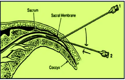
Anatomical landmarks(Fig. 8)
The posterior superior iliac spines form an equilateral triangle with the sacral hiatus. Intergluteal fold is not an ideal landmark because it will not always
correspond to the midline. Left forefinger placed in coccyx tip, hiatus corresponds to the second crease of finger. Palpation of this membrane gives a characteristic feel of a membrane under tension similar to that of a fontanelle, the point of puncture is at the midpoint of this triangular space
TECHNIQUE(Fig. 9)
more than 2 -3 mm so as to ensure that the entire bevel is within the sacral canal.
Confirmation of space:
Whoosh test: this is done by injecting 7ml of air via the needle and auscultating just proximal to injection site, where a whoosh sound is heard.
[image:43.612.112.516.464.723.2]Swoosh test: Auscultation at a site just proximal to hiatus, while injecting local anaestheic produces a swoosh sound. The swoosh test is useful in children which avoids air injection into the epidural space. Air injection may cause a patchy block, venous air embolism and a rare complication of pneumocephalus if a large volume of air is injected.
Injection of drug:
After gentle aspiration, the drug is injected over a period of 60-90sec, irrespective of the volume injected (0.023-0.33ml/sec). The syringe should be repeatedly aspirated during the course of injection
The patient is monitored for any change in BP or heart rate. Faster injection causes increased cephalad spread resulting in high block and respiratory problems. Transient increase in intracranial pressure with transient loss of consciousness or headache can occur.
On the other hand, too slow an injection increase the chances of lateralisation of the block or a lower level of anaesthesia since the drug tends to leak through the foramina or increase the risk of needle displacement
Indications:
Ideal for lower abdominal or lower limb surgeries
Emergency: testicular torsion, strangulated hernia repair, paraphimosis, wound debridement of pelvis or lower limbs
Repair of inguinal hernia, umbilical hernia, hydrocele, orchidopexy, anorectal, genitourinary surgery, pelvic or hip or lowerlimb surgery, phimosis.
Contraindications:
Hydrocephalus, seizure disorders, vertebral osteosynthesis.
Local skin infection, pilonidal sinus near hiatus, major sacral malformation - meningomyelocele, meningitis.
Spinabifida occulta is not a contraindication
COMPLICATIONS
Possibilities due to errors in technique:
i. Subcutaneous injection
ii. Vascular puncture: 10-15%. Since epidural veins are valveless, injection is immediately followed by convulsions, arrhythmias, hypotension, respiratory depression.
iv. Bone marrow or rectal or intra osseous injection.
Complete or partial failure of the block
More common in > 7 years old
Success rate increases or failure rate decreases with experience
Lateralisation: occurs in 1 in 1000 cases
When caudal is performed in lateral decubitus, 50% have a level of anaesthesia 2 dermatomes higher on the dependent side.
When the injection is given slowly, the difference may be more than 4 dermatomes ; this may be due to the presence of a complete plica mediana dorsalis
Unanaesthetised dermatomes: L5, S1 may go unanaesthetised because of the large size.
Other complications are Vomiting, epidural infection or meningitis, and shivering.
REVIEW OF LITERATURE
Fifty-three children (1–72 mo) scheduled for inguinal hernia repair were caudally injected with either S(+)-Ketamine 1mg/kg alone (Group K) or with additional Clonidine (Group C1 =1 µg/kg) Group C2 =2 µg/kg) during Sevoflurane anesthesia via a laryngeal mask. The mean duration of postoperative analgesia was 13.3 ± 9.2 h in Group K, 22.7 ±3.5 h in Group C1, and 21.8 ± 5.2 h in Group C2. Groups C1 and C2 received significantly fewer analgesics in the postoperative period than Group K. 2. Caudal bupivacaine supplemented with caudal or intravenous
Clonidine in children undergoing hypospadias repair: T. G. Hansen,
S. W. Henneberg, S.Walther‐Larsen, J. Lund and M. Hansen et al .
British journal of Anaesthesia 2004; 92: 223–7 Forty‐six children (ASA I or II) aged 24–104 months received standardized premedication with midazolam, a general anaesthetic and a caudal block with Bupivacaine 0.25%, 0.5 ml/ kg. The children were randomized in a
double‐blind fashion to two groups: the i.v. group received Clonidine 2
µg/ kg i.v. and simultaneously the same volume of saline caudally. The caudal group received Clonidine 2 µg/ kg caudally and a similar volume of saline i.v. They concluded that the analgesic effect of clonidine 2 µg /kg as an adjunct to caudal block with Bupivacaine 0.25%, 0.5 ml/ kg is similar whether administered i.v. or caudally.
3.Analgesic effect of clonidine added to bupivacaine 0.125% in
paediatric caudal blockade. Joshi W, Connelly NR, Freeman K, Reuben SS. Paediatric Anaesthesia. 2004 Jun;14(6):483-6. 36 children undergoing elective surgery were studied. Following anaesthetic induction, a caudal was placed (1 mg/kg of bupivacaine 0.125%) with an equal volume of either clonidine (2µ.kg) or saline. There was no difference in analgesic duration between groups. There were significantly more children who vomited during the first 24 postoperative hours in the clonidine group than in the saline group (eight in clonidine, two in saline; P < 0.05).In this study, they do not recommend adding clonidine (2 microg.kg(-1)) to a bupivacaine (0.125%) caudal block in children undergoing surgery.
4. Clonidine increases duration of bupivacaine caudal analgesia for
ureteroneocystostomy: a double-blind prospective trial Tripi PA, Palmer JS, Thomas S, Elder JS. Journal of Urology. 2005
second caudal block was performed with half of the original dose of medications. They concluded that the addition of Clonidine to Bupivacaine significantly increases the duration of caudal analgesia and decreases postoperative parenteral morphine requirements in children undergoing ureteroneocystostomy.
5.A Comparison of Single-Dose Caudal Clonidine, Morphine,or Hydromorphone Combined with Ropivacaine in Pediatric Patients Undergoing Ureteral Reimplantation Thomas R. Vetter, MD Daniel Carvallo, MD Jodie L. Johnson, MD Michael S. Mazurek, MD Robert G. Presson, Jr., Anesthesia Analgesia 2007;104:1356 –63
Patients aged 6 mo to 6 yr were evenly and randomly enrolled in a double-blind manner. Patients received a single caudal dose of 2 mcg/kg of Clonidine, 10 mcg/kg of hydromorphone, or 50 mcg/kg of morphine, combined with 1.0 mL/kg of 0.2% Ropivacaine with epinephrine. No difference was observed in pain scores, total morphine use, time to first oral intake or discharge home. No postoperative respiratory depression, e xcessive sedation, hypotension, or bradycardia was identified.
The study was conducted with forty-six children, aged 1 to 8 years scheduled for open reduction of the hip. Group I caudal block was with bupivacaine 0.25% 1.4 mL/kg and group 2 caudal block was with clonidine 2 µg/kg plus bupivacaine 0.25% 1.4mL/kg. Anesthesia was maintained with sevoflurane 1-1.5 vol%. Group 2 had longer duration of analgesia than group 1 (13.06 vs 7.65 h). The blood pressure and cardiac rate were lower in group 2 (p<.05Group 2 had longer sedation. They concluded that adding clonidine (2 µg/kg) to bupivacaine (0.25% 1.4 ml/kg) provided good post-surgical analgesia without significant haemodynamic changes.
7.The Effect of Bupivacaine-Clonidine Combination in Quality and Duration of Caudal Block in Children between 2-7 Years Old SEYEDHEJAZI M, MD, KALAMI L, MD NEGARGER S, MD
ASLANADADI S, NAGIZADEH S, MD Journal of Tabriz University
of Medical Sciences, Vol. 30 No. 2,Summer 2008: 100 children aged 2-7 years, scheduled for elective lower abdominal surgery, were assigned in two equal groups to receive 0.25% bupivacain 1ml/kg combined with either clonidine 2μg/kg (group 1)or 1ml normal saline (group 2). 3 patients (6%) in group 1(first 1-2hours) and 21patients(42%) in group 2(up to
there were significant differences between two groups in the mean of systolic blood pressure (P<0.01), diastolic blood pressure(P<0.013). and heart rate(P<0.001) it was concluded that when added to bupivacaine, clonidine improves the efficacy and lengths of caudal analgesia in children.
8.Addition of Clonidine or Dexmedetomidine to Bupivacaine
prolongs caudal analgesia in children A. M. El-Hennawy, A. M.
Abd-Elwahab, A. M. Abd-Elmaksoud, H. S. El-Ozairy and S. R. Boulis Br J
Anaesth 2009; 103: 268–74 Sixty patients (6 months to 6 yr) were
randomly assigned into three groups. each patient received a single caudal
dose of Bupivacaine 0.25% (1 ml/kg) combined with either
Dexmedetomidine 2 µg/kg in normal saline 1 ml, Clonidine 2 µg/ kg in
normal saline 1 ml, or corresponding volume of normal saline according
to group assignment. Addition of Dexmedetomidine or Clonidine to
caudal Bupivacaine significantly promoted analgesia time 16hr (14–18)
and 12 (3–21) h, respectively] than the use of Bupivacaine alone[5 (4–6)
h].However, there was no statistically significant difference between
dexmedetomidine and clonidine as regards the analgesia time
9. Low Dose IntrathecalClonidine With Bupivacaine Improves Onset
And Duration Of Block With Haemodynamic Stability: Archna Koul, Deepanjali Pant, Jayshree Sood et al Indian Journal of
undergoing inguinal hernia repair were randomly given caudal injection with 0.75 ml.kg -1 of Bupivacaine (0.25%) and Clonidine 2 µg.kg -1 in Group C or 0.75 ml.kg -1 of Bupivacaine (0.25%) alone in Group B after induction of anaesthesia. Postoperatively duration of analgesia, OPS score (observational pain / discomfort scale), Sedation score, heart rate and blood pressure were recorded. In this study duration of analgesia was significantly longer (p< 0.001) in Group C (10.25 hours) as compared to 4.55 hours in Group B. Bradycardia, hypotension and sedation were not observed in Group C. The addition of Clonidine in caudal blocks prolongs postoperativepain relief in children and is safe alternative to Bupivacaine alone in paediatric daycare surgeries
0.2% provides a longer duration of analgesia and sedation compared with caudal morphine 30 µg /kg in Bupivacaine 0.2% without significant side-effects
11.Caudal Ropivacaine-Clonidine :A better postoperative analgesic
approach Sukhminder Jit Singh Bajwa, Jasbir Kaur, Sukhwinder Kaur Bajwa, Geetika Bakshi et al . Indian journal of anesthesia 2010 54(3)
:226-230 A total of 44 ASA-I paediatric patients between the ages of 1 and 9 years, scheduled for elective hernia surgery, were enrolled in this randomised double-blind study. The caudal block was administered with Ropivacaine 0.25%(Group I) and Ropivacaine 0.25% and Clonidine 2 µg/kg (Group II) after induction of general anaesthesia.They concluded that, a caudal block with 0.25% of isobaric Ropivacaine combined with 2 µg/kg of Clonidine provides efficient analgesia intra-operatively and prolonged duration of analgesia post-operatively
group received 15µg of Clonidine (group2), 30µg Clonidine (group3), 37.5µ of Clonidine (group4) made to 3ml volume with13.5mg of 0.5% hyperbaric Bupivacaine. The onset of block was fast , duration of analgesia & motor block was longer in all Clonidine groups.
MATERIALS AND METHODS Study design:
This is a prospective, randomised, double blind, comparative study, done to compare the efficacy and safety of caudal Clonidine with Bupivacaine and plain Bupivacaine for intraoperative and postoperative analgesia in children.
The study was carried out in 60 children at Govt.Rajaji hospital, Madurai for surgeries of lower abdomen and perineum lasting for 45 – 90 minutes, in the year 2009-2010.
Inclusion criteria:
The children in the age group of 1 -10 years and weighing 5-20Kgs were selected for the study. Only patients of ASA I physical status were chosen to avoid the influence of the associated diseases. They were divided into 2 groups of 30 each. Group B received caudal Bupivacaine and group BC received caudal Clonidine with Bupivacaine.
i. H/o allergic reaction to local anaesthetics
ii. Bleeding diathesis
iii. Neurological disease
iv. Local sepsis
v. Grossly abnormal sacral anatomy
Preanaesthetic evaluation:
1. History
2. Clinical examination
3. Relevant investigations – haemoglobin, urine analysis
4. Informed consent from parents
5. All children were kept nil per oral for 6 hours prior to surgery
Induction and maintenance:
After recording the pulse, blood pressure and saturation, an intravenous line was secured and isolyte-P infusion started. Mask induction was done with incremental doses of Halothane and 70% Nitrous oxide in Oxygen
area was thoroughly cleaned and draped. A 23G needle was inserted in the hiatus at an angle of 45°.A distinct pop off was felt once the sacrococcygeal membrane was pierced, after which the angle of the needle was lowered to 20° and advanced to 2-3mm. After negative aspiration for blood and CSF, the drug was injected. Following injection, the patient was placed in the supine position and 10minutes later, the surgeons were allowed to proceed. Anaesthesia maintained with 0.5% Halothane and 70%N₂O in O₂ and patient breathing spontaneously.
Group I :
These children received 1ml/Kg of 0.25% Bupivacaine ( 0.9% NaCl as diluent ) total dose not exceeding 3mg/Kg
Group II:
These children received Clonidine 2µg/Kg with 0.25% Bupivacaine to a total volume of 1ml/Kg.Total volume not exceeding 20ml
Intraoperative monitoring:
Heart rate, blood pressure, saturation were monitored.
At the beginning of skin closure, anaesthesia was discontinued.
Postoperative monitoring: wasdone at every 15 minutes for 3 Hours i. Heart rate
ii. Saturation (SpO₂) iii. Blood pressure
iv. 4 point sedation score v. Objective pain scale score vi. First supported standing
Side effects like nausea, vomiting, bradycardia, respiratory depression were noted.
If the child complained of pain or if the objective pain scale score was more than 11,rectal Paracetamol(15mg/Kg) was given as rescue analgesic.
4 point sedation score
1 – Asleep, not arousable by verbal contact 2 – Asleep, arousable by verbal contact 3 – Drowsy/ not sleeping
OBJECTIVE PAIN SCALE SCORE
PARAMETERS FINDING POINTS
SYSTOLIC BP
+ 10% before operation + 20% before operation + 30% before operation
1 2 3
CRYING
Not crying
Crying but responds to tender loving care (TLC)
Crying and no response to TLC
1 2 3 MOVEMENTS None Restless Thrashing 1 2 3 AGITATION
Patient asleep or calm Mildly agitated Hysterical 1 2 3 COMPLAINS OF PAIN
Asleep or states no pain Cannot localize pain Can localize pain
OBSERVATIONS AND RESULTS
The observation was recorded for both the groups as shown in the master chart
Statistical tools
The information collected regarding all the selected cases were recorded in a Master Chart. Data analysis was done with the help of computer using Epidemiological Information Package (EPI 2008).
Using this software, range, frequencies, percentages, means, standard deviations, chi square and 'p' values were calculated. Kruskul Wallis chi-square test was used to test the significance of difference between quantitative variables. A 'p' value less than 0.05 is taken to denote significant relationship.
RESULTS
Table 1 shows the demographic variables of the patients selected for the study.(Graph 1, 2, 3)
GRAPH 1: AGE DISTRIBUTION
Table-1 DEMOGRAPHIC VARIABLES
Variable GroupB MEAN±SD
Group BC MEAN ±SD
‘p’ value
Significance
Age(years) 2.83±1.51 2.67±1.49 0.638 Not significant Sex Male Female 12 18 16 14
0.4376 Not significant
Weight (kg)
12.97±3.21 12.47±3.51 0.5279 Not significant
Hence the two groups were comparable with respect to age, sex and weight
DURATION OF SURGERY: (table 2)
The duration of surgery was around 40-70 min in both the groups with a mean duration of 49.33 ±8.58min in group B and 50.67±8.68min in group BC which is not statistically significant.
[image:62.612.106.571.108.353.2]GRAPH 3 : WEIGHT
GRAPH 4 : DURATION OF SURGERY
TABLE 2: DURATION OF SURGERY
Group B MEAN±SD Group BC MEAN±SD ‘p’ value 49.33±8.58 50.67±8.68 0.5187
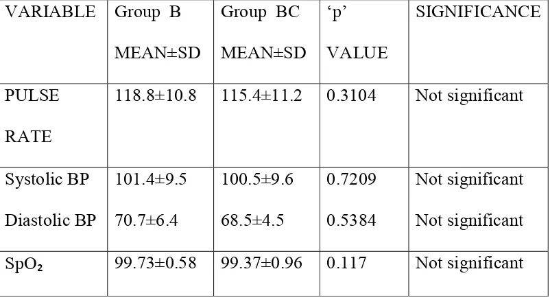
BASELINE HAEMODYNAMIC PARAMETERS(Table 3): The preoperative pulse rate, blood pressure, Hb saturation in the two groups were comparable in both groups.
TABLE 3: BASELINE HAEMODYNAMIC PARAMETERS
VARIABLE Group B MEAN±SD
Group BC MEAN±SD ‘p’ VALUE SIGNIFICANCE PULSE RATE
118.8±10.8 115.4±11.2 0.3104 Not significant
[image:64.612.106.508.190.285.2] [image:64.612.107.508.503.723.2]DURATION OF ANALGESIA( table 4 &5) :
[image:65.612.93.508.471.592.2]The time elapsed from the onset of caudal block to the objective pain scale score >11 was around 165min - 230min in group B with a mean of 196.5 ± 16.6min. In group BC duration of analgesia ranged from 265min to 345min with a mean of 300.2 ± 21.9min which is statistically significant.(Graph 4 & 5)
TABLE 4 : DURATION OF ANALGESIA
GROUP MEAN±SD (min)
P SIGNIFICANCE
B 196.5±16.6
0.0001 Significant BC 300.2±21.9
TABLE 5: OBJECTIVE PAIN SCALE SCORE
B 6.54±0.25 0.0001
Significant BC 5
GRAPH 4 : DURATION OF ANALGESIA
GRAPH 5 : OBJECTIVE PAIN SCALE SCORE
SEDATION:
[image:67.612.107.510.347.441.2]As seen in table 6, the sedation score in BC group was 2.49 ± 0.11 as compared to 2.97±0.09 in group B which shows that the quality of sedation was better in group BC than group B.(Graph 6)
TABLE 6 : SEDATION SCORE
GROUP MEAN±SD p SIGNIFICANCE
B 2.97±0.09
0.0001 Significant BC 2.49±0.11
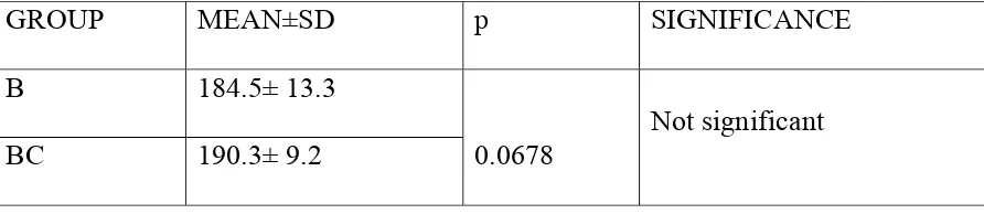
MOTOR RECOVERY (table 7):
GRAPH 6 : 4 POINT SEDATION SCORE
GRAPH 7: MOTOR RECOVERY
[image:69.612.93.539.222.319.2]
TABLE 7: TIME OF FIRST SUPPORTED STANDING
GROUP MEAN±SD p SIGNIFICANCE
B 184.5± 13.3
0.0678
Not significant
BC 190.3± 9.2
HAEMODYNAMIC PARAMETERS (table 8) PULSE RATE:
The change in the pulse rate between the two groups was significant. The average pulse rate was 116.7±9.3 /minute in group B and 104.4±7.7 / minute in group BC. This is statistically significant between the two groups.(graph 8)
BLOOD PRESSURE:
The mean systolic BP was 100.8±6.6mmHg group B and 98.3±6.8mmHg in group which is not statistically significant. Diastolic BP was 68.5± 4.5mmHg in group B and 67±3.3mmHg in group BC which is not statistically significant.(graph 9 & 10)
SPO₂ in group was around 98.93±0.91 % in group B and 98.5±0.94% in group BC which is statistically not significant.(graph 11)
GRAPH 8: PULSE RATES AT VARIOUS INTERVALS
GRAPH 9 : SYSTOLIC BP
GRAPH 10: DIASTOLIC BP
GRAPH 11:SpO₂
0 10 20 30 40 50 60 70 80 90 100 110
Pre operative SPO2 Post operative SPO2
TABLE 8: HAEMODYNAMIC PARAMETERS VARIABLE Group B
MEAN±SD
Group BC MEAN±SD
p
VALUE
SIGNIFICANCE
Pulse rate 116.7±9.3 104.4±7.7 0.0001 Significant Systolic
Diastolic
100.8±6.6 68.5±4.5
98.3±6.8 67±3.3
0.1294 0.1675
Not significant Not significant
SpO₂ 98.93±0.91 98.5±0.94 0.0778 Not sigmificant
SIDE EFFECTS:
DISCUSSION
Pain is an unpleasant subjective sensation which can only be experienced and not expressed, especially in children; who would seem to conceal their feelings when suffering from pain. The primary reason to treat or prevent pain is humanitarian. This is even more important in children who rely completely on their parents or care givers for their well being.
The concept of postoperative pain relief and its utilisation in the paediatric age group has improved dramatically over the recent years. The society of Paediatric anaesthesia, at its 15th annual meeting in 2001
Ideal anaesthetic technique should be targeted at three sites – the periphery, the sensory flow in nerves and cells in the central nervous system. The administration of an analgesic before any tissue damage takes place could interfere with and reduce the magnitude of nociception and thereby prevent a state of hyersensitisation.
The various methods of providing pain relief have some side effects which prohibit their use, for eg, narcotics in children, because of their respiratory depression, the objection to needles in the case of parenterally administered analgesics.
Caudal route was chosen for the study as it is the simplest and safest techniques in paediatric surgery with a success rate of allowing single shot injection of local anaesthetics and additives that prolong the postoperative analgesia. By this regional technique, problems and complications of intubation and polypharmacy due to general anaesthesia are avoided.
The two groups were comparable with respect to age, sex, weight and duration of surgery. The preoperative pulse rate, blood pressure and saturation was also comparable.
HAEMODYNAMIC STABILITY:
significantly low in group BC but not to the extent of haemodynamic instability.
SEDATION: Sedation and hence patient comfortability as assessed from 4 point sedation score was better in group BC compared to group B
ANALGESIA:
Pain intensity was assessed in this study by objective pain scale score (OPSS). The OPS score is a reliable and sensitive tool in evaluating postoperative pain in children.
The mean duration of analgesia for caudal Bupivacaine was 196.5 minutes. (165minutes - 230minutes). In group BC duration of analgesia was 300.2 minutes (265-345 minutes).
MOTOR RECOVERY:
Motor recovery was assessed from the time for first supported standing postoperatively which was similar in both the groups.
compared with plain Bupivacaine or Bupivacaine plus 1:200000 epinephrine.
Similarly, Lee JJ et al in their study used Clonidine in the dose of 2 µg/kg in children under going orthopaedic surgery and found significant prolongation of analgesia (9.8 hrs) as compared to Bupivacaine (5.2 hrs). The results of the study done, is comparable to the above similar studies with respect to analgesic effects of Clonidine.
In contrast to the study done in 36 chidren by Joshi W, Connelly NR, Freeman K, Reuben SS, in which there was no significant increase in the duration of analgesia but there was vomiting in 8 cases in clonidine group, this study shows significant prolongation of the duration of analgesia and only 2 cases reported to have vomiting.
SUMMARY
Group I : caudal 1ml/Kg of 0.25% Bupivacaine
Group II : caudal Clonidine 2µg/Kg with Bupivacaine as above.
All children were induced and maintained with halothane and 70% N₂O in O₂. This study shows that
1.Clonidine when added to caudal Bupivacaine provided longer duration of analgesia when compared to Bupivacaine alone.
2. Patient comfortability in terms of sedation and quality of analgesia is better in Clonidine - Bupivacaine group
3. Cardiovascular stability was similar in both the groups
4. Duration of motor blockade were similar in both the groups.
CONCLUSION
BIBLIOGRAPHY
1. Anatomy for anaesthetist – Haroid Ellis 7th edition, pg 113-117
2. Arthur D.S.; N/c NICOLL R.(1986): local anaesthetic techniques in paediatric surgery: Br. J. Anaesth: 58:771
3. Arthur C.Guyton. M.D.(1981) – Text book of Medical Physiology, sixth edition, somatic sensation : p 600-609
4. Bromage P. (1978) Epidural analgesia : Philadelphia Sanders 259 5. International association for study of pain. Subcommittee on
Taxonomy. Pain terms ; a list of definitions and notes on usage pain 1979:6:249-252
6. Understanding paediatric anaesthesia- Rabecca Jacob. Practical Paediatric regional Aanaesthesia 89-92
7. M.J. Cousins &P.O. Briden baugh – Neural blockade in clinical anaesthesia and management of pain,4th edition.
8. European Journal of clinical Pharmacology 13, 97-101 (1978) Pharmacokinetics and Side-Effects of Clonidine. A Keriinen, S. Nyk~inen, and J. Taskinen
10.Ronald D. Miller, Anaesthesia 6th edition, 1725-1726. Adjuvants to local anaesthetics.
11.Robert.K. Stoelting, Pharmacology and Physiology in anaesthetic practice, 4th edition; 340 – 344, Sympatholytics
12.Wylie and Churchill-Davidson’s, A practice of anaesthesia, 7th edition ; 601, Adjuvants to local anaesthetics.
13.G. Edward Morgan, Jr, Clinical Anaesthesiology, 4th edition; 283, Adjuvants to anaesthesia.
14. Bergendahl HTG, De Negri P, Ivani G, Eksborg S, Lonnqvist PA. "Increased postoperative blood pressure stability with continuous epidural infusion of clonidine in children." Anesthesia-Analgesia(2002)
15.Ivani G, Bergendahl HT, Lampugnani E, Eksborg S, Jasonni V, Palm C, Mattioli G, Podesta E, Famularo A, Lonnqvist PA (1998). "Plasma levels of clonidine following epidural bolus injection in children. "
Acta Anaesthesiololgica Scandanavica 42(3): 306-11
16.Bergendahl HT, Eksborg S, Lonnqvist PA (1997). "Low-dose intravenous clonidine in children: plasma concentrations and haemodynamic response. " Acta Anaesthesiologica Scandanavica 41(3): 381-4
August 1, 2002 Author: Bharti, N; Madan, R; Kaul, H L; Khokhar, S K; Mishra, S
18.Hospital Medicine. 1998 Mar;59(3):221-3. The role of clonidine in anaesthesia. Sanderson PM, Eltringham R. Gloucestershire Royal Hospital Gloucester.
19.Caudal anaesthesia with 0.375% Ropivacaine or 0.375% Bupivacaine for paediatric patients M.J. DA CONCEICAO & L.COELHO British
Journal of Anaesthesia 1998; 80: 507-508
20.The Efficacy and Safety of a Clonidine/Bupivacaine Combination in Caudal Blockade for Pediatric Hernia Repair W. Klimscha, MD, A. Chiari, MD, A. Michalek-Sauberer, MD, E. Wildling, MD, A. Lerche, MD, C. Lorber, MD, H. Brinkmann, MD, and M. Semsroth, MD. Anaesthesia analgesia vol 86, 54-61; 1998
21.Analgesia for circumcision in a paediatric population: comparison of caudal bupivacaine alone with bupivacaine plus two doses of clonidine P. Sharpe FRCA, J.R. Klein FRCA,DipIMCRCSEd J.P.Thompson BSc,FRCA, S.C.Rushman FRCA, J.Sherwin DA,FFARCSI, J.G. Wandless BSc,FRCA , D. Fell FRCA Paediatric
anaesthesia, Volume 11, Issue 6, pages 695–700, November 2001 .
clonidine. Sharpe, P. Frca; Klein, J. R. Frca, Dipimcrcsed; Thompson, J. P. Bsc, Frca; Rushman, S. C. Frca; Sherwin, J. Da, Ffarcsi; Wandless, J. G. Bsc, Frca; Fell, D. Frca . Paediatric Anaesthesia.