Copyrightq1995, American Society for Microbiology
Mutations in the DNA-Binding and Dimerization Domains of
v-Rel Are Responsible for Altered
k
B DNA-Binding Complexes
in Transformed Cells
RADMILA HRDLICˇ KOVA´ , JIRˇ I´ NEHYBA,ANDHENRY R. BOSE, JR.*
Department of Microbiology and the Cell Research Institute, The University of Texas at Austin, Austin, Texas 78712-1095
Received 25 October 1994/Accepted 21 February 1995
The c-relproto-oncogene encodes a member of the Rel/NF-kB family of transcription factors. The oncogenic viral form, v-rel, transduced by avian reticuloendotheliosis virus T, induces lymphoid tumors. v-Rel transfor-mation may be mediated directly by binding of v-Rel to cognate DNA sites, resulting in altered gene expression, and/or indirectly by releasing Rel/NF-kB transcription factors from cytoplasmic retention molecules, resulting in their translocation to the nucleus and the inappropriate expression of genes under kB control. v-Rel-transformed cell lines of different phenotypes contained v-Rel as well as endogenouskB DNA-binding proteins in nuclear extracts. Kinetic analysis with avian leukosis virus-transformed B-cell lines expressing v-Rel or c-Rel indicated that the presence of endogenous kB DNA-binding proteins in the nucleus is temporally correlated with the relocalization of v-Rel to the cytoplasm. Supershift analysis of these DNA-binding com-plexes revealed that v-Rel was present in all of the nuclear DNA-binding comcom-plexes heterodimerized with c-Rel, NF-kB1, and other proteins. In contrast, c-Rel-transformed cells exhibited a less-complex pattern of nuclear kB DNA-binding complexes, and the nuclear appearance of these endogenous complexes was not observed. Studies with c-/v-Rel hybrids suggest that the induction of the endogenouskB DNA-binding complexes is the result of the mutations in the C-terminal region of the Rel homology (RH) domain of v-Rel. Moreover, v-Rel differed from c-Rel in its DNA-binding specificity. The altered DNA-binding specificity of v-Rel was associated with mutations located in the N-terminal part of the RH domain of v-Rel. These results suggest that two different regions of v-Rel (both located in the RH domain) influence the formation of kB DNA-binding complexes differently.
The proto-oncogene c-rel belongs to the family of Rel/ NF-kB transcription factors. This family consists of related genes which are involved in the regulation of transcription during differentiation and developmental processes. In addi-tion to c-rel, other vertebrate members include nfkb1, nfkb2, rela, and relb. The Rel/NF-kB family is regulated by a small group of inhibitors (IkBs) which serve to sequester these tran-scription complexes in the cytoplasm. The mechanism by which this family of transcription factors is regulated suggests that it evolved to be able to respond rapidly to external signals. Rel/ NF-kB transcription factors are present in inactive cytoplasmic complexes, and following the appropriate regulatory signal, they are released to the nucleus, where they participate in the transcriptional regulation of a large set of genes (reviewed in references 3 and 46).
Rel/NF-kB and IkB proteins have two structural features that have coevolved to promote DNA-protein and protein-protein interactions: the Rel homology (RH) domain and ankyrin repeats. The RH domain in c-Rel, NF-kB1 (p105/p50), NF-kB2 (p100/p52), RelA (p65), and RelB mediates DNA binding, dimerization, and interactions with IkB. At the C-distal region of the RH domain is a nuclear localization se-quence. The C termini of c-Rel, RelA, and RelB contain trans-activation domains. c-Rel and RelA also contain cytoplasmic retention sequences in their C termini. By contrast, the C termini of NF-kB1/p105 and NF-kB2/p100 contain ankyrin
repeats that are proteolytically removed, generating NF-kB1/ p50 and NF-kB2/p52. Ankyrin repeats are repetitive elements also found in IkB-a, IkB-g, and Bcl-3. Two functions have been associated with the IkB ankyrin repeats, (i) inhibition of DNA binding of Rel/NF-kB factors in vitro and (ii) cytoplas-mic retention of Rel/NF-kB factors in vivo (reviewed in refer-ences 6, 31, and 46).
Rel/NF-kB transcription factors bind to DNA as ho-modimers or heterodimers. The RH domain is responsible for the DNA-binding property of the Rel/NF-kB transcription fac-tors (28, 39). The C-distal part of the RH domain is necessary for dimerization, which is indispensable for DNA binding (12, 49). The N-terminal part of the RH domain is involved in direct interactions with DNA. The RXXRXRXXC motif was shown to contact DNA directly (45). Specifically, two amino acids in or near this motif which contact DNA were identified (48). A secondary b-turn-b structure was proposed for this region of the RH domain (48). In accordance with the struc-tural analysis, comparative studies of the DNA-binding speci-ficity of NF-kB1/p50 and RelA showed that four amino acids in this region are responsible for the different DNA-binding spec-ificity of these two transcription factors (21, 68).
REV-T, which expresses the v-rel oncogene, induces a fatal lymphoma in young birds (58). v-rel originated as a result of a nonhomologous recombination event involving a retrovirus and the c-rel proto-oncogene. Subsequently, tumorigenic mu-tations were selected for during the replication of REV-T (64, 72). The transduction of c-rel resulted in the deletion of 2 N-terminal and 118 C-terminal acids, which were replaced by 11 and 18 env-derived amino acids, respectively. On the basis of a comparison with chicken and turkey c-Rel, 13 amino acid * Corresponding author. Mailing address: Department of
Microbi-ology, The University of Texas at Austin, Austin, TX 78712-1095. Phone: (512) 471-5525. Fax: (512) 471-2130. Electronic mail address: [email protected].
3369
on November 9, 2019 by guest
http://jvi.asm.org/
substitutions and 3 small in-frame deletions were identified in v-Rel (15, 72). Despite these differences, v-Rel shares two essential properties with c-Rel: (i) binding tokB sites and (ii) formation of large molecular complexes that contain several proteins, including NF-kB1, NF-kB2, IkB-a, and the constitu-tive form of heat shock protein 70 (10). The deletion of the C terminus of c-Rel to produce v-Rel resulted in the removal of a cytoplasmic anchor sequence and transactivation domain II (30, 57). v-Rel, however, retains transactivation domain I, which possesses significant transactivation potential (60, 68, 73). Mutational analyses indicates that DNA binding, complex formation, and transactivation are necessary for the full trans-formation potential of v-Rel (24, 32, 45, 52, 60, 69, 73).
Both c-Rel and v-Rel transform avian cells when overex-pressed from a heterologous retroviral promoter. However, c-Rel transforms lymphoid cells and fibroblasts much less ef-ficiently than v-Rel does (34, 42, 55, 56). Studies with c-/v-Rel chimeric proteins indicate that alterations in both the RH domain and C-terminus contribute to the enhanced transfor-mation potential of v-Rel (8, 25, 33, 56). These oncogenic differences between v-Rel and c-Rel are probably due to changes in domains responsible for interacting with other pro-teins and/or DNA, thereby resulting in an oncogenic protein with altered responsiveness to cellular regulatory constraints. The mutations in the central part of v-Rel were shown to decrease the IkB-a-mediated inhibition of v-Rel DNA binding (25). The C-terminal deletion of c-Rel, which removed the cytoplasmic anchor and transactivation domain II, is associated with increased nuclear localization that correlates with trans-formation (33). The contribution of the other mutations, how-ever, which have impaired the responsiveness of v-Rel to cel-lular regulatory pathways has not been identified.
Transformation by v-Rel is likely to be mediated by altered gene expression, most probably involving genes normally reg-ulated by c-Rel and other members of the Rel/NF-kB family. Several Rel/NF-kB-regulated genes have been shown to be upregulated as a consequence of v-Rel expression. These in-clude the high-mobility-group protein 14b, the major histo-compatibility complex (MHC) genes class I and II, the inter-leukin-2 receptor (IL-2R), nfkb1, and Ikba (9, 34, 35, 42). Two non-mutually-exclusive mechanisms, which are associated with transformation by v-Rel, have been proposed to explain the alterations in Rel/NF-kB-controlled gene expression (23, 29). A direct mechanism predicts that v-Rel bindskB elements in the transformed cells and either transactivates or represses transcription. An indirect mechanism, on the other hand, pro-poses that v-Rel competes for binding to IkBs in the cytoplasm, thereby releasing endogenouskB DNA-binding proteins to the nucleus. The requirement for DNA binding and transactiva-tion by v-Rel supports the model that v-Rel directly partici-pates in altered gene expression by binding tokB sites. How-ever, the presence of the majority of v-Rel in cytoplasmic complexes containing the inhibitory proteins IkB-a and the NF-kB1 and NF-kB2 precursors suggests a role for an indirect mechanism in v-Rel-mediated transformation. The direct mechanism predicts the presence of v-Rel in DNA-binding complexes; in contrast, the indirect mechanism predicts that v-Rel induces the appearance of endogenouskB DNA-binding proteins in the nucleus. v-Rel and c-Rel were both shown to be found in nuclear extracts (1, 9, 14, 25, 36, 41, 45, 70), but the differences between c-Rel and v-RelkB DNA-binding speci-ficity and the possible induction of endogenouskB DNA-bind-ing proteins have not been evaluated.
In this study, we demonstrate that initially after infection of cells with retroviruses expressing v-rel, v-Rel becomes the ma-jorkB DNA-binding protein in the nucleus. As the infection
proceeds, the majority of v-Rel assumes a cytoplasmic location. Temporally correlated with the relocalization of v-Rel to the cytoplasm, newkB DNA-binding complexes can be detected in the nucleus. This is also the time at which NF-kB1, NF-kB2, and IkB-a transcription becomes elevated in v-Rel-trans-formed cells (35, 42). These new complexes contain v-Rel heterodimerized with c-Rel and other Rel/NF-kB family mem-bers. The proteins capable of forming nuclearkB DNA-bind-ing complexes in cells overexpressDNA-bind-ing or transformed by c-Rel were different from those observed in v-Rel-expressing cells. The differences in the nuclear DNA-binding specificities be-tween v-Rel and c-Rel and the ability of v-Rel to increase the number of endogenous kB DNA-binding complexes in the nucleus are due to mutations located in two different regions in the RH domain. The presence of v-Rel itself, as well as the appearance of endogenouskB DNA-binding proteins in the nucleus of v-Rel-expressing cells, suggests that both direct and indirect mechanisms contribute to the transformation of cells by v-Rel.
MATERIALS AND METHODS
Plasmids.All recombinant DNA techniques were performed by standard procedures (59). The REV-T based retroviral vector REV-0 was described pre-viously (34, 56). REV-C and REV-TW express chicken c-rel (15) and v-rel (19), respectively (34). Five c/v-rel chimeras were constructed by crossing the parental genes in the common internal HindIII and HindII sites (56). ctt-rel was con-structed in a similar way by exchanging the v-rel C-terminal region with the C terminus of c-Rel in their common HindIII site. c-rel mutants lacking the C-terminal sequence encoding the last 55 amino acids (DXba) were described previously (33, 56). c-rel50a and c-relb were constructed by site-directed mu-tagenesis with a Muta-gene Phagemid in vitro Mumu-tagenesis Kit (Bio-Rad Labo-ratories, Inc., Richmond, Calif.). c-rel50a encodes c-rel with an oncogenic amino acid alteration (Met-20 to Phe), and c-relb has the v-rel-like amino acid Gly instead of Asp-82 in the N-terminal region of the RH domain. All mutated rel genes were cloned into the retroviral vector REV-0. pCSV11S3 contains an infectious genome of chicken syncytial virus (CSV) (27). The retroviral vector expressing C-terminally deleted v-Rel dH172 was a kind gift of T. D. Gilmore (60).
Chickens, cell lines, and tissue culture procedures.Cells were cultured in Dulbecco’s modified Eagle’s medium supplemented with 5% calf serum (Hy-Clone Laboratories, Inc., Logan, Utah), 5% chicken serum (GIBCO Biologicals, Grand Island, N.Y.), and antibiotics. DT95 is an avian B-cell line derived from a chicken with an avian leukosis virus-induced lymphoid leukosis (2). v-rel-transformed lymphoid cell lines 123/12, SS.1, 160/2, 160/8, 123/6, and 123/6T were described previously (34). Cell lines A27, B4, and B5 were derived from splenic tumors obtained from chickens infected with the REV-0-based retrovi-ruses expressing c-Rel50a (A7) or c-Relb (B4 and B5). Chicken embryo fibro-blasts were prepared from 10-day-old embryos. Secondary cultures of chicken embryo fibroblasts were used for transfection of plasmid DNAs by a calcium phosphate method as described previously (34). Viruses were harvested between 5 and 7 days after transfection, and infectious titers of Rel-expressing viruses and CSV were determined by an immunochemical titration assay (34, 65).
Antibodies.Cell surface protein expression was examined by indirect immu-nofluorescence with specific monoclonal antibodies. Monoclonal antibody HY19 detects the avian immunoglobulin M (IgM) protein (5). TCR-2 (18, 20) recog-nizes the avian 90-kDaa/bT-cell receptor. CT4 and CT8 (16) are specific for the avian homologs of CD4 and CD8, respectively. CMTDI and CMTDII recognize activated chicken macrophages (26). HY87 is specific for avian c-Rel and v-Rel (34). Helper virus (CSV) expression was detected in titration assays with mono-clonal antibody HY83 (34). Anti-mouse Ig class-specific antibodies were pur-chased from Southern Biotechnology Associates, Inc., Birmingham, Ala. Anti-chicken c-Rel-specific rabbit antiserum 1096 was the kind gift of N. R. Rice. Additional polyclonal and monoclonal antibodies were used in electrophoretic mobility shift assays (EMSAs). The basic characteristics of these antibodies are shown in Table 1.
Indirect immunofluorescence.Indirect immunofluorescence staining was per-formed as follows. Cells were fixed and permeabilized for 5 min with methanol-acetone (1:1), washed twice with phosphate-buffered saline (PBS), and incubated for 30 min with PBS–0.1% bovine serum albumin (BSA). The cells were then incubated for 1 h with hybridoma supernatant fluids containing monoclonal antibody, washed with PBS, and incubated for 1 h with fluorescein isothiocya-nate-conjugated goat anti-mouse antibodies directed against specific subclasses. They were washed with PBS with a final wash overnight and mounted with Fluoromount-C (Southern Biotechnology Associates). The intensity of staining was analyzed under a microscope.
Western analysis.Western immunoblot analysis was performed as described
on November 9, 2019 by guest
http://jvi.asm.org/
previously (34). Briefly, harvested cells were washed, resuspended in buffer, and boiled in sodium dodecyl sulfate (SDS) sample buffer, and proteins were sepa-rated on an SDS-polyacrylamide gel with a Mini-PROTEAN II apparatus (Bio-Rad Laboratories). The proteins were transferred to nitrocellulose and sequentially reacted with monoclonal antibody HY87, goat anti-mouse IgG1 biotinylated antibody, and streptavidin-linked alkaline phosphatase (Boehringer Mannheim, Corp., Indianapolis, Ind.). They were visualized by an enzymatic reaction with 5-bromo-4-chloro-3-indolyl phosphate and 4-nitroblue tetrazolium chloride as substrates (Boehringer Mannheim). Biotinylated molecular weight markers (Bio-Rad Laboratories) were used.
EMSAs.For EMSAs, nuclear and cytoplasmic extracts were prepared from 5
3106
cells as previously described (61). Briefly, harvested and PBS-washed cells were pelleted and resuspended in 200ml of cold solution A [10 mM N-2-hydroxyethylpiperazine-N9-2-ethanesulfonic acid (HEPES pH 7.9), 10 mM KCl, 0.1 mM EDTA, 0.1 mM ethylene glycol-bis(b-aminoethyl ether)-N,N,N9,N9 -tetraacetic acid (EGTA)] with 4 mM phenylmethylsulfonyl fluoride and 0.1 mM dithiothreitol. The cells were incubated for 15 min on ice. Then, 12ml of 10% Nonidet P-40 was added, the mixture was vortexed, and nuclei were separated by a 1-min centrifugation at 3,0003g. An 180-ml portion of the cytoplasmic fraction was transferred to a new tube and mixed with 60ml of 75% glycerol and 60ml of solution B (50 mM HEPES [pH 7.9], 2 M KCl, 5 mM EDTA, 5 mM EGTA). The nuclei were washed twice with solution A and resuspended in 100ml of solution C (20 mM HEPES [pH 7.9), 400 mM KCl, 1 mM EDTA, 1 mM EGTA, 15% glycerol) with phenylmethylsulfonyl fluoride and dithiothreitol. The cytoplasmic and nuclear fractions were shaken for 15 min at 48C and centrifuged for 10 min at 10,0003g. The supernatant fluids were aliquoted and stored at2708C. For binding assays, oligonucleotide probes were prepared by annealing of comple-mentary primers followed by primer extension with the Klenow fragment of DNA polymerase I (Boehringer Mannheim) in the presence of [a-32P]dCTP and deoxynucleoside triphosphates. The following coding-strand templates were used: palindromickB, 59-AATTCAGGGGAATTCCCCTAAGCTTGAGCT-39; avian MHC class I-derived site, 59-TCGAGGTGCTCGAAGGGCCGGGGGT TCCCACACCAC-39; and avian c-rel promoter kB site, 59-AATTCACGGG AAATTCCCTAAGCTTGAGCT-39(the rule indicateskB sites). The unincor-porated radioactivity was separated on a G-25 Sephadex column. The binding-reaction mixture was mixed with water plus 1 mM phenylmethylsulfonyl fluoride (to a final volume of 20ml), 10% acetylated BSA, 0.1% Nonidet P-40 (with 20 mM dithiothreitol), 4ml of 53binding buffer (100 mM Tris-HCl [pH 7.4], 0.2 mM EDTA, 0.5 M KCl, 25% glycerol), 0.5mg of eukaryotic DNA, and 2ml of cell extract. A 2-ml portion of the probe (105
cpm) was added after 10 min of preincubation on ice. The reaction mixture was incubated for 30 min on ice. For supershift, 1ml of rabbit antiserum was added and incubation was continued for 20 min on ice and then for 10 min at 378C. The complexes were separated from the free DNA probe by electrophoresis through a 4% polyacrylamide gel in 0.53 TBE (TBE is 0.09 M Tris-borate plus 2 mM EDTA [pH 8.0]). The gels were analyzed by autoradiography.
In vitro transcription and translation.c-rel and v-rel were cloned into modified pTZ-18R, to create the expression plasmids pc-rel2 and pv-rel2, respectively (56). Transcription templates were prepared by linearization of the expression plasmids by BssHII. The proteins were produced with the TNT T7-coupled wheat germ extract system (Promega, Madison, Wis.). Synthesis of c-Rel and v-Rel was verified by Western blot analysis.
RESULTS
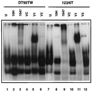
The nature of the oncogenic Rel protein determines the pattern ofkB DNA-binding complexes found in the nucleus of Rel-transformed cells.To determine whether differences exist among thekB DNA-binding complexes in the nuclei of v-Rel-transformed cells expressing different phenotypes, the kB DNA-binding activities in six v-Rel-transformed cell lines were analyzed. As shown in Fig. 1A, lymphoid cell lines expressing either the IgM or TCR-2 receptor, as well as cell lines which fail to express either of these receptors, demonstrated a similar pattern of nuclearkB DNA-binding complexes. In all of the cell lines analyzed, multiplekB DNA-binding complexes were detected. Each cell type contained nuclearkB DNA-binding complexes with similar sizes, although the relative levels of the
kB DNA-binding complexes were different in each of the cell lines analyzed. Three v-Rel cell lines representing each phe-notypic group were then analyzed by a supershift assay with an antiserum (VC) directed against the C terminus of v-Rel to identify which complexes contain v-Rel (Fig. 1B, lanes 1 to 9). Only a small portion of the total kB DNA-binding proteins detected in the nucleus in these lymphoid cell lines was capable of reacting with this antiserum.
To compare the pattern of nuclearkB DNA-binding pro-teins in v-Rel-transformed cell lines with the pattern associated with c-Rel transformation, several additional cell lines were developed. Because stable c-Rel-transformed lymphoid cell lines are not available, cell lines were derived from splenic tumors of animals infected with retroviruses expressing a vari-ant of c-Rel with point mutations in the N-terminal part of the RH domain. In contrast to the v-Rel-transformed cell lines, mutant c-Rel-transformed cells exhibited a different set ofkB DNA-binding complexes (Fig. 1B, lanes 10 to 18). The fast-migrating kB DNA-binding nuclear complexes identified in v-Rel-transformed cell lines were not detected in mutant c-Rel-transformed cells.
[image:3.612.57.297.84.203.2]v-Rel-transformed cell lines may express phenotypic mark-ers found on B cells at different stages of differentiation, T cells, or monocytes (10). By contrast, c-Rel-derived cell lines express markers found on T lymphocytes (33). To demonstrate that the differences in the nuclearkB DNA-binding complexes observed in v-Rel- and c-Rel-transformed cells were not due the long time required to establish transformed cell lines with a specific phenotype, splenic cells infected in vitro by retrovi-ruses expressing either c-Rel or v-Rel were analyzed 10 days after infection. The analysis of kB DNA-binding activity in nuclear extracts from these cells indicated that the differences in the kB DNA-binding complexes observed in c-Rel- and v-Rel-transformed cells were not due to different target cell specificities (Fig. 2). In a fashion analogous to the situation with the established cell lines, v-Rel-transformed splenic cells exhibited a more complex pattern of kB DNA-binding com-plexes than did c-Rel-expressing cells. Only a limited number of these complexes were supershifted with c-Rel or anti-v-Rel C-terminus-specific sera. In addition, no differences in the nature of the nuclear kB DNA-binding complexes were observed in cells expressing wild-type c-Rel, in contrast to the cell lines expressing the oncogenic c-Rel variant containing amino acid substitutions in the RH domain. These single-amino-acid substitutions therefore did not contribute to the differences in the nature of the nuclearkB DNA-binding com-plexes observed in c-Rel- and v-Rel-transformed cells. How-ever, the difficulties encountered in growing CSV-infected splenic cells in vitro and the heterogeneity of splenic cultures prompted us to continue this analysis with the established avian leukosis virus-transformed cell line DT95. This avian TABLE 1. Antibodies used in EMSA
Name Character Antigen Amino
acidsa Specificityb Reference
1647 Polyclonal v-Rel 2–11 v-Rel This studyc 1967 Polyclonal v-Rel 1–12 v-Rel 73 VC Polyclonal v-Rel 485–503 v-Rel This studyc 1648 Polyclonal v-Rel 488–503 v-Rel This studyc 10H Monoclonal c-Rel 350–582 c-Rel 42 1801 Polyclonal c-Rel 571–581 c-Rel 44 V1 Polyclonal v-Rel 115–292 v-Rel, c-Rel This studyc V2 Polyclonal v-Rel 293–458 v-Rel, c-Rel This studyc V3 Polyclonal v-Rel 425–437 v-Rel, c-Rel This studyc AL-1 Polyclonal NF-kB1 1–175 NF-kB1 42
a
Amino acids designate the Rel/NF-kB epitope in the immunogen used for developing the antibody.
b
The specificity of serum to Rel/NF-kB proteins was tested by immunopre-cipitation and/or EMSA of in vitro translated proteins.
c
Antisera 1647 and 1648 were developed by immunization by peptides cou-pled to keyhole limpet hemocyanin. Antiserum VC was derived against fusion proteins expressed from GEX3X expression vector in bacteria. Antisera V1 and V2 were derived against trpE/v-rel fusion proteins expressed from pJH11 and pJH12 in bacteria (62). Antiserum V3 was obtained after immunization with peptide coupled to BSA.
on November 9, 2019 by guest
http://jvi.asm.org/
B-cell line supports v-Rel expression and exhibits changes which correlate with Rel transformation (33–35, 56). To pro-vide additional epro-vidence that the DNA-binding complexes in REV-T-transformed cells correspond to those in DT95 cells, a direct comparison of the complexes recognized by various anti-Rel sera was performed (Fig. 3). The nature of the antisera
used in these studies is described in Table 1. The results indi-cated that thekB DNA-binding complexes in DT95 cells and splenic cell lines expressing v-Rel were indistinguishable. This experiment confirms that the analysis of DNA-binding com-plexes in Rel-expressing DT95 cells reflects those that appear during the transformation of splenic cells by v-Rel.
The appearance of nuclearkB DNA-binding complexes is different in c-Rel- and v-Rel-expressing lymphoid cell lines.
[image:4.612.93.532.73.261.2]The presence of a more complex pattern ofkB DNA-binding
FIG. 1.kB DNA-binding complexes in cytoplasmic and nuclear extracts in Rel-transformed cell lines. (A) The establishment of six v-Rel-transformed cell lines was described previously (34). Phenotypic analysis was done by indirect immunofluorescence with monoclonal antibodies specific to surface receptors. Briefly, cell lines 123/12 and SS.1 expressed IgM receptor; cell line 160/2 is TCR-21CD41CD81, and cell line 160/8 is TCR-21CD42CD81; cell lines 123/6 and 123/6T do not express IgM or TCR receptors or CD4/CD8 surface molecules. However, cell line 123/6T is positive for macrophage-specific antigens CMTDI and CMTDII. Cytoplasmatic (cy) and nuclear (n) extracts from these cell lines were analyzed by EMSA with the palindromic variant of the IL-2Ra kB site. (B) Nuclear extracts from REV-TW (v-Rel) cell lines 123/12, 160/2, and 123/6T, together with three REV-C-based (c-Rel mutants) T-cell lines, A27, B4, and B5 (see Materials and Methods), were analyzed by EMSA. Western blot analysis demonstrated that A27, B4, and B5 expressed a protein with a molecular weight corresponding to c-Rel (data not shown). DNA-binding reactions to the palindromickB site were carried out in the presence of unrelated antiserum (U), anti-c-Rel-specific rabbit serum 1096 (C), or anti-v-Rel-specific rabbit serum VC (T). B and S represent bound and supershiftedkB probe, respectively.
[image:4.612.81.271.443.603.2]FIG. 2.kB DNA-binding proteins in cytoplasmic and nuclear extracts of c-Rel- and v-Rel-transformed splenic cells 10 days after infection. The spleens from 4-week-old chickens were passed through nylon mesh and placed in 50 ml of culture medium. Four days later, mononuclear leukocytes were separated by centrifugation of the cell suspension on Isolymph (Gallard-Schlesinger Indus-tries, Inc., New York, N.Y.). The fractionated cells were infected by REV-C(CSV) or REV-TW(CSV) at a multiplicity of infection of 1. Lanes 1 to 4, the transformed cells were harvested 10 days after infection, and cytoplasmic (cy) and nuclear (n) extracts were prepared and analyzed by EMSA with the palin-dromickB site as described in the legend to Fig. 1. Lanes 5 to 10, supershift analysis was performed with the nuclear extracts in the presence of unrelated antiserum (U), anti-c-Rel-specific rabbit serum 1096 (C), or anti-v-Rel-specific rabbit serum VC (T). B and S represent bound and supershiftedkB probe, respectively. C designates extracts from REV-C(CSV)-infected cells, and TW designates extracts from REV-TW(CSV)-infected cells.
FIG. 3. Nuclear extract from DT95 expressing v-Rel (lanes 1 to 6) was com-pared with nuclear extract from the v-Rel-transformed cell line 123/6T (lanes 7 to 12). The DNA-binding reaction was performed with unrelated antiserum (U) (lanes 1 and 7), with anti-c-Rel C-terminus-specific antibody (lanes 2 and 8), with anti-v-Rel serum derived against the N terminus (lanes 3 and 9), with anti-v-Rel serum against the C terminus (lanes 4 and 10), and with anti-v-Rel sera V1 (lanes 5 and 11) and V2 (lanes 6 and 12).
on November 9, 2019 by guest
http://jvi.asm.org/
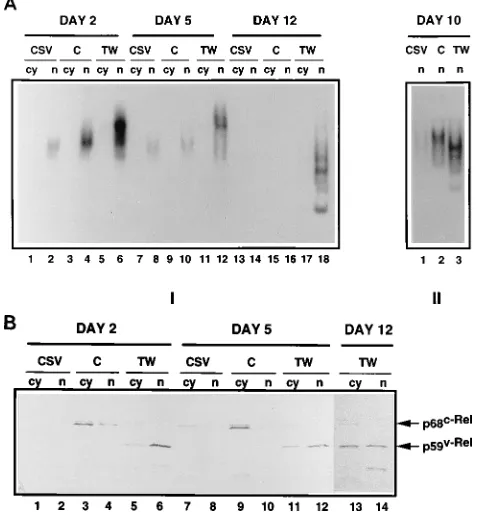
[image:4.612.337.530.478.669.2]proteins in the nuclei of v-Rel-transformed cells than in the nuclei of c-Rel-transformed cells suggested that this difference may be the consequence of the expression of v-Rel in lymphoid cells. To test this hypothesis, DT95 cells were coinfected with REV-C (c-rel) or REV-TW (v-rel) and the helper virus CSV or with CSV alone. The cells were harvested at 2, 5, and 12 days after infection, and cytoplasmic and nuclear fractions were analyzed by Western analysis and EMSAs with two different
kB sites. A palindromic variant (GGGGAATTCCCC) of the IL-2Ra kB DNA-binding site (50) was used, since this element binds the majority of Rel/NF-kB family members and is rec-ognized by v-Rel (4). The secondkB motif used in this study corresponds to thekB site upstream of the avian MHC class I promoter (GGGGGTTCCC) (43), which is also recognized by proteins in v-Rel-transformed cells (9).
Two days after infection, DT95 cells infected with REV-C or REV-TW exhibited increased nuclear DNA-binding activity to the palindromic variant of the IL-2Ra kB site when compared with DT95 cells infected with CSV (Fig. 4A). Five days after infection, the level of the proteins withkB DNA-binding ac-tivity decreased substantially in the infected cells expressing REV-C or REV-TW. This decrease inkB DNA-binding activ-ity was accompanied by a decrease in the total amount of Rel in the nuclear fraction and a concomitant relocalization of Rel to the cytoplasm (Fig. 4B). The relocalization of Rel is likely to be mediated by an increase in the levels of NF-kB1, NF-kB2, and IkB-ain cells transformed by oncogenic Rel proteins (35, 42). Twelve days after infection, newkB DNA-binding
com-plexes were detected in the nucleus of REV-TW-infected cells. During this time, there was a gradual redistribution of v-Rel, with the cytoplasmic compartment containing approximately half of the total detected in the cells (Fig. 4B). c-Rel expression declined in a parallel culture of DT95 cells infected with REV-C as a result of a pronounced cytotoxic effect induced by c-Rel in this particular experiment (Fig. 4B). For this reason, a comparison of the nuclear kB DNA-binding activity 10 days after infection from another experiment is shown (Fig. 4A). The nuclei of v-Rel-expressing cells contained more proteins capable of formingkB DNA-binding complexes than did the nuclei of c-Rel-expressing cells, and the size of thekB DNA-binding complexes were also different. These observations are consistent with the analysis of v-Rel- and c-Rel-transformed cell lines, as well as splenic cells (Fig. 1 and 2).
Despite the accumulation of c-Rel in the cytoplasm of DT95 cells 5 and 12 days after infection (Fig. 4B),kB DNA-binding activity was not detected in cytoplasmic extracts obtained from c-Rel-expressing cells. However, the appearance ofkB DNA-binding proteins in the cytoplasm was detected in DT95 cells expressing v-Rel 12 days after infection. This coincided with the redistribution of v-Rel to the cytoplasm (Fig. 4A). Similar observations were made with c-Rel- and v-Rel-transformed cell lines. In contrast to c-Rel-transformed cells, a significant amount of cytoplasmickB DNA-binding activity was found in v-Rel-expressing cells (Fig. 1A and 2).
The same extracts that were used to identify thekB DNA-binding complexes by using the IL-2RagenekB site were also analyzed with the kB site derived from the promoter of the avian MHC class I gene (Fig. 5). The level ofkB DNA-binding proteins capable of binding either the MHC class IkB site or the IL-2Rasite were equivalent 10 days after v-Rel infection. By contrast to the analysis with a palindromic variant of the IL-2Ra kB site, different DNA-binding complexes were not detected in nuclei of c-Rel-expressing cells at any time after infection. Collectively, these results indicate that both v-Rel and c-Rel recognize a similar kB site but display different DNA-binding specificities.
[image:5.612.363.490.67.252.2]The analysis of DNA-binding proteins present in nuclearkB DNA-binding complexes.At 2 and 5 days after infection, anti-c-Rel-specific serum 1096 and anti-v-Rel-specific serum (VC),
[image:5.612.58.298.75.331.2]FIG. 4. Kinetic analysis ofkB DNA-binding activity in the DT95 lymphoid cell line expressing c-Rel or v-Rel. DT95 cells were infected with c-Rel (C)- and v-Rel (TW)-expressing retroviruses at a multiplicity of infection of 2. The parallel culture was infected with CSV helper virus (CSV). Cells were harvested 2, 5, and 12 days after infection and analyzed by EMSA and Western blot analysis. (A) Cytoplasmic (cy) or nuclear (n) extracts were incubated with palindromic variant of IL-2Ra kB site, and DNA-binding complexes were analyzed by EMSA (panel I). Analysis of nuclear extracts from another experiment 10 days after infection is shown (panel II). (B) The same extracts as in panel A were analyzed by Western blot analysis with anti-Rel monoclonal antibody HY87. This experiment was repeated three times with similar results.
FIG. 5. DNA-binding specificity of proteins in DT95 cells infected with ret-roviruses expressing CSV (CSV), c-Rel (C), and v-Rel (TW). Nuclear extracts from the experiment described in the legend to Fig. 4A, panel II, were used for EMSA of protein complexes binding to the IL-2Ra kB site (lanes 1 to 3) and MHC class IkB site (lanes 4 to 6).
on November 9, 2019 by guest
http://jvi.asm.org/
directed against the C terminus, supershifted the majority of thekB DNA-binding activity in nuclear extracts from REV-C-or REV-TW-infected DT95 cells (Fig. 6). In contrast, at 12 days after infection, anti-v-Rel serum (VC) supershifted only a small fraction of the proteins in the new nuclear DNA-binding complexes. This observation is in agreement with the results obtained with v-Rel-transformed cell lines and splenic cells (Fig. 1 and 2).
To further characterize the nature of thekB DNA-binding complexes which appear in the nucleus of DT95 cells
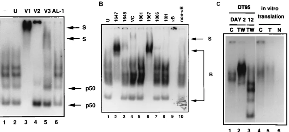
express-ing v-Rel, supershift experiments were performed with various antisera (Fig. 7). Three rabbit polyclonal antisera, V1, V2, and V3, directed against different regions of v-Rel protein (which are also shared with c-Rel), were used. Rabbit polyclonal an-tiserum V1, raised against amino acids 115 to 292 of the v-Rel RH domain, supershifted all the proteins capable of forming nuclearkB DNA-binding complexes detected in DT95 cells 12 days after infection (Fig. 7A, lane 3). Rabbit polyclonal anti-serum V2, derived against amino acids 293 to 458 of v-Rel, recognized the majority of the DNA-binding proteins present in the slowly migrating complexes. The fast-migrating kB DNA-binding complexes, however, failed to supershift (lane 4). Another rabbit polyclonal antiserum (V3), derived against a short peptide (amino acids 425 to 437 of v-Rel; the C-terminal part of the antigen used for production of V2 anti-serum), detected only the slowly migrating DNA-binding com-plexes (lane 5). The two fast-migrating comcom-plexes were partially supershifted with antiserum derived against the N terminus of NF-kB1 (lane 6). In contrast to the antisera which recognize v-Rel and c-Rel, antiserum against NF-kB1 did not significantly interfere with the DNA binding of the proteins present in the slowly migrating complexes. These results sug-gest thatkB DNA-binding complexes contained v-Rel or c-Rel proteins and that the two fast-migrating complexes also con-tained NF-kB1.
[image:6.612.59.296.70.224.2]The epitopes recognized by V1, V2, and V3 are present in both c-Rel and v-Rel. The peptide used to prepare the V3 antiserum represents a conserved domain shared by v-Rel and c-Rel (86% homology) but has no homology with avian RelA or other Rel/NF-kB family members. This analysis therefore does not define whether v-Rel and/or c-Rel is present in these DNA-binding complexes. Further analysis involved specific an-ti-v-Rel sera raised against the N- or C-terminal part of this
FIG. 6. Supershift analysis of DNA-binding proteins from nuclear extracts from DT95 cells infected with CSV (CSV) or retroviruses expressing either c-Rel (C) or v-Rel (TW). The nuclear extracts from the experiment described in the legend to Fig. 4 were analyzed with unrelated antiserum (U), anti-c-Rel rabbit serum 1096 (C), or anti-v-Rel rabbit serum VC (T). B and S represent bound and supershiftedkB probe, respectively.
FIG. 7. Nuclear DNA-binding proteins in a palindromic variant of IL-2Ra kB DNA-binding complexes. Detailed analysis of nuclear extracts from DT95 cells infected with REV-TW 2 and 12 days after infection is shown. (A) The DNA-binding reaction was performed without antiserum (lane 1), with unrelated antiserum (lane 2), with rabbit anti-v-Rel sera V1, V2, and V3 (lanes 3 to 5), or with rabbit anti-NF-kB1 antiserum AL-1 (lane 6). A detailed description of the specificities of these antisera is given in Table 1. S represents supershifted probe. The complexes in which NF-kB1/p50 was detected are designated p50. (B) A DNA-binding reaction was performed with unrelated antiserum (U), with anti-v-Rel sera derived against the N terminus (lanes 2 and 6), with anti-v-Rel serum derived against the C terminus (lanes 3 and 4), or with anti-c-Rel C-terminus-specific antibodies (lanes 5, 7, and 8). The same nuclear extracts were incubated with labeled probe in the presence of a 100-fold excess of cold competitor, i.e., the palindromic variant of IL-2Ra kB site (kB), or with the same amount of unrelated DNA (non-kB) (lanes 9 and 10). B and S represent bound and supershiftedkB probe, respectively. (C) DNA binding of proteins from DT95 expressing c-Rel (C) or v-Rel (TW) at days 2 and 12 (lanes 1 to 3) was compared with in vitro translated c-Rel (C) and v-Rel (T) (lanes 4 and 5). The DNA-binding assay was also done with an unprogrammed wheat germ lysate (lane 6).
on November 9, 2019 by guest
http://jvi.asm.org/
[image:6.612.72.547.426.643.2]oncoprotein and three antisera raised against the unique C terminus of c-Rel (Fig. 7B). Surprisingly, two different anti-v-Rel N-terminal sera were able to supershift all thekB DNA-binding complexes in v-Rel-expressing cells in a fashion anal-ogous to the V1 antiserum, which is directed against the N-terminal part of the RH domain (Fig. 7B, lanes 2 and 6). However, two different antisera directed against the C termi-nus of v-Rel were able to supershift only the more slowly migrating DNA-binding complexes (lanes 3 and 4). Similarly, the three different antisera raised against the C-terminal por-tion of c-Rel detected only a minor fracpor-tion of the slowly migrating complexes present in the nucleus of v-Rel-expressing cells (lanes 5, 7, and 8). Finally, a comparison of the migration ofkB complexes, using in vitro translated v-Rel and c-Rel, with the migration of the DNA-binding complexes in early and later stages after infection was performed (Fig. 7C). The migration of c-Rel and v-Rel bound to kB sequences on day 2 after infection corresponded to the migration of the in vitro trans-lated proteins (lanes 1, 2, 4, and 5). However, the migration of v-Rel-containingkB DNA-binding complexes (Fig. 7B, lanes 2 and 6), which appeared 12 days after infection of DT95 cells, dramatically changed (Fig. 7C, lane 3).
Collectively, these results suggest that the nature of the nuclear DNA-binding complexes changes with time. Soon after infection of cells with retroviruses expressing either c-Rel or v-Rel, DNA-binding complexes were composed principally of these exogenous proteins. The observation that the migration of these DNA-binding complexes corresponded to the migra-tion of in vitro translated c-Rel and v-Rel proteins bound tokB oligonucleotides supports the suggestion that c-Rel or v-Rel is the principal component of thekB complexes detected in the infected cell 2 to 5 days after infection. Coincident with the appearance of other nuclear kB DNA-binding proteins, the migration of v-Rel-containing DNA-binding complexes dra-matically changed between 5 and 10 days after infection. In addition, the ability of the antisera directed against the C terminus of v-Rel to recognize v-Rel decreased, suggesting that some of the epitopes in the C terminus of v-Rel were masked as a result of the interaction of v-Rel with other Rel/NF-kB family members and/or other proteins. Consistent with this conclusion, NF-kB1 and v-Rel were detected in the fast-mi-grating complexes, but the identities of other v-Rel partners remain to be determined. In the later stages of infection, the association of other proteins with c-Rel could reduce the abil-ity of antiserum raised against the c-Rel C terminus to recog-nize this protein in the DNA-binding complexes. The super-shift analysis would therefore underestimate the amount of c-Rel in the nucleus that is capable of binding DNA.
Mutations in the RH domain of v-Rel influence the nature of the nuclearkB DNA-binding complexes in Rel-expressing lym-phoid cell lines. Kinetic experiments conducted with DT95 cells (Fig. 4 and 5) demonstrated that c-Rel- and v-Rel-ex-pressing cells differed in two aspects: (i) v-Rel induces the appearance of new endogenous nuclearkB DNA-binding com-plexes, and (ii) v-Rel has a different specificity of DNA binding from that of c-Rel. v-Rel contains mutations in the RH domain as well as in the C-terminal transactivation and cytoplasmic anchoring domains, which are absent in c-Rel (15, 72). These alterations can be divided into three mutation clusters on the basis of the functional criteria shown in Fig. 8A (25, 33, 56). To define which mutations in v-Rel are responsible for the differ-ences in kB DNA-binding activity observed between c-Rel-and v-Rel-expressing cells, a set of retroviruses expressing c/v-Rel chimeric proteins was used (Fig. 8B). These six c/v-c/v-Rel hybrids represent all possible combinations of the three muta-tion clusters in v-Rel. c-Rel with a delemuta-tion of the last 55 amino
acids (DXba) was also used. DT95 cells were infected with the retroviruses described above, as well as with retroviruses ex-pressing c-Rel or v-Rel. Cells were harvested at different days after infection, and cytoplasmic and nuclear fractions were prepared and analyzed by EMSA with three differentkB sites. The analysis ofkB DNA-binding activity in the nuclear ex-tracts obtained from cells expressing the c/v-Rel chimeric pro-teins revealed that the altered DNA-binding specificity of v-Rel and the ability to induce endogenous nuclearkB DNA-binding activity are associated with distinct domains in v-Rel. Analysis of cytoplasmic and nuclear extracts for DNA binding with the IL-2Ra gene kB palindromic site showed that the appearance of the new endogenouskB DNA-binding activity is most prominent in c/v-Rel hybrids with v-Rel-like mutations in the central region of the RH domain (ctc-Rel, ttc-Rel and ctt-Rel) (Fig. 9). These Rel hybrids induced the fast-migrating complexes previously detected in nuclear extracts prepared from cells expressing v-Rel. Interestingly, two of these hybrids (ttc-Rel and ctc-Rel) also increased thekB DNA-binding ac-tivity detected in the cytoplasmic fraction. Moreover, cooper-ation between mutcooper-ations in the second and third domain was observed. The hybrid ctt-Rel induced the appearance of the slowly migrating complexes (Fig. 9, lane 11) that were also detected in v-Rel-expressing cells. These complexes were not detected in cells expressing the cct-Rel or ctc-Rel, chimeras with the second or third v-Rel domain alone (lanes 9 and 11). c-Rel with a deletion of the last 55 amino acids (DXba) did not induce the appearance of endogenouskB DNA-binding activ-ity (data not shown).
To determine whether a v-Rel mutant which lacks both transactivation domains is capable of inducing the appearance of nuclear endogenouskB-binding complexes, a v-Rel mutant (designated dH172) containing a deletion of the 172 C-termi-nal amino acids was used. This mutant not only lacks transac-tivation ability but also has an altered DNA-binding activity and is completely nontransforming (60). The nuclei of DT95-infected cells expressing dH172 do not contain endogenouskB DNA-binding activity (Fig. 10). The failure of dH172 to induce the appearance of endogenouskB DNA-binding activity may be the consequence of the lack of transactivating activity or may be due to changes in its DNA-binding ability relative to v-Rel.
By contrast, mutations in the N-terminal region of the RH domain were responsible for the altered DNA-binding speci-ficity of v-Rel relative to c-Rel. We have used three different
kB sites to compare the DNA binding of v-Rel, c-Rel, and their hybrids by using a c-rel promoter kB site GGGAAATTCC (13a), the IL-2Ra site GGGGAATTCCCC (50), and the MHC class I kB site GGGGGTTCCC (43) (Fig. 11). The analysis of nuclear extracts from DT95 cells 3 days after infec-tion showed that v-Rel and tct-Rel, proteins with v-Rel muta-tions in the N-terminal part of the RH domain, bind the c-rel promoter site with the same efficiency as c-Rel does (Fig. 11, lanes 1 to 3). These proteins, however, bind to the palindromic variant of the IL-2Rasite with better efficiency than c-Rel does (lanes 6 to 8). Moreover, v-Rel and tct-Rel bind to the MHC class I kB site better than c-Rel does (lanes 11 to 13). Like tct-Rel, increasedkB DNA binding was also observed in cells expressing tcc-Rel and ttc-Rel, which express Rel at a level comparable to or lower than that for c-Rel-expressing cells (data not shown). c-Rel and c/v-Rel hybrids with mutated sec-ond and third domains (cct-Rel and ctt-Rel) failed to bind to the MHC class I-derivedkB site. Together, these results indi-cate that the DNA-binding specificity of v-Rel to three sepa-ratekB sites can be attributed to mutations in the N-proximal region of the RH domain of v-Rel.
on November 9, 2019 by guest
http://jvi.asm.org/
DISCUSSION
The pattern of kB DNA-binding complexes in v-Rel- and c-Rel-transformed cells is different.This study compared the nature ofkB DNA-binding proteins which are present in nu-clear extracts obtained from a number of different cell lines transformed by v-Rel or c-Rel. Although differences in thekB DNA-binding proteins between individual cell lines were ob-served, the pattern of kB DNA-binding complexes detected was determined principally by whether the cells were overex-pressing v-Rel or c-Rel. A similar analysis was also conducted with nuclear extracts obtained from spleen cells 10 days after infection with retroviruses expressing either v-Rel or c-Rel. The nature of the kB DNA-binding proteins detected in the nucleus of these cells was also determined by whether the cells were overexpressing v-Rel or c-Rel. The major difference in thekB DNA-binding patterns observed between cell lines ex-pressing the highly transforming v-Rel and the less oncogenic
c-Rel was the presence of additional kB DNA-binding com-plexes in v-Rel-expressing cells.
All of thekB DNA-binding complexes observed in v-Rel-transformed cells contained v-Rel. c-Rel and NF-kB1/p50, het-erodimerized with v-Rel, were also found in different com-plexes in v-Rel-transformed cells. The migration ofkB DNA-binding complexes, as well as the presence of NF-kB1/p50 in the two fast-migrating complexes, resembles the pattern ofkB DNA-binding complexes found in nuclear extract of B lympho-cytes in late stages of differentiation (47, 51). However, the supershift analysis indicates that all of the DNA-binding com-plexes present in the nuclei of v-Rel-expressing cells contain v-Rel. Antisera specific against the N terminus of v-Rel re-moved all of the proteins from the nuclear extracts capable of forming kB DNA-binding complexes. This observation sug-gests that otherkB DNA-binding proteins (for example,
[image:8.612.155.459.69.434.2]NF-kB1/p50) are associated with v-Rel in heterodimers in the
FIG. 8. Functional domains of c-Rel, positions of mutations in v-Rel, and structures of parental and mutant Rel proteins used in this study. (A) The RH domain is divided into a DNA-binding region consisting of exons 0 to 3 and a dimerization region containing exons 4 and 5. The small black box in the DNA-binding domain represents the amino acid sequence RXXRXRXXC that is in direct contact with DNA. The open ellipse indicates the position of a nuclear localization sequence, and the solid ellipse marks the position of multiple kinase recognition sites. A region necessary for binding of IkB-ais indicated. In the C-terminal region of c-Rel, two transactivation domains (horizontal line pattern) overlap (cross-hatched pattern) with a cytoplasmic anchoring region (vertical line pattern). This definition of c-Rel functional domains is based primarily on studies of c-Rel (7, 15, 30, 37, 38, 45, 54, 55, 57), but some data obtained from the analysis of other Rel/NF-kB proteins were taken into account (12, 49). Mutations by which v-Rel differs from both turkey and chicken c-Rel (15, 32, 72) are indicated as follows: solid boxes, additions of amino acids encoded by env-derived nucleic acid sequence and fused to the truncated N or C terminus of c-Rel; open ellipses, 13 single-amino-acid substitutions and three small internal deletions. The mutations are grouped into three clusters by long black ovals intended to contain mutations that affect a similar structural-functional domain. (B) Protein structures of mutant Rel proteins derived from parental c-Rel and v-Rel. The RH domain (exons 0 to 6a) is indicated by the darker pattern, while the less highly conserved regions at the ends of the protein are in the lighter pattern. The names of specific Rel proteins and the corresponding viruses that encode them are on the right.
on November 9, 2019 by guest
http://jvi.asm.org/
DNA-binding complexes. This explanation is consistent with the fact that cells transformed with a v-Rel temperature-sen-sitive mutant undergo apoptosis soon after the DNA-binding ability of v-Rel is lost at the nonpermissive temperature (71). The identities of otherkB DNA-binding proteins in the nu-clear DNA-binding complexes of v-Rel-transformed cells are unknown. We are in the process of developing antisera specific for the other avian members of the Rel/NF-kB family.
Induction of endogenouskB DNA-binding proteins by v-Rel is preceded by the redistribution of v-Rel from the nucleus to the cytoplasm. The kinetics of the induction of kB DNA-binding complexes revealed that in the first few days after infection, both c-Rel and v-Rel were the predominant compo-nents in the nuclear complexes. However, at later stages of infection, additional v-Rel- and other Rel/NF-kB-containing
kB DNA-binding complexes appeared in the nuclear extracts obtained from v-Rel-expressing cells. This observation was temporally correlated with a decrease in the relative amount of v-Rel in the nucleus as a result of the relocalization of v-Rel from the nuclear to the cytoplasmic compartment. This sug-gests that the relocalization of v-Rel to the cytoplasm and the formation of complexes of v-Rel with IkB proteins may lead to the release of endogenous cytoplasmic Rel/NF-kB transcrip-tion factors, permitting their nucleus translocatranscrip-tion. In v-Rel-expressing cells, dramatic changes occur at this time. The ap-pearance of these endogenouskB DNA-binding factors in the nucleus of these cells correlates with the proliferation differ-ence between c-Rel- and v-Rel-transformed cells and their ability to inducekB target genes, including NF-kB1 and
NF-kB2 (35, 42). The proliferation rate of v-Rel-expressing cells increases 10-fold at approximately the same time as the endo-genous nuclearkB DNA-binding complexes can be detected (34). These observations support the hypothesis that an indi-rect mechanism may play a significant role in transformation by v-rel.
Although c-Rel resides in complexes with other Rel family members in the cytoplasm of the transformed cells (22, 40, 53),
kB DNA-binding proteins were not detected in nuclear ex-tracts of cells overexpressing c-Rel. Mutations in the C-termi-nal region of the RH domain of v-Rel were found to be prin-cipally responsible for the ability of v-Rel to induce these endogenous nuclearkB DNA-binding complexes. The C-ter-minal part of the RH domain of NF-kB1/p50, as well as v-Rel, was shown to contain the elements responsible for dimeriza-tion (12, 45, 49, 69). In addidimeriza-tion, oncogenic mutadimeriza-tions in this region of v-Rel may protect it from subsequent mutations, which could disrupt its ability to oligomerize and bind DNA (54). Therefore, it is likely that mutations in the C-terminal part of the RH domain of v-Rel have enhanced its ability to dimerize with other Rel/NF-kB partners. The enhanced ability of v-Rel to dimerize with these proteins may mediate the induction of endogenouskB DNA-binding complexes by two mechanisms: (i) v-Rel may more strongly dimerize with the inhibitory precursors NF-kB1 and NF-kB2 in the cytoplasmic compartment, thereby releasing other Rel/NF-kB transcription factors to the nucleus, and/or (ii) v-Rel may, by heterodimer-ization, mediate the DNA binding of other Rel/NF-kB part-ners in the nucleus.
[image:9.612.319.550.70.185.2]Moreover, the C-terminal region of the RH domain of v-Rel
FIG. 9. Analysis ofkB DNA-binding proteins in DT95 expressing c/v-Rel chimeric proteins. DT95 cells were infected at a multiplicity of infection of 2 with retroviruses expressing the designated Rel chimeric proteins. The cells were harvested between 7 and 20 days after infection, and cytoplasmic (cy) and nuclear (n) extracts were analyzed by EMSA for DNA binding to the palin-dromic variant of the IL-2Ra kB site. Abbreviations of Rel proteins overex-pressed by retroviruses described in the legend to Fig. 8: C, c-Rel; TCC, tcc-Rel; TCT, tct-Rel; TTC, ttc-Rel; CCT, cct-Rel; CTT, ctt-Rel; CTC, ctc-Rel; TW, v-Rel.
[image:9.612.71.288.70.205.2]FIG. 10. Analysis of DNA-binding complexes in DT95 cells expressing dH172 (v-Rel with C-terminal deletion) or v-Rel (TW). DT95 cells were infected at a multiplicity of infection of 2 with retroviruses expressing designated Rel proteins. The cells were harvested 2 and 10 days after infection, and nuclear extracts were analyzed by EMSA for DNA binding to the palindromic variant of the IL-2Ra kB site. CSV indicates extracts from CSV-infected DT95 cells.
FIG. 11. Analysis of DNA-binding specificity of c/v-Rel chimeric proteins. DT95 cells were infected at a multiplicity of infection of 2 with retroviruses expressing the designated Rel chimeric proteins. The cells were harvested 3 days after infection, and nuclear extracts were analyzed by EMSA for DNA binding to the c-rel promoterkB site GGGAAATTCC (lanes 1 to 5). The same extracts were analyzed with the palindromic variant of the IL-2Ra kB site GGGGAAT TCCCC (lanes 6 to 10) and the MHC class I-derivedkB site GGGGGTTCCC (lanes 11 to 15). Abbreviations of Rel proteins overexpressed by retroviruses described in the legend to Fig. 8: C, c-Rel; TW, v-Rel; TCT, tct-Rel; CCT, cct-Rel; CTT, ctt-Rel.
on November 9, 2019 by guest
http://jvi.asm.org/
[image:9.612.121.237.471.678.2]contributes to changes in thekB DNA-binding complexes in an additive manner during the later stages of infection (Fig. 9). The relatively poor induction of Ikba mRNA in cells expressing v-Rel in comparison to c-Rel, although both are equally stabi-lized by IkB-a, may result in an enhanced ability of v-Rel to compete for the pool of IkB-a in the cell (35). The third mutation cluster in v-Rel (the truncated and mutated C-termi-nal transactivation domain) was found to be responsible for impaired induction of Ikba mRNA (data not shown). These mutations, present in hybrids cct-Rel, tct-Rel, and ctt-Rel, may also cooperate in the induction of endogenouskB DNA-bind-ing proteins in combination with other mutated regions of v-Rel. Finally, it is likely that the differential DNA-binding specificity, which maps to the N terminus of the RH domain of v-Rel, is responsible for the altered transcription of genes regulated by Rel/NF-kB transcription factors (see below). Rel/ NF-kB genes are under the transcription control of Rel/NF-kB factors in an autoregulatory fashion (47a, 66, 67). Therefore, the N-terminal part of the RH domain of v-Rel may provide a distinct pool of Rel/NF-kB proteins capable of forming kB DNA-binding complexes as a result of its changed DNA-bind-ing specificity.
In contrast to c-Rel-expressing cells, a significant amount of cytoplasmic v-Rel exhibited kB DNA-binding activity. This increasedkB DNA-binding activity of v-Rel was enhanced in ctc-Rel and ttc-Rel, two hybrids with the C-terminal part of the RH domain of v-Rel and the entire C terminus of c-Rel. It appeared that the DNA-binding activity of these proteins is so strong that cytoplasmic IkBs were not able to efficiently pre-vent their binding to DNA. This hypothesis is in agreement with the observation that this domain in v-Rel is associated with the unresponsiveness of v-Rel to IkB-a-mediated DNA-binding inhibition (25). v-Rel is able to stabilize IkB-a as efficiently as c-Rel, so that the ability of v-Rel to interact with IkB-a does not appear to be altered (35). Therefore, it is probable that the enhanced dimerization and DNA-binding activity of v-Rel results in an impaired ability of IkB-a to inhibit DNA binding by v-Rel.
The nontransforming mutant of v-Rel lacking 172 C-termi-nal amino acids was unable to induce the appearance of new endogenous kB DNA-binding complexes. This deletion re-moved transactivation domain I of v-Rel and altered its DNA-binding properties (60). The absence of the transactivation ability of v-Rel may be responsible for its failure to induce the transcription of Rel/NF-kB members required for the forma-tion of the kB DNA complexes detected in the nucleus of v-Rel-transformed cells. However, c-Rel, with full transactiva-tion activity, did not inducekB DNA-binding complexes, de-spite its ability to induce the transcription of NF-kB1 and NF-kB2 early after infection (35, 42). Therefore, it is likely that the mutations in the C-terminal region of the RH domain of v-Rel and the extreme C terminus of v-Rel are both necessary for induction of endogenouskB DNA-binding complexes.
Mutations in the N-terminal region of the RH domain of v-Rel are responsible for its differential DNA-binding speci-ficity.The ability of c-Rel and v-Rel to bind to three different
kB sites in vivo was evaluated. v-Rel bound each site with comparable affinity, but c-Rel was able to effectively bind to only two sites (the palindromickB site upstream of the IL-2Ra gene, and the c-rel promoterkB site). The altered DNA-bind-ing specificity of v-Rel mapped to the N-terminal part of the RH domain of v-Rel. This observation is consistent with pre-vious results which suggested that two different functions of c-Rel are altered by mutations in the N- and C-terminal re-gions of the RH domain of v-Rel (25, 33, 56). In addition to the deletion of two amino acids from the N terminus of c-Rel and
their replacement by 11 env-derived amino acids, this region has three point mutations leading to amino acid changes, i.e., Met-20 to Thr, Asp-82 to Gly, and Arg-97 to Glu (72). All three mutations contribute significantly to the oncogenic po-tential of v-Rel (8, 33). The amino acid change Met-20 to Thr lies inside the RXXRXRXXC motif which directly contacts DNA (45). Moreover, this amino acid is in a position that corresponds to one of four amino acids responsible for the differential DNA-binding specificities of NF-kB1/p50 and RelA (21). The importance of the relative positions of specific amino acids in the DNA-binding motif for DNA-binding spec-ificity was demonstrated for the Ets-related transcription fac-tors (11). Therefore, the amino acid change Met-20 to Thr may contribute to the altered DNA-binding specificity of v-Rel by directly modulating its ability to interact with DNA.
The second and third amino acid changes in the N-terminal region of v-Rel (Asp-82 to Gly and Arg-97 to Glu) are approx-imately 50 bp downstream from the domain shown to be re-sponsible for the different DNA-binding specificities of
NF-kB1/p50 and RelA. This region shares extensive sequence homology with other Rel/NF-kB family members. The muta-tion at Asp-82 to Gly is adjacent to the conserved (100%) Cys-83, but the function of this region remains to be defined. However, the previous analysis may provide some clues to which function was altered by these two mutations. c-Rel with any one of three mutations in the N-terminal region of v-Rel fails to cooperate with C-terminally truncated mutants in trans-formation (33). This observation suggests that the second and third amino acid changes altered the same function as did the first one, i.e., the DNA-binding specificity of v-Rel. Interest-ingly, NF-kB1/p50 and NF-kB2/p52 were recently shown to have different DNA-binding specificities (17). However, these two proteins have identical amino acid sequences in the region associated with the DNA-binding specificity of NF-kB/p50 and RelA (13, 63). Therefore, the amino acid sequence responsible for specific DNA–Rel/NF-kB interactions is not limited to this region, and the second and third point mutations in the N terminus of the RH domain of v-Rel may change the DNA-binding specificity of c-Rel, despite the fact that these muta-tions are located outside of the RXXRXRXXC motif. The identification of altered DNA-binding specificity of v-Rel com-pared with c-Rel suggests that v-Rel may recognize a different spectrum ofkB genes from those recognized by c-Rel, which may in effect contribute to the transformation event. In agree-ment with this prediction, the N-terminal mutations responsi-ble for these changes have been shown to abolish the c-Rel-mediated cytotoxicity seen in avian lymphoid cell lines (56).
In conclusion, the induction of endogenouskB DNA-bind-ing proteins associated with oncogenic mutations in the RH domain of v-Rel suggests a role for other kB DNA-binding proteins in v-Rel oncogenesis. However, the presence of v-Rel associated with endogenouskB DNA-binding complexes in the nuclei of transformed cells implies that both a direct mecha-nism and an indirect mechamecha-nism are involved in v-Rel-medi-ated alterations of gene expression.
ACKNOWLEDGMENTS
This work was supported by Public Health Service grant CA33192 from the National Cancer Institute and by Texas Advanced Research Program 003658-470.
We thank T. D. Gilmore for the gift of the retroviral expression vector dH172. We also thank M. D. Cooper and R. R. Dietert for gifts of monoclonal antibodies specific for avian T lymphocytes and mac-rophage surface markers, N. R. Rice for the rabbit anti-chicken c-Rel-specific antiserum 1096, and C. Ge´linas for the rabbit antisera 1801 and 1967. We also thank J. D. Schaltzle for careful reading of the manuscript.
on November 9, 2019 by guest
http://jvi.asm.org/
REFERENCES
1. Abbadie, C., N. Kabrun, F. Bouali, J. Smardova, D. Ste´helin, B.
Vanden-bunder, and P. J. Enrietto.1993. High levels of c-rel expression are associ-ated with programmed cell death in the developing avian embryo and in bone marrow cells in vitro. Cell 75:899–912.
2. Baba, T. W., B. P. Giroir, and E. H. Humphries. 1985. Cell lines derived from avian lymphomas exhibit two distinct phenotypes. Virology 144:139–151. 3. Baeuerle, P. A., and T. Henkel. 1994. Function and activation of NF-kB in
the immune system. Annu. Rev. Immunol. 12:141–179.
4. Ballard, D. W., W. H. Walker, S. Doerre, P. Sista, J. A. Molitor, E. P. Dixon,
N. J. Peffer, M. Hannink, and W. C. Greene. 1990. The v-rel oncogene encodes akB enhancer binding protein that inhibits NF-kB function. Cell
63:803–814.
5. Barth, C. F., and E. H. Humphries. 1988. A nonimmunosuppressive helper virus allows high efficiency of B cell lymphomas by reticuloendotheliosis virus strain T. J. Exp. Med. 167:89–108.
6. Beg, A. A., and A. S. Baldwin, Jr. 1993. The IkB proteins: multifunctional regulators of Rel/NF-kB transcription factors. Genes Dev. 7:2064–2070. 7. Beg, A. A., S. M. Ruben, R. I. Scheinman, S. Haskill, C. A. Rosen, and A. S.
Baldwin, Jr.1992. IkB interacts with the nuclear localization sequences of the subunits of NF-kB: a mechanism for cytoplasmic retention. Genes Dev.
6:1899–1913.
8. Bhat, G. V., and H. M. Temin. 1990. Mutational analysis of v-rel, the onco-gene of reticuloendotheliosis virus strain T. Oncoonco-gene 5:625–634. 9. Boehmelt, G., A. Walker, N. Kabrun, G. Mellitzer, H. Beug, M. Zenke, and
P. J. Enrietto.1992. Hormone-regulated v-rel estrogen receptor fusion pro-tein: reversible induction of cell transformation and cellular gene expression. EMBO J. 11:4641–4652.
10. Bose, H. R., Jr. 1992. The Rel family: models for transcriptional regulation and oncogenic transformation. Biochim. Biophys. Acta 1114:1–17. 11. Bosselut, R., J. Levin, E. Adjadj, and J. Ghysdael. 1993. A single amino-acid
substitution in the Ets domain alters core DNA binding specificity of Ets1 to that of the related transcription factors Elf1 and E74. Nucleic Acids Res.
21:5184–5191.
12. Bressler, P., K. Brown, W. Timmer, V. Bours, U. Siebenlist, and A. S. Fauci. 1993. Mutational analysis of the p50 subunit of NF-kB and inhibition of NF-kB activity by trans-dominant p50 mutants. J. Virol. 67:288–293. 13. Capobianco, A. J., D. Chang, G. Mosialos, and T. D. Gilmore. 1992. p105,
the NF-kB p50 precursor protein, is one of the cellular protein complexes with v-Rel oncoprotein in transformed chicken spleen cells. J. Virol. 66: 3758–3767.
13a.Capobianco, A. J., and T. D. Gilmore. 1991. Repression of the chicken c-rel promoter by vRel in chicken embryo fibroblasts is not mediated through a consensus NF-kB site. Oncogene 6:2203–2210.
14. Capobianco, A. J., and T. D. Gilmore. 1993. A conditional mutant of vRel containing sequences from the human estrogen receptor. Virology 193:160– 170.
15. Capobianco, A. J., D. L. Simmons, and T. D. Gilmore. 1990. Cloning and expression of a chicken c-rel cDNA: unlike p59v-rel, p68c-relis a cytoplasmic protein in chicken embryonic fibroblasts. Oncogene 5:257–265.
16. Chan, M. M., C.-L. H. Chen, L. L. Ager, and M. D. Cooper. 1988. Identifi-cation of the avian homologues of CD4 and CD8 antigens. J. Immunol.
140:2133–2138.
17. Chang, C.-C., J. Zhang, L. Lombardi, A. Neri, and R. Dalla-Favera. 1994. Mechanism of expression and role in transcriptional control of the proto-oncogene NFKB-2/LYT-10. Oncogene 9:923–933.
18. Chen, C.-L. H., J. Cihak, U. Lo¨sch, and M. D. Cooper.1988. Differential expression of two T cell receptors, TcR1 and TcR2, on chicken lymphocytes. Eur. J. Immunol. 18:539–543.
19. Chen, I. S. Y., T. W. Mak, J. J. O’Rear, and H. M. Temin. 1981. Character-ization of reticuloendotheliosis virus strain T DNA and isolation of a novel variant of reticuloendotheliosis virus strain T by molecular cloning. J. Virol.
40:800–811.
20. Cihak, J., H. W. Lo¨ms Ziegler-Heitbrock, H. Trainer, I. Schranner, M. Merkenschlager, and U. Lo¨sch.1988. Characterization and functional prop-erties of a novel monoclonal antibody which identifies a T cell receptor in chickens. Eur. J. Immunol. 18:533–537.
21. Coleman, T. A., C. Kunsch, M. Maher, S. M. Ruben, and C. A. Rosen. 1993. Acquisition of NF-kB1-selective DNA binding by substitution of four amino acid residues from NF-kB1 into RelA. Mol. Cell. Biol. 13:3850–3859. 22. Davis, N., W. Bargmann, and H. R. Bose, Jr. 1990. Identification of protein
complexes containing the c-rel proto-oncogene product in avian hematopoi-etic cells. Oncogene 5:1109–1115.
23. Davis, N., S. Ghosh, D. L. Simmons, P. Tempst, H.-C. Liou, D. Baltimore,
and H. R. Bose, Jr.1991. Rel-associated pp40: an inhibitor of the Rel family of transcription factors. Science 253:1268–1271.
24. Diehl, J. A., and M. Hannink. 1993. Heterologous C-terminal sequences disrupt transcriptional activation and oncogenesis by p59v-rel
. J. Virol. 67: 7161–7171.
25. Diehl, J. A., T. A. McKinsey, and M. Hannink. 1993. Differential pp40IkBb inhibition of DNA binding by rel proteins. Mol. Cell. Biol. 13:1769–1778. 26. Dietert, R. R., M. A. Qureshi, W. C. Gause, K. A. Trembicki, and J. M.
Marsh.1987. Detection of a macrophage population in the thyroids of obese strain chickens using monoclonal antibodies, p. 109–123. In W. T. Weber, and D. L. Ewert (ed.), Avian immunology. Alan R. Liss, Inc., New York. 27. Filardo, E. J., M. F. Lee, and E. H. Humphries. 1994. Structural genes, not
the LTRs, are the primary determinants of reticuloendotheliosis virus A-in-duced runting and bursal atrophy. Virology 202:116–128.
28. Ghosh, S., A. M. Gifford, L. R. Riviere, P. Tempst, C. P. Nolan, and D.
Baltimore.1990. Cloning of the p50 DNA binding subunit of NF-kB: ho-mology to rel and dorsal. Cell 62:1019–1029.
29. Gilmore, T. D. 1991. Malignant transformation by mutant Rel proteins. Trends Genet. 7:318–322.
30. Gilmore, T. D., and H. M. Temin. 1988. v-rel oncoproteins in the nucleus and in the cytoplasm transform chicken spleen cells. J. Virol. 62:703–714. 31. Grilli, M., J. J.-S. Chiu, and M. J. Lenardo. 1993. NF-kB and Rel:
partici-pants in a multiform transcriptional regulatory system. Int. Rev. Cytol. 143: 1–62.
32. Hannink, M., and H. M. Temin. 1989. Transactivation of gene expression by nuclear and cytoplasmic rel proteins. Mol. Cell. Biol. 9:4323–4336. 33. Hrdlicˇkova´, R., J. Nehyba, and E. H. Humphries.1994. In vivo evolution of
c-rel oncogenic potential. J. Virol. 68:2371–2382.
34. Hrdlicˇkova´, R., J. Nehyba, and E. H. Humphries.1994. v-rel induces expres-sion of three avian immunoregulatory surface receptors more efficiently than c-rel. J. Virol. 68:308–319.
35. Hrdlicˇkova´, R., J. Nehyba, A. Roy, E. H. Humphries, and H. R. Bose, Jr.
1995. The relocalization of v-Rel from the nucleus to the cytoplasm coincides with induction of expression of Ikba and nfkb1 and stabilization of IkB-a. J. Virol. 69:403–413.
36. Kabrun, N., J. W. Hodgson, M. Doemer, G. Mak, B. R. Franza, Jr., and P. J.
Enrietto.1991. Interaction of the v-rel with an NF-kB DNA binding sites. Proc. Natl. Acad. Sci. USA 88:1783–1787.
37. Kamens, J., P. Richardson, G. Mosialos, R. Brent, and T. Gilmore. 1990. Oncogenic transformation by vRel requires an amino-terminal activation domain. Mol. Cell. Biol. 10:2840–2847.
38. Kerr, L. D., J. I. Inoue, N. Davis, E. Link, P. A. Baeuerle, H. R. Bose, Jr., and
I. M. Verma.1991. The Rel-associated pp40 protein prevents DNA binding of Rel and NF-kB: relationship with IkBband regulation by phosphoryla-tion. Genes Dev. 5:1464–1476.
39. Kieran, M., V. Blank, F. Logeat, J. Vandekerckhove, F. Lottspeich, O. Le
Bail, M. B. Urban, P. Kourilsky, P. A. Baeuerle, and A. Israe¨l.1990. The DNA binding subunit of NF-kB is identical to factor KBF1 and homologous to the rel oncogene product. Cell 62:1007–1018.
40. Kochel, T., J. F. Mushinski, and N. R. Rice. 1991. The v-rel and c-rel proteins exist in high molecular weight complexes in avian and murine cells. Onco-gene 6:615–626.
41. Kochel, T., and N. R. Rice. 1992. v-rel- and c-rel-protein complexes bind to the NF-kB site in vitro. Oncogene 7:567–572.
42. Kra´lova´, J., J. D. Schatzle, W. Bargmann, and H. R. Bose, Jr.1994. Trans-formation of avian fibroblasts overexpressing the c-rel proto-oncogene and a variant of c-rel lacking 40 C-terminal amino acids. J. Virol. 68:2073–2083. 43. Kroemer, G., R. Zoorob, and C. Auffray. 1990. Structure and expression of a
chicken MHC class I gene. Immunogenetics 31:405–409.
44. Kumar, S., and C. Ge´linas. 1993. IkBa-mediated inhibition of v-Rel DNA binding requires direct interaction with the RXXRXRXXC Rel/kB DNA binding motif. Proc. Natl. Acad. Sci. USA 90:8962–8966.
45. Kumar, S., A. B. Rabson, and C. Ge´linas. 1992. The RxxRxRxxC motif conserved in all Rel/kB proteins is essential for the DNA-binding activity and redox regulation of the v-Rel oncoprotein. Mol. Cell. Biol. 12:3094–3106. 46. Liou, H.-C., and D. Baltimore. 1993. Regulation of the NF-kB/rel
transcrip-tion factor and IkB inhibitor system. Curr. Opin. Cell Biol. 5:477–487. 47. Liou, H.-C., W. C. Sha, M. L. Scott, and D. Baltimore. 1994. Sequential
induction of NF-kB/Rel family proteins during B-cell terminal differentia-tion. Mol. Cell. Biol. 14:5349–5359.
47a.Liptay, S., R. M. Schmid, E. G. Nabel, and G. J. Nabel. 1994. Transcriptional regulation of NF-kB2: evidence forkB-mediated positive and negative au-toregulation. Mol. Cell. Biol. 14:7695–7703.
48. Liu, J., M. Sodeoka, W. S. Lane, and G. L. Verdine. 1994. Evidence for a non-a-helical DNA binding motif in the Rel homology region. Proc. Natl. Acad. Sci. USA 91:908–912.
49. Logeat, F., N. Israe¨l, R. Ten, V. Blank, O. Le Bail, P. Kourilsky, and A.
Israe¨l.1991. Inhibition of transcription factors belonging to the rel/NF-kB family by a transdominant negative mutant. EMBO J. 10:1827–1832. 50. Lowenthal, J. W., D. W. Ballard, H. Bogerd, E. Bo¨hnlein, and D. C. Greene.
1989. Tumor necrosis factor-alpha activation of the IL-2 receptor-alpha gene involves the induction ofkB-specific DNA binding proteins. J. Immunol.
142:3121–3128.
51. Miyamoto, S., M. J. Schmitt, and I. M. Verma. 1994. Qualitative changes in the subunit composition ofkB-binding complexes during murine-B-cell dif-ferentiation. Proc. Natl. Acad. Sci. USA 91:5056–5060.
52. Morrison, L. E., G. Boehmelt, and P. J. Enrietto. 1992. Mutations in the
rel-homology domain alter the biochemical properties of v-rel and render it
transformation defective in chicken embryo fibroblasts. Oncogene 7:1137– 1147.