0022-538X/95/$04.0010
Copyrightq1995, American Society for Microbiology
Specific Proteolytic Cleavage of Recombinant Norwalk
Virus Capsid Protein
MICHELE E. HARDY, LAURA J. WHITE, JUDITH M. BALL,ANDMARY K. ESTES*
Division of Molecular Virology, Baylor College of Medicine, Houston, Texas 77030
Received 31 May 1994/Accepted 7 December 1994
Norwalk virus (NV) causes epidemic outbreaks of acute nonbacterial gastroenteritis in humans. The NV capsid is made up of a single protein, and expression of the capsid protein in baculovirus recombinants results in spontaneous assembly of the protein into virus-like particles (X. Jiang, M. Wang, D. Y. Graham, and M. K. Estes, J. Virol. 66:6527–6532, 1992). We have investigated whether the NV capsid protein undergoes a specific proteolytic cleavage. Recombinant NV (rNV) particles were digested with trypsin to determine if a specific cleavage occurred. A predominant band with a molecular weight of approximately 32,000 (32K protein) was observed when trypsin-treated rNV was electrophoresed on sodium dodecyl sulfate-polyacrylamide gels. De-termination of the N-terminal sequence of this band showed that a trypsin-specific cleavage occurred at amino acid residue 227. Early studies identified two proteins with molecular weights of 59,000 and 30,000 (59K and 30K proteins) in the stool of NV-infected volunteers that were reactive with postinfection antiserum. (H. B. Greenberg, J. R. Valdesuso, A. R. Kalica, R. G. Wyatt, V. J. McAuliffe, A. Z. Kapikian, and R. M. Chanock, J. Virol. 37:994–999, 1981). We hypothesized that the 32K rNV cleavage product might be analogous to the 30K soluble protein detected in stools of NV-infected volunteers. Immunoprecipitation of soluble protein from these stool extracts with a rabbit polyclonal antiserum made against rNV, and Western blot detection with a mouse polyclonal antiserum made against rNV, revealed a single band with an apparent molecular weight of 30,000 that migrated similarly to the trypsin cleavage product observed in vitro. The N terminus of this band was identical to that of the 32K cleavage product of rNV capsid protein. These data show that the 30K protein in stool is produced by specific cleavage of the NV capsid protein in vivo. Trypsin cleavage of isolated soluble rNV 58K capsid protein and of assembled particles showed that only soluble 58K capsid protein is susceptible to cleavage. The presence of a large amount of soluble capsid protein may influence the immune response to or pathogenicity of NV infections.
Norwalk virus (NV) is an agent causing epidemic outbreaks of nonbacterial acute gastroenteritis in humans (21). Since the first description of the Norwalk agent by immune electron microscopy in 1972 (22), virus particles that appear morpho-logically similar in electron micrographs to NV have been associated with outbreaks of acute gastroenteritis. These agents, which have been named according to the geographical location of the outbreak, include the Hawaii agent (41), the Snow Mountain agent (7, 31), the Taunton agent (6), and more recently the Desert Shield (24), Toronto (formerly minireovi-rus) (26), and Southampton (23) viruses. A number of similar agents in the United Kingdom and Japan also have been de-scribed (33, 34, 37). Definitive classification of these viruses was elusive until recently, as these agents remain refractory to cultivation in cell culture systems and animal models.
Early biochemical studies suggested NV was a calicivirus on the basis of the presence of a single structural protein with a molecular weight of 59,000 (59K protein) (15). The NV ge-nome was recently cloned and found to consist of a 7.6-kb single-stranded RNA of positive polarity (17, 19, 28), and the genome organization of NV predicted by the nucleotide se-quence is similar to the organization of established animal caliciviruses for which the entire genome sequence is known, i.e., feline calicivirus and rabbit hemorrhagic disease virus (5, 29). A similar genome organization was shown for a
Norwalk-like virus isolated in the United Kingdom (23). Subsequent analysis of many other morphologically similar viruses has in-dicated that many, if not all, also are human caliciviruses (1, 24–26, 30).
The NV genome has three open reading frames (ORFs) predicted to encode proteins with molecular weights of 193,000, 56,600, and 22,500 (19). The first ORF encodes a polyprotein that shows sequence homology to picornavirus 2C helicase, 3C protease, and RNA-dependent RNA polymerase. The second ORF encodes a protein with an apparent molec-ular weight of 58,000 (58K protein), and expression of a cDNA encompassing the second and third ORFs in a recombinant baculovirus system results in spontaneous assembly of the 58K capsid protein into virus-like particles that can be purified with a high yield (18). The availability of these recombinant NV (rNV) particles has made possible the development of reagents and diagnostic assays that have contributed significantly to defining the epidemiology and serologic relationships of NV and NV-like viruses (11, 13, 16, 39).
Despite recent major advances in defining the clinical sig-nificance of NV and NV-like viruses, data concerning the bi-ology of NV are limited because of the difficulties in studying this virus. There is no cell culture system that supports virus replication, and there is no animal model in which the virus can be passaged. Very low concentrations of virus are excreted in stool, and it is estimated that as much as 50% of the excreted antigen is soluble protein (10, 15). Original biochemical data for NV showed that two proteins present in the stool of NV-infected volunteers were reactive with convalescent-phase an-tisera, a virion-associated 59K protein and a soluble 30K
pro-* Corresponding author. Mailing address: Division of Molecular Virology, Baylor College of Medicine, One Baylor Plaza, Houston, TX 77030. Phone: (713) 798-3585. Fax: (713) 798-3586. Electronic mail address: mestes@bcm.tmc.edu.
1693
on November 9, 2019 by guest
http://jvi.asm.org/
tein (15). The source of the 30K protein was not identified, but the protein was suggested to be either capsid derived or an antigenic nonstructural protein made in excess by the virus during infection. A similar protein composition was reported for the Snow Mountain agent (27), with a single virion-associ-ated 62K protein and a large amount of soluble antigen that remained uncharacterized. A monoclonal antibody made against the Snow Mountain agent by in vitro immunization reacted with the 62K capsid protein but not with soluble pro-tein, suggesting that either the soluble antigen was not capsid derived or it lacked the epitope in the viral capsid recognized by this monoclonal antibody (38).
A specific proteolytic cleavage of viral capsid proteins has clearly been established to be important for virus infectivity or pathogenesis in the life cycles of several viruses which replicate primarily on mucosal surfaces. Because NV also presumably replicates on a mucosal surface, we investigated whether the NV capsid protein undergoes a specific proteolytic cleavage, using baculovirus-expressed rNV particles.
MATERIALS AND METHODS
Expression and purification of rNV particles.rNV particles were prepared and purified essentially as previously described (18). Spodoptera frugiperda (Sf9) insect cells (23106cells per ml) were infected with a baculovirus recombinant (Bac-rNV C8) (18) expressing the capsid protein of NV at a multiplicity of infection of 1 and harvested at 7 days postinfection. rNV particles released into the medium were purified by centrifugation for 2 h in an SW28.1 rotor (Beck-man) at 26,000 rpm. Pellets were suspended in water, layered onto 10 to 50% sucrose gradients, and centrifuged for 1 h at 25,000 rpm in an SW28 rotor. Peak fractions then were pooled and pelleted for 2 h at 26,000 rpm, and the pellets were suspended in MilliQ water and stored at 48C. rNV particle integrity was confirmed by negative-stain electron microscopy (1% ammonium molybdate), and the protein concentration was determined with a Pierce BCA protein assay reagent system (Rockford, Ill.).
Protease digestions.TPCK (N-tosyl-L-phenylalanine chloromethyl ketone)-trypsin and TLCK (Na-p-tosyl-L-lysine chloromethyl ketone)-chymotrypsin were purchased from Worthington Biochemical (Freehold, N.J.). Stock solutions were prepared in concentrations of 2 mg/ml in 0.001 N HCl, and serial dilutions were made in trypsin diluent buffer (pH 8.5) containing 140 mM NaCl, 5 mM KCl, 400 mM Na2HPO4, 400mM KH2PO4, 220 mM glucose, and 180 mM NaHCO3. Digestions of rNV particles with proteases were performed for 30 min at 378C in reaction volumes of 10 to 20ml. Following incubation, reactions were electro-phoresed on sodium dodecyl sulfate (SDS)–12% polyacrylamide gels, and bands were visualized by staining with silver nitrate.
Concentration of soluble protein from stool extracts.Approximately 10 ml of stool from an NV-infected volunteer (no. 547) (10) was diluted 1:2 in 0.01 M phosphate-buffered saline (PBS). This 50% suspension was extracted twice with an equal volume of 1,1,2-tricholoro-1,1,2 trifluoroethane (DuPont, Wilmington, Del.), and the virus particles in the aqueous phase were centrifuged for 3 h in a Beckman 50.2 Ti rotor at 40,000 rpm. Soluble proteins in the supernatant were concentrated by the addition of saturated ammonium sulfate to a final concen-tration of 60% and precipitated overnight at 48C. The precipitate was pelleted for 30 min in a Beckman JA-17 rotor at 4,500 rpm. The supernatant was decanted, and the pellet was suspended in 2 ml of PBS and dialyzed for 24 h at 48C against PBS. The dialysate was recovered and kept at 48C prior to immunoprecipitation. Immunoprecipitation and Western blot analysis of NV protein from stool. Immunoprecipitation and Western blot analysis were performed essentially as previously described (3). NV-specific protein was immunoprecipitated from stool extracts with a rabbit polyclonal antiserum made against rNV particles (18). Immunoprecipitation reaction mixtures were electrophoresed on SDS–12% polyacrylamide gels and then transferred to nitrocellulose for Western blot detection of precipitated proteins. The detection serum was a mouse polyclonal antiserum made against rNV particles (18). Immunoprecipitation reactions with ammonium sulfate-concentrated soluble protein from stool extracts were per-formed in the same manner, but following electrophoretic separation by SDS-polyacrylamide gel electrophoresis (PAGE), proteins were transferred to Immo-bilon polyvinylidene difluoride membranes (Bio-Rad, Hercules, Calif.) for N-terminal microsequencing.
Trypsin treatment and sucrose gradient centrifugation.TPCK-trypsin from bovine pancreas tissue attached to beaded agarose (Sigma, St. Louis, Mo.) was used in analysis of rNV capsid protein that was to be subjected to sucrose gradient centrifugation. Digestions were performed in 10 mM Tris (pH 8.0) for 4 to 8 h at 308C because of the slower kinetics of the insoluble enzyme. Following incubation, the trypsin was removed by low-speed centrifugation, and the super-natant containing digested rNV capsid protein was layered onto continuous 10 to 50% sucrose gradients. Gradients were centrifuged for 1 h at 35,000 rpm in a
TLS-55 rotor (Beckman), and fractions were collected by bottom puncture and analyzed by SDS-PAGE.
RESULTS
Trypsin treatment results in a specific cleavage of rNV cap-sid protein. Because specific proteolytic cleavage of a viral capsid protein is a common property in the life cycles of other enteric viruses, we investigated whether a specific cleavage might occur in the rNV capsid protein. Purified rNV particles were incubated with equal volumes of serial twofold dilutions
of trypsin beginning at 500mg/ml for 30 min at 378C. A single
predominant cleavage product with an apparent molecular weight of approximately 32,000 was observed on SDS–12% polyacrylamide gels (Fig. 1). Several intermediate bands with molecular weights of approximately 40,000, 35,000, and 30,000 resulted from the reactions performed with the lower concen-trations of trypsin, but the 32K band still was the predominant product. The origin of these other bands was not determined, but they may represent intermediates in the cleavage pathway. To determine the site of cleavage producing the predomi-nant 32K product, the trypsin digestion was repeated and, following gel electrophoresis, the proteins were transferred to an Immobilon polyvinylidene difluoride membrane for N-ter-minal microsequencing. Sequence analysis of the first 10 amino acids (aa) of the N terminus of the cleavage product showed a trypsin-specific cleavage at the C terminus of lysine residue 227 of the rNV capsid protein (see Fig. 4A). On the basis of the location of the N terminus, the cleavage product has a pre-dicted molecular weight of 32,100. In a previous study, immu-noprecipitation of an NV-baculovirus recombinant-infected cell lysate revealed a band with a molecular weight of approx-imately 34,000, the N terminus of which was 8 amino acid residues upstream (aa 219) from the site of the trypsin cleavage (18). When rNV particles were digested with chymotrypsin, a similar product with a molecular weight of approximately 34,000 was observed (data not shown). The N terminus of this
FIG. 1. SDS-PAGE of trypsin-treated and buffer-treated rNV particles. rNV particles (1mg) were incubated with decreasing concentrations of trypsin for 30 min at 378C and then electrophoresed on an SDS–12% polyacrylamide gel. Bands were visualized by staining with silver nitrate. Lane 1, buffer-treated rNV; lanes 2 through 8, rNV incubated with equal volumes of trypsin at the following concentrations: 500mg/ml (lane 2), 250mg/ml (lane 3), 125mg/ml (lane 4), 62.5 mg/ml (lane 5), 31.2mg/ml (lane 6), 15.6mg/ml (lane 7), and 7.8mg/ml (lane 8). Lane 9 contains 1mg of trypsin alone. Arrows indicate full-length 58K protein and the 32K trypsin cleavage product.
on November 9, 2019 by guest
http://jvi.asm.org/
product was not determined, but the product may be analogous to the 34K soluble protein detected in the insect cell lysates.
The 30K soluble protein present in stool is a cleavage prod-uct of the NV capsid protein. On the basis of the apparent molecular weight of the cleavage product obtained by trypsin digestion of rNV capsid protein, we hypothesized that this cleavage product might be the source of the 30K soluble pro-tein detected in the stools of NV-infected volunteers. A stool extract from an infected volunteer was prepared for immuno-precipitation of NV-specific proteins. One milliliter of stool supernatant containing soluble protein was immunoprecipi-tated with a rabbit polyclonal antiserum made against rNV particles. The immunoprecipitation reaction mixture was elec-trophoresed on an SDS–12% polyacrylamide gel and then transferred to nitrocellulose. Trypsin-treated and untreated rNV particles also were included in the gel, and NV-specific proteins on the membrane were detected by Western blotting using a mouse polyclonal antiserum made against rNV. A single band was detected in the immunoprecipitate from sol-uble protein in the stool supernatant, and this band migrated similarly to the trypsin cleavage product of rNV observed in vitro (Fig. 2). This experiment was repeated, and the results were confirmed with two additional samples from different volunteers (data not shown). These data showed that the NV-specific soluble protein excreted in the stools is derived from the NV capsid. Although the migration of the soluble protein and that of the rNV trypsin cleavage product in the gel were similar, it still was not clear if these two bands originated from the same region of the capsid protein. To obtain enough of this protein to determine the N-terminal amino acid sequence, soluble protein in 10 ml of stool extract was concentrated by ammonium sulfate precipitation and then immunoprecipitated and transferred to an Immobilon polyvinylidene difluoride membrane for N-terminal microsequencing. The N-terminal 10 aa of the soluble protein immunoprecipitated from the stool were identical to those of the trypsin cleavage product of rNV capsid protein.
Only soluble rNV capsid protein is susceptible to trypsin cleavage.We next examined the effect of trypsin cleavage on rNV particle integrity. Specifically, we asked if the 32K cleav-age product remained associated with particles or if the capsid protein was simply converted into soluble protein. For these experiments, the trypsin digestion was performed with trypsin attached to beaded agarose to facilitate removal of the enzyme following proteolytic digestion. Trypsin-treated rNV protein or buffer-treated controls then were analyzed by sucrose gradient centrifugation. SDS-PAGE analysis of fractions collected from each gradient showed that for trypsin-treated rNV, the 32K cleavage product was detected only in the top fractions of the gradients containing soluble protein, suggesting that this prod-uct was not particle associated (Fig. 3B). However, for the buffer-treated rNV control, a significant amount of soluble 58K protein also was present in the top of the gradient (Fig. 3A). This was not surprising, as previously we had observed that some rNV particles are converted to soluble protein as a con-sequence of ultracentrifugation during rNV purification pro-cedures (15a). Because some uncleaved 58K protein was present in the fractions containing intact rNV particles when the 32K product was in the soluble fraction (Fig. 3B), we next asked if both the soluble and particle-associated 58K proteins were susceptible to trypsin cleavage. To address this question, rNV particles were subjected to sucrose gradient
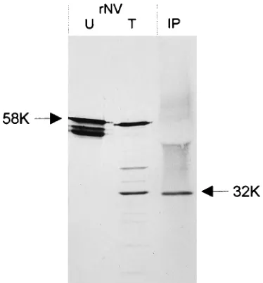
[image:3.612.80.267.70.271.2]centrifuga-FIG. 2. Immunoprecipitation and Western blot detection of NV-specific sol-uble protein from stool extracts. U, untreated rNV; T, trypsin-treated rNV; IP, soluble protein immunoprecipitated from a stool extract from a volunteer with a rabbit polyclonal antiserum made against rNV particles. Immunoprecipitated proteins were detected by Western blotting with mouse polyclonal antisera made against rNV. Arrows indicate the single band detected in the immunoprecipitate, which migrated similarly to the 32K trypsin cleavage product of rNV particles, and uncleaved 58K rNV capsid protein.
FIG. 3. SDS-PAGE analysis of trypsin-treated rNV particles and soluble rNV capsid protein. Sucrose gradient fractions of buffer-treated rNV (A) and trypsin-treated rNV (B) were analyzed by SDS-PAGE. (C) Trypsin treatment of soluble 58K rNV protein from fraction 2 of the gradient shown in panel A and trypsin treatment of assembled 58K rNV protein from fraction 9 of the gradient shown in panel A. Arrows indicate the positions of the 58K protein band and the 32K trypsin cleavage product.
on November 9, 2019 by guest
http://jvi.asm.org/
[image:3.612.316.556.70.394.2]tion to separate the 58K soluble protein from the 58K protein in assembled particles. Fraction 9 (Fig. 3A), containing the particle-associated 58K protein, and fraction 2 (Fig. 3A),
con-taining only soluble 58K protein, were each treated with 10mg
of trypsin per ml for 30 min at 378C, and the reaction mixtures
were electrophoresed on polyacrylamide gels. The results showed that the rNV capsid protein from the particle fraction of the gradient was resistant to trypsin cleavage, while trypsin treatment of the soluble 58K protein produced the 32K cleav-age product (Fig. 3C).
The trypsin cleavage site is conserved among other human caliciviruses.Subsequent to the cloning and sequencing of the NV genome, there has been a rapid increase in the number of sequences available for the capsid proteins of other Norwalk-like viruses (4, 12, 24–26, 40). Sequence alignments of capsid proteins from different strains show regions of sequence con-servation and divergence that essentially correspond to those defined for the capsid proteins of animal caliciviruses (32). The largest number of conserved amino acids is found in region 1 between aa 1 and 280 (8, 26). Alignment of the amino acid sequences of available Norwalk-like viruses in the region where the trypsin cleavage occurs (aa 199 to 245) shows that the lysine residue at position 227 is conserved in all but one human calicivirus sequenced to date (Fig. 4B). The one excep-tion is the recently described Bristol virus (14), which has an arginine at this position, still a site for trypsin-specific activity. Alignment with animal caliciviruses for which capsid sequences are available shows this residue to be conserved in San Miguel sea lion virus serotypes 1 and 4 (32) but not in feline calicivirus or rabbit hemorrhagic disease virus (5, 29). This alignment also shows that the N-terminal 3 amino acid residues of the 34K product detected in recombinant baculovirus-infected cell ly-sates (aa 219 to 221) are completely conserved among the
human caliciviruses. The amino acid residue at position 218, the C terminus at which the cleavage producing the 34K prod-uct presumably occurs, is a phenylalanine. This phenylalanine residue also is conserved among all of the human caliciviruses, with the exception of the Hawaii strain, which has a tyrosine residue at this position. Both of these residues are potential sites for chymotrypsin cleavage.
DISCUSSION
For viruses that replicate on mucosal surfaces, and particu-larly in the gastrointestinal tract, proteolytic cleavage of outer capsid proteins often plays an important role in the replication cycle of the virus. Data obtained in this study show that the NV capsid protein also undergoes a specific proteolytic cleavage, and this cleavage product is the source of the soluble protein detected in stools of infected volunteers. The N terminus of the soluble protein immunoprecipitated from stool and that of the cleavage product generated by digestion of rNV protein in vitro are identical. The apparent molecular weight of the sol-uble protein previously was reported to be approximately 30,000, although the migration in polyacrylamide gels in this study was closer to 32,000. These slight differences in electro-phoretic migration may simply reflect differences in SDS-PAGE conditions, or they may result from comigration of the antibody light chain used in the immunoprecipitation. Alter-natively, there may be trimming of C-terminal residues of the capsid protein in vivo that makes it slightly smaller.
Our data also show that only soluble 58K rNV capsid protein is susceptible to trypsin cleavage, while assembled rNV cles are resistant. Previous characterization of the rNV parti-cles showed that they are stable at pH 3 for 10 min but that they are not stable at pH 10 (18). We have further determined
FIG. 4. (A) Location of the trypsin cleavage site in rNV particles with respect to the full-length capsid protein. The arrow indicates the lysine residue at amino acid residue 227 where the trypsin-specific cleavage occurs. (B) Alignment of aa 199 to 245 of the capsid proteins of NV and other Norwalk-like human caliciviruses for which capsid sequences are available. Numbering is according to that for the NV sequence. The bold arrow indicates the site of trypsin cleavage conserved among these viruses. The light arrow shows the cleavage site that produces a 34K product in recombinant baculovirus-infected cell lysates as described previously (18). Alignments were generated by using the PILEUP program of the Genetics Computer Group (9).
on November 9, 2019 by guest
http://jvi.asm.org/
[image:4.612.142.468.71.330.2]that the rNV particles are not stable at pH 8.5 (data not shown), the pH of the buffer used in the initial trypsin diges-tions. At this pH, the assembled 58K protein unfolds into soluble protein. This observation is consistent with complete conversion of the full-length protein to the 32K cleavage prod-uct as the particles unfold under alkaline conditions. It is es-timated that as much as 50% of the NV antigen excreted in stool is soluble protein (10, 15), making it necessary to perform immunoelectron microscopy to detect NV particles. We have shown that the soluble protein in stool is a specific cleavage product of the capsid protein; therefore, it is possible that the events leading to cleavage of the NV capsid protein in vivo mimic those observed in vitro with the 58K rNV capsid protein. It may be that a large percentage of the NV released from infected cells is dissociated as it is excreted into a more alkaline environment in the intestine. This dissociation and cleavage would explain the paucity of NV particles detectable by elec-tron microscopy. Because the cleavage occurs at a specific site, it also is likely that the soluble protein remains folded in some structural form such that the other 16 potential trypsin cleav-age sites present in the linear sequence are not exposed.
Because assembled rNV particles are resistant to trypsin digestion, proteolytic cleavage is probably not important for the infectivity of the virus, but the large amount of soluble protein produced may have an effect on the immune response to NV and the pathophysiology of infection. A recent clinical study has confirmed and extended the results of early studies of NV infection in volunteers (10, 20), showing that the presence of preexisting antibody generally does not correlate with resis-tance to disease and that higher levels of antibody correlate with disease susceptibility (10). Furthermore, the risk of both infection and illness following challenge with NV was higher for volunteers who possessed prechallenge antibody, and the levels of immunoglobulin G, immunoglobulin A, and immuno-globulin M in prechallenge serum were higher for those who developed infection or illness (39). What role the soluble pro-tein plays in the biologic consequences of NV infection is not clear, but it may influence the immune response and clinical outcome of NV infection.
One possibility is that a large amount of soluble protein induces antibodies that are nonneutralizing, and these may be the predominant antibodies produced following infection with NV. This would offer an explanation for the absence of a protective effect of prechallenge antibody. What region of the NV capsid protein induces neutralizing antibodies is not known because of the lack of an in vitro virus neutralization assay. The three-dimensional structure of rNV particles has been determined to a resolution of 22 Å (2.2 nm) by electron cryomicroscopy and computer image processing (35). It has
been proposed on the basis of similarities with other T53
single-stranded RNA viruses that the N-terminal 250 amino acid residues make up a shell domain (S) and that the remain-ing C-terminal residues form the arch-like protrudremain-ing domains (P1 and P2). It may be that neutralizing antibodies are mainly directed to the S domain and nonneutralizing antibodies are mainly directed to the arches. This idea is consistent with the 32K trypsin cleavage product containing the C terminus of the capsid protein. If the structural predictions for the region of the linear amino acid sequence that makes up the shell domain are correct, then the trypsin cleavage site at amino acid residue 227 would be buried in assembled particles and not accessible to cleavage. It is likely that the trypsin cleavage site becomes exposed as particles swell or unfold under alkaline conditions. Trypsin cleavage of rNV capsid protein in vitro results in deg-radation of the N terminus to products with lower molecular weights, and this is likely what happens to the N terminus of
the capsid protein in vivo, since only the C-terminal 32K cleav-age product was found as soluble protein in stool extracts.
A second possibility is that a rapid and amplified secondary antibody response to the soluble protein directs an immune system-mediated pathology that results in the clinical manifes-tations of NV infection. However, the question of why some individuals who lack or have lower levels of preexisting anti-body get infected and become ill still remains. It would be of interest to see if those individuals who were asymptomatic but shed virus as determined by enzyme-linked immunosorbent assay (10) excreted larger numbers of virus particles and less soluble protein than those individuals with gastroenteritis. Worth noting are studies that have shown a positive correla-tion between the presence of preexisting serum antibody and resistance to NV infection in children in the Third World (2, 36). These results might reflect different physiological condi-tions of the gut that result in a lower level of virus dissociation and subsequently a smaller amount of soluble protein. Inter-estingly, more particles are often seen in children than in adults (8a). Since volunteer studies of NV infection have in-volved only adults, the conflict in these results may reside in differences between the two age groups. Finally, it has been suggested that the susceptibility to NV infection may be ge-netically determined, and this explanation cannot be ruled out (21).
The complete conservation of the trypsin cleavage site in all of the human caliciviruses sequenced to date suggests that there is a biologic role for the soluble protein. Definitive stud-ies to address this question will be difficult in the absence of an animal model or cell culture system for the human calicivi-ruses.
ACKNOWLEDGMENTS
This work was supported by Public Health Service grants AI-30448-04 and T32 DK-07664-03 from the National Institutes of Health.
REFERENCES
1. Ando, T., M. N. Mulders, D. C. Lewis, M. K. Estes, S. S. Monroe, and R. I. Glass.1994. Comparison of the polymerase region of small round structured virus strains previously characterized in three serotypes by solid-phase im-mune electron microscopy. Arch. Virol. 135:217–226.
2. Black, R. E., H. B. Greenberg, A. Z. Kapikian, K. H. Brown, and S. Becker. 1982. Acquisition of serum antibody to Norwalk virus and rotavirus and relation to diarrhea in a longitudinal study of young children in rural Ban-gladesh. J. Infect. Dis. 145:483–489.
3. Burns, J. W., H. B. Greenberg, R. D. Shaw, and M. K. Estes. 1988. Func-tional and topographical analyses of epitopes on the hemagglutinin (VP4) of the simian rotavirus SA11. J. Virol. 62:2164–2172.
4. Carter, M. J., I. D. Milton, and C. R. Madeley. 1991. Caliciviruses. Med. Virol. 1:177–186.
5. Carter, M. J., I. D. Milton, J. Meanger, M. Bennett, R. M. Gaskell, and P. C. Turner.1992. The complete nucleotide sequence of a feline calicivirus. Virology 190:443–448.
6. Caul, E. O., C. Ashley, and J. V. Pether. 1979. ‘‘Norwalk’’-like particles in the epidemic gastroenteritis in the U.K. Lancet ii:1292. (Letter.)
7. Dolin, R., R. C. Reichman, K. D. Roessner, T. S. Tralka, R. T. Schooley, W. Gary, and D. Morens.1982. Detection by immune electron microscopy of the Snow Mountain agent of acute viral gastroenteritis. J. Infect. Dis. 146: 184–189.
8. Estes, M. K., and M. E. Hardy. Norwalk virus and other enteric caliciviruses.
In M. J. Blaser, P. D. Smith, J. I. Ravdin, H. B. Greenberg, and R. L.
Guerrant (ed.), Infections of the gastrointestinal tract, in press. Raven Press, New York.
8a.Estes, M. K. Unpublished observation.
9. Genetics Computer Group. 1994. Program manual for the Wisconsin pack-age, version 8. Genetics Computer Group, Madison, Wis.
10. Graham, D. Y., X. Jiang, T. Tanaka, A. R. Opekun, H. P. Madore, and M. K. Estes.1994. Norwalk virus infection of volunteers: new insights based on improved assays. J. Infect. Dis. 170:34–43.
11. Gray, J. J., X. Jiang, P. Morgan Capner, U. Desselberger, and M. K. Estes. 1993. Prevalence of antibodies to Norwalk virus in England: detection by enzyme-linked immunosorbent assay using baculovirus-expressed Norwalk virus capsid antigen. J. Clin. Microbiol. 31:1022–1025.
on November 9, 2019 by guest
http://jvi.asm.org/
12. Green, J., J. P. Norcott, D. Lewis, C. Arnold, and W. G. Brown. 1991. Norwalk-like viruses: demonstration of genomic diversity by polymerase chain reaction. J. Clin. Microbiol. 31:3007–3012.
13. Green, K. Y., J. F. Lew, X. Jiang, A. Z. Kapikian, and M. K. Estes. 1993. Comparison of the reactivities of baculovirus-expressed recombinant Nor-walk virus capsid antigen with those of the native NorNor-walk virus antigen in serologic assays and some epidemiologic observations. J. Clin. Microbiol. 31:2185–2191.
14. Green, S. M., K. E. Dingle, P. R. Lambden, E. O. Caul, C. R. Ashley, and I. N. Clarke.1994. Human enteric caliciviridae; a new prevalent SRSV group defined by RNA-dependent RNA polymerase and capsid diversity. J. Gen. Virol. 75:1883–1888.
15. Greenberg, H. B., J. R. Valdesuso, A. R. Kalica, R. G. Wyatt, V. J. McAuliffe, A. Z. Kapikian, and R. M. Chanock.1981. Proteins of Norwalk virus. J. Virol. 37:994–999.
15a.Hardy, M. E., and M. K. Estes. Unpublished observation.
16. Hyams, K. C., J. D. Malone, A. Z. Kapikian, M. K. Estes, X. Jiang, A. L. Bourgeois, S. Paparello, and K. Y. Green.1993. Norwalk virus infection among Desert Storm troops. J. Infect. Dis. 167:986–987.
17. Jiang, X., D. Y. Graham, K. Wang, and M. K. Estes. 1990. Norwalk virus genome cloning and characterization. Science 250:1580–1583.
18. Jiang, X., M. Wang, D. Y. Graham, and M. K. Estes. 1992. Expression, self-assembly, and antigenicity of the Norwalk virus capsid protein. J. Virol. 66:6527–6532.
19. Jiang, X., M. Wang, K. Wang, and M. K. Estes. 1993. Sequence and genomic organization of Norwalk virus. Virology 195:51–61.
20. Johnson, P. C., J. J. Mathewson, H. L. DuPont, and H. B. Greenberg. 1990. Multiple-challenge study of host susceptibility to Norwalk gastroenteritis in US adults. J. Infect. Dis. 161:18–21.
21. Kapikian, A. Z., and R. M. Chanock. 1990. Norwalk group of viruses, p. 671–693. In B. N. Fields (ed.), Virology. Raven Press, New York. 22. Kapikian, A. Z., R. G. Wyatt, R. Dolin, T. S. Thornhill, A. R. Kalica, and
R. M. Chanock.1972. Visualization by immune electron microscopy of a 27-nm particle associated with acute infectious nonbacterial gastroenteritis. J. Virol. 10:1075–1081.
23. Lambden, P. R., E. O. Caul, C. R. Ashley, and I. N. Clarke. 1993. Sequence and genome organization of a human small round-structured (Norwalk-like) virus. Science 259:516–519.
24. Lew, J. F., A. Z. Kapikian, X. Jiang, M. K. Estes, and K. Y. Green. 1994. Molecular characterization and expression of the capsid protein of a Nor-walk-like virus recovered from a Desert Shield troop with gastroenteritis. Virology 200:319–325.
25. Lew, J. F., A. Z. Kapikian, J. Valdesuso, and K. Y. Green. 1994. Molecular characterization of Hawaii virus and other Norwalk-like viruses: evidence for genetic polymorphism among human caliciviruses. J. Infect. Dis. 170:535– 542.
26. Lew, J. F., M. Petric, A. Z. Kapikian, X. Jiang, M. K. Estes, and K. Y. Green. 1994. Identification of minireovirus as a Norwalk-like virus in pediatric patients with gastroenteritis. J. Virol. 68:3391–3396.
27. Madore, H. P., J. J. Treanor, and R. Dolin. 1986. Characterization of the Snow Mountain agent of viral gastroenteritis. J. Virol. 58:487–492. 28. Matsui, S. M., J. P. Kim, H. B. Greenberg, W. Su, Q. Sun, P. C. Johnson,
H. L. DuPont, L. S. Oshiro, and G. R. Reyes.1991. The isolation and characterization of a Norwalk virus-specific cDNA. J. Clin. Invest. 87:1456– 1461.
29. Meyers, G., C. Wirblich, and H. J. Thiel. 1991. Rabbit hemorrhagic disease virus—molecular cloning and nucleotide sequencing of a calicivirus genome. Virology 184:664–676.
30. Moe, C. L., J. Gentsch, G. Grohmann, T. Ando, S. S. Monroe, X. Jiang, J. Wang, M. K. Estes, Y. Seto, C. Humphrey, S. Stine, and R. I. Glass.1994. Application of PCR to detect Norwalk virus in fecal specimens from out-breaks of gastroenteritis. J. Clin. Microbiol. 32:642–648.
31. Morens, D. M., R. M. Zweighaft, T. M. Vernon, G. W. Gary, J. J. Eslien, B. T. Wood, R. C. Holman, and R. Dolin.1979. A waterborne outbreak of gastro-enteritis with secondary person-to-person spread: association with a viral agent. Lancet i:964–966.
32. Neill, J. D. 1992. Nucleotide sequence of the capsid protein gene of two serotypes of San Miguel sea lion virus: identification of conserved and non-conserved amino acid sequences among calicivirus capsid proteins. Virus Res. 24:211–222.
33. Oishi, I., K. Yamazaki, Y. Minekawa, H. Nishimura, and T. Kitaura. 1985. Three-year survey of the epidemiology of rotavirus, enteric adenovirus, and some small spherical viruses including ‘‘Osaka-agent’’ associated with infan-tile diarrhea. Biken J. 28:9–19.
34. Okada, S., S. Sekine, T. Ando, Y. Hayashi, M. Murao, K. Yabuuchi, T. Miki, and M. Ohashi.1990. Antigenic characterization of small, round-structured viruses by immune electron microscopy. J. Clin. Microbiol. 28:1244–1248. 35. Prasad, B. V. V., R. Rothnagel, X. Jiang, and M. K. Estes. 1994.
Three-dimensional structure of baculovirus-expressed Norwalk virus capsids. J. Virol. 68:5117–5125.
36. Ryder, R. W., N. Singh, W. C. Reeves, A. Z. Kapikian, H. B. Greenberg, and R. B. Sack.1985. Evidence of immunity induced by naturally acquired rota-virus and Norwalk rota-virus infection on two remote Panamanian islands. J. Infect. Dis. 151:99–105.
37. Taniguchi, K., S. Urasawa, and T. Urasawa. 1979. Virus-like particle, 35 to 40 nm, associated with an institutional outbreak of acute gastroenteritis in adults. J. Clin. Microbiol. 10:730–736.
38. Treanor, J., R. Dolin, and H. P. Madore. 1988. Production of a monoclonal antibody against the Snow Mountain agent of gastroenteritis by in vitro immunization of murine spleen cells. Proc. Natl. Acad. Sci. USA 85:3613– 3617.
39. Treanor, J. J., X. Jiang, H. P. Madore, and M. K. Estes. 1993. Subclass-specific serum antibody responses to recombinant Norwalk virus capsid antigen (rNV) in adults infected with Norwalk, Snow Mountain, or Hawaii virus. J. Clin. Microbiol. 31:1630–1634.
40. Wang, J., X. Jiang, H. P. Madore, J. Gray, U. Desselberger, T. Ando, Y. Seto, I. Oishi, J. F. Lew, K. Y. Green, and M. K. Estes.1994. Sequence diversity of small, round-structured viruses in the Norwalk virus group. J. Virol. 68:5982–5990.
41. Wyatt, R. G., R. Dolin, N. R. Blacklow, H. L. DuPont, R. F. Buscho, T. S. Thornhill, A. Z. Kapikian, and R. M. Chanock.1974. Comparison of three agents of acute infectious nonbacterial gastroenteritis by cross-challenge in volunteers. J. Infect. Dis. 127:709–714.