0022-538X/95/$04.0010
Copyrightq1995, American Society for Microbiology
Stem Cell Factor Binding to Retrovirus Primer Binding Site Silencers
MICHAEL YAMAUCHI,† BRETON FREITAG, CHARLOTTE KHAN, BRENT BERWIN,‡ ANDERIC BARKLIS*
Vollum Institute for Advanced Biomedical Research and Department of Microbiology and Immunology, Oregon Health Sciences University, Portland, Oregon 97201
Received 5 July 1994/Accepted 21 October 1994
Using modified nuclear lysis and binding conditions, we have examined the binding of an embryonal carcinoma (EC) cell factor, binding factor A, to a stem cell-specific silencer which acts at the DNA level and overlaps the Moloney murine leukemia virus (M-MuLV) proline primer binding site (PBS). Following our protocol, we found that in vitro binding of factor A correlated with the in vivo activity of the M-MuLV silencer. Factor A bound specifically to the wild-type silencer element at room temperature and 30&C, but not at 4&C, and bound 10-fold better to the full-length silencer than to a minimal silencer core element. The factor was enriched in nuclear compared with cytosolic extracts and in undifferentiated EC cells compared with differentiated cells in which the silencer is nonfunctional. Salt and ion requirements for factor A binding were investigated, and partial purification steps indicated the factor to be a heparin-Sepharose-binding moiety of greater than 100 kDa. To examine possible relationships between silencer and PBS activities, sequences representing phenyl-alanine, isoleucine, lysine-1,2, lysine-3, methionine, and tryptophan PBS DNA fragments were tested in vivo for stem cell-specific repression of M-MuLV expression and in vitro in DNA binding assays. Of these PBS elements, only the lysine-1,2 PBS DNA fragment showed consistently high levels of repression. Interestingly, the lysine-1,2 PBS DNA fragment also formed a complex with an EC cell factor with characteristics similar to those of factor A. However, the two factors did not cross-compete in binding studies, suggesting that they may be different but related factors. Our results suggest that expression of Mason-Pfizer monkey virus, visna virus, and spumavirus, which use the lysine-1,2 PBS, may be inhibited in undifferentiated stem cells.
Embryonal carcinoma (EC) cells, such as F9 cells (4), derive from spontaneous gonadal tumors or from the inner cell mass of embryos at the 64- to 128-cell stage (24). They are undif-ferentiated and are developmentally the earliest culturable mammalian cell line. Moloney murine leukemia virus (M-MuLV) infects EC cells with extremely low efficiency (3, 10–12, 15), while differentiated cell lines such as 3T3 fibroblasts are readily infected by M-MuLV (33). The repression of M-MuLV in EC cells occurs after the viral integration step of the repli-cation cycle (3, 5, 10, 11, 30). Infection of EC cells with M-MuLV leads to integration of proviral DNA into the genome at normal levels, but virus-specific RNA is detected at less than 1% of the level of infected fibroblast cells (10, 11, 30). A wild-type (wt) DNA element referred to as the negative regu-latory element (19) or the repressor binding site (RBS) (25) has been demonstrated to be critical for part of the repression effect (3, 5, 9, 16–19, 25, 32). Previous experiments with re-combinant M-MuLV vectors showed that the RBS is sufficient to repress expression from M-MuLV, simian virus 40 early, and adenovirus major late promoters in EC cells in an orientation-and position-independent (upstream or downstream) fashion (25). However, there appears to be a diminution of the repres-sion effect when the RBS is separated from its target promoter by more than 1 to 2 kbp (5, 16). Interestingly, stem cell-specific silencing mediated by the M-MuLV RBS has been difficult to demonstrate in transient transfections but has been readily observed in transient infections, stable infections, and stable transfections (16, 19, 25). One possible explanation for these
results is that the amount of DNA used in transient transfec-tions is greater than the amount of repressor present in the cell. Alternatively, there may be a requirement for integration into the host chromosome in order for the repression effect to occur. Regardless, after an initial 10- to 100-fold repression exerted by the RBS, RBS-associated promoter regions gradu-ally become methylated (5, 16) and are repressed an additional 10- to 100-fold. Consequently, the eventual level of repression can be 100- to 10,000-fold relative to M-MuLV expression in differentiated 3T3 fibroblast cells (3).
With regard to the RBS sequence, the element was identi-fied first by a single point mutation (G to A at M-MuLV nucleotide [nt] 160 according to the numbering of Shinnick et al. [28]) in a recombinant M-MuLV-derived retrovirus which was not repressed in EC cells (3). This mutation (RBSB2) is located in the M-MuLV primer binding site (PBS) (M-MuLV nt 146 to 163), which binds a cellular tRNA (tRNAProfor wt M-MuLV) to prime first-strand synthesis during reverse tran-scription (33). However, the silencer function of this region was shown to be distinct from the reverse transcription role, since silencing occurs at the DNA level, in the absence of viral proteins, and a recombinant retrovirus with a PBS-glutamine (PBSQ) (7) substituted for the wt PBS-proline (PBSP) was replication competent but lacked the repressor function (13, 25). By using a PBSQrecombinant retrovirus backbone, PBS-derived DNAs were tested for repressor function, and it was shown that the minimal repressor site (RBSWT-18) overlapped the M-MuLV PBSP for 17 of its 18 bp and that few point mutations were compatible with repression (16). However, it was also found that the addition of the 11-bp wt sequence onto the 39end of the wt RBS (RBSWT-28; M-MuLV nt 147 to 174) appeared to increase repression levels by a variable amount (16).
The above-described data suggested that an EC cellular factor mediated the M-MuLV repression via the RBS at the
* Corresponding author. Phone: (503) 8098. Fax: (503) 494-6862.
† Present address: Department of Biology, Massachusetts Institute of Technology, Cambridge, MA 02139.
‡ Present address: Molecular and Cellular Biology Program, Uni-versity of Wisconsin, Madison, WI 53706.
1142
on November 9, 2019 by guest
http://jvi.asm.org/
DNA level, and a putative repressor factor called the negative regulatory element binding factor (19) or binding factor A (25) was identified in exonuclease III protection assays (19) and DNA mobility shift assays (25). However, the RBS binding factor (referred to here as factor A) was extraordinarily sensi-tive to binding conditions and was refractory to standard DNase protection and methylation interference mapping pro-tocols. Because of its sensitivity, it previously had been difficult to assess factor A with regard to a number of parameters, such as cell specificity, optimal binding conditions, factor size, and binding site size. In our current work we are investigating the optimization and characterization of factor A binding condi-tions, and we now show that in vitro factor A binding correlates well with in vivo results. We also have investigated the repres-sion effects of other PBS variants and have found that the lysine-1,2 PBS sequence (PBSK2) acts as a stem cell-specific silencer, binding a factor which is related to but distinct from factor A.
MATERIALS AND METHODS
Cell extracts.Nuclear extracts were prepared from tissue culture cells essen-tially as described by Dignam et al. (8) with the modifications of Baeuerle and Baltimore (1, 2), which permit the fractionation of nuclear and cytosolic extracts, as well as a postnuclear fraction, obtained as resuspended pellet material from centrifugations used to clear cytosolic extracts. Cell lysis was carried out with 10 strokes of a Wheaton Dounce homogenizer (type A pestle) in Dignam buffer A (10 mM N-2-hydroxyethylpiperazine-N9-2-ethanesulfonic acid [HEPES] [pH 7.9], 1.5 mM MgCl2, 10 mM KCl, 0.5 mM dithiothreitol [DTT], 0.1 mM
phenyl-methylsulfonyl fluoride [PMSF], 5mg of leupeptin per ml, 2.5mg of pepstatin A per ml, 2mg of benzamidine per ml, and 10mg of aprotinin per ml). The nuclear-D, cytosolic, and postnuclear fractions were dialyzed quickly (twice for 2 h each at 48C) against Dignam buffer D (20 mM HEPES [pH 7.9], 100 mM KCl, 20% [vol/vol] glycerol, 0.2 mM EDTA, 0.5 mM PMSF, 0.5 mM DTT) in order to retain active protein. To increase yields of binding factor A DNA binding activ-ity, some nuclear extracts (nuclear-C extracts) were prepared without dialysis against modified Dignam buffer D and were left in Dignam buffer C (20 mM HEPES [pH 7.9], 25% [vol/vol] glycerol, 0.3 M NaCl, 1.5 mM MgCl2, 0.2 mM
EDTA, 0.5 mM PMSF, 0.5 mM DTT). Protein concentrations were measured by the method of Bradford (6).
Electrophoretic mobility shift assay (gel shift assay).The preparation of double-stranded (ds) radioactive oligonucleotide probes and the gel shift proce-dure were essentially those of Thornell et al. (31), with modifications. For probes, sense strand oligonucleotides were end labeled for 45 min at 378C with [g-32P]ATP and T4 polynucleotide kinase (Gibco) in 20ml of 50 mM Tris (pH
7.4)–10 mM MgCl2–1 mM spermidine–100mg of bovine serum albumin (BSA)
per ml–10 mM DTT (the reaction volume was adjusted to 20ml with distilled water). After kinase inactivation (5 min at 708C), a 10% excess of complementary strands was added, and strand pairs were incubated at 508C for 30 min and then allowed to cool to room temperature. The ds radioactive probes were gel purified on a 12% native acrylamide gel in Tris-borate, and fragments were eluted (16 to 20 h at room temperature in 1 M NaCl–20 mM Tris [pH 7.5]–1 mM EDTA). The eluted probes were ethanol precipitated, dried, resuspended (25,000 cpm/ml) in distilled water, and stored at2208C. Oligonucleotide probes were as follows (only one strand for each is designated): a wt RBS probe (RBSWT-28) (59GGG
GGCTCGTCCGGGATCGGGAGACCCC 39), an RBS point mutant (RBSB2)
(59GGGGGCTCGTCCGaGATCGGGAGACCCC 39), a control Sp1 transcrip-tion factor probe (Sp1) (59GCTCGCCCCGCCCCGATCGAAT 39), a 28-mer primer PBS lysine-1,2 probe (PBSK2-28) (59GGCGCCCAACGTGGGGCCGG
GAGACCCC 39) (20), and 18-mer probes representing the wt M-MuLV PBS (RBSWT-18) (59GGGGGCTCGTCCGGGATC 39), a phenylalanine PBS (PBSF)
(59TCCCGGGTTTCGGCACCA 39) (29), an isoleucine PBS (PBSI) (59TCCC
CGTACGGGCCACCA 39) (20), lysine-2 (PBSK2-18) (59 GCCCCACGTTGG
GCGCCA 39) (20) and lysine-3 (PBSK3) (59GTCCCTGTTCGGGCGCCA 39)
(33) PBSs, a methionine PBS (PBSM) (59TCCTCACACGGGGCACCA 39)
(29), and a tryptophan PBS (PBSW) (59ATCACGTCGGGGTCACCA 39) (33).
Typical binding reactions were carried out with an excess of probe and 5 to 10 ml of cellular extract (the amount of extract varied because of the addition of various other components which were tested [see below]) in a total volume of 20 ml of Thornell binding buffer (25 mM HEPES [pH 7.9], 1 mM EDTA, 10% [vol/vol] glycerol, 5 mM DTT, 0.5 mM PMSF) with an additional 25 ng of poly(dI-dC) perml, 5 mM NaCl, 5 mM KCl, 3 mM MgCl2, and 0.1 mM ZnCl2.
Some reaction mixtures contained 3ml of fetal calf serum (FCS), which was added to increase the signal of the factor A band, and various concentrations of salts (0 to 1 M NaCl and 0 to 90 mM KCl), divalent cations (0 to 10 mM [each] CaCl2, CuSO4, FeSO4, MgCl2, MnCl2, and NiSO4), nucleotides (0 to 10 mM
[each] cyclic AMP [cAMP], ATP, GTP, and GTPgS), and detergents (0 to 1%
[each] deoxycholate, Nonidet P-40, Sarkosyl, sodium dodecyl sulfate, and Triton X-100) were tested during our investigations. For competitions, various amounts (15 to 200 ng) of unlabeled ds and single-stranded forms of the oligonucleotides used for probes were added 20 min prior to the addition of the radioactive probe. Unless indicated otherwise, binding reaction mixtures were preincubated at 308C for 20 minutes, and then ds probes were added (50,000 cpm, 0.5 ng per reaction mixture) and mixtures were incubated at 308C for an additional 20 min. After incubations, binding reactions were run on 6% native acrylamide gels (31.0 cm by 38.5 cm by 0.4 mm) at 800 V for approximately 3 h in a Tris-glycine buffer (5 mM Tris [pH 8.5], 38 mM glycine, 0.2 mM EDTA). After electrophoresis, gels were transferred to used X-ray films for support and were exposed for 3 to 7 days at 2808C with intensifying screens.
Partial protein purifications.Binding factor A was partially purified from F9 nuclear extracts by Sephadex G-200 (Pharmacia), heparin-Sepharose (Pharma-cia), and DNA-affinity chromatographies. Sephadex gel filtration was at 48C with a 20-ml (bed volume) column and a 9-ml void volume. Crude nuclear extract was loaded in Dignam buffer D (without glycerol) at a gravity flow rate of 0.5 ml/min. Sephadex G-200 fractions (1 ml) were concentrated rapidly by centrifugation on Microcon-30 microconcentrators (Amicon) at 48C in an Eppendorf 5415 micro-centrifuge at 12,000 rpm for 30 min, and fractions were snap frozen on dry ice and stored at2808C.
For heparin-Sepharose chromatography, crude nuclear extracts were applied to a 10-ml (bed volume) heparin-Sepharose column in Dignam buffer D (without glycerol) as the loading buffer, and proteins were eluted from the resin by increasing the KCl concentration of the running buffer in steps (0 mM, 100 mM, 300 mM, 600 mM, and 1 M KCl). Heparin-Sepharose fractions were desalted and concentrated with Centricon-100 microconcentrators by adding the fractions to the concentrators and spinning at 48C in a Sorvall SS-34 rotor at 6,500 rpm for 30 min. The concentrated heparin-Sepharose fractions were then diluted with Dignam buffer D (with glycerol) and reconcentrated as described above. The dilution-reconcentration step was performed three times, each time with approx-imately a 1:5 dilution of concentrated sample to buffer D. The final volume of each fraction was approximately one-half of the starting fraction volume.
For DNA affinity chromatography, a 59biotinylated RBSWT-28oligonucleotide
was used. The DNA affinity resin was prepared by annealing 2.5mg each of the antisense strand biotinylated RBSWT-28oligonucleotide and its complementary
strand and binding the ds product to streptavidin-agarose resin (Sigma) equili-brated in MY running buffer [25 mM HEPES (pH 7.9), 0.5 mM EDTA, 10% glycerol, 5 mM DTT, 0.5 mM PMSF, 3 mM MgCl2, 0.1 mM ZnCl2, 25 ng of
poly(dI-dC) perml]. Chromatography binding reactions were performed in a total volume of 2.5 ml [500ml (bed volume) of DNA affinity resin, 500ml of F9 nuclear extract, 400ml of 53Thornell binding buffer, 300ml of bandshift salts, 300ml of FCS, 100ml of 500-ng/ml poly(dI-dC), 50ml of PMSF, and 350ml of H2O], and reaction mixtures were incubated at 308C with periodic agitation. The
binding mixture was centrifuged (15 s at 90 3g), and the supernatant was collected as the flowthrough fraction. The resin was washed with MY buffer containing increasing amounts of KCl (0 mM, 200 mM, 400 mM, 600 mM, 800 mM, and 1 M). Fractions were desalted and concentrated in the same manner as for heparin-Sepharose fractions. The resin was washed with 5 ml of MY running buffer plus 0.02% sodium azide and stored at 48C.
Recombinant retroviruses.BlankIsle is a plasmid derived by inserting the 702-bp EcoRV-to-ScaI fragment of pBR322, with BamHI linkers, into the unique BamHI site of PBSQ(16, 25). WTIsle is a subclone of BlankIsle
contain-ing M-MuLV nt 147 to 174 at the old pBR322 SspI site. B2Isle is similar to WTIsle but has the previously described single-base-pair RBSB2mutation (3).
The PBSIsle and miniWTIsle constructs are subclones containing the following different tRNA PBSs at the old pBR322 SspI sites (only the sense strand is shown): PBSF, 59TCCCGGGTTTCGGCACCA 39(29); PBSI, 59TCCCCGTA
CGGGCCACCA 39(from RTVL-1) (20); PBSK2, 59GCCCCACGTTGGGCG
CCA 39(from Mason-Pfizer monkey virus, visna virus, and spumavirus) (20); PBSK3, 59 GTCCCTGTTCGGGCGCCA 39 (from human immunodeficiency
virus) (33); PBSM, 59TCCTCACACGGGGCACCA 39(29); and PBSW, 59AT
CACGTCGGGGTCACCA 39(from Rous sarcoma virus and avian myeloblas-tosis virus) (33). The F9 EC and NIH 3T3 cell lines were grown as described previously (3, 32), and virus infections, selections with G418 (Gibco), and titer determinations (given as 3T3/F9 ratios [restriction indices], where higher num-bers indicate greater viral repression) were performed as described previously (3, 23). Cloning and sequencing were by standard protocols (22, 27).
RESULTS
Characteristics of factor A binding to the M-MuLV RBS. The stem cell-specific silencer element called the RBS or neg-ative regulatory element originally was identified as a cis-active element in M-MuLV that repressed expression at the DNA level in EC or embryonal stem cells (19, 25). Subsequently, an EC cell factor, binding factor A, was identified by exonuclease III protection assays and gel mobility shift assays with a wt RBS probe (RBSWT) (19, 25). However, by either assay, factor A
on November 9, 2019 by guest
http://jvi.asm.org/
binding was very sensitive and required specific binding and gel electrophoresis conditions (19, 25). Because of these difficul-ties, additional attempts to characterize factor binding to the RBS and related sequences were hampered.
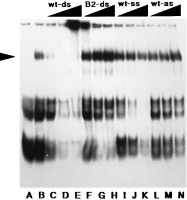
In order to circumvent the above-described problem, we have undertaken efforts to optimize and analyze factor A bind-ing conditions. Because we found that factor A did not tolerate extensive Dounce homogenization or dialysis, we modified our original protocol for extract preparation as described in the Materials and Methods. By using this protocol, lysates were prepared from F9 EC cells, which demonstrate the RBS-me-diated repression, and from 3T3 fibroblasts, which are un-affected by the RBS (3, 9, 19, 25). As shown in Fig. 1, three major cellular complexes form with the 28-bp RBSWT probe (RBSWT-28): a pair of fast-migrating nonspecific complexes (referred to as complexes B and C) and the low-mobility factor A complex, indicated by the arrowhead. At 48C, factor A ap-peared to bind equally well to the RBSWT-28probe (Fig. 1, lane E) and a 1-bp mutant probe (RBSB2) (lane F), but at room temperature or 308C, the factor A specificity for the RBSWT-28 probe was apparent (lanes G to N). In comparison with com-plexes B and C, factor A appeared to be enriched in nuclear versus cytosolic extracts (compare lane A with lanes G, I, K, and M in Fig. 1), as might be expected for a factor which acts at the DNA level. Consistent with the observation that RBS
repression is specific for EC or embryonal stem cells, factor A was enriched greatly in F9 (Fig. 1, lanes G, I, K, and M) versus 3T3 (lanes Q, R, U, and W) nuclear extracts. Similarly, we have observed factor A in extracts of the PCC4 EC cell line (25), in which the RBS is active, but not in other cell lines which do not demonstrate RBS repression (16), such as mouse SLK.3 and 10T1/2, rat PC12, and human HeLa and 293 cells (data not shown).
Previous competition and interference assays demonstrated the specificity of factor A binding to the RBSWT-28probe, as did assays with RBSWT-28versus RBSB2probes (Fig. 1). Nev-ertheless, it was of interest to test competitors in binding assays with lysates produced by our modified protocol. Re-sults of these experiments are shown in Fig. 2. Unlabeled ds RBSWT-28DNA effectively competed for factor A binding to the RBSWT-28probe (Fig. 2, lanes C to E), whereas unlabeled ds RBSB2(lanes F to H) and single-stranded RBSWT-28(lanes I to N) DNAs were poor competitors. Because the RBS se-quence overlaps the M-MuLV tRNA PBS, it also was of in-terest to investigate whether factor A might recognize some feature of a tRNA molecule. However, neither total calf liver tRNA (up to 400 ng, a 200-fold excess of tRNA to probe) nor purified tRNAPro(up to 500 ng, a 250-fold excess of tRNA to probe) (kindly provided by D. Dignam) competed for factor A binding (data not shown). Similarly, purified proline tRNA syn-thetase (40mg/ml) (kindly provided by D. Dignam) showed no binding to the RBSWT-28probe on gel shifts (data not shown).
With regard to optimization of factor A binding, the pres-ence of 100mM zinc (Fig. 1, lanes K and M) versus 10mM zinc (lanes G and I) appeared to improve binding, while a reducing agent such as DTT had no apparent effect (Fig. 1, compare lanes I and G and compare lanes M and K). The effects of other salts, divalent cations, chelators, phosphorylation inhib-itors, nucleotides, and detergents also were tested. In
particu-FIG. 1. RBS binding factors in F9 and 3T3 cells. The ds RBSWT-28 and
RBSB2(28-mer) probes were prepared as described in Materials and Methods
and used at 50,000 cpm (approximately 2 ng) per binding reaction. The se-quences of these probes were as follows.
RBSWT-28: 59 GGGGG CTCGT CCGGG ATCGG GAGCA CCC39
39CCCCC GAGCA GGCCC TCGCC CTCGT GGG 59
RBSB2: 59 GGGGG CTCGT CCGaG ATCGG GAGCA CCC39
39CCCCC GAGCA GGCtC TAGCC CTCGT GGG 59
RBSWT-28(lanes A, C, E, G, I, K, M, O, Q, S, U, and W) or RBSB2(lanes B, D,
F, H, J, L, N, P, R, T, V, and X) was incubated for 20 min with extracts at room temperature (lanes A to D, G to N, and Q to X) or at 48C (lanes E, F, O, and P). Reaction mixtures contained extracts from F9 cytosol (lanes A and B) (6.5 mg/ml), 3T3 cytosol (lanes C and D) (2.8 mg/ml), F9 nuclei (lanes E to N) (1.3 mg/ml), or 3T3 nuclei (lanes O to X) (1.5 mg/ml). In some cases, DTT was added to a final concentration of 1 mM (lanes I, J, S, and T), and ZnCl2was added to
[image:3.612.64.289.70.251.2]a final concentration of 10mM (lanes A to J and O to T) or 100mM (lanes K to N and U to X). Binding reactions were terminated by addition of loading dye, and free and complexed probes were separated by electrophoresis on a 6% native acrylamide–Tris-glycine gel and autoradiographed. Because large gels (31.0 cm by 38.5 cm by 0.4 mm) were used for electrophoresis, free probe is not shown. The factor A band is indicated by the arrowhead, and the nonspecific B and C complexes appear as bands of increasing mobilities.
FIG. 2. Factor A binding competition studies. RBSWT-28(lanes B to N) or
RBSB2(lane A) probe was incubated for 20 min at room temperature with F9
nuclear extract (1.3 mg/ml). Binding reactions were performed without compet-itor DNA (lanes A and B) or with increasing amounts of the following unlabeled competitors: RBSWT-28(ds) (5 ng [lane C], 15 ng [lane D], or 45 ng [lane E]),
RBSB2(ds) (5 ng [lane F], 15 ng [lane G], or 45 ng [lane H]), RBSWT-28(sense
strand [ss] only) (5 ng [lane I], 15 ng [lane J], or 45 ng [lane K]), and RBSWT-28
(antisense strand [as] only) (5 ng [lane L], 15 ng [lane M], or 45 ng [lane N]). Binding reactions, electrophoresis, and autoradiography were as described in the legend to Fig. 1. The factor A band is indicated by the arrowhead.
on November 9, 2019 by guest
http://jvi.asm.org/
[image:3.612.338.529.72.276.2]lar, we found that factor binding occurred at 0 to 150 mM NaCl but was abolished at 250 mM NaCl. Divalent cations provided as MgCl2and CaCl2permitted detection of the factor A com-plex at 3 mM (with MgCl2enhancing the factor A band at this concentration), but they inhibited binding of factor A at 10 mM (data not shown). Binding was eliminated by 6 mM EDTA, detergents (0.1% Sarkosyl or deoxycholate), and 10 mM N-ethylmaleimide (unless it was preblocked by DTT) but was detected at only slightly reduced levels with 10 mM EGTA [ethylene glycol-bis(b-aminoethylether)-N,N,N9,N9-tetraacetic acid] and was unaffected by up to 1 mM sodium vanadate or sodium fluoride, 1 to 10 mM cAMP, 0.1 to 1 mM ATP or GTP, or 0.1 mM GTPgS(data not shown).
In other studies, on the basis of the observation that exten-sive dialysis either diminished the factor A band signal or abolished it outright, we modified the lysis procedure by leav-ing the extracts in Dignam buffer C and eliminatleav-ing the dialysis against Dignam buffer D, as described in Materials and Meth-ods. The nuclear extracts which were kept in Dignam buffer C (nuclear-C extracts) yielded a more reproducible factor A band than did the nuclear extracts which were dialyzed against Dignam buffer D (nuclear-D extracts) (data not shown). Sur-prisingly, we also found that the addition of serum to our
standard binding reaction mixtures increased the signal of the factor A band approximately 10-fold. Serum itself showed no RBSWT-28or RBSB2binding activity (data not shown), but as illustrated in Fig. 3, FCS enhanced the factor A complex (lane E versus lane B), while it had no effect on the B or C complex (Fig. 3) or on the F9 nuclear Sp1 site binding activity (data not shown). The enhancement of the factor A band also was ob-served with rabbit serum, horse serum, and FCS that had been extensively dialyzed or that had been heat treated for 1 h at 508C, suggesting that the enhancement effect was a nonspecific one. However, factor A signals were not increased by up to 9 mg of BSA per ml, and 9 mg of gelatin per ml produced only a twofold increase in the signal. These results imply that factor A is stabilized by either a specific heat-resistant component or a nonspecific mixture of nondialyzable components in serum, and we currently are testing these hypotheses.
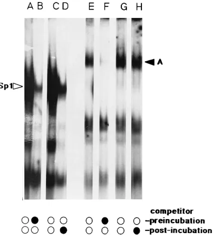
[image:4.612.144.211.63.398.2]One possible explanation for the repeatedly observed sensi-tivity of factor A is that it might possess an unusually high rate of dissociation from its DNA-binding site. To test this, rates were examined by adding ds competitor DNAs to binding reaction mixtures either 5 min before or 20 min after probe addition. As shown in Fig. 4, the F9 Sp1 site binding activity (14, 26) was inhibited effectively both when competitor was added before the probe (lane B) and when it was added after the probe (lane D). However, ds RBSWT-28competitor DNA competed with RBSWT-28 probe for factor A only when the competitor was added before the probe (lane F) and not when it was added after the probe (lane H). These results suggest that the Sp1 site binding activity dissociates from its probe within 5 min after competitor addition (lane D), while factor A remains probe bound for over 20 min (lane H). Thus, it does
FIG. 3. Serum effects on factor A detection. RBSWT-28 (lanes B and E),
RBSB2(lanes A and D), or JCV, a control dsDNA (sense, 59GAGCT CATGC
[image:4.612.329.545.71.307.2]TTGGC TGGCA GCCAT CCCT) (lanes C and F) probes were incubated at 308C (following preincubations for 20 min at 308C without probe) with F9 nuclear extract (0.28 mg/ml) in the absence (lanes A to C) or presence (lanes D to F) of 3ml of FCS (Gibco). Electrophoresis and autoradiography were as described in the legend to Fig. 1. The factor A complex is indicated by the arrowhead, and free probe is shown at the gel bottom.
FIG. 4. Binding factor dissociation rates. Sp1 (sense, 59GCTCG CCCCG CCCCG ATCGA AT 39(lanes A to D) or RBSWT-28(lanes E to H) probe was
incubated with F9 nuclear extract (2.1 mg/ml) for 20 min at 308C. Fifteen nanograms of unlabeled Sp1 or RBSWT-28 competitor was either not added
(lanes A, C, E, and G), added 5 min prior to probe addition (lane B [Sp1 competitor] and lane F [RBSWT-28competitor]), or added 20 min after probe
addition (lane D [Sp1 competitor] and lane H [RBSWT-28competitor]). For lane
D, binding continued for 5 min after competitor addition, while for lane H, binding continued for 20 min after competitor addition. Gel electrophoresis and autoradiography was as described in the legend to Fig. 1.
on November 9, 2019 by guest
http://jvi.asm.org/
not appear that the sensitivity of the factor A complex is due to an inordinately high dissociation rate.
To further characterize factors which bind to the M-MuLV RBS, F9 nuclear lysates were passed over a Sephadex G-200 gel filtration column and binding activity from the column was monitored by gel shift assay. By following this protocol, we found that binding activity eluted from the column near the void volume, implying a size of greater than 100 kDa for factor A (data not shown). In a separate enrichment step, crude F9 nuclear extracts were applied to a heparin-Sepharose column, from which factor A eluted in the 600 mM KCl wash with a 5-to 10-fold enrichment (data not shown). In addition 5-to Seph-adex G-200 and heparin-Sepharose chromatographies, alter-nate factor A enrichment steps have been tested. Factor A was present in the flowthrough fraction of nuclear lysate-loaded DEAE-Sepharose columns, and precipitates in the 40 to 50% ammonium sulfate fractionation cut, but these steps yielded only a slight enrichment in specific binding activity. Initial attempts at affinity chromatography with RBSWT-28multimers covalently attached to Sepharose 4B via a cyanogen bromide catalyst showed little enrichment of factor A in high-salt washes. Subsequent attempts with a 59biotinylated RBSWT-28 sequence in conjunction with streptavidin-agarose beads showed that factor A bound and eluted in a 600 mM KCl wash (data not shown), although these attempts so far have yielded too little protein to permit accurate estimation of enrichment levels. With all our purification efforts, we have found that two characteristics of factor A have hampered purification. First, binding activity rapidly decays in high-salt solutions. Second,
partially purified factor A binding activity in dilute solutions is sensitive to freeze-thaw episodes and diminishes over time, even at 2808C. Nevertheless, immediate Centricon desalting and concentration of partially purified fractions have improved retention of factor A binding activity, suggesting that further purification should be possible.
Effects of other PBS sequences.The fact that the M-MuLV RBS could not be delineated more than 1 bp smaller than the wt PBSP(16) suggested that there might be something about a tRNA PBS which affects repression. It was shown that a PBSQ substitution for the PBSPeliminated RBS-mediated repression of M-MuLV in EC cells (16, 25), but other PBS sequences were not tested for the repression phenomenon. To examine potential effects of other PBS sequences on EC cell repression of M-MuLV, we employed a parental construct (BlankIsle) which uses the nonrepressed PBSQ and possesses a unique
SspI site for insertion of test sequences (16) (Fig. 5). For test
sequences we inserted PBS sequences which can anneal to tRNA acceptors for phenylalanine (PBSF) (29), isoleucine (PBSI) (present in RTVL-1) (20), lysine-1,2 (PBSK2) (present in Mason-Pfizer monkey virus visna virus, and spumavirus) (20), lysine-3 (PBSK3) (present in human immunodeficiency virus) (33), methionine (PBSM) (29), and tryptophan (PBSW) (present in Rous sarcoma virus and avian myeloblastosis virus) (33), as well as the RBSWT-18, RBSWT-28, and RBSB2 se-quences (Fig. 5).
[image:5.612.60.297.72.213.2]Analysis of stem cell-specific repression effects of test se-quences followed our previously established methods (3, 16, 25, 32). Virus stocks were made in Psi2 packaging cells (23) and used for parallel infections of 3T3 fibroblasts (as a control
FIG. 5. Recombinant retrovirus constructs. BlankIsle (16) is a recombinant retrovirus construct based on the vectors PBSQ (25) and MP10 (3). As a provirus it is 5,370 bp and contains the following elements: an intact M-MuLV 59long terminal repeat (LTR) up to the KpnI site at proviral nt 480 (viral nt 32); 59 noncoding sequences corresponding to M-MuLV viral nt 32 to 212 from an endogenous murine retrovirus (7), which alters the M-MuLV PBS from one for proline (PBSP) to one for glutamine (PBSQ); M-MuLV nt 212 to 563; a 702-bp EcoRV-to-ScaI ‘‘island’’ fragment from pBR322, which was BamHI linkered and inserted into a created BamHI site; the M-MuLV splice acceptor (viral nt 5409 to 5768); the neomycin gene (NEO) from Tn5, including the bacterial promoter; simian virus 40 (viral nt 160 to 0 to 5154) and pBR322 (nt 3102 to 2521) origins of replication; and the 39end of M-MuLV from viral nt 7197 through the 39LTR. As a virus, BlankIsle uses a glutamine tRNA primer and is not repressed in undifferentiated EC cells (16). The constructs WTIsle and B2Isle differ from BlankIsle in that fragments from wt M-MuLV (nt 147 to 174) or from the nonrepressed B2 mutant were inserted into the unique SspI site in the BlankIsle pBR322 island. The miniWTIsle construct is similar to WTIsle except that it contains an insert of only MuLV PBS sequences and does not possess M-MuLV nt 164 to 174. PBSIsleF, PBSIsleI, PBSIsleK2, PBSIsleK3, PBSIsleM, and PBSIsleW were derived from BlankIsle by insertion into the SspI site of PBS sequences corresponding to tRNAs for phenylalanine, isoleucine, lysine-1,2, lysine-3, methionine, and tryptophan, respectively. For each insert the indicated initial T residue is derived from the SspI juncture sequence, as is the 39AT for
the B2Isle, WTIsle, and miniWTIsle constructs. TABLE 1. Viral infectivitiesa
Expt and construct
Titer % WTIsle
repression
3T3 F9 3T3/F9
1
WTIsle 137,500 6 22,917 100
B2Isle 75,000 1,213 62 0.3
MiniWTIsle 600,000 502 1,195 5.2
PBSIsleF 35,500 296 110 0.5
PBSIsleI 65,000 963 67 0.3
PBSIsleK2 101,000 15 6,733 29
PBSIsleK3 27,750 245 113 0.5
PBSIsleM 65,000 2,388 27 0.2
2
WTIsle 165,000 18 9,167 100
B2Isle 105,000 1,260 83 0.9
MiniWTIsle 212,000 95 2,232 24.5
PBSIsleF 72,500 75 966 10.5
PBSIsleI 47,500 1,140 42 0.5
PBSIsleK2 165,000 15 11,000 120
PBSIsleK2 240,000 90 2,667 29
PBSIsleK3 120,000 170 706 7.7
PBSIsleK3 82,500 120 688 7.5
PBSIsleM 50,000 640 78 0.9
PBSIsleW 23,000 340 68 0.7
PBSIsleW 27,500 420 65 0.7
a
DNA constructs were converted into recombinant retrovirus stocks by tran-sient transfection followed by infection of into Psi2 packaging cells (23). Identical Psi2 supernatants were used to infect NIH 3T3 and F9 cells. Titers are expressed as the number of G418-resistant colonies formed per milliliter of viral superna-tant. The ratio of 3T3 to F9 titers (the restriction index) is given to compare F9 restriction of different constructs within the same experiment, with 3T3 titers as a standard. The percentage of WTIsle repression was calculated as 100 times the restriction index of the experimental construct divided by the restriction index of WTIsle in the same experiment.
on November 9, 2019 by guest
http://jvi.asm.org/
[image:5.612.315.554.407.654.2]for absolute levels of infectious virus) and F9 cells. By com-parison of virus titers (number of G418-resistant colonies per milliliter of virus), it was possible to assess repression levels in EC cells. Consistent with previous results (16), we found that the RBSWT-28 element in WTIsle caused a 100- to 300-fold reduction in virus titers in EC cells relative to that caused by the 1-bp RBSB2mutant sequence in B2Isle (Table 1). Addi-tionally, the minimal 18-bp RBSWT-18sequence in miniWTIsle repressed expression about 20-fold relative to repression by B2Isle in parallel experiments but 4- to 20-fold less than the 28-bp RBSWT-28element (Table 1). Of the alternative 18-bp PBS sequences, the isoleucine, methionine, and tryptophan PBS elements in PBSIsleI, -M, and -W consistently showed no repression effect, while those in PBSIsleF and -K3 demon-strated moderate (2- to 10-fold) but variable levels of repres-sion. Follow-up experiments using a recombinant M-MuLV with a PBSK3replacement for the natural PBSP(tvPBS-Lys-3; kindly provided by F. S. Pedersen) (21), also showed the ly-sine-3 PBS to have a small but detectable (threefold) repres-sion of M-MuLV expresrepres-sion in EC cells (data not shown). However, in contrast to the other alternative PBS inserts, the lysine-1,2 tRNA PBS of PBSIsleK2 consistently gave titers in EC cells that were repressed over 30-fold relative to those with
B2Isle (Table 1). These results indicate that the PBSK2present in Mason-Pfizer monkey virus, visna virus, and spumavirus can repress expression in EC cells, at least in the context of the BlankIsle backbone.
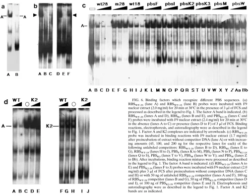
To extend our in vivo results, factor binding to variant RBS and PBS sequences was examined by gel shift assay (Fig. 6). An RBSWT-18 probe corresponding to the insert in miniWTIsle formed three complexes with F9 nuclear lysates (Fig. 6a, lane B), one of which had the same mobility as the corresponding RBSWT-28 factor A band (lane A). However, the factor A complex detected by RBSWT-18was about 10-fold less intense than that detected by RBSWT-28, a reduction which could be due to the difference in probe fragment sizes but nonetheless correlates well with in vivo results.
[image:6.612.57.561.82.477.2]With regard to alternative PBS sequences, we found no evidence of a factor A-like complex with any of our PBSF, PBSI, PBSK3, PBSM, and PBSWprobes (data not shown). How-ever, with 18- and 28-bp PBSK2 probes, a complex with a mobility similar to that of factor A was observed (Fig. 6b, lane C). Interestingly, PBSK2 levels of the low-mobility complex detected by the PBSK2 probe were increased by addition of serum to binding reaction mixtures (compare lanes F and C) in a fashion similar to what we have seen for factor A (lanes A
FIG. 6. Binding factors which recognize different PBS sequences. (a) RBSWT-28 (lane A) and RBSWT-18(lane B) probes were incubated with F9
nuclear extract (2.0 mg/ml) for 20 min at 308C in the presence of 3ml of FCS and processed as described in the legend to Fig. 1. The factor A band is indicated. (b) RBSWT-28(lanes A and D), RBSB2(lanes B and E), and PBSK2-28(lanes C and
F) probes were incubated with F9 nuclear extract (2.4 mg/ml) for 20 min at 308C in the absence (lanes A to C) or presence (lanes D to F) of 3ml of FCS. Binding reactions, electrophoresis, and autoradiography were as described in the legend to Fig. 1. Factor A and K2 complexes are indicated by arrowheads. (c) RBSWT-28
probe was incubated in binding reactions with F9 nuclear extract (1.7 mg/ml) after preincubation of extract without competitor DNA (lane A) or with increas-ing amounts (45, 100, and 200 ng for the respective lanes for each) of the following unlabeled competitors: RBSWT-28(lanes B to D), RBSB2(lanes E to
G), RBSWT-18(lanes H to J), PBSF(lanes K to M), PBSI(lanes N to P), PBSK2-18
(lanes Q to S), PBSK3(lanes T to V), PBSM(lanes W to Y); and PBSW(lanes Z
to Bb). After incubations, binding reaction mixtures were processed as described in the legend to Fig. 1. The factor A band is indicated. (d) RBSWT-28(lanes A to
E) and PBSK2-28(lanes F to J) probes were incubated with F9 nuclear extract (2.0
mg/ml) plus 3ml of FCS after preincubation without competitor DNA (lanes C and H) or with 50 ng of unlabeled RBSWT-28competitor (lanes A and F), 100 ng
of RBSWT-28competitor (lanes B and G), 50 ng of PBSK2-28competitor (lanes D
and I), or 100 ng of PBSK2-28competitor (lanes E and J). Electrophoresis and
autoradiography were as described in the legend to Fig. 1. Factor A and K2 bands are as indicated.
on November 9, 2019 by guest
http://jvi.asm.org/
and D). Despite this similarity, competition studies suggest that factor A and the K2 factor which complexes with PBSK2 are not identical. As shown in Fig. 6c, factor A binding to the RBSWT-28 probe was inhibited efficiently by the RBSWT-28 competitor (lanes B to D), less well by RBSWT-18(lanes H to J), and not at all by the 1-bp mutant RBSB2 (lanes E to G), PBSK2(lanes Q to S), or any of the other ds PBS competitors. This result was confirmed in cross-competition studies between RBSWT-28and the 28-bp PBSK2sequence, PBSK2-28(Fig. 6d). Factor A (Fig. 6d, lane C) was inhibited well by RBSWT-28 (lanes A and B) but not by PBSK2-28(lanes D and F), while the K2 factor (lane H) was inhibited by PBSK2-28(lanes I and J), but not by RBSWT-28(lanes F and G). The fact that RBSWT and PBSK2did not cross-compete but represent PBS sequences which repress M-MuLV expression in EC cells (Table 1) and bind to factors with similar mobilities leaves open the possi-bility that factors A and K2 may be different, but related, stem cell binding factors.
DISCUSSION
The aim of our studies was to investigate the RBS-mediated repression phenomenon of M-MuLV in undifferentiated EC cells. Specifically, experiments were designed to characterize the RBS binding factor A and to determine the relationship between stem cell-specific RBS sequences and retrovirus tRNA PBS sequences. Previous studies (16, 19, 25) showed that factor A bound specifically to the M-MuLV RBS, but no other correlations between factor binding and in vivo repres-sion were observed. However, after optimization of factor iso-lation and binding protocols, additional data support the no-tion that factor A is the cellular factor which mediates repression at the M-MuLV RBS. First, factor A was enriched in F9 nuclear extracts versus cytosolic extracts (Fig. 1), consis-tent with its action at the DNA level (3, 5, 16–19, 25). Second, the factor was enriched in F9 cells versus 3T3 cells (Fig. 1), consistent with the cell type specificity of RBS repression (16, 19, 25). Additionally, our radiolabeled RBSWT-28ds oligonu-cleotide bound factor A approximately 10-fold better than the radiolabeled RBSWT-18ds oligonucleotide (Fig. 6a). This ob-servation is in agreement with infectivity studies which showed that the full-length sequence in WTIsle was 10- to 20-fold more efficient in stem cell-specific silencing than the short version in miniWTIsle (Table 1). The fact that sequences present in WTIsle but not in miniWTIsle (M-MuLV nt 164 to 174) can influence the observed repression phenomenon may explain why the myeloproliferative sarcoma virus does not appear to be expressed in EC cells (32): myeloproliferative sarcoma virus is identical to M-MuLV at nt 147 to 163 but possesses two T nucleotides in place of the one C nucleotide at M-MuLV nt 164, immediately 39of the M-MuLV PBS.
It has been shown that factor A is very unstable (19, 25), and thus some standard studies have not been possible to perform. To combat this instability, we have varied conditions both for preparation of protein extracts and for DNA binding. Specif-ically, we have observed that by shortening the times of dialysis of the crude protein extracts against Dignam buffer D or elim-inating dialysis altogether, factor A bands on gel shift assays were enhanced. We have shown that performance of binding reactions at 308C increased the RBSWT-28versus RBSB2probe specificity of factor A binding but did not decrease the intensity of the band (Fig. 1), that tRNAProdid not compete with and tRNA synthetase did not bind the RBSWTprobe, that addition of serum to reaction mixtures enhanced the binding of factor A (Fig. 3) but that gelatin or BSA had little or no effect on binding (data not shown), and that 3 mM Mg21and 100mM
Zn21enhanced binding (Fig. 1), while other cations,
nucleoti-des, or detergents either had no effect or impaired factor A binding. Furthermore, Sepharose G-200 column chromatogra-phy results indicated that factor A is larger than 100 kDa and is enriched in the 600 mM KCl washes of both heparin-Sepha-rose and DNA affinity columns (data not shown). These results suggest that purification and identification of factor A should be possible.
Although maximal RBS repression requires sequences out-side the M-MuLV PBS and tRNA does not compete for factor A binding, the 17-of-18-bp overlap of the PBS and the core RBS has suggested that PBS and RBS functions may be inter-twined. We (3) and others (32) have shown that recombinant M-MuLV vectors with glutamine tRNA PBSs do not suffer stem cell-specific repression. However, to ascertain whether PBSP or PBSQ is atypical, PBS sequences for phenylalanine (PBSF), isoleucine (PBSI), lysine-1,2 (PBSK2), lysine-3 (PBSK3), methionine (PBSM), and tryptophan (PBSW) were tested for in vivo silencing function and in vitro binding activity. Of these sequences, PBSI, PBSM, and PBSWshowed no silencing activ-ity, while the phenylalanine and lysine-3 PBS sequences re-duced viral infectivities 2- to 10-fold relative to that with the RBS mutant construct, B2Isle (Table 1). Interestingly, the PB-SIsleK2 virus consistently showed reduced titers in EC cells (Table 1), and the PBSK2 probe formed a complex with a mobility and response to serum that were similar to those of factor A (Fig. 6). However, we observed that the RBSWTand PBSK2 probes did not cross-compete (Fig. 6). These results suggest that factor A and the factor which binds to the PBSK2 probe (factor K2) may be different but related factors, and they raise the possibility that expression of viruses which use the tRNA lysine-1,2 PBS sequence (Mason-Pfizer monkey virus, visna virus, and spumavirus) (20) may also be repressed in undifferentiated stem cells.
ACKNOWLEDGMENTS
We are grateful to David Dignam for kindly providing purified preparations of proline tRNA and the proline tRNA synthetase. We also are thankful to Finn Skou Pedersen for the gifts of retrovirus vectors tvPBS-Pro, tvPBS-Gln-1, and tvPBS-Lys-3. The advice and assistance of laboratory members Robert Gray and Rachel Lee are gratefully acknowledged.
This work was supported by grant 1 R01 CA53332-01 from the National Cancer Institute.
REFERENCES
1. Baeuerle, P. A., and D. Baltimore. 1988. Activation of DNA-binding activity in an apparently cytoplasmic precursor of the NF-kbtranscription factor. Cell 53:211–217.
2. Baeuerle, P. A., and D. Baltimore. 1988. Ikb: a specific inhibitor of the NF-kbtranscription factor. Science 242:540–546.
3. Barklis, E., R. C. Mulligan, and R. Jaenisch. 1986. Chromosomal position or virus mutation permits retrovirus expression in embryonal carcinoma cells. Cell 47:391–399.
4. Bernstine, E., M. Hooper, S. Grandchamp, and B. Ephrussi. 1973. Alkaline phosphatase activity in mouse teratoma. Proc. Natl. Acad. Sci. USA 70:3899–3903. 5. Berwin, B., and E. Barklis. 1993. Retrovirus-mediated insertion of expressed and non-expressed genes at identical chromosomal locations. Nucleic Acids Res. 21:2399–2407.
6. Bradford, M. M. 1976. A rapid and sensitive method for the quantitation of protein utilizing the principle of protein-dye binding. Anal. Biochem. 72: 248–254.
7. Colicelli, J., and S. Goff. 1987. Isolation of a recombinant murine leukemia virus utilizing a new primer tRNA. J. Virol. 57:37–45.
8. Dignam, J. D., R. M. Lebovitz, and R. G. Roeder. 1983. Accurate transcrip-tion initiatranscrip-tion by RNA polymerase II in a soluble extract from isolated mammalian nuclei. Nucleic Acids Res. 11:1475–1489.
9. Feuer, F., M. Taketo, R. Hanecak, and H. Fan. 1989. Two blocks in Moloney murine leukemia virus expression in undifferentiated F9 embryonal carci-noma cells as determined by transient expression assays. J. Virol. 63:2317– 2324.
on November 9, 2019 by guest
http://jvi.asm.org/
10. Gautsch, J., and M. Wilson. 1983. Restriction of Moloney murine leukemia virus growth in tetratocarcinoma: involvement of factors other than DNA methylation. Cold Spring Harbor Conf. Cell Proliferation 10:363–378. 11. Gautsch, J., and M. Wilson. 1983. Delayed de novo methylation in
tetrato-carcinoma suggests additional tissue-specific mechanisms for controlling gene-expression. Nature (London) 301:32–37.
12. Gorman, C., P. Rigby, and D. Lane. 1985. Negative regulation of viral enhancers in undifferentiated embryonic stem cells. Cell 42:519–526. 13. Grez, M., E. Akgun, F. Hilberg, and W. Ostertag. 1990. Embryonic stem cell
virus, a recombinant murine retrovirus with expression in embryonic stem cells. Proc. Natl. Acad. Sci. USA 87:9202–9206.
14. Jackson, S., and R. Tjian. 1989. Purification and analysis of RNA polymerase II transcription factors by using wheat germ agglutinin affinity chromatog-raphy. Proc. Natl. Acad. Sci. USA 86:1781–1785.
15. Jaenisch, R., and A. Berns. 1977. Tumor virus expression during mammalian embryogenesis, p. 267–314. In M. Sherman (ed.), Concepts in mammalian embryogenesis. MIT Press, Cambridge, Mass.
16. Kempler, G., B. Freitag, B. Berwin, O. Nanassy, and E. Barklis. 1993. Characterization of the Moloney murine leukemia virus stem cell-specific repressor binding site. Virology 193:690–699.
17. Loh, T., L. Sievert, and R. Scott. 1987. Proviral sequences that restrict retroviral expression in mouse embryonal carcinoma cells. Mol. Cell. Biol.
7:3775–3784.
18. Loh, T., L. Sievert, and R. Scott. 1988. Negative regulation of retrovirus expression in embryonal carcinoma cells mediated by an intragenic domain. J. Virol. 62:4086–4095.
19. Loh, T., L. Sievert, and R. Scott. 1990. Evidence for a stem cell-specific repressor of Moloney murine leukemia virus expression in embryonal carci-noma cells. Mol. Cell. Biol. 10:4045–4057.
20. Lo¨wer, L., J. Lo¨wer, C. Tondera-Koch, and R. Kurth. 1993. A general method for the identification of transcribed retrovirus sequences (R-U5 PCR) reveals the expression of the human endogenous retrovirus loci HERV-H and HERV-K in tetratocarcinoma cells. Virology 192:501–511. 21. Lund, A., M. Duch, J. Lovmand, P. Jorgensen, and F. S. Pedersen. 1993.
Mutated primer binding sites interacting with different tRNAs allow efficient murine leukemia virus replication. J. Virol. 12:7125–7130.
22. Maniatis, T., E. F. Fritsch, and J. Sambrook. 1982. Molecular cloning: a laboratory manual. Cold Spring Harbor Laboratory, Cold Spring Harbor, N.Y.
23. Mann, R., R. Mulligan, and D. Baltimore. 1983. Construction of a retrovirus packaging mutant and its use to produce helper-free defective retrovirus. Cell 33:153–159.
24. Martin, G. R. 1980. Tetratocarcinomas and mammalian embryogenesis. Sci-ence 209:768–776.
25. Petersen, R., G. Kempler, and E. Barklis. 1991. A stem cell-specific silencer in the primer-binding site of a retrovirus. Mol. Cell. Biol. 11:1214–1221. 26. Prince, V., and P. Rigby. 1991. Derivatives of Moloney murine leukemia
virus capable of being transcribed in embryonal carcinoma stem cells have gained a functional Sp1 binding site. J. Virol. 65:1803–1811.
27. Sanger, F., S. Nicklen, and A. R. Coulsen. 1977. DNA sequencing with chain-terminating inhibitors. Proc. Natl. Acad. Sci. USA 74:5463–5467. 28. Shinnick, T., R. Lerner, and J. Sutcliffe. 1981. Nucleotide sequence of
Moloney murine leukemia virus. Nature (London) 293:543–548. 29. Sprinzl, M., T. Hartmann, J. Weber, J. Blank, and R. Zeidler. 1989.
Com-pilation of tRNA sequences and sequences of tRNA genes. Nucleic Acids Res. 17:R1–R172.
30. Stewart, C., H. Stuhlman, D. Ja¨hner, and R. Jaenisch.1982. De novo meth-ylation, expression and infectivity of retroviral genomes introduced into embryonal carcinoma cells. Proc. Natl. Acad. Sci. USA 79:4098–4192. 31. Thornell, A., B. Hallberg, and T. Grundstro¨m.1988. Differential protein
binding in lymphocytes to a sequence in the enhancer of the mouse retro-virus SL3-3. Mol. Cell. Biol. 8:1625–1637.
32. Weiher, H., E. Barklis, W. Ostertag, and R. Jaenisch. 1987. Two distinct sequence elements mediate retroviral gene expression in embryonal carci-noma cells. J. Virol. 61:2742–2746.
33. Weiss, R., N. Teich, H. Varmus, and J. Coffin. 1985. RNA tumor viruses. Cold Spring Harbor Laboratory, Cold Spring Harbor, N.Y.