2007
Mechanism of cap-independent translation by long
distance kissing stem-loop interactions in plant viral
RNAs
Aurélie Mamisoa Rakotondrafara
Iowa State UniversityFollow this and additional works at:https://lib.dr.iastate.edu/rtd
Part of theMolecular Biology Commons, and thePlant Pathology Commons
This Dissertation is brought to you for free and open access by the Iowa State University Capstones, Theses and Dissertations at Iowa State University Digital Repository. It has been accepted for inclusion in Retrospective Theses and Dissertations by an authorized administrator of Iowa State University Digital Repository. For more information, please contactdigirep@iastate.edu.
Recommended Citation
Rakotondrafara, Aurélie Mamisoa, "Mechanism of cap-independent translation by long distance kissing stem-loop interactions in plant viral RNAs" (2007).Retrospective Theses and Dissertations. 15929.
by
Aurélie Mamisoa Rakotondrafara
A dissertation submitted to the graduate faculty in partial fulfillment of the requirements for the degree of
DOCTOR OF PHILOSOPHY
Major: Molecular, Cellular, and Developmental Biology
Program of Study Committee: W. Allen Miller, Major Professor
Adam J. Bogdanove Kristen Johansen
Alan Myers Marit Nilsen-Hamilton
Iowa State University Ames, Iowa
2007
3274871 2007
UMI Microform Copyright
All rights reserved. This microform edition is protected against unauthorized copying under Title 17, United States Code.
ProQuest Information and Learning Company 300 North Zeeb Road
P.O. Box 1346 Ann Arbor, MI 48106-1346
TABLE OF CONTENTS
ABSTRACT iii
CHAPTER 1. GENERAL INTRODUCTION 1
CHAPTER 2. OSCILLATING KISSING STEM-LOOP INTERACTIONS MEDIATE SCANNING-DEPENDENT TRANSLATION BY A VIRAL CAP-INDEPENDENT TRANSLATION ELEMENT
37
CHAPTER 3. TRANS-REGULATION OF TRANSLATION BY A VIRAL SUBGENOMIC RNA
83
CHAPTER 4. COMPETING VIRAL MRNAS SHOW DIFFERENT EFFICIENCY IN PROMOTING CAP-INDEPENDENT TRANSLATION UNDER LOW FACTOR AVAILABILITY
121
CHAPTER 5. GENERAL CONCLUSIONS 150
APPENDIX A. RNA SEQUENCES IMPLICATED IN CAP-INDEPENDENT TRANSLATION
156
APPENDIX B. IN VITRO ANALYSIS OF TRANSLATION ENHANCERS 168
APPENDIX C. PREPARATION AND ELECTROPORATION OF OAT PROTOPLASTS FROM CELL SUSPENSION CULTURE
191
ABSTRACT
While the presence of a 5’ cap and a 3’ poly(A) tail are key elements for efficient translation of eukaryotic mRNAs, many viral mRNAs lack one or both of such structures and yet translate efficiently. Owing to the absence of a 5’ cap, Barley yellow dwarf virus (BYDV, Luteoviridae family) genomic and subgenomic RNA1 rely on a cap-independent translation element (BTE), which is an RNA domain capable of promoting translation. In contrast to an internal ribosome entry site (IRES), the BTE is located at the 3’ end of the viral mRNA, while translation initiation occurs at the 5’ end proximal AUG. For its activity, the BTE requires a cis-acting element within the 5’ untranslated region (UTR) of the RNA, with which it shares a five base sequence complementarity. We propose that the BTE recruits part of the translation machinery and the long distance base pairing of the BTE to this BTE-complementary loop (BCL) – across 4000 bases - facilitates delivery of the factors from the 3’ UTR to the 5’ end, where translation initiates.
Just as with cellular mRNAs, BTE-mediated translation requires 5’ end-dependent ribosome scanning to reach the initiation codon. In such a scheme, the long distance interaction must be continuously disrupted by the scanning ribosome, and reformed to allow the delivery of the factors to the next 43S ribosomal complex. We have provided the first evidence suggesting that the initial entry of the ribosomal complex occurs at the 5’ end of the uncapped viral mRNA. Due to the structure nature of the BYDV 5’ UTRs, the efficiency of ribosome scanning remains highly dependent on the BTE for the delivery of the initiation factors, which are necessary for unwinding of the local 5’ end structure. In such a view, the 5’ UTR become a rate limiting-factor under low factor availability.
CHAPTER 1: GENERAL INTRODUCTION
HISTORICAL PERSPECTIVE
If the DNA is really our thread of Ariadne, the labyrinths out of which it is expected to lead
us are truly inscrutable” Erwin Chargaff (1971).
Following the discovery of the double-helix structure of the DNA (1), attention was shifted in trying to decipher the coding scheme of protein synthesis (reviewed by (2). It was soon clear that the order of the four bases in the DNA must direct protein synthesis from a set of 20 amino acids. The first model for protein synthesis was drawn by Gamow, father of the Big Bang theory, who hypothesized that it occurred directly on the DNA in a key-and-lock model (3). In such context, the different cavities formed by four contiguous bases within the double helix dictated the nature of the amino acids. The proposed model raised on important coding dilemma: all amino acid arrangements should be permitted, and the codons should not overlap. More confusions arose with the discovery of the protein machine makers, identified as the microsomal particles (and later called ribosomes) in the cytoplasm (4). How could the genetic material locked in the nucleus direct protein synthesis in the cytoplasm?
But what was the messenger? The ribosomal RNAs bear the obvious features of the perfect message: they are the most abundant and stable RNAs in the cells, and importantly, located within the site of protein synthesis. In such a view, each ribosome directed with its RNAs the synthesis of one specific protein in a one gene-one ribosome-one protein model. In the following years, it was soon clear that the model could not explain the constant size and homogeneity of the nucleotide composition of the ribosomal RNA, while the protein and DNA size vary greatly in length and between species (6). The early perception by Jacob and Monod (1961) that the protein synthesis can be turned on and off in response to external condition, led them to hypothesize that the ribosomes were just non-specialized structures, which receive an mRNA of very short half-life, which will determine the sequences of amino acids for protein production (7). That was the first concept of RNA turnover as mechanism of control of gene expression. Rapidly, the existence of the unstable message separate from the ribosomal mRNA was independently proven by several scientists (6,8). The first biochemical proof that RNA directs protein synthesis was provided by Niremberg et al. (1961). A poly(U) stretch of RNA resulted in the synthesis of poly-phenylalanine protein (9). The discovery of adaptor molecules that carry the amino acid and match to the RNA nucleotide sequence (10) contributed to putting all the pieces of protein synthesis together. The flow of the genetic information from DNA→RNA→protein was deciphered, and much of the translational apparatus was characterized.
consists of four major steps: initiation, elongation, termination and recycling. We will focus on the complexity of translation initiation event. It is of great importance to understand that the ability of an mRNA to recruit the translation machinery determines its translational fate.
This chapter reviews (i) the major steps of translation initiation, (ii) global and selective controls of translation initiation, (iii) the alternative mechanisms of translation utilized by some RNA viruses to circumvent host regulation, (iv) Barley yellow dwarf virus RNA as model system for non-canonical translation.
TRANSLATION INITIATION
The basic steps of translation initiation in eukaryotes and prokaryotes are evolutionary conserved (13,14). It involves (i) association/dissociation of the small and large ribosomal subunits, (ii) selection of the initiator aminoacyl -tRNA (iii) selection of the correct initiation start on the mRNA, and (iv) joining of the large subunit at the start codon. However, each system involves disparate processes in the recruitment of the ribosomal subunit and in the mechanism of selection of the initiation site. The prokaryotic translation mechanism is based on a simple rRNA-mRNA interaction and requires only three translation factors to direct protein synthesis from each cistron of the polycistronic message. In contrast, the eukaryotic protein machinery involves multi-protein complex of at least 12 initiation factors for the translation of only one cistron per message, as summarized in Table 1.
In bacteria
Dalgarno (SD), preceding the start codon (13,15). The 4-5 nt SD motif is sufficient to recruit the ribosome subunit by direct base pairing to the 3’ end of the 16S rRNA. In addition to the base pairing, the SD is strategically located about 5-7nt upstream of the AUG, which allows the start codon to be positioned right in the P-site of the small subunit (11). The translation initiation factors IF1, IF2 and IF3 bound to the 30S ribosomal subunit, facilitate the assembly of the mRNA onto the ribosome subunit, the correct selection of the initiation site, the binding of initiator tRNA into the P-site of the small ribosome, and the joining of the larger 50S subunit (13). The regulation of the translation initiation is mediated by conformational changes of the mRNA secondary structure or protein binding, which either exposes or masks the SD for ribosome entry.
In Eukaryotes
and ribosomes are unable to bind and initiate efficient translation of circularized cellular mRNAs (17).
The anchoring point for ribosome entry in eukaryotes is a 7 methylguanosine cap structure at the 5’ end of the mRNA, which is acquired during the course of transcription (18-20). Another key feature of mRNA is a 50-300 or longer nucleotide-long poly-adenosine stretch at the 3’ end. These two elements are involved in (i) RNA stability, protection against exonucleases, (ii) RNA export from the nucleus, (iii) check-point for RNA integrity prior to translation, and (iii) as described here, in RNA translation efficiency (21).
Step 1: Assembly of the translation machinery onto the mRNA
One of the first steps in initiation is the formation of the 43S pre-initiation complex, which consists of the eIF2/GTP/initiator met-tRNA ternary complex bound to the 40S ribosomal subunit (22)(Figure 1). This involves first (i) the selection of the correct initiator tRNA from the pool of tRNAs and elongator met-tRNA, which is mediated by eIF2, and (ii) the delivery of the met-tRNA to the 40S small ribosomal subunit, which is maintained separated from the large subunit. The complex is then stabilized by additional factors - eIF1, eIF1A, eIF5 and eIF3 - which will play crucial roles in downstream steps, and is then ready to be presented to the mRNA via eIF3 (23,24).
Table 1. List of the eukaryotic initiation factors (eIFs) involved in recruitment of the translation machinery
Translation
factors Function Binds Reference
43S complex
eIF1 Stabilizes binding of the ternary complex to the 40S ribosome
Required for scanning and selection of correct start codon. Has anti-subunit association activity
Met-tRNAimet, eIF2, eIF5, eIF3
(25)
eIF1A Acts synergistically with eIF1 eIF2, eIF5B, eIF3 (16) eIF2 Selects the correct initiator met-tRNA.
Binds the 40S subunit as a ternary complex with Met-tRNAimet and GTP.
Irreversibly hydrolyzes GTP at the recognition of the correct AUG codon
40S subunit, eIF1, eIF1A, eIF2B, eIF3, eIF5
(16))
eIF2B Guanine nucleotide exchange factor of eIF2
Catalyses the regeneration of GTP-bound form of eIF2
eIF2 (16)
eIF5 Promotes GTPase activity of ribosome bound-eIF2 Interacts with eIF1A for the selection of the correct AUG Promotes mRNA binding to the 43S complex
eIF1, eIF1A, eIF2, eIF2B, eIF31, eIF4G1
(26)
eIF5B Stabilizes Met-tRNAimet on ribosome Hydrolyzes GTP for correct 80S assembly
GTP, eIF1A (27)
eIF3 Enhances assembly of the 43S complex Recruits the 43S complex to the mRNA Promotes scanning and AUG recognition Prevents premature joining of the 60S subunit
40S subunit, eIF1, eIF1A eIF2, eIF4B, eIF4G2, eIF1A1, eIF51
(23,24)
48S complex eIF4F/eIFiso4F
* Multisubunit component Assembles the translation machinery at the 5’ end of the RNA
Assures processivity of ribosome scanning
(11,16)
eIF4E/eIFiso4E *
Recognizes the 5’ cap 5’cap, eIF4G (28)
eIF4G/eIFisoG *
Scaffold protein: recruits the 43S complex through eIF3 Enhances eIF4E interaction to cap
Enhances eIF4A helicase activity
eIF4E, eIF4A, eIF4B, eIF3, PABP
(29,30)
eIF4A** RNA ATP-helicase
Unwinds mRNA to facilitate binding of the 40S subunit and processive scanning
mRNA, eIF4G,
eIF4B3, eIF4H3 (16)
eIF4B (eIF4H***)
Promotes eIF4A ATPase and helicase activity mRNA, eIF3, PABP, eIF4A3
(31)
PABP Increase the affinity of 4F to the mRNA
Brings the 3’-5’ together through eIF4G interaction Stabilizes mRNA
3’ poly(A) tail, eIF4G, eIF4B
(32), (33)
* part of eIF4F in animals but not in plants
The proposed role of eIF4F in initiation is to increase the mRNA competency by (i) binding the 5’ end, (ii) melting the secondary structure around the 5’ cap to facilitate attachment of the 40S ribosomal subunit and (iii) bridging the ribosomal subunit to the mRNA (16) .
The eIF4F complex, which is absent in bacteria, consists of three subunits: eIF4E - the 5’ cap-binding subunit, eIF4G - the multi-interacting protein, and eIF4A - the RNA ATP-dependent helicase, the activity of which is additionally promoted by eIF4B (29). In plants, eIF4F consists of only eIF4G and eIF4E, and has an isoform - eIFiso4F - composed of eIFiso4G and eIFiso4E. Compared to eIF4F, eIFiso4F is different in size, cellular abundance, and function (34,35). eIF4G is the central organizer of the assembly of the ribosomal subunit on the mRNA (30). It binds eIF4E and eIF4A, and enhances each of their functions, which are to stabilize the 4F-mRNA interaction and to enhance unwinding of the 5’ cap-proximal structure, respectively. eIF4G also interacts with the poly(A) tail binding protein, PABP, which in turn binds the 3’ poly(A) tail, bringing the mRNA in to a closed-loop conformation (Fig. 1) (36,37). The physical circularization of the mRNA (38) has been proposed to serve several roles, including (i) increasing stability of the mRNA, (ii) enhancement of the translation factor interactions with the mRNA, which, as a result, becomes highly competent to bind the 40S subunit, and (iii) recycling of the terminating ribosomes back to the 5’ end (27,39). Once eIF4F is positioned, the 43S preinitiation complex is ready to load via eIF3-eIF4G interaction.
is enhanced by the presence of eIF4G/eIF4A alone (40), and strongly stimulated by the cooperative interaction between the full combination of eIF4E, eIF4G, eIF4A and PAPB, following the recognition of the 5’ cap by eIF4E (16). Indeed, the requirement for eIF4F for initiation varies greatly among mRNAs, and is proportional to the amount of secondary structure downstream of the cap (25). mRNAs with long and highly structured 5’ UTRs (referred to as weak mRNAs) are more eIF4F-dependent - in order to unwind the RNA structure for scanning to occur - compared to those with less structured and short 5’ UTRs (referred to as strong mRNAs) (41). Examples of strong mRNAs include the housekeeping mRNAs, required for maintenance of normal cell functions, which are constitutively and advantageously translated (42). As we will see, the change in the availability of eIF4E/eIF4G can affect both global and selective regulation of translation. While its shortage causes shut down of general translation, its overexpression selectively induces translation of the highly structured mRNAs, which are strongly dependent on eIF4F availability. It is noteworthy that a majority of the weak mRNAs are involved in cell growth, apoptosis, tumor suppression. And there is accumulating evidence linking over-expression of those mRNA to oncogenesis (43).
Step 2: Recognition of the correct initiation start site
position to the 5’ end, but also by its local sequence context (45-47). In mammalian cells, the optimal context of a start codon is a purine at base -3 and +4 from the AUG (+1 corresponds to the A of the initiation start) (A/G)CCAUGG, as shown in bold (45-47). In plants, the optimal context is (A/C)AAAUGGC in dicots and (A/G)CCAUGG in monocots (Joshi et al., 1997). An AUG in a poor context is often bypassed and the ribosomal complex continues scanning.
The recognition of the correct AUG is coordinated by the initiation factors eIF1, eIF2 and eIF5 (22,48). eIF1 is proposed to maintain the ribosomal complex in an “open” configuration to facilitate scanning, and destabilize it when assembled at an incorrect codon (25). When the correct initiation codon is reached, the ribosomal complex adopts a “scanning incompetent closed” conformation, which prevents any further movement of the complex and accommodates a perfect anticodon-codon match of the met-tRNA to the mRNA. The recognition of the initiation codon coincides with (i) the release of eIF1 from the complex, (ii) the specific interaction of the α subunit of eIF2 with base G at position -3 of
the AUG, and (iii) the irreversible hydrolysis of the GTP bound to eIF2, catalyzed by eIF5, which releases the met-tRNA into the ribosomal P site (22). As a result, the 48S ribosomal complex is irreversibly committed to initiation at the selected AUG. The additional hydrolysis of a GTP bound to eIF5B (49) promotes joining of the 60S subunit to form the translationally competent 80S ribosome and protein synthesis starts.
GLOBAL AND SELECTIVE CONTROLS OF TRANSLATION
stresses such as viral infection (12). Regulation of translation, which can occur at various stages of translation, results in a turning off or on of protein synthesis in response to external stimuli. The main step for control is at the level of initiation. We can distinguish (i) global control of translation, which affects equally the general pool of mRNAs, and (ii) selective control of translation, which targets only a subset of mRNAs without affecting general protein synthesis (50,51).
Global control
Global control of translation is implemented by changes in the activity of the general components of the translation complex, such as eIF2 and eIF4E. Their regulation is mediated primarily by changes in their phosphorylation state (reviewed in (51).
GDP-bound eIF2 (52). This rapidly results in a deprivation of the ternary complex, which leads to a general decrease of translation.
The second target in the global control of translation is the formation of eIF4F complex, targeted by the translation repression proteins, 4EBPs, which have been primarily characterized in mammals (review (50). 4EBP competes with eIF4G for the same binding site on eIF4E. The 4EBP-eIF4E interaction prevents the assembly of the eIF4F complex, and therefore results in a general inhibition of cap-dependent translation. 4E-BP affinity to eIF4E is regulated by its phosphorylation state. Hypo-phosphorylated 4E-BP binds eIF4E, while its hyper-phosphorylated form loses affinity to eIF4E and therefore favors eIF4E/eIF4G interaction. The phosphorylation state of 4E-BP is controlled by the mTOR signal transduction pathway that activates the protein kinase in response to extracellular stimuli such as growth factors, hormones, cell stress, and amino acid depravation (28). A positive regulation of translation in found in vertebrates, where eIF4E is also directly targeted for phosphorylation by MnK1 kinase, which is bound to eIF4G. Phosphorylated eIF4E has an increased affinity to eIF4G and to the cap, which results in higher translation efficiency (28).
Specific control
Protein-mediated control. One example of protein-mediated inhibition is by the iron regulatory proteins (IRP1 and IRP2), which modulate the uptake, storage and transport of iron to the cells by influencing mRNA translation or stability. The IRPs have a specific binding site, the iron response element (IRE), which forms a 28 nucleotide stem-loop structure and is located either in the 5’ or 3’ UTR of the target mRNA. Under low iron condition, IRP binds to the 5’ proximal end of ferritin mRNA, which is responsible for iron storage. The binding of IRP hinders entry of the 43S pre-initiation complex and therefore inhibits translation (53) Also, IRP binds to the 5 IREs in the 3’ UTR of transferrin mRNA, which is responsible for iron transport. The binding of IRP stabilizes the mRNA by protecting it from endonucleases, and results in active translation of the mRNA, necessary for transport of iron outside the cell. At high iron level, IRP are released from both mRNAs to allow translation of ferritin and turn off transferring expression as its mRNA is degraded.
A similar mechanism is observed for the inhibition of anterior expression of the caudal mRNA during Drosophila development via blocking of the cap-structure for eIF4F binding (54). The repression of caudal mRNA translation is mediated by the binding of a bicoid transcription factor protein to the 3’ UTR of the mRNA, that is followed by interaction with 4E-HP - a cap-binding protein that cannot bind eIF4G. The interaction of 4E-HP to bicoid transcription factor and the cap represses translation.
RNA-mediated control. The recently discovered RNA-mediated control of translation
by small RNAs relies on base pairing to the target mRNAs - normally within the UTRs - followed by the assembly of the Argonaute proteins, which constitute the RNA induced silencing complex (RISC).
The fate of the target mRNA depends on the nature of the Argonaute protein and the degree of complementarity to the small RNA. This could either trigger specific RNA degradation or arrest of translation at different stages. A perfect match operated by siRNA results in endonuclease cleavage of the RNA. While the mechanism of repression of translation remains under debate, it is proposed that an imperfect interaction with the 3’ UTR, which is normally observed for miRNA, can trigger both accelerated RNA degradation, through deadenylation of the mRNA, or repression of translation. Repression of translation can occur (i) at the initiation stage, with the miRNA interfering with the formation of the 48S complex only in capped-mediated translation (58), possibly by targeting eIF4E (59); or by binding of the Argonaute domain to the 5’ cap of the mRNA, excluding it from eIF4E interaction (60), and (ii) at the elongation phase, as observed in C. elegans for the mRNA target of Let 7, Lin 4 regulatory of developmental timing (61,62). It has been observed that while translation is repressed, the mRNAs remain in the polysome fraction.
As part of the focus of this dissertation, we will describe a novel mechanism of RNA-mediated regulation of translation, which induces differential repression of translation of its target mRNAs.
ALTERNATIVE MECHANISMS OF TRANSLATION INITIATION
for the cap and yet translate efficiently. These strategies involve (i) IRES or internal ribosome entry site elements, which tether the translation complex directly to the AUG in a 5’ end-independent manner, and/or (ii) CITE or cap-independent translation elements, which require scanning from the 5’ end.
As we will see, cap-independent translation confers several lines of translational advantages to viral RNAs and a growing number of reported cellular mRNAs.
Internal ribosome entry sites (IRES)
With the exception of certain plant viruses, cap-independent translation is facilitated by internal ribosome entry site (IRES) elements, which are RNA domains that vary greatly in sequence, length, and structure, and are capable of recruiting the 40S ribosomal subunit directly to close proximity of the initiation site (63,64). This strategy obviates the need for ribosome scanning from the 5’ end of the mRNA, and requires different sets of translation factors, from all to none, and/or trans-acting factors, depending on the virus and its affinity to the ribosomal subunit.
of eIF4E in adenovirus and influenza virus-infected cells, which reduces its affinity to eIF4G (63).
Animal IRESes can be classified in at least four groups: the type I and II of picornaviral IRESes, the Hepatitis C virus IRES, and the highly divergent Cricket paralysis virus IRES element (64,65). In the type I IRES found in enteroviruses and rhinoviruses, the 40S ribosomal subunit binds to the IRES and reaches the initiation codon located about 40-150 nt downstream by scanning. The central domain of eIF4G, which contains the binding site for eIF3 and eIF4A, is sufficient for ribosome recruitment. In the type II IRES found in cardioviruses and aphtoviruses, the 40S subunit first binds near or at the initiation site and does not require scanning. This class includes the Encephalomyocarditis virus IRES. In contrast to those of Picornaviridae, the IRES in Hepatitis C virus (Flaviviridae) bypasses the need for the eIF4 complex for the recruitment of the ribosomal complex, and is dependent only on eIF3 and eIF2-bound to the GTP for full assembly of the 80S complex (66,67). The initiation codon is placed directly at the P-site in the ribosome (68). Even more extreme, the Cricket paralysis virus IRES (Dicistroviridae), located at the intergenic region of two open reading frames (ORFs), assembles the elongation-competent ribosomes independently of initiation factors, met-tRNA or an AUG (69,70). The secondary structure of the IRES mimics a tRNA structure, which lures the ribosome and places the non-canonical initiation codon into the A-site of the subunit.
IRESes have also been identified in few plant viral RNAs including members of the picorna-like Potyviridae and Comoviriade families, Crucifer-infecting tobamovirus, and Potato leafroll virus (71). In contrast to animal viruses, these IRESes are less complex,
It is noteworthy that a growing number of cellular mRNAs are being found to harbor an IRES in their 5’ UTR (63). The cellular IRES-containing mRNAs use both cap-dependent and cap-independent translation to ensure continuous expression of their encoded proteins. These cellular mRNAs are involved primarily in the regulation of cell metabolism, growth control, differentiation, proliferation and apoptosis. The IRES is proposed to be activated in response to a broad range of environmental stresses and/or metabolic processes, which coincides with a repression of cap-dependent translation. Such an alternative mechanism of translation must have physiological importance for the cell as it provides an additional level of control of gene expression.
Cap-independent translation elements (CITE)
the 5’ end, where ribosomes initiate scanning, just as in cellular mRNAs (78-80). To this date, at least six classes of CITEs could be proposed (Figure 2), which include: the Satellite tobacco necrosis virus (STNV) translation enhancer domain (TED) (76,81); the Barley yellow dwarf virus (BYDV) and BYDV-like cap-independent translation elements (BTE), which are found in the genus Luteovirus (82,83) and Umbravirus of the Luteoviridae family, and in the Dianthovirus (84) and Necrovirus (85) genera of the Tombusviridae; the translation elements within the remaining genera of the Tombusviridae family, which include the Tomato bushy stunt virus (TBSV) (86,87), the Maize necrotic streak virus (MNSV) (88), Turnip crinkle virus (TCV) (89), Hibiscus chlorotic ringspot virus (HCRSV) (90), the
Panicum mosaic virus (PMV) (91), and PMV-like translation elements; and the Blackcurrant
reversion nepovirus (BRV) (92) translation element in the picornavirus-like Comoviridae family.
The following paragraphs, which include my contribution to a manuscript entitled “Cap-independent translation of plant viral RNAs” by Elizabeth Pettit Kneller, Aurélie M. Rakotondrafara and W. Allen Miller, submitted and accepted by Virus Research, review the features of each of the CITEs.
vitro of STNV and heterologous RNAs (81). Most likely, TED adopts an extended stem loop structure for its translation activity (96).
The TED was shown to bind specifically and directly to eIF4E (26 kDa) and eIFiso4E (28 kDa) (76). TED inhibited translation of cap- or TED-containing mRNAs when added in trans, whereas no trans-inhibition was caused by mutant TED sequences that lost their ability to bind to translation factors (97). Trans-inhibition by TED was reversed by the addition of eIF4F or eIFiso4F (76). The addition of free cap analogue m7GTP to the translation extract did not inhibit TED-mediated translation, suggesting that the TED may not compete with the cap for binding of eIF4E (76). To achieve translation initiation at the 5’-proximal AUG, TED must interact with the STNV RNA 5’ UTR (81).
Figure 2. The putative classes of 3’ cap-independent translation elements. Purple box: STNV- Satellite tobacco necrosis virus translation enhancer domain; Green box: BTEs- Barley yellow dwarf virus and BYDV-like translation elements
Yellow boxes: the Tombusviridae translation elements, including that of TBSV - Tomato bushy stunt virus; MNSV - Maize necrotic streak virus; PMV - Panicum mosaic virus; not represented are the Hibiscus chlorotic ringspot virus and Turnip crinkle virus translation elements
Both the STNV 5’ UTR and 3’ TED have sequences complementary to 18S rRNA (81) which may facilitate recruitment of the 40S ribosomal subunit by direct base pairing (76). The eIF4F complex recruited by the TED would then assemble the translation complex at the 5’end of the mRNA either by interaction of both ends with the 40S subunit or by tertiary structure of the mRNA (76).
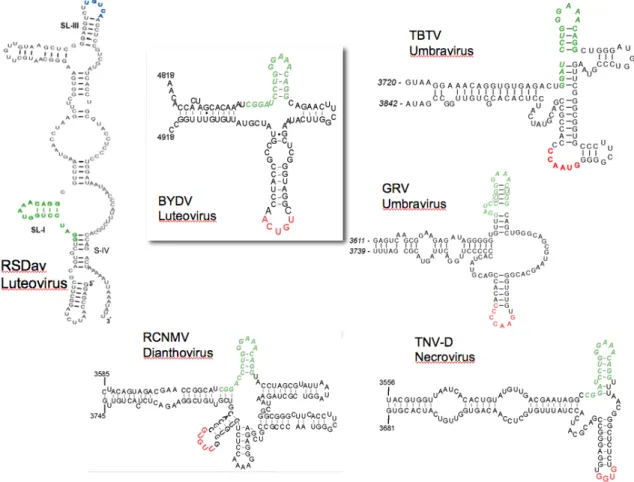
The Barley yellow dwarf virus (BYDV-) and BYDV-like cap-independent translation elements. BYDV and other members of genus Luteovirus (98), including Soybean dwarf virus, Bean leafroll virus, and the recently described Rose spring dwarf associated virus, all
Figure 3. The Barley yellow dwarf (BYDV) and BYDV-like translation elements.
While there is no prototype (Figure 3), we define BTEs as cap-independent translation elements with at least two structural features: (i) a consensus 17 nt sequence, GGAUCCUGGGAAACAGG that includes a stem-loop (SL-I, paired bases underlined) with a GNRNA loop motif (99), and (ii) a loop (not in SL-I) that can base pair to a loop in the 5’ UTR of the RNA. BTEs are functionally indistinguishable from the STNV TED, but bear no similarity in sequence or secondary structure to TED (83). An invariant hexamer, GAUCCU, within the 17 nt conserved sequence, is complementary to 18S rRNA at exactly the same distance from the 3’ end of 18S rRNA as is the RNA binding site (Shine-Dalgarno binding site) of prokaryotic 16S ribosomal RNA (83). It is still unclear whether in addition to recruiting initiation factors that facilitate ribosome recruitment, the BTE may bind the 40S subunit directly.
The mechanism of regulation of the Barley yellow dwarf virus cap-independent translation element or BTE-mediated translation as a model for 3’ mediated translation is the focus of my dissertation.
represents the core translation-specific element (86,100), but requires the flanking RIII and RIV regions that are also essential for replication (100).
The R3.5 adopts a Y-shaped structure with three extended stems (S-A, B and SL-C) that is highly conserved in genus Tombusvirus (87). Mutations on either stem loop impaired RNA translatability (87). 3’-5’ communication is mediated by base pairing of the SL-B R3.5 to a complementary stem loop, SL3, within the 5’ T-shaped domain, TSD, essential for replication (87,101). The interaction involves 9 nt that extend from the terminal loop to stem sequence. Sequence complementarity rather than primary sequence is important for this 3’-5’ communication (87). Thus, like BTE-containing viruses, base pairing between loops in the 3’ and 5’ UTRs is necessary. In fact, Fabian and White found potential base pairing between 3’ and 5’ UTRs in most genera of the Tombusviridae family (87).
Interestingly, the Maize necrotic streak virus 3’ CITE, related to the Tombusvirus genus, mediates cap-independent translation in vitro from the gRNA and both sgRNAs. Rather than adopting a Y-shaped structure, it forms a long stem loop, and the loop has sequence complementary to a loop within the TSD (88). On the other hand, the maintenance of the T-shaped structure for the Panicum mosaic virus translation element (Panicovirus
genus) is critical for its function (91). Interestingly, a similar PMV-like element was
discovered in Pea enation mosaic virus RNA2, which belongs to the Umbravirus genus
(Wang Z., personal communication).
cooperation may not involve base pairing interaction as no potential complementary sequence was identified (87,89).
Interestingly, the 5’ UTR of the CP sgRNA of TCV mediates translation more efficiently than the gRNA 5’ UTR. This makes sense because the level of accumulation of the CP is at least 100-fold greater than other gene products. Qu and Morris (2000) propose that the primary role of the TCV 3’ TE is to ensure efficient translation of viral RNA in competition with the host mRNA, while the 5’ UTRs modulate the level of expression to control the switch from early to late stages of viral life cycle (89). Yoshii et al. (2004) showed that the recessive eIF4G mutant in the cum2 gene of Arabidopsis thaliana prevents translation of TCV RNA. However, TCV translatability was not affected in the cum1 mutant line that had lost eIF4E (102) or with mutation within eIFiso4E gene (103). Thus TCV RNA translation may be eIF4G- but not eIF4E-dependent.
The Blackcurrant reversion virus translation element. Blackcurrant reversion virus (BRV) is in the Nepovirus genus of Comoviridae family. This plant virus family resembles the
animal Picornaviridae family in that the RNAs, encodes a single large polyprotein, have a
viral protein genome linked at their 5’ end instead of a cap, and a poly(A) tail at their 3’ end.
The unique feature of the viral RNA is that it bears (i) a 3’ cap-independent translation
element in its 3’ UTR, which requires a complementary stem-loop in the 5’ end, conserved
among nepoviruses (92), and (ii) a 5’ leader with IRES activity (104) - base pairing to a
BARLEY YELLOW DWARF VIRUS (BYDV)
On a biological point of view, Barley yellow dwarf virus (BYDV, Luteovirus genus, Luteoviridae family) is one of the most economically important diseases in cereal crops
worldwide (105,106). One main characteristic of the Luteoviridae family is their tissue specificity – they are confined in the phloem- and insect transmissibility (107). BYDV is transmitted in a circulative manner and uses the aphids just as a vector. Along with the plant sap, the virus reaches the hindgut of the aphids and penetrates into the haemocoel. From the haemocoel, the virus migrates to and accumulates into the accessory salivary glands. During aphid feeding, the virus particles are released into the plant phloem along with the saliva, and infection can start. BYDV disrupts the transport of plant nutrients, which induces chlorosis, plant stunting and poor fertilization. Ultimately the viral infection, if not controlled, can lead to substantial decrease in crop yield.
BYDV genome organization
The sgRNAs are 3’-coterminal with the genomic RNA. BYDV produces three sgRNAs of different sizes during cell infection, that are not packaged, but serve rather as transient transcripts, and as we will see, regulators of gene expression. sgRNA1 is the mRNA for ORF3, ORF4 and ORF5, whose expression is critical in the late phase of viral life cycle, but dispensable for viral RNA replication (110,111). ORF3 encodes the coat protein. ORF4, which overlaps ORF3 sequence in a different open reading frame, codes for a 17 kDa protein that appears to be involved in virus movement. ORF5 - which is required for aphid transmission (112) - is expressed as an extended C-terminal in-frame fusion of the coat protein. sgRNA2 is the potential mRNA for ORF6, which remains undetectable and of unknown function (113), and sgRNA3 does not encode for any ORF and its function remains unknown. As we will see in the next chapter, rather being a transcript, sgRNA2 plays the role of a regulator of gene expression.
Gene expression strategies
Due to the limited size of their genomes, BYDV has evolved an array of strategies to expand its coding capacity, and to ensure efficient expression of its genes, while complying with host translation rules (114). These include (Figure 4):
dissertation, their production regulates the proper timing and level of protein accumulation.
(ii) frame-shifting recoding event, in which less than 5% of the ribosome skip a stop codon by changing reading frame – in this case by slipping back one base relative to the initial ORF being translated ( -1 frameshift) and resume elongation in a different frame. This results in one fusion protein from two overlapping reading frames. The ribosomal frameshifting event plays a regulatory function in maintaining a particular ratio between the ORF1 and ORF1+ORF2 products (116,117). It is regulated by a long-distance frameshift element, which must base pair to a stem loop adjacent to the frameshifting site across 4000 bases (116,117).
(iii) leaky scanning mechanism, in which a portion of ribosomes bypass the ORF3 AUG, which is in a poor context, and continue scanning until they reach the downstream ORF4 AUG.
(iv) readthrough event, which involves the redefinition of a stop codon as an amino acid, for the production of the fusion CP-ORF5 protein.
(v) And as previously mentioned and the topic of this dissertation, cap-independent translation, obviating the need for the 5’ cap.
FOCUS OF MY Ph.D. DISSERTATION RESEARCH
As described above, BYDV relies on the cap-independent translation element or BTE located in the 3’ end of the mRNA to facilitate expression of viral proteins from the genomic RNA (gRNA) and sgRNAs in vivo and in vitro (83). The BTE folds into a roughly cruciform secondary structure of about 100 nt with three major stem loops (SL-I, SL-II, SL-III) and a terminal stem or “stalk” (S-IV) that allows it to protrude from the viral genome (82); E. Pettit, unpublished). For its function, the BTE must communicate with the 5’ end of the mRNA. It achieves this through the base pairing between the loop of SL-III in the BTE and a five base loop sequence in the 5’ UTRs of BYDV gRNA (78) and sgRNA1 (E. Pettit, unpublished). A single point mutation within either loop disrupted base pairing and abolished translation and replication. Compensatory mutations predicted to restore the kissing-stem loop base pairing also restored cap-independent translation and replication activity (78).
classical ribosome scanning from the 5’ end, as in normal cap-dependent translation. We propose that the kissing interaction is repeatedly disrupted by the scanning of the ribosomes and re-formed in an oscillating process that regulates ribosome entry onto the RNA.
Chapter 3 “ Trans-regulation of translation by a viral subgenomic RNA” focuses on the competition between the genomic and subgenomic RNAs with each other for the cellular translation machinery. In addition to its in cis stimulatory activity, the BTE inhibits both cap-dependent and BTE-dependent translation when added in trans (83). This inhibition is reversed by addition of eIF4F, suggesting that the BTE-mediated repression may be based on a sequestration of the translation factors. In this study, we further explored the role of BYDV sgRNA2 with its BTE in the 5’ end, in the repression of translation of replication gene from the gRNA while allowing translation of structure genes from sgRNA1. I found that the gRNA and sgRNA1 tuned their expression efficiencies via the 5’-3’ long-distance kissing interaction. Their translation in presence of sgRNA2 is controlled by the proximity to the 5’ end of the mRNA of the stem loop that interacts with the 3’ BTE. This research reveals a new level of control of sgRNA gene expression, a new role for a viral sgRNA and a new mechanism for RNA-mediated regulation of translation.
the first evidence supporting that the initial entry of ribosome is at the 5’ end of the BYDV mRNAs, following the delivery of the translation factors by the BTE from the 3’ UTR.
Appendix A covers a short study on RNA sequences implicated in cap-independent translation.
Appendix B focuses on methods for in vitro analysis of translation enhancers
REFERENCES
1. Watson, J. D., and Crick, F. H. C. (1953) Nature 171, 737-738 2. Judson, H. F. (1996)
3. Gamow. (1954) Nature 173, 318
4. Siekevitz, P., and Zamecnick, D. (1981) Journal of Cell Biology 91, 53-65 5. Crick, F. H. C. (1958) Society for Experimental Biology Symposium XII, 138-163 6. Brenner, S., Jacob, F., and Meselson, M. (1961) Nature 190, 576-581
7. Jacob, F., and Monod, J. (1961) Journal of Molecular Biology 3, 318-356
8. Gros, F., Hiatt, H., Gilbert, W., Kurland, C. G., Risebrough, R. W., and Watson, J. D. (1961) Nature 190, 581
9. Nirenberg, M. W., and Matthaei, J. H. (1961) PNAS (47), 1588-1602 10. Holley, R. W. (1965) JAMA 194, 868-871
11. Kapp, L. D., and Lorsch, J. R. (2004) Annu Rev Biochem 73, 657-704
12. Matthews, M. B., Sonenberg, N., and Hershey, J. W. B. (2007) Translation Control in Biology and Medicine, 1
13. Hershey, J. W. B., and Merrick, W. C. (2000) The pathway and mechanism of initiation of protein synthesis. In: Sonenberg, N., Hershey, J. W. B., and Mathews, M. B. (eds). Translational Control of Gene Expression, Cold Spring Harbor Laboratory Press, Cold Spring Harbor, NY
14. Kozak, M. (1999) Gene 234(2), 187-208
15. Jackson, R. J. (2005) Biochem Soc Trans 33, 1231-1241
16. Marintchev, A., and Wagner, G. (2004) Q Rev Biophys 37(3-4), 197-284 17. Kozak, M. (1979) Nature 280, 82-85
18. Furuichi, Y., Morgan, M., Shatkin, A. J., Jelinek, W., Salditt-Georgieff, M., and Darnell, J. E. (1975) PNAS 72, 1904-1908
19. Muthukrishnan, S., Both, g. W., Furuichi, Y., and Shatkin, A. J. (1975) Nature 255, 33-37
20. Both, G. W., Furuichi, Y., Muthukrishnan, S., and Shatkin, A. J. (1975) Cell 2, 185-195
21. Gallie, D. R. (1991) Genes Dev. 5(2108), 2108-2116
22. Pestova, T. V., Lorsch, J. R., and Hellen, C. U. (2007) Translation Control in Biology and Medicine, 87-128
23. Hinnebusch, A. G. (2006) Trends Biochem Sci 10, 553-562
24. Kolupaeva, V. G., Unbehaun, A., Lomakin, I. B., Hellen, C. U., and Pestova, T. V. (2005) RNA 4, 470-486
25. Pestova, T. V., and Kolupaeva, V. G. (2002) Genes Dev 16(22), 2906-2922
26. Yamamoto, Y., Singh, C. R., Marintchev, A., Hall, N. S., Hannig, E. M., Wagner, G., and Asano, K. (2005) PNAS 45, 16164-16169
27. Preiss, T., and Hentze, M. W. (2003) Bioessays 25(12), 1201-1211
28. Gingras, A.-C., Raught, B., and Sonenberg, N. (1999) Annu Rev. Biochem. 68, 913-963
32. Kahvejian, A., Svitkin, Y. V., Sukarieh, R., M'Boutchou, M. N., and Sonenberg, N. (2005) Genes Dev 19(1), 104-113
33. Karim, M. M., Svitkin, Y. V., Kahvejian, A., De Crescenzo, G., Costa-Mattioli, M., and Sonenberg, N. (2006) PNAS 25, 9494-9499
34. Gallie, D. R., and Browning, K. S. (2001) J Biol Chem 276(40), 36951-36960 35. Gallie, D. R. (2007) Translation Control in Biology and Medicine, 747-775 36. Preiss, T., and Hentze, M. (1999) Curr. Opinion Genet. Dev. 9, 515-521 37. Gallie, D. R. (2002) Plant Mol Biol 50, 949-970
38. Wells, S. E., Hillner, P. E., Vale, R. D., and Sachs, A. B. (1998) Mol Cell 2(1), 135-140
39. Gallie, D. R. (1998) Gene 216(1), 1-11
40. Ali, I. K., and Jackson, R. J. (2001) Cold Spring Harb Symp Quant Biol. 66, 377-387 41. Svitkin, Y. V., Pause, A., Haghighat, A., Pyronnet, S., Witherell, G., Belsham, G. J.,
and Sonenberg, N. (2001) RNA 7(3), 382-394
42. Eisenberg, E., and Levanon, E. Y. (2003) Trends Genet. 7, 362-365
43. Mamane, Y., Petroulakis, E., LeBacquer, O., and Sonenberg, N. (2006) Oncogene 25, 6416-6422
44. Kozak, M. (2002) Gene 299(1-2), 1-34
45. Kozak, M. (1989) Mol Cell Biol 9, 5073-5080
46. Kozak, M. (1986) Cell (Cambridge, Mass. 44(2), 283-292 47. Kozak, M. (1991) J Biol Chem 266, 19867-19870
48. Algire, M. A., and Lorsch, J. R. (2006) Curr Opin Chem Biol. 10, 480-486
49. Lee, J. H., Pestova, T. V., Shin, B. S., Cao, C., Choi, S. K., and Dever, T. E. (2002) Proc Natl Acad Sci U S A 99(26), 16689-16694
50. Gebauer, F., and Hentze, M. W. (2004) Nat Rev Mol Cell Biol 5(10), 827-835
51. Hentze, M. W., Gebauer, F., and Preiss, T. (2007) Translation Control in Biology and Medicine, 269-295
52. Pavitt, G. D. (2005) Biochem Soc Trans 33, 1487-1492
53. Gray, N. K., and Hentze, M. W. (1994) EMBO J 13(16), 3882-3891
54. Cho, P. F., Gamberi, C., Cho-Park, Y. A., Cho-Park, I. B., Lasko, P., and Sonenberg, N. (2006) Curr Biol. 16, 2035-2041
55. Pillai, R. S., Bhattacharyya, S. N., and Filipowicz, W. (2007) Trends Cell Biol. 17, 118-126
56. Jackson, R. J., and Standart, N. (2007) Sci STKE 367, 1-13
57. Valencia-Sanchez, M. A., Liu, J., Hannon, G. J., and Parker, R. (2006) Gene Dev. 20, 515-524
58. Thermann, R., and Hentze, M. W. (2007) Nature 447, 875-878
59. Humphreys, D. T., Westman, B. J., Martin, D. I., and Preiss, T. (2005) PNAS 102, 16961-16966
60. Kiriakidou, M., Tan, G. S., Lamprinaki, S., De Planell-Saguer, M., Nelson, P. T., and Mourelatos, Z. (2007) Cell 129, 1141-1151
61. Olsen, P. H., and Ambros, V. (1999) Dev Biol 216(2), 671-680
64. Doudna, J. A., and Sarnow, P. (2007) Translation Control in Biology and Medicine, 129-153
65. Jang, S. K. (2006) Virus Res 119, 2-15
66. Pestova, T. V., Shatsky, I. N., Fletcher, S. P., Jackson, R. J., and Hellen, C. U. (1998) Genes Dev 12(1), 67-83
67. Kieft, J. S., Zhou, K., Jubin, R., and Doudna, J. A. (2001) RNA 7(2), 194-206 68. Fraser, C. S., and Doudna, J. A. (2007) Nat Rev Microbiology 1, 29-38
69. Wilson, J. E., Pestova, T. V., Hellen, C. U., and Sarnow, P. (2000) Cell 102(4), 511-520
70. Wilson, J. E., Powell, M. J., Hoover, S. E., and Sarnow, P. (2000) Mol Cell Biol 20(14), 4990-4999
71. Kneller, E. L., Rakotondrafara, A. M., and Miller, W. A. (2006) Virus Res 119, 63-75 72. Skulachev, M. V., Ivanov, P. A., Karpova, O. V., Korpela, T., Rodionova, N. P.,
Dorokhov, Y. L., and Atabekov, J. G. (1999) Virology 263(1), 139-154
73. Jaag, H. M., Kawchuk, L., Rohde, W., Fischer, R., Emans, N., and Prufer, D. (2003) Proc Natl Acad Sci U S A 100(15), 8939-8944
74. Zeenko, V., and Gallie, D. R. (2005) J Biol Chem 280(29), 26813-26824 75. Miller, W. A., and White, K. A. (2006) Ann. Rev. Phytopathol. 44, (In press)
76. Gazo, B. M., Murphy, P., Gatchel, J. R., and Browning, K. S. (2004) J Biol Chem 279(14), 13584-13592
77. Wang, S., Guo, L., Allen, E., and Miller, W. A. (1999) RNA 5, 728-738 78. Guo, L., Allen, E., and Miller, W. A. (2001) Mol. Cell 7, 1103-1109 79. Fabian, M. R., and White, K. A. (2006) RNA in press
80. Rakotondrafara, A. M., Polacek, C., Harris, E., and Miller, W. A. (2006) Rna 12(10), 1893-1906
81. Meulewaeter, F., Van Montagu, M., and Cornelissen, M. (1998) RNA 4(11), 1347-1356
82. Guo, L., Allen, E., and Miller, W. A. (2000) RNA 6, 1808-1820
83. Wang, S., Browning, K. S., and Miller, W. A. (1997) EMBO J 16(13), 4107-4116 84. Mizumoto, H., Tatsuta, M., Kaido, M., Mise, K., and Okuno, T. (2003) J Virol
77(22), 12113-12121
85. Meulewaeter, F., van Lipzig, R., Gultyaev, A. P., Pleij, C. W., Van Damme, D., Cornelissen, M., and van Eldik, G. (2004) Nucleic Acids Res 32(5), 1721-1730
86. Wu, B., and White, K. A. (1999) J Virol 73(11), 8982-8988
87. Fabian, M. R., and White, K. A. (2004) J Biol Chem 279(28), 28862-28872 88. Scheets, K., and Redinbaugh, M. G. (2006) Virology 350(1), 171-183 89. Qu, F., and Morris, T. J. (2000) J Virol 74(3), 1085-1093
90. Koh, D. C.-Y., Liu, D. X., and Wong, S.-M. (2002) J Virol 76, 1144-1143
91. Batten, J. S., Desvoyes, B., Yamamura, Y., and Scholthof, K. B. (2006) FEBS Lett 580(11), 2591-2597
92. Karetnikov, A., Keränen, M., and Lehto, K. (2006) Virology 354, 178-191
93. Timmer, R. T., Benkowski, L. A., Schodin, D., Lax, S. R., Metz, A. M., Ravel, J. M., and Browning, K. S. (1993) J Biol Chem 268(9504), 9504-9510
95. Meulewaeter, F., Van Montagu, M., and Cornelissen, M. (1998) Rna 4(11), 1347-1356
96. van Lipzig, R., Gultyaev, A. P., Pleij, C. W., van Montagu, M., Cornelissen, M., and Meulewaeter, F. (2002) Rna 8(2), 229-236
97. van Lipzig, R., Van Montagu, M., Cornelissen, M., and Meulewaeter, F. (2001) Nucleic Acids Res 29(5), 1080-1086
98. Domier, L. L., McCoppin, N. K., Larsen, R. C., and D'Arcy, C. J. (2002) J Gen Virol 83(Pt 7), 1791-1798
99. Legault, P., Li, J., Mogridge, J., Kay, L. E., and Greenblatt, J. (1998) Cell 93(2), 289-299
100. Oster, S. K., Wu, B., and White, K. A. (1998) J Virol 72(7), 5845-5851 101. Wu, B., Vanti, W. B., and White, K. A. (2001) J Mol Biol 305(4), 741-756
102. Yoshii, M., Nishikiori, M., Tomita, K., Yoshioka, N., Kozuka, R., Naito, S., and Ishikawa, M. (2004) J Virol 78(12), 6102-6111
103. Lellis, A. D., Kasschau, K. D., Whitham, S. A., and Carrington, J. C. (2002) Curr Biol 12(12), 1046-1051
104. Karetnikov, A., and Lehto, K. (2007) J Gen Virol 88, 286-297
105. Miller, W. A., and Rasochova, L. (1997) Annu. Rev. Phytopathol. 35, 167-190
106. Lister, R. M., and Ranieri, R. (1995) Distribution and economic importance of barley yellow dwarf. In: D'Arcy, C. J., and Burnett, P. (eds). Barley yellow dwarf: 40 years of progress, APS Press, St. Paul
107. Power, A. G., and Gray, S. M. (1995) Aphid transmission of barley yellow dwarf viruses: Interactions between viruses, vectors, and host plants. In: D'Arcy, C. J., and Burnett, P. A. (eds). Barley Yellow Dwarf: 40 Years of Progress, APS Press, St. Paul, MN
108. Miller, W. A., Dinesh-Kumar, S. P., and Paul, C. P. (1995) Critic Rev Plant Sci 14, 179-211
109. Mohan, B. R., Dinesh-Kumar, S. P., and Miller, W. A. (1995) Virology 212, 186-195 110. Koev, G., and Miller, W. A. (2000) J. Virol. 74, 5988-5996
111. Dinesh-Kumar, S. P., Brault, V., and Miller, W. A. (1992) Virology 187, 711-722 112. Chay, C., Smith, D. M., Vaughan, R., and Gray, S. M. (1996) Phytopathology 86,
370-377
113. Shen, R., Rakotondrafara, A. M., and Miller, W. A. (2006) J Virol 80(20), 10045-10054
114. Dreher, T. W., and Miller, W. A. (2006) Virology 344(1), 185-197 115. Miller, W. A., and Koev, G. (2000) Virology 273, 1-8
116. Paul, C. P., Barry, J. K., Dinesh-Kumar, S. P., Brault, V., and Miller, W. A. (2001) J. Mol. Biol. 310, 987-999
CHAPTER 2. OSCILLATING KISSING STEM-LOOP INTERACTIONS MEDIATE
5’ SCANNING-DEPENDENT TRANSLATION BY A VIRAL 3’
CAP-INDEPENDENT TRANSLATION ELEMENT
A paper published in
RNA (2006) 12: 1893-1906
Aurélie M. Rakotondrafara, Charlotta Polacek, Eva Harris and W. Allen Miller
Author contributions: AMR performed all experiments. CP and EH provided the dengue
viral constructs. AMR and WAM designed, analyzed and interpreted the data and wrote the
papers.
ABSTRACT
The 3’ untranslated regions (UTRs) of a group of novel uncapped viral RNAs allow efficient
translation initiation at the 5’-proximal AUG. A well-characterized model is the Barley
yellow dwarf virus class of cap-independent translation element (BTE). It facilitates
translation by forming kissing stem-loops between the BTE in the 3’ UTR and a
BTE-complementary loop in the 5’ UTR. Here we investigate the mechanisms of the long–
distance interaction and ribosome entry on the RNA. Upstream AUGs or 5’ extensions of the
5’ UTR inhibit translation, indicating that, unlike internal ribosome entry sites in many viral
RNAs, the BTE relies on 5’ end-dependent ribosome scanning. Cap-independent translation
occurs when the kissing sites are moved to different regions in either UTR, including outside
of the BTE. The BTE can even confer cap-independent translation when fused to the 3’ UTR
and 5’ ends. Thus, the BTE serves as a functional sensor to detect sequences capable of
long-distance base pairing. We propose that the kissing interaction is repeatedly disrupted by
the scanning ribosome and re-formed in an oscillating process that regulates ribosome entry
on the RNA.
INTRODUCTION
Initiation of translation for cellular mRNAs can be broken down into three steps (Kozak,
2002; Kapp & Lorsch, 2004). The 40S ribosomal subunit with the initiation factor complex
enters the capped mRNA at the 5’ terminus (step 1), scans the 5’ untranslated region (UTR)
to reach the 5’ proximal AUG (step 2) at which the 60S subunit joins (step 3) and protein
synthesis ensues. The recognition of the 5’ cap by the cap-binding protein component
(eIF4E) of initiation complex eIF4F enhances ribosome recruitment to the mRNA (von der
Haar et al., 2004). The other component of eIF4F, initiation factor 4G (eIF4G) orchestrates
the assembly of the scanning machinery (Kapp & Lorsch, 2004). eIF4F recruits the RNA
helicase eIF4A (Oberer et al., 2005) (not a subunit of eIF4F in plants), which unwinds the 5’
region to favor the attachment and subsequent scanning of the eIF3-bound 40S-ribosomal
subunit onto the unstructured RNA (Pestova & Kolupaeva, 2002; Siridechadilok et al., 2005;
Jivotovskaya et al., 2006).
Many viral RNAs have structures within their 5’ or 3’ UTRs that recruit the host
translation machinery in the absence of a 5’ cap and/or a poly(A) tail (Dreher & Miller, 2006;
Kneller et al., 2006). With the exception of certain plant viral RNAs, cap-independent
translation is facilitated by a highly structured internal ribosome entry site (IRES), which
2001). IRESes recruit the 40S ribosomal subunit to an internal region of the RNA in close
proximity to the initiation codon, without ribosomal scanning from the 5’ end (Sarnow et al.,
2005).
Here we report on a different type of cap-independent translation element discovered in
certain uncapped non-polyadenylated plant viral and satellite RNAs including the
Luteoviruses (Wang et al., 1997) and members of the Tombusviridae family (Timmer et al.,
1993; Wu & White, 1999; Mizumoto et al., 2003; Meulewaeter et al., 2004; Shen & Miller,
2004; Batten et al., 2006; Scheets & Redinbaugh, 2006). Unlike IRES-mediated translation,
the cap-independent translation element is present in the 3’ untranslated region, yet
translation initiation occurs at the AUG closest to the 5’ end of the mRNA (Kneller et al.,
2006). Through a long-distance kissing stem-loop interaction between the 3’
cap-independent translation element and the 5’ UTR, the 3’ element mediates translation
initiation at the 5’ end of the mRNA. Presumably, the long-distance base pairing facilitates
the delivery of the ribosomes and/or initiation factors to the 5’ proximal AUG (Guo et al., 2001; Fabian & White, 2006).
At least three different classes of 3’ cap-independent translation element, which show no
apparent similarity in sequence or structure to each other, have been identified (review by
Miller & White, 2006). Typical members of each class include Satellite tobacco necrosis
virus (STNV, genus Necrovirus) (Timmer et al., 1993), Barley yellow dwarf virus (BYDV,
genus Luteovirus) (Wang et al., 1997), and Tomato bushy stunt virus (TBSV, genus
Tombusvirus) (Wu & White, 1999). Here, we focus on the BYDV(-like) cap-independent
The genome of BYDV is an uncapped, non-polyadenylated positive-sense RNA of 5677
nts that encodes six open reading frames (ORFs) (Fig. 1A). The minimal in vitro functional
BYDV translation element (BTE) spans nts 4809-4918 and forms a roughly cruciform
secondary structure with three major stem loops (SL-I, SL-II, SL-III) flanked by stem IV
(Fig. 1B). Additional sequences in the 3’ UTR, including a domain that functionally replaces
a poly(A) tail, are required for full cap-independent and poly(A) tail-independent translation
in plant cells (Wang et al., 1997; Guo et al., 2001).
The 3’ BTE facilitates cap-independent translation initiation at the 5’ proximal AUG via
base pairing of the loop of stem-loop III (SL-III) of the BTE to a complementary loop, SL-D
(that we refer to as a BTE-complementary loop or BCL) located within the 5’ UTR of the
viral genomic RNA. A single point mutation within the middle of the five bases of either of
these kissing loops disrupts base pairing and abolishes translation both in cells and in wheat
germ extract; while compensatory double mutations that restore base pairing restore
translation (Guo et al., 2001). The BTE can also potentially base pair to a BCL in the 5’
UTR of BYDV subgenomic RNA1, which serves as the mRNA for the coat protein and other
ORFs downstream of ORFs 1 and 2 (Fig. 1B). However, the BCL in the 5’ UTR of sgRNA1
does not facilitate translation in the context of full-length genomic RNA, because no ORFs
downstream of the sgRNA1 BCL are translated from genomic RNA (Allen et al., 1999).
It remained to be determined (i) the constraints in sequences, structures and position of
the long distance kissing-loop interaction to support cap-independent translation, (ii) how the
ribosomes enter the mRNA, and (iii) whether the long-distance kissing loop in the 3’ BTE
must be located within the context of the BTE or can be separated from the intrinsic
Here we show that (i) the possible sequences of the long-distance kissing loops that
support cap-independent translation are limited, even if complementarity is maintained, (ii)
ribosomes must enter from the 5’ terminus of the RNA via the BCL-BTE kissing interaction,
and scan to the 5’ proximal AUG just as on a cellular capped RNA, and (iii) the
long-distance base pairing structures can be uncoupled from the BTE structure, and even replaced
entirely by complementary sequences from an unrelated viral RNA in vitro. Thus, the 3’
BTE can be used as a functional sensor of long-distance interactions of any RNA of interest,
even if the RNA is not normally involved in translation. Because the ribosome must scan
through the BCL to reach the start codon, the RNA structure must oscillate between kissing
stem-loops and disrupted kissing. We propose that this regulates entry of the translation
initiation machinery onto the viral RNA.
RESULTS
Sequence specificity required for the BYDV end-to-end interaction. Among different
viruses, the kissing loops in the 3’ and 5’ UTRs, which are involved in long-distance
interaction, vary in sequence and length with no obvious consensus (Edgil & Harris, 2006;
Miller & White, 2006). The BYDV kissing interaction, required for cap-independent
translation, involves a five base sequence that tolerated an exchange of the middle base of
each loop to maintain complementarity (Guo et al., 2001). The five bases involved in base
pairing in each loop are identical, except for the middle base (UGACA:UGUCA). Thus,
primary sequence as well as complementarity may be important for this long-distance
interaction. This led us to ask whether more sequence variation in the kissing loop could be
in the 5’ UTR with complementary sequences containing three base changes,
UCUGA:UCAGA, (altered bases in bold), or 10-base complementary tracts,
UCAAUAUGCC:GGCAUAUUGA (Fig. 2A). The 10-base tracts, called cyclization
sequences (CS), are located naturally at opposite ends of the 11kb dengue virus (DEN)
genome and are known to base pair (Khromykh et al., 2001). The mutations were tested in
the context of a reporter construct (LUC869) that was used previously to demonstrate the
long-distance interaction (Fig. 2) (Guo et al., 2001). In LUC869 firefly luciferase open
reading frame is flanked by the BYDV genomic 5’ and 3’ UTRs (nts 1-142; nts 4809-5677).
The expression efficiency of each mutant was measured both in oat protoplasts (in vivo) and
in wheat germ extract (in vitro). Despite retaining complementarity, neither set of altered
kissing loop sequences allowed translation in vivo or in vitro (Fig. 2B). In both cases
translation was similar to the level obtained with a four-base insertion in the BamHI site of
the BTE that we showed previously renders the BTE completely non-functional (BTEBF)
(Wang et al., 1997). The BTEBF mutation serves as a negative control throughout this
report.
The 3’ BTE must perform at least two functions: mediate the recruitment of the
translational machinery, and communicate with the 5’ end where translation initiates. The
need for the latter function can be eliminated by placing the BTE in the 5’ UTR, from which
it can mediate cap-independent translation in vitro (Wang et al., 1997; Guo et al., 2000). We
refer to any BTE mutant capable of mediating cap-independent translation from the 5’ UTR
as having the core cap-independent translation activity. These mutants may or may not have
the long-distance base pairing capability. To determine whether the above alternative
the BTE, the BTE mutants (LIII-3; LIII-CS) were tested in the 5’ UTR context of the
luciferase reporter gene with non-viral sequences in the 3’ UTR (5’ BTE-LUC, Fig 2C).
Translation of these uncapped BTE-LUC RNAs was at least as efficient as the “wild type” 5’
BTE-LUC RNA (Fig. 2C), indicating that the mutations within loop III of the BTE did not
interfere with the cap-independent translation activity of the BTE. Thus, the 3 and
LIII-CS mutants lost ability to mediate translation from the 3’ UTR due to an inability to interact
productively with the 5’ UTR, or because the compensating mutants in the 5’ BCL (absent in
the constructs with the BTE in the 5’ UTR) disrupted translation.
Structural flexibility in the 5’ UTR but not in the BTE. Previously, we showed that the
stem portions of the kissing stem loops can be exchanged between the SL-III and the 5’ BCL,
SL-D (Guo et al., 2001). Here, we test whether the stem is necessary at all. Mutations were
introduced to disrupt the base pairing in the stem portion of the BTE SL-III (m1,
SIII-m2, Fig. 3A). Although the BTE loop III bases were predicted to remain single-stranded and
available for base pairing to the 5’ end, no translation activity above background level
(obtained with the BTEBF construct) was observed in cells or in wheat germ extract (Fig.
3B). The BTE mutants also failed to promote cap-independent translation above background
level when placed in the 5’ UTR context of the luciferase gene (Fig. 3C), revealing that
disruption of the stem III structure abolished the core cap-independent translation function of
the BTE, even in the 5’ UTR context that lacks a requirement for the long-distance kissing
interaction. The double compensatory mutation, predicted to restore the BTE stem III
structure (SIII-r), rescued the translation of the LUC869 RNA fully in vitro and to one third
200-fold, respectively, greater than the expression obtained for the mutants with the disrupted
stem III. As expected, double mutant SIII-r facilitated full translation in the 5’ BTE-LUC
context (SIII-r, Fig. 3C). Thus, the stem portion of SL-III tolerates changes in the primary
sequence as long as the stem remains intact. Disruption of the stem causes structural changes
sufficient to abolish cap-independent translation in any context.
Unlike SL-III in the BTE, disruption of the stem flanking the BCL in the 5’ UTR
(SD-m1, -m2, Fig. 3D), still allowed significant translation of the uncapped reporter
transcript in vivo (Fig. 3E), while the double compensatory mutant that restored the stem-D
(SD-r) translated as efficiently as wild type LUC869 RNA. Thus, regardless of the flanking
structure, the BCL bases are sufficient to support the long-distance RNA:RNA interaction
with the 3’ BTE.
Ribosome scanning from the 5’ end is required for cap-independent translation.
Previously we reported that addition of a highly stable stem-loop at the extreme 5’ end of the
gRNA abolished both BTE- and cap-mediated translation (Guo et al., 2001), presumably by
blocking access of the 5’ end for ribosome binding (Kozak, 1989). Such a major change to
the 5’ UTR could prevent BTE-BCL interactions or affect translation in unpredicted ways.
Thus, we further examined the possibility of 5’ end-dependency of the BTE-mediated
cap-independent translation with different alterations of the 5’ UTR. An out-of-frame AUG
(uAUG) was inserted just upstream of the 5’ BCL in the LUC869 reporter construct context,
creating a small upstream ORF (uORF) of 29 codons (Fig. 4A). If scanning occurs, the
ribosomes would encounter the uORF before reaching the start codon of the luciferase gene,
Luciferase expression in vivo was compared to that of the wild type RNA and a
capped BTEBF RNA construct, in which translation is cap-mediated and thus
scanning-dependent (Fig. 4B). The presence of the uAUG reduced cap- and BTE-mediated translation
of luciferase to 13% and 27%, respectively, of that obtained from the wild type LUC869
RNA (capped uAUG-BTEBF, uAUG). The reduction of the luciferase expression correlated
with the accumulation of a 2.9kDa peptide encoded by the uORF, except for the
uAUG-BTEBF construct in which no translation above background of either ORF is expected (Fig.
4C). In this construct, both the uAUG and the LUC AUG had the same sequence context
(GCGCAUGG). We next placed the uAUG in the optimal Kozak context for efficient
initiation (GCCACCAUGG) (Kozak, 1991). This construct abolished luciferase expression
(Kozak uAUG, Fig 4B). These data support that cap-independent translation mediated by the
3’ BTE relies on a ribosome scanning mechanism that obeys conventional Kozak rules,
starting from a region upstream of the 5’ kissing site (BCL), most likely the 5’ end itself
(Guo et al., 2001), just as on a cellular capped mRNA.
Increasing distance of the BTE-complementary loop from the 5’ end reduces
translation efficiency. In BYDV gRNA, the BCL is located at base positions 103-107 of the
5’ UTR, and is the fourth stem-loop from the 5’ end (Guo et al., 2000). In contrast, in all
other 3’ TE-bearing RNAs including BYDV sgRNA1 and other luteoviruses, the BCL is
located within 10-30 nucleotides of the 5’ UTR in the 5’ proximal stem-loop (Fig. 5A). This
raises the question of the importance of the position of the BCL relative to the 5’ end for the
efficiency of the BTE-mediated cap-independent translation. We propose that the 3’-5’