Int. J. Electrochem. Sci., 7 (2012) 4545 - 4558
International Journal of
ELECTROCHEMICAL
SCIENCE
www.electrochemsci.org
Determination of Lead and Cadmium in the Presence of
Quercetin–5’–sulfonic Acid by Adsorptive Stripping
Voltammetry with a Hanging Mercury Drop Electrode and a
Nafion–coated Mercury Film Electrode
Edgar Nagles*, Verónica Arancibia*, Roxana Ríos
Pontificia Universidad Católica de Chile, Facultad de Química, Vicuña Mackenna 4860, Santiago– 7820436, Chile.
*
E-mail: darancim@uc.cl; ernagles@uc.cl
Received: 3 February 2012 / Accepted: 4 April 2012 / Published: 1 May 2012
The use of a hanging mercury drop electrode (HMDE) and a glassy carbon electrode modified with Nafion–Hg (NHgFE) to determine Pb(II) and Cd(II) by adsorptive stripping voltammetry in the presence of quercetin–5’–sulfonic acid (QSA) is reported. With a HMDE it is possible to determine Pb(II) and Cd(II) individually because the peak current of Pb–QSA is maximum at pH 6.1 (Epeak –0.50 V), while the peak current of the Cd–QSA complex is maximum at pH 8.9 (Epeak –0.66 V). The linear calibration curves ranged from 0.5–40.0 µg L–1 for Pb(II) and 0.5–45.0 µg L–1 for Cd(II). The detection limits (3) were estimated to be around 0.3 and 0.1 µg L–1 and the relative standard deviations were 2.0 and 1.7 % at the 9.4 µg L–1 level of Pb(II) and Cd(II) with 30 s of accumulation (n=7) (CQSA 2.7 µmol L–1; Eads –0.10 V). On the other hand, it is found that with the prepared Nafion– mercury film electrode Pb(II) and Cd(II) can be analyzed simultaneously at pH 6.1 (Epeak –0.55 V, – 0.73 V). The detection limits were 0.2 µg L–1 for both metal ions, with a linear range until 18.0 µg L–1. The methods were validated using synthetic sea water (ASTM D665) spiked with several metal ions, and reference material for measuring of elements in water (TMDA–61) and waste waters (SPS–WW1). Finally, the method was successfully applied to the determination of Pb(II) and Cd(II) in tap water after UV digestion.
Keywords: Adsorptive stripping voltammetry; Nafion–coated mercury film electrode; Pb(II) and Cd(II) determination; Quercetin–5`–sulfonic acid; Water analysis
1. INTRODUCTION
bioaccumulation along the food chain. The development of new methods for quantifying traces of these elements is required. Trace metal ions have been determined with success by anodic stripping voltammetry (ASV) due to its low limit of detection (LoD), selectivity and relatively inexpensive instrumentation. The most widely used electrode material in ASV is mercury. Some of the advantages include reproducible surface, high hydrogen overpotential, high sensitivity, high selectivity, low instrumentation cost, and the ability to dissolve many metals, which aids in the preconcentration process 1. Different electrodes, such as Hg–coated glassy carbon 2–4, Hg–film supported on wax-impregnated carbon paste 5, Hg(II)–modified multiwalled carbon nanotubes 6, mercury film screen printed carbon or graphite 7–9, and in recent years solid amalgams 10 have been applied to the determination of Pb(II) and Cd(II) by ASV. The detection limits obtained are between 0.0094 to 5.0 µg L–1 for Pb(II) and 0.0018 to 2.0 µg L–1 for Cd(II). These and others electrodes were shown to be extremely useful for adsorptive stripping measurements of trace nickel, cobalt, molybdenum, vanadium, and chromium, but the large majority of the published applications of simultaneous determination of Pb(II) and Cd(II) using modified electrodes utilize ASV. In adsorptive voltammetry the metal ions must be converted into stable complexes with adequate surface-active ligands to be adsorbed on the working electrode by means of a nonelectrolytic process prior to the voltammetric scan, and the detection limit of AdSV is also usually better than ASV 11. Different ligands like morin 12, morin–5–sulfonic acid 13, quercetin 14, dopamine 15,16, carbidopa 17, formazone 18, 2–hydroxybenzaldehyde benzoylhydrazone 19, 2–acetylpyridine salicyloylhydrazone 20, 4,5– dihydroxy–3–(p–sulfophenylazo)–2,7–naphthalene disulfonic acid 21, thymolphthalexone 22, and others have been adequate for determining Pb(II) in natural waters. On the other hand, oxine [23,24], 1–(2–pyridylazo)–2,7–dihydroxynaphthalene [25], 2–acetylpyridine salicyloylhydrazone 26, ammonium 2–amino–cyclopentene dithiocarboxylate 27, calcein blue [28], glyoxylic acid thiosemicarbazide [29], –mercaptocarboxylic acids [30], 2,5–dimercapto–1,3,4–thiadiazole [31], and 2–mercapto–5–phenylamino–1,3,4–thiadiazole [32] have been used as complexing agents for the voltammetric determination of Cd(II), while 2–mercaptobenzothiazole 33 and 1–phenylpropane–1– pentylsulfonylhydrazone–2–oxime 34 have been studied for the adsorptive collection of complexes with Pb(II) and Cd(II) on a hanging mercury drop electrode (HMDE).
In this study we investigated the simultaneous determination of Pb(II) and Cd(II) using 3,5,7,3´,4´–pentahydroxy–5´–sulfoflavone (quercetin–5’–sulfonic acid, QSA) as adsorbing and complexing ligand. Comparisons will be made of HMDE and HgFE. With the purpose of getting higher sensitivity and selectivity, anionic surfactants such as sodium dodecyl sulfate (SDS) and cationic surfactants such as cetylpyridinium bromide (CPB) were added to the solution.
2. EXPERIMENTAL PART
2.1. Apparatus
electrode (NHgFE, disc diameter of 2 mm), were used as working electrodes with a Ag/AgCl/KCl 3 mol L–1 reference electrode,and a platinum wire auxiliary electrode. Solutions were deoxygenated with high purity nitrogen. The pH measurements were carried out with an Orion–430 digital pH/mV meters equipped with combined pH glass electrode. UV–irradiation of water samples was carried out in quartz tubes using a 705 UV–digester (Metrohm).
2.2. Reagents and solutions
Water used for sample preparation, dilution of the reagents, and rinsing purposes was obtained in a Milli–Q system (18.2 MΩ. Millipore, USA). All the chemicals (nitric acid, acetic acid, etc.) were analytical grade from Merck (Darmstadt, Germany). Standard Pb(II) and Cd(II) solutions were prepared by diluting commercial standards containing 1000 mg L–1, Merck (Darmstadt, Germany). Quercetin–5’–sulfonic acid (QSA) was synthesized as reported by Kopacz 35 and the solutions were prepared in methanol:water 1:1. The stock solutions of CPB and SDS (from Aldrich) were prepared by dissolving the reagent in water.
Britton Robinson (BR) buffer solutions were used to investigate pH in the 4.1–9.5 range. These buffers (0.4 mol L–1) were prepared by mixing equal volumes of orthophosphoric acid, acetic acid, and boric acid, adjusting to the required pH with 2.0 mol L–1 NaOH solution. Once the pH was shortened, acetic/acetate solutions were used in the range 4.0–7.0 (0.4 mol L–1). Synthetic sea water (ASTM D665, Aldrich) spiked with ICP multi–element standard solution IV (Merck), certified reference water (TMDA–61. Environment Canada), and certified waste water level 1 (SPS–WW1, Norway) were used for validation measurements.
2.3. Procedure for preparation of NHgFE
The glassy carbon disk electrode was polished with water slurry of 0.3 µm Al2O3, rinsed with ethanol and water in an ultrasonic bath and dried with N2. The electrode was placed in a 5% Nafion solution under rotation for 300 s and then the solvents were left to evaporate at room temperature for 15 min.
For preplating the electrode with a mercury film it was immersed in a 200 mg L–1 Hg(II) solution and the Hg film was formed by holding the working electrode potential at –1.30 V for 150 s. The same electrode was used in a series of measurements.
2.4. Sample preparation
2.5. Procedure
Ten mL of deionized water (or tap water samples), 0.5 mL of Britton–Robinson buffer solution (0.4 mol L–1) or 0.5 mL of acetate buffer solution (0.4 mol L–1), 5–13 µL of quercetin–5’–sulfonic acid solution (2.9 mmol L–1), and aliquots of Pb(II) and/or Cd(II) solution (0.5 mg L–1) were pipetted into the voltammetric cell. The solution was purged with argon (saturated with water vapor) for 5 minutes in the first cycle and for 60 s for each successive cycle. Then, after eliminating some drops (size:8), a new mercury drop was extruded to initiate the preconcentration step for a given tads and Eads at a stirring speed of 1800 rpm. After an equilibration time of 10 s, the adsorptive voltammogram was recorded, while the potential was scanned from –0.10 to –1.0 V using square wave modulation with 10 mV step amplitude, 20 mV pulse amplitude, and a frequency of 25 Hz (sweep rate 0.248 V s–1). Each voltammogram was repeated three times. The calibration curves were obtained and linear regression and detection limits were calculated. The proposed method was applied to the determination of lead and cadmium in tap water; in order to eliminate matrix effects the standard addition method was used. All data were obtained at room temperature (~25 °C).
3. RESULTS AND DISCUSSION
3,5,7,3´,4´–pentahydroxy–5´–sulfoflavone (QSA) (pK1 = 7.4; pK2 = 8.4; pK3 = 10.5) has good aqueous solubility and forms complexes with a metal:ligand stoichiometry of 1:1 with Pb(II) and of 1:2 with Cd(II) that are poorly soluble in water (10–4–10–5 mol dm–3, 20 °C). In the complexes the metal is chelated by three C–OH and four C=O, and the sulfonic group contributes a negative charge 36. On the other hand, with Pb(II) and quercetin in methanol have been reported three complexes with 1:2, 1:1, and 2:1 stoichiometry (M:L), with the following stability constants: log 7.71 ± 0.43, log 4.87 ± 0.04, and log 8.23 ± 0.05 for PbQ2, PbQ, and Pb2Q respectively. For the best sensitivity in the simultaneous determination of Pb(II) and Cd(II) using the HMDE the influence of different parameters such as pH, CQSA, Eads and tads was investigated.
3.1. Effect of operational parameters 3.1.1. Effect of pH variation
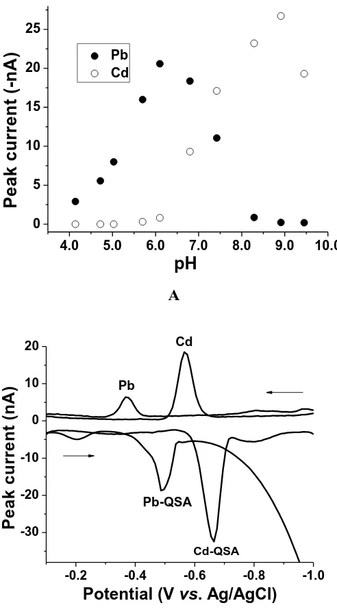
the Cd–QSA complex is seen at –0.66 V. In anodic voltammograms (Edep = –1.20 V), the oxidation peak of the Pb(0) is seen at –0.38 V while the oxidation peak of the Cd(0) is seen at –0.57 V. The peak currents obtained with AdSV are higher than ASV. The peak current of Pb–QSA at pH 6.1 was not affected by the addition of 50 µgL–1 of Cd(II) and the peak current of Cd–QSA at pH 8.9 was not affected by the addition of 50 µgL–1 of Pb(II). At all the pH values it was not possible to see both signals with good sensitivity. For this reason further measurements were carried out individually for Pb(II) at pH 6.1 and Cd(II) at pH 8.9.
A
B
Figure 1. (A). Effect of pH on the peak current of the Pb–QSA and Cd–QSA complexes. (B). Adsorptive and anodic stripping voltammograms at pH 6.1 and 8.9. Conditions: Pb(II), Cd(II) 9.4 μg L−1; CQSA: 2.5 μmol L−1; Eads: −0.10 V; Edep: −1.20 V; tacc: 30s; step amplitude: 10 mV; pulse amplitude: 20 mV and frequency: 25 Hz.
4.0 5.0 6.0 7.0 8.0 9.0 10.0
0 5 10 15 20 25
Pb Cd
P
e
a
k
c
u
rr
e
n
t
(-n
A
)
pH
-0.2 -0.4 -0.6 -0.8 -1.0
-30 -20 -10 0 10 20
P
e
a
k
c
u
rr
e
n
t
(n
A
)
Potential (V vs. Ag/AgCl)
Pb
Cd
Pb-QSA
[image:5.596.171.417.213.661.2]
0.0 1.0 2.0 3.0 4.0 5.0
0 5 10 15 20 25 30
Pb Cd
P
e
a
k
c
u
rr
e
n
t
(-n
A
)
C
QSA(
molL
-1)
[image:6.596.144.411.116.347.2]3.1.2. Effect of Quercetin–5’–sulfonic acid concentration (CQSA)
Figure 2. Effect of CQSA on the peak current at pH 6.1 (Pb) and 8.9 (Cd). Conditions: Pb(II), Cd(II) 9.4 μgL−1; tads: 30 s; Eads: −0.10 V. Others conditions as in Fig. 1.
Fig. 2 shows the effect of the variation of CQSA on the peak current at pH 6.1 for Pb(II) and at pH 8.9 for Cd(II). The experimental conditions were: Pb(II), Cd(II) 9.4 μg L−1; Eads = –0.10 V and tads = 30 s. The peak current increased with increasing CQSA up to 2.7 µmol L–1 for Pb(II) (M:L ratio of 1:60) and 2.0 µmol L–1 for Cd(II) (M:L ratio 1:24). At concentrations higher than 4.5 µmol L–1 the peak current decreased slightly with increasing concentration of QSA, probably due to the competition of QSA with complexes for adsorption on the HMDE. An optimum ligand concentration of 2.7 µmol L–1 was used for further experiments.
3.1.3. Effect of accumulation potential (Eads)
Figure 3. Effect of accumulation potential on the peak current at pH 6.1 (Pb) and 8.9 (Cd). Conditions: Pb(II), Cd(II) 9.4 μg L−1; CQSA: 2.7 μmol L−1; tads: 30 s. Others conditions as in Fig. 1.
3.1.4. Effect of accumulation time (tads)
Figure 4. Effect of accumulation time on the peak current at pH 6.1 (Pb) and 8.9 (Cd). Conditions: Pb(II), Cd(II) 9.4 μg L−1; CQSA: 2.7 μmol L−1; Eads: –0.1 V. Others conditions as in Fig. 1.
0.0 -0.2 -0.4 -0.6 -0.8 -1.0
0 8 16 24 32
Pb Cd Pb Cd Pb Cd
P
e
a
k
c
u
rr
e
n
t
(-n
A
)
E
ads
(V
vs
Ag/AgCl)
0
60
120
180
240
300
360
0
40
80
120
160
Pb
Cd
P
e
a
k
c
u
rr
e
n
t
(-n
A
)
[image:7.596.125.442.441.720.2]
Fig. 4 shows the effect of accumulation time on the stripping peak current of the Pb–QSA and Cd–QSA complexes at pH 6.1 and 8.9 over the 0–330 s range. The experimental conditions were: Pb(II), Cd(II) 9.4 μg L−1; CQSA = 2.7 μmol L−1 and Eads = –0.1 V. Peak current increases with increasing accumulation prior to the potential scan, indicating that the Pb(II) and Cd(II) complexes are readily adsorbed on the HMDE. Peak current increased with time up to 300 s. However, considering the speed of the measurement, tads of 30 or 90 s were used for further studies, but in the analysis of real samples higher times can be used to achieve good sensitivity.
3.1.5. Effect of SDS and CPB concentration
The effect of the SDS concentration on the peak current of the Pb(II) and Cd(II) complexes was investigated in the 0.0–1.7 µmol L–1 range. The peak current of Pb(II) increased about 25 % when SDS concentration increased from 0.0 to 0.6 μmol L–1, and then decreased slowly, while the peak current of the Cd(II) complex decreases rapidly as SDS concentration increases. At pH 6.1 the Pb–QSA complex is positively charged, Pb(Q4L)+. SDS and the negatively charged Pb complex–SDS micelles may be adsorbed on the surface of the mercury electrode, whose charge is positive, by electrostatic attraction, while the Cd–QSA complex is negatively charged, Cd(H3Q)2=. SDS concentration had no influence on peak potential.
The effect of the presence of CPB was examined in the 0.0–2.0 μmolL–1 range. It is seen that, in contrast with the effect of SDS, in the presence of small amounts of cationic surfactant the peak current of the Pb–QSA and Cd–QSA complexes has no positive effect.
3.1.6. Effect of instrumental variables (frequency, step potential and amplitude)
A linear increase of the peak current of the complex was seen when the size of the mercury drop varied from 0.25 to 1.25 mm2, and 0.50 mm2 was adopted as optimum, with larger sizes not convenient because mercury drops fall more frequently.
The peak current of the Pb–QSA and Cd–QSA complexes increased as the frequency increased from 10 to 50 Hz. However, at frequencies of 50 Hz there was a deterioration of the peak shape and the background, so 25 Hz was adopted as optimum.
Peak current increased linearly with step potential variations from 1 to 10 mV and pulse amplitude from 5 to 50 mV, so 10 mV and 20 mV were adopted as optimum for step potential and pulse amplitude, respectively.
3.2. Analytical parameters
resulted in a double peak due to the formation of the different complexes causing distortion in the calibration curve and yielding two slopes. As illustrated in Fig. 5A, this effect was not observed at 30 s, yielding a linear range of 0.5–40.0 µg L–1. The limit of detection (LoD) was 0.3 µg L–1 Pb at the 3 level and the relative standard deviation was 2.0 % at the 9.4 µg L–1 level with 30 s of accumulation (n=7). In the presence of SDS the LoD was lower (0.2 µg L–1). These values were similar to others reported for Pb(II) determination by adsorptive voltammetry using HMDE in the presence of different ligands. For instance, Shams et al. 12 used morin and got a LoD of 0.8 µg L–1; Babaei et al. 22 used thymolphthalexone and got 0.7 µg L–1; Abbasi et al. 21 used 4,5–dihydroxy–3–(p–sulfophenylazo)– 2,7–naphthalene disulfonic acid and got 0.11 µg L–1 for Pb(II). Lower LoD were achieved by Rajabi et al. 15 using dopamine (0.06 µg L–1); Gholivand et al. 17 using carbidopa (0.06 µg L–1); Arancibia et al. 13 using morin–5–sulfonic acid (0.04 µg L–1), and Espada-Bellido et al. 19 using 2– acetylpyridine salicyloylhydrazone (0.04 µg L–1).
For the Cd–QSA complex at accumulation times of 30 and 90 s (pH 8.9), the linear ranges were 0.3–45.0 and 0.3–28.0 µg L–1, respectively (Fig. 5B). The LoD were 0.1 and 0.07 µgL–1 Cd at the 3 level for 30 and 90 s, respectively, and the relative standard deviation was 1.7 % at the 9.4 µg L–1 level with 30 s of accumulation (n=7). These LoD were lower compared to others reported for AdSV of Cd(II) using HMDE. For instance, Shemirani et al. 37 used 4-amino-5-methyl-2.4-dihydro-3H-1,2,4-triazol-3-tion as ligand and got a DL of 1.7 µg L–1 for Cd(II). Ensafi et al. 38 used xylenol orange to simultaneously determine Cd(II) and Zn(II) getting a DL of 1.7 and 1.8 µg L–1, respectively. Babaei et al. 22 used thymolphthalexon and got a DL of 0.9 µg L–1. Lower LoD were achieved by Gholivand et al. 39–41, who used 2,2-dithiosalicylic acid, N,N'-bis(salicylaldehydo)4-carboxyphenylenediamine, and captopril, getting DLs of 0.3, 0.03 and 0.034 µg L–1, respectively.
A
A
-0.1 -0.2 -0.3 -0.4 -0.5 -0.6 -0.7
-40 -30 -20 -10 0
0.0 20.0 40.0 0
30 60 90
P
e
a
k
c
u
rr
e
n
t
(-n
A
)
Pb (gL-1)
C
u
rr
e
n
t
(-n
A
)
B
Figure 5. Adsorptive voltammograms and calibration curves: (A) Pb(II) at pH 6.1; (B) Cd(II) at pH 8.9. Conditions: CQSA: 2.7 μmol L−1; Eads: –0.1 V; tads: 30 s, 90 s. Others conditions as in Fig. 1.
3.3. Studies with Nafion–coated Mercury Film Electrode (NHgFE)
With the HMDE it is possible to determine Pb(II) and Cd(II) without interference among them, but two signals were not found with the similar sensitivity because the optimum pH for each is different. Later measurements were carried out using a Nafion coated mercury film electrode (NHgFE) and applying the optimal analytical conditions obtained with HMDE: CQSA of 2.7 µmol L–1, Eads of – 0.10 V and tads of 30 s. A new study in function of pH was carried out using acetate buffer in the range 4.0–7.0 (0.4 mol L–1), obtaining current slightly higher at pH 6.1. Fig. 6 shows adsorptive voltammograms for solutions of increasing Pb(II) and Cd(II) concentration after 30 s accumulation. With this electrode, reduction of the ligand was not observed, yielding sharp and well defined peaks for Pb(II) at –0.55 V and Cd(II) at –0.73 V. The results clearly show that the negative Nafion film greatly minimizes the adsorption of free QSA which at pH 6.1 it could prevent the accumulation of the Cd-QSA complex. The LoD were 0.2 µg L–1 for both metal ions, with a linear range up to 18.0 µg L–1. These results were better than others reported for ASV of Pb(II) and Cd(II) using different modified electrodes. For instance, Fairulnizal et al. 7 got LoD of 1.0 and 2.0 µg L–1 for Pb(II) and Cd(II) using a mercury film screen-printed carbon electrode. Wang et al. 10 reported LoD of 0.342 and 0.551 µg L–1 using a nafion coated nanosized Ag-Hg amalgam on a glassy carbon electrode. Using a bismuth modified carbon paste electrode Svancara et al. 42 reported LoD of 0.8 and 1.0 µg L–1, whereas Kang et al. 43 used a bismuth-modified carbon nanotube electrode obtaining LoD of 1.3 and 0.7 µg L–1 and Luo et al. 44 using a bismuth/poly(p-aminobenzene sulfonic acid) film electrode obtained LoD of 0.8 and 0.63 µg L–1 for Pb(II) and Cd(II), respectively. Using an antimony film electrode Hocevar et al. 45 got LoD of 0.3 and 1.1 µg L–1 for Pb(II) and Cd(II), whereas Swain et al. 4 reported high LoD
-0.2 -0.4 -0.6 -0.8 -1.0
-60 -50 -40 -30 -20 -10 0
0.0 15.0 30.0 45.0
0 15 30 45 60
30 s 90 s
P
e
a
k
c
u
rr
e
n
t
(-n
A
)
Cd (gL-1)
C
u
rr
e
n
t
(n
A
)
[image:10.596.151.414.90.310.2]
using a boron-doped diamond thin-film electrode (5.0 and 1.0 µg L–1 for Pb(II) and Cd(II)). The best LoD were obtained by Narayanan et al. 6, who prepared Hg(II)-modified multi-walled carbon nanotubes immobilized on the graphite rod dispersed in Nafion for the ASV of Pb(II) and Cd(II), getting LoD of 0.94 and 1.8 ngL–1, respectively.
Figure 6. Adsorptive voltammograms and calibration curves at pH 6.1 (acetate buffer). Conditions: CQSA: 2.7 μmol L−1; Eads: –0.1 V; tads: 30 s. Others conditions as in Fig. 1.
3.4. Interference studies and validation of the method
A 10 mL aliquot of synthetic sea water (ASTM D665) fortified with a standard solution containing metal ions such as Ag, Al, Cd, Co, Cr, Cu, Fe, Ni, Pb, Sr, Zn (5.0 µg L–1) and 0.5 mL of 0.4 mol L–1 BR buffer of pH 6.1 were added to the electrochemical cell. Due to the important presence of metal ions unusual in natural water samples, aliquots of QSA were added until the Pb–QSA peak current reached a maximum (CQSA: 3.6 µmol L
–1
). The analysis of Pb(II) (HMDE) was carried out using the standard addition method. The result obtained for Pb(II) was 4.8 ± 0.3 µg L–1 (tads: 30 s; Eads: –0.10 V). With this sample the DL obtained was 0.9 µg L−1 and the plot was linear until 30.0 µmol L–1. The method for Cd(II) was validated with another aliquot of synthetic sea water fortified with metal ions at pH 8.9 (CQSA: 3.6 µmol L–1, tads: 30 s; Eads: –0.10 V). The results obtained for Cd was 4.9 ± 0.1 µg L–1. The presence of all these metal ions in the same concentration of analytes not interferes in the determination. Using the same electrode the usefulness of the present method was also evaluated by analysis of Pb(II) in certified reference waste water (SPS–WW1) containing certified values of Al 2.0; As 0.1; Cd 0.02; Co 0.06; Cr 0.2; Cu 0.4; Fe 1.0; Mn 0.4; Ni 1.0; P 1.0; Pb 0.1; V 0.1, and Zn 0.6 mg L–1. This analysis was carried out with 10.0 mL of deionized water, 200 µL of sample, 0.5 mL of 0.4 mol L–1 BR buffer and 13 µL of QSA 2.9 mmol L–1. Three replicate analyses were performed for each sample. The value obtained was 1.8 ± 0.01 µg L–1 (–3.7 % RE) and the plot was also linear until 30.0 µg L–1. These measurements were made in the presence of SDS 0.7 µmol L–1, giving higher values and
0.2 0.0 -0.2 -0.4 -0.6 -0.8 -1.0
-12.0 -10.0 -8.0 -6.0 -4.0 -2.0
0.0 5.0 10.0 15.0
0.0 4.0 8.0 12.0
P
e
a
k
c
u
rr
e
n
t
/-
A
g L-1
C
u
rr
e
n
t
(
A
)
[image:11.596.161.416.169.372.2]
slopes in the presence of anionic surfactant. On the other hand, the method for Cd(II) with the HMDE was evaluated by validating with certified reference waste water (SPS–WW1) and certified reference water (TMDA–61) containing Al 57.9; As 34.4; Cd 58; Co 63; Cu 63.5; Fe 79.7; Mn 75.7; Ni 57.5; Pb 61.4; V 71.1; Zn 71.3 µg L–1 and others. The experimental conditions were: CQSA: 3.6 µmol L–1, tads: 30 s; Eads: –0.10 V, adding 800 µL SPS–WW1 or 600 µL of TMDA–61 and 0.5 mL BR buffer of pH 8.9. The results obtained were 1.3 ± 0.3 and 3.2 ± 0.2 µg L–1 respectively (–7.8 and 2.2 % RE). On the other hand, the method using the NHgFE electrode was validated using certified reference waste water. The experimental conditions were: CQSA: 3.6 µmol L–1, tads: 30 s; Eads: –0.10 V, adding 1.0 mL of waste water and 0.5 mL of 0.4 mol L–1 acetate buffer (total volume 10 mL), getting 10.7 ± 0.2 and 1.9 ± 0.1 µg L–1 for Pb(II) and Cd(II), respectively (7.0 and –5.0 % RE).
3.4. Analysis of Pb and Cd in tap water
The proposed method was applied to the determination of Pb(II) and Cd(II) in domestic tap water previously digested with UV radiation in the presence of H2O2 solution. These analyses (10–mL samples) were carried out in the presence of SDS 0.7 µmol L–1 (pH 6.1; CQSA: 2.7 µmol L–1, tads: 30 s; Eads: –0.10 V). To check the reliability of the method the samples were analyzed by ICP–AES, however the results obtained with this technique were below 10 μg L–1, which is the detection limit of this procedure. The value obtained was 9.0 ± 0.2 µg L–1 for Pb(II) (n=5) and Cd(II) was not detected. The levels are below the limit proposed by the EPA for Pb(II) tap water (15 µg L–1).
4. CONCLUSIONS
The simultaneous analysis of Pb(II) and Cd(II) by adsorptive stripping voltammetry was the aim of this study. It is not possible with the HMDE as work electrode and quercetin–5’–sulfonic acid as ligand. The respective complexes are formed, but the charge is not adequate at the same pH for accumulation on the electrode. For the analysis of Pb(II) and Cd(II) it is necessary to realize measurements at pH 6.1 and 8.9 respectively. However, replacing the HMDE by nafion/mercury film electrode two signals are observed and it is possible the simultaneous analysis at pH 6.1. The presence of nafion in the electrode avoid the interference of free ligand. The methods showed excellent sensibility, selectivity and relative rapidity. The presence of SDS always increases the peak current of the Pb–QSA complex and the slope of the ipeak vs. Pb(II) concentration plot, but the exact ipeak ratio in the presence and absence of SDS is dependent on analyte concentration and the sample's matrix.
ACKNOWLEDGEMENTS
References
1. S. B. Khoo, S. X. Guo,Electroanalysis, 14 (2002) 813.
2. J. L. Hardcastle, C. E. West, R. G. Compton, Analyst, 127 (2002) 1495.
3. M. M. Neto, M. M. Rocha, I. M. Campos, Portugaliae Electrochim. Acta, 19 (2001) 57. 4. E. A. McGaw, G. M. Swain, Anal. Chim. Acta, 575 (2006) 180.
5. B. S. Sherigara, Y. Shivaraj, R. J. Mascarenhas, A. K. Satpati, Electrochim. Acta, 52 (2007) 3137. 6. S. J. R. Prabakar, C. Sakthivel, S. S. Narayanan, Talanta, 85 (2011) 290.
7. M. Fairulnizal, I. E. Tothill, Sains Malaysiana, 40 (2011) 1153.
8. R. Güell, G. Aragay, C. Fontás, E. Anticóc, A. Merkoci, Anal. Chim. Acta, 627 (2008) 219. 9. I. Palchetti, S. Laschi, M. Mascini, Anal. Chim. Acta, 530 (2005) 61.
10. D. Guo, J. Li, J. Yuan, W. Zhou, E. Wang, Electroanalysis, 22 (2010) 69. 11. R. Kalvoda, M. Kopanica, Pure & Appl. Chem., 61 (1989) 97.
12. E. Shams, A. Babaei, M. Soltaninezhad, Anal. Chim. Acta, 501 (2004) 119. 13. V. Arancibia, E. Nagles, S. Cornejo, Talanta, 80 (2009) 184.
14. O. A. Farghly, H. M. Wadood, H. A. Mohamed, Alexandria J. Pharm. Sci., 17 (2003) 43. 15. M. Rajabi, A. Asghari, M. H. Zavvar, J. Anal. Chem., 65 (2010) 511.
16. A. Asghari, N. Saadatjou, M. Rajabi, Chemia Analityczna, 53 (2008) 365. 17. M. B. Gholivand, F. Ahmadi, A. Sohrabi, Electroanalysis, 19 (2007) 2465. 18. C. Locatelli, Electroanalysis, 17 (2005) 140.
19. E. Espada–Bellido, M. D. Galindo–Riano, A. Aouarram, M. García–Vargas, Anal. Sci., 25 (2009) 903.
20. E. Espada–Bellido, M. D. Galindo–Riano, M. Garcia–Vargas, M. J. Hazardous Materials, 166 (2009) 1326.
21. S. Abbasi, M. Allahyari, Z. Taherimaslak, D. Nematollahi, F. Abbasi, Int. J. Electrochem. Sci., 4 (2009) 602.
22. A. Babaei, E. Shams, A. Samadzadeh, Anal. Sci., 22 (2006) 955. 23. C. M. G. van den Berg, J. Electroanal. Chem., 215 (1986) 111. 24. M. Strozik, W. W. Kubiak, Z. Kowalski, Chem. Anal. 40 (1995) 1.
25. Z. Q. Zhang, S. Z. Chen, H. M. Lin, H. Zhang, Anal. Chim. Acta, 272 (1993) 227.
26. J. A. Jurado–González, M. D. Galindo–Riaño, M. García–Vargas, Anal. Chim. Acta, 487 (2003) 229.
27. A. A. Ensafi, K. Zarei, Talanta, 52 (2000) 435.
28. K. Yokoi, M. Mizumachi, T. Koide, Anal. Sci., 11 (1995) 257.
29. L. J. Zhou, Q. C. Zhu, J. M. You, H. N. Li, Feuxi Huaxue, 23 (1995) 243. 30. I. Turyan, D. Mandler, Anal. Chem., 66 (1994) 58.
31. C. Li, B. D. James, J. Rumble, R. J. Magu, Mikrochim. Acta, 111 (1988) 175. 32. P. Suciu, M. Vega, L. Roman, J. Pharm. Biomed. Anal., 23 (2000) 99.
33. S. Abbasi, K. Khodarahmiyan, F. Abbasi, Food Chem., 128 (2011) 254.
34. E. N. Iliadoua, S. T. Girousia, U. Dietzeb, M. Ottob, A. N. Voulgaropoulos, C. G. Papadopoulos, Analyst, 122 (1997) 597.
35. M. Kopacz, J. Anal. Chem., 58 (2003) 225.
36. A. Kuniar, M. Kopacz, D. Nowak, J. Coord. Chem., 61 (2008) 1005. 37. F. Shemirani, M. Rajabi, J. Anal. Chem., 62 (2007) 878.
38. A. A. Ensafi, A. Benvidi, T. Khayamian, Anal. Lett., 37 (2004) 449.
39. M. B. Gholivand, A. Pourhossein, M. Shahlaei, Turkish J. Chem., 35 (2011) 839. 40. M. B. Gholivand, S. Bahrami, S. Abbasi, A. Sohrabi, Electroanalysis, 20 (2008) 1367. 41. M. B. Gholivand, H. R. Nassab, A. R. Mosavat, Electroanalysis, 17 (2005) 1985.