This is a repository copy of Analysis of Mechanical Forces Used During Laparoscopic Training Procedures.
White Rose Research Online URL for this paper: http://eprints.whiterose.ac.uk/130399/
Version: Accepted Version
Article:
Jones, D, Jaffer, A, Nodeh, AA et al. (2 more authors) (2018) Analysis of Mechanical Forces Used During Laparoscopic Training Procedures. Journal of Endourology, 32 (6). pp. 529-533. ISSN 0892-7790
https://doi.org/10.1089/end.2017.0894
© 2018, Mary Ann Liebert, Inc. This is an author produced version of a paper accepted for publication in Journal of Endourology. Final publication is available from Mary Ann Liebert, Inc., publishers https://doi.org/10.1089/end.2017.0894. Uploaded in accordance with the publisher's self-archiving policy.
[email protected] https://eprints.whiterose.ac.uk/ Reuse
Items deposited in White Rose Research Online are protected by copyright, with all rights reserved unless indicated otherwise. They may be downloaded and/or printed for private study, or other acts as permitted by national copyright laws. The publisher or other rights holders may allow further reproduction and re-use of the full text version. This is indicated by the licence information on the White Rose Research Online record for the item.
Takedown
If you consider content in White Rose Research Online to be in breach of UK law, please notify us by
Mechanical Forces in Minimally Invasive Surgery: An Analysis of Surgical
Experience
Analysis of mechanical forces on a porcine ureter during laparoscopic simulated training
Dominic Jones, School of Mechanical Engineering, University of Leeds
Ata Jaffer, Specialist Urology Registrar, St. James’s University Hospital, Leeds
Chandra Shekhar Biyani, Consultant Urologist, St. James’s University Hospital,
Leeds
Peter Culmer, School of Mechanical Engineering, University of Leeds
Address forCorrespondence:
Chandra Shekhar Biyani
Consultant Urologist & Hon Senior Lecturer
Department of Urology, St James’s University Hospital
Leeds Teaching Hospitals NHS Trust, Beckett Street, Leeds, LS9 7TF
Telephone: 0113 206 6017
Fax: 0113 206 4920
Email: [email protected]
Key words: laparoscopy, mechanical force, simulation, ureter
Abbreviations
MIS = Minimally Invasive Surgery
Introduction
The concept of minimally invasive surgery (MIS) was introduced in 1901 when
George Kelling performed the first laparoscopic procedure on dogs1. Development in
technology allowed MIS to gain popularity in the 1990’s whereby it posed formidable
competition to existing open techniques. Since the 90’s, MIS has completely
revolutionized surgery2. One of the pitfalls of MIS however, as indeed with most new
technological advances in surgery, is the accompanied learning curve.
During open operative procedures, the surgeon receives direct haptic feedback
when manipulating tissues and is therefore able to regulate the amount of exerted
forces, so that they are sufficient to prevent tissue slipping out of the instrument, yet
not excessive to prevent tissue damage. Moreover, direct vision and
three-dimensional visual cues are available; hand-eye coordination is therefore preserved.
With the advent of MIS, long rigid instruments have been introduced between the
surgeon’s hands and the tissue, and therefore the direct feedback of mechanical forces is lost. The current instrumentation obstructs the perception of forces,
velocities, and displacements of the tissues and the proprioception required for
motor performance is distorted3. With direct haptic feedback, the trainee is able to
perform laparoscopic tasks more consistently4. This is likely to be a result of better
differentiation of tissue types with the use of direct vision as well as tactile feedback5.
The steep learning curve required to overcome these obstacles posed by MIS has
long been recognized as a potential hurdle for trainee surgeons especially given the
static training models currently in place. Although virtual reality simulation has the
potential to offer important advantages in the area of training for new skills and
procedures, evidence on the transfer of skills from the simulated environment to the
operating theatre is still limited, especially in advanced surgical procedures6. The
direct feedback from tissue handling is diminished in MIS and therefore the
discrepancy between ‘safe’ and potentially ‘traumatic’ mechanical forces applied to
tissues is far more discrete as compared to traditional approaches in surgery. Given
that most virtual reality simulators used for training currently lack realistic haptic
feedback7, trainees find it difficult to safely differentiate between varying forces
applied to tissues.
In this study, we look to compare mechanical forces applied to ex-vivo porcine tissue
through laparoscopic instruments by novice, junior and expert surgeons to assess
of mechanical force. If so, this would highlight the need to reinforce the importance of
mechanical forces applied to tissues at an early stage of training to ensure safe
handling and minimal trauma.
Materials and Methods
Thirty-four participants with different levels of experience in laparoscopy participated
in the experiment. In the UK, once a student graduates from medical school, a
further 2-years of foundation training is carried out to acquire the general
competencies to work as a junior hospital doctor. This will involve working on wards
with nurses and allied health professionals and delivering day to day medical care to
in and out patients. Having completed the required foundation in the practice of
hospital medicine, the next stage involves 2 years of core training either in surgery or
medicine. Core surgical training lasts two years and provides training in a hospital in
a range of surgical specialties and trainees are expected to take the examination to
achieve membership of the Royal College of Surgeons (MRCS) or equivalent. For
surgical specialty training, core trainees are invited to apply for the specialty training
post through a national selection process. If successful, trainees are allocated a
national training programme number and join a regional “rotation” as a Specialty
Trainee (ST3 – designating the fact that is the third year of a seven-year formative
training programme and finish as ST7. STs are often called registrars [resident]). The
participants were divided into three groups (Novices, Intermediate and Experts)
defined by their position in the medical training pathway. Novices included junior
doctors who had been qualified for 1-2 years and had completed at least 4 months
training in surgery, Intermediate as surgeons who were in surgical specialty training
(UK: Specialty Registrar, US: Resident), and Experts were defined as surgeons who
had completed their training (UK: Consultants, US: Attending). This did not correlate
directly with specific training on MIS skills, yet was a good indication. The study was
approved by the local research committee.
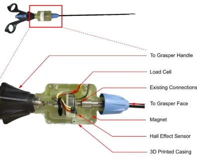
Instrumented Graspers
The laparoscopic graspers used in the experiment were curved dissectors (Surgical
Innovations), modified to provide sensing of both grasping force and grasper face
angle. This sensing module was positioned between the handle of the instrument
It contained a 200N load cell (LCM201-200N, Omega) and positioned between the
existing mechanism to measure the grasp forces, and a Hall Effect sensor coupled to
a moving magnet to measure the movement of the linkage, thereby measuring the
grasper face angle. The sensors were calibrated across a range of values and
achieved resolutions of 0.005N and 0.1° respectively. The instrumentation was
housed in a custom casing fabricated by 3D printing. The full instrumentation module
weighed 90g. The data was logged and recorded by a custom data acquisition
software at 100Hz (Labview).
Experimental Protocol
The Instrumented Graspers were used to analyse grasping forces in a simulated
surgical environment. A portable laparoscopic box trainer (Eosim) was used in
conjunction with a webcam (C920 HD Pro, Logitech) to replicate the visual
environment of MIS (Error! Reference source not found.). Porcine ureter samples
were divided into ~50mm sections, and spatulated from the distal end. The samples
were then affixed within the simulated environment. Participants were then asked to
grasp the sample in three positions with each hand, both dominant and
non-dominant. Each position was designated to a task, specifically grasping a total of 1, 5
or 10 times. Before the measurements were started, an overview was provided to
the novices to explain how to perform the task.
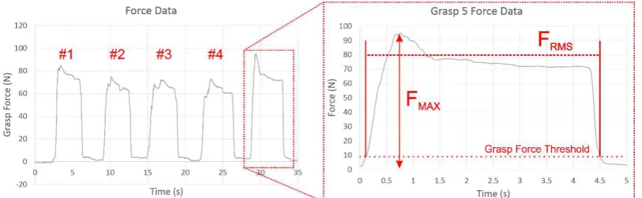
The grasp forces and grasper face angle were recorded, with a video of each task
also saved. Grasps were identified by analysing the force and position data,
selecting peaks which surpassed both force and position thresholds. For each grasp,
the peak force (Fmax), and mean force (Frms) were calculated, illustrated in Figure
3. using typical data for a grasping task. To eliminate bias of differing dominant
hands, an Edinburgh Handedness Survey (EHS) was completed by each participant.
Results
The grasping study consisted of 34 participants, of which 8 were experts, 10
intermediates, and 16 novices completed a total of 32 grasps over the six tasks.
Significant correlation was observed between Fmax and Frms (Pearson Correlation,
Error! Reference source not found. shows a summary of grasps across tasks for
the three skill levels. The associated grasp metric Fmax is shown in Table 1. A
one-way ANOVA was conducted to compare the effect of the three different Tasks on
Peak force, yielding no significant differences (F (2,1084) = 0.28, p = 0.753).
A Two-Way ANOVA was conducted to compare the effect of surgeon’s experience
on the Peak Grasp forces. This showed that there was a statistically significant
difference with those more experienced applying consistently lower mechanical
forces (F (2,1084) = 21.36, p < 0.0005). A similar test was conducted to assess the
relationship between experience and handedness however this did not show a
statistically significant interaction between dominant and non-dominant hands (F
(1,1084) = 0.06, p = 0.806). The interaction effect (Training X Hand) was significant
(F (2,1084) = 5.66, p = 0.004), therefore assessment of handedness in individual
groups was performed.
In individual training groups the effect of Dominant Hand is significant in the Novice
(significantly lower, F (1,510) = 6.70, p = 0.010) and Consultant (significantly higher,
F(1,250) = 9.601, p < 0.020). The Intermediate group showed no significant
difference between the hands (Error! Reference source not found.).
Discussion
The advent of endoscopic, laparoscopic, robotic, and image-guided percutaneous
techniques to manage patients is only the beginning of an ever-expanding array of
minimally invasive modalities available. Such modalities have reduced haptic
feedback as compared to conventional open techniques8,9 and therefore the
demands placed upon surgeons in training to be able to differentiate between the
subtleties of safe and excessive mechanical forces is significantly greater. There is
evidence to suggest that experience is the most important factor in allowing the
surgeon to develop a safer sense of mechanical forces applied to tissues10. This
evidence was echoed by the results obtained from our study where we have shown
that there are significant discrepancies in the mechanical forces applied to tissue
between novice/intermediate trainees as compared to experts. Horeman et al also
observed similar findings11. Whereas the novice/intermediate group where applying
significantly higher forces onto tissue with increased variability over grasp time, the
expert group showed a far greater level of force consistency with significantly
In vivo, there is a direct, graded response between forces applied and tissue
damage with liver and small bowel being most susceptible and ureter most robust12.
In addition, certain laparoscopic complications can be attributed directly to tissue
handling. One study analysing the errors during laparoscopic cholecystectomies
showed that graspers were the most frequently involved instrument in erroneous
task performance: 70 out of 189 errors (37%) in 20 procedures. Importantly, 14 out
of the 70 grasping errors (20%) were due to excessive forces and all of them
required corrective action13. A further study investigating erroneous task
performance during 977 laparoscopic operations performed by 20 surgeons,
graspers came third in frequency of causing complications (53%), after coagulators
and dissectors. Because of the delicate arterial supply of the ureter, it is very
important to handle the ureter with minimal force. Clearly, the threshold of safe
mechanical pressure that one can apply is dependent on the type of tissue surface
and therefore further work is required to firstly identify these thresholds and secondly
to incorporate a feedback mechanism to alert the operating surgeon.
As such systems are currently not in place, surgeons find the ability to differentiate
between safe and dangerous levels of tissue handling in laparoscopic surgery very
challenging. It is felt by some that there is currently an insufficient training model in
place for surgical trainees to develop this skill14. In years gone by, trainees were
able to learn and develop open surgery in the operating theatre however the
complexities of MIS are such that these conventional training methods cannot be
replicated. Many have looked to simulation to fill this void in surgical and this has
been an area which has grown in recent times15. Several training models have been
proposed with the virtual reality simulators being very popular in surgical training16.
These can provide basic skills training in a controlled environment free of pressure of
the operation on patients. They can also offer objective performance assessment
without the need for monitored human supervision and directly measure multiple
aspects of a subject's psychomotor performance on specific laparoscopic skills. The
major flaw with current virtual reality simulators however is that the majority lack the
feel of realistic haptic feedback17. As discussed earlier, this is crucial in allowing
trainees to develop an understanding of safe grasping forces and is therefore an
area which is deficient in current training models. There clearly needs to be
improvement within this area of training if future MIS surgeons are to perform safer
Analysis of the forces acquired with the instrumented grasping system has given us
a quantitative insight into the mechanics of surgical grasping. Specifically, it has
suggested a relationship between the training level of the surgeon and the forces
imparted on the tissue. This demonstrates a need for further training in surgeons
until a consistent low force can be applied to tissues. Whether such measures could
be used as an indicator of surgeon proficiency is unclear, however it has the
potential to be used to determine whether more training is needed for surgeons.
Future work would characterise the tissues commonly involved in MIS to find the
safe levels of force which can be applied.
References
1. Litynski GS. Laparoscopy - The Early Attempts: Spotlighting Georg Kelling and
Hans Christian Jacobaeus. JSLS : Journal of the Society of Laparoendoscopic
Surgeons. 1997;1(1):83-85.
2. Kelley WE. The Evolution of Laparoscopy and the Revolution in Surgery in the
Decade of the 1990s. JSLS : Journal of the Society of Laparoendoscopic
Surgeons. 2008;12(4):351-357.
3. Ottermo MV, Ovstedal M, Langø T, Stavdahl O, Yavuz Y, Johansen TA, Mårvik
R. The role of tactile feedback in laparoscopic surgery. Surg Laparosc Endosc
Percutan Tech.2006;16(6):390-400.
4. Zhou M, Tse S, Derevianko A, Jones DB, Schwaitzberg SD, Cao CGL. Effect of
Haptic Feedback in Laparoscopic Surgery Skill Acquisition. Surgical Endoscopy.
2012;26(4):1128-1134.
5. Tholey G, Desai JP, Castellanos AE. Force Feedback Plays a Significant Role in
Minimally Invasive Surgery: Results and Analysis. Annals of Surgery.
2005;241(1):102-109.
6. Yiannakopoulou E, Nikiteas N, Perrea D, Tsigris C. Virtual reality simulators and
training in laparoscopic surgery. International Journal of Surgery. 2015
Jan;13:60-4.
7. Våpenstad C, Hofstad EF, Langø T, Mårvik R, Chmarra MK. Perceiving haptic
8. Klatzky RL, Lederman SJ, Hamilton CL, Grindley M, Swendsen RH. Feeling
textures through a probe: Effects of probe and surface geometry and exploratory
factors. Percept Psychophys. 2003;65:613–631
9. Brydges R, Carnahan H, Dubrowski A. Surface exploration using laparoscopic
surgical instruments: The perception of surface
roughness. Ergonomics. 2005;48:874–894
10. Susmitha WK, Mathew G, Devasahayam SR, Perakath B, Velusamy SK. Factors
influencing forces during laparoscopic pinching: Towards the design of virtual
simulator. International Journal of Surgery. 2015;18:211-215
11. Horeman T, Rodrigues SP, Jansen FW, Dankelman J, and Dobbelsteen JJ.
“Force measurement platform for training and assessment of laparoscopic skills” Surgical Endoscopy. 2009;24(5): 3102-3108.
12. De S, Rosen J, Dagan A, Hannaford B, Swanson P, Sinanan M. Assessment of
Tissue Damage due to Mechanical Stresses. The International Journal of
Robotics Research. 2007;26(11-12):1159-1171
13. Joice P, Hanna GB, Cuschieri A. Errors enacted during endoscopic surgery- a
human reliability analysis. Applied Ergonomics.1998 Dec;29(6):409-14
14. Nácul MP, Cavazzola LT, De Melo MC. Current Status of Residency Training in
Laparoscopic Surgery in Brazil: A Critical Review. Arquivos Brasileiros de
Cirurgia Digestiva : ABCD = Brazilian Archives of Digestive Surgery.
2015;28(1):81-85.
15. Agha RA, Fowler AJ. The Role and Validity of Surgical Simulation. International
Surgery. 2015;100(2):350-357.
16. Buckley CCE, Nugent E, Ryan D, Neary PC. Virtual Reality – A New Era in
Surgical Training, Virtual Reality in Psychological, Medical and Pedagogical
Applications. Dr. Christiane Eichenberg (Ed.). InTech. 2012. DOI: 10.5772/46415.
Available from:
https://www.intechopen.com/books/virtual-reality-in-psychological-medical-and-pedagogical-applications/virtual-reality-a-new-era-in-surgical-training
17. Botden SM, Jakimowicz JJ. What is going on in augmented reality simulation in
laparoscopic surgery? Surg. Endosc. 2009;23:1693-1700.
Figure 1- A Photo indicating the structure of the instrumentation module of the
Graspers
Figure 2 - The test setup in use, showing the simulated environment and positioning
of the participants.
Figure 3 - Example of typical Grasping data. Indicates the Peak Force (FMAX) and the
force threshold at which grasps were detected.
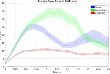
Figure 4 - The average grasp for each skill level (±SEM)
Figure 5 - Mean peak forces (FMAX) ±95%CI for all tasks against training level
[image:10.595.73.469.380.695.2]Table 1 – Mean values of FMAX for each task, (Hand (No. of Grasps))
Figure 6- A Photo indicating the structure of the instrumentation module of the
Figure 7 - The test setup in use, showing the simulated environment and positioning
of the participants.
Figure 8 - Example of typical Grasping data. Indicates the Peak Force (FMAX) and the
[image:11.595.75.528.361.501.2]Figure 9 - The average grasp for each skill level (±SEM)
Figure 10 - Mean peak forces (FMAX) ±95%CI for all tasks against training level
TASK FMAX
(MEAN±SEM)
NON-DOMINANT
1
19.69±4.53
NON-DOMINANT
5
18.16±2.09
NON-DOMINANT
10
23.24±2.29
DOMINANT 1 22.81±5.39
DOMINANT 5 24.41±2.75