This is a repository copy of Inhibiting ABCG2 could potentially enhance the efficacy of hypericin-mediated photodynamic therapy in spheroidal cell models of colorectal cancer. White Rose Research Online URL for this paper:
http://eprints.whiterose.ac.uk/134100/ Version: Accepted Version
Article:
Khot, MI, Perry, SL orcid.org/0000-0002-2561-8195, Maisey, T et al. (5 more authors) (2018) Inhibiting ABCG2 could potentially enhance the efficacy of hypericin-mediated photodynamic therapy in spheroidal cell models of colorectal cancer. Photodiagnosis and Photodynamic Therapy, 23. pp. 221-229. ISSN 1572-1000
https://doi.org/10.1016/j.pdpdt.2018.06.027
© 2018 Elsevier B.V. This manuscript version is made available under the CC-BY-NC-ND 4.0 license http://creativecommons.org/licenses/by-nc-nd/4.0/.
eprints@whiterose.ac.uk https://eprints.whiterose.ac.uk/
Reuse
This article is distributed under the terms of the Creative Commons Attribution-NonCommercial-NoDerivs (CC BY-NC-ND) licence. This licence only allows you to download this work and share it with others as long as you credit the authors, but you can’t change the article in any way or use it commercially. More
information and the full terms of the licence here: https://creativecommons.org/licenses/
Takedown
If you consider content in White Rose Research Online to be in breach of UK law, please notify us by
Inhibiting ABCG2 could potentially enhance the
efficacy of Hypericin-mediated photodynamic
therapy in spheroidal cell models of colorectal
cancer
M. Ibrahim Khot
*,1, Sarah L. Perry
1, Thomas Maisey
1,
Gemma Armstrong
1, Helen Andrew
1, Thomas A. Hughes
1,
Nikil Kapur
2and David G. Jayne
11. School of Medicine, St. James’s University Hospital, University of Leeds, Beckett St., Leeds, LS9 7TF, UK
2. School of Mechanical Engineering, University of Leeds, Leeds, LS2 9JT, UK
*Corresponding author: M. Ibrahim Khot Email: ummik@leeds.ac.uk
Address: Section of Translational Anaesthesia and Surgical Sciences, Level 9, Wellcome Trust Brenner Building,
St. James’s University Hospital, University of Leeds, Beckett St., Leeds,
ABSTRACT
Background: Photodynamic Therapy (PDT) is an attractive modality for treating solid cancers. This study evaluates the efficacy of Hypericin-PDT as a cytotoxic
therapy in colorectal cancer (CRC), using 2D cell cultures and 3D multicellular
tumour spheroids.
Methods: Spheroids were generated through forced-floating and agitation-based techniques. 2D and spheroid models of HT29 and HCT116 CRC cells were
incubated with Hypericin (0–200nM) for 16 hours. Cultures were irradiated with
light (1J/cm2) and cytotoxicity assessed using Propidium Iodide fluorescence.
Expression of ABCG2 protein was assessed by immunoassays in 2D and spheroid
cultures. The effect of ABCG2 inhibition, using 10μM Ko143, on cytotoxicity
following Hypericin-PDT was evaluated.
Results: Hypericin-PDT produced a significant reduction in HT29 (p<0.0001) and HCT116 (p<0.0001) cell viability in 2D cultures, with negligible non-phototoxicity.
Spheroids were more resistant than 2D cultures to Hypericin-PDT (HT29: p=0.003,
HCT116: p=0.006) and had a greater expression of ABCG2. Inhibition of ABCG2 in
spheroids with Ko143 resulted in an enhanced Hypericin-PDT effect compared to
Hypericin-PDT alone (HT29: p=0.04, HCT116: p=0.01).
Conclusions: Hypericin-PDT has reduced efficacy in CRC spheroids as compared to 2D cultures, which maybe attributable through upregulation in ABCG2. The
Keywords: Colorectal cancer, Hypericin, Photodynamic Therapy, ABCG2,
INTRODUCTION
Photodynamic Therapy (PDT) involves the administration of a
tumour-retaining photosensitiser (PS), followed by light administration to generate reactive
oxygen species (ROS) that cause necrosis and apoptosis, depending on the type
and concentration of PS, light dose and tissue sensitivity [1–3]. Hypericin, a
photoactive compound found in St. John’s Wort (Hypericum perforatum) [4], has
attracted interest through its diverse range of medicinal applications, including PDT
in pre-clinical cancer studies [5–12]. Hypericin possesses several advantages over
other photosensitisers, including a wide light absorption spectrum, low
photobleaching, high quantum yield and negligible non-phototoxicity [8,13–15].
Pre-clinical evaluation of anti-cancer therapeutics has traditionally used
two-dimensional (2D) monolayer cancer cell cultures, which are simple and
reliable. However, they fail to replicate the diversity and complexity of in vivo
cancers [16,17]. Solid tumours exhibit heterogeneity in access to oxygen, nutrients
and essential growth factors leading to diversity in intra-tumoural cellular
proliferation, survival and response to anti-cancer treatment [18], which cannot be
reproduced in 2D cell cultures [19,20]. Three-dimensional (3D) multicellular tumour
spheroids simulate a more realistic in vivo cancer model [21] incorporating the
cellular interactions that are crucial to signalling pathways, and the heterogeneous
distribution of oxygen and metabolites that influence proliferation and survival
[22,23]. Spheroids also provide useful information on the spatiotemporal
Chemotherapy is routinely given post-operatively to colorectal cancer
(CRC) patients at high-risk of recurrence, but with a survival advantage of only
10% [25,26]. Alternative treatment strategies are therefore required to improve
outcomes. PDT is one such strategy, supported by clinical evidence of efficacy: the
outcome for patients with lung [27] and bladder [28] cancers has been shown to be
improved by the addition of PDT as compared to surgery alone. PDT also has a
role in palliation and does not interact with other adjuvant therapy [29].
The aim of the current investigation is to compare the efficacy of
Hypericin-PDT in 2D and spheroid CRC cultures and to explain any differences observed in
MATERIALS AND METHODS
Materials
Hypericin was obtained from Molecular Probes® by Life TechnologiesTM
(Eugene, Oregon, USA) and prepared as a 100μM stock solution in ethanol. 1mL
aliquots of the stock solution was stored in the dark. Ko143 hydrate, an inhibitor
of ABCG2, was purchased from Sigma Aldrich (Gillingham, UK) and was
prepared as a 1mg/mL stock solution in DMSO and stored as 100μL aliquots.
Cell line and culturing conditions
Human colon cancer cell lines, HCT116 and HT29, were obtained from
the European Collection of Authenticated Cell Cultures (Salisbury, UK). Both cell
lines were cultured in Roswell Park Memorial Institute (RPMI) 1640 Medium plus
GlutaMAXTM (Gibco® by Life TechnologiesTM, Paisley, UK) supplemented with
10% (v/v) Fetal Bovine Serum (FBS) (Sigma-Aldrich). Cell cultures were
maintained at 37°C/5% CO2/95% relative humidity. Upon 80-90% confluency,
cell cultures were washed with Dulbecco’s Phosphate-Buffered Saline (DPBS,
Gibco® by Life TechnologiesTM) and incubated for 5 minutes with 0.05% (v/v)
trypsin and 0.5% (v/v) ethylenediaminetetraacetic acid (EDTA, Gibco® by Life
TechnologiesTM) in DPBS. Cell medium containing 10% (v/v) FBS was added to
trypsinised cells and the cell suspensions centrifuged at 400g for 5 minutes. The
supernatant was discarded and the pelleted cells resuspended in fresh medium,
seeded into 75cm2 tissue culture flasks (Corning Inc., New York, USA) and
Photodynamic therapy
For 2D cell cultures, 5x10⁴ cells were seeded per well into 96-well tissue
culture plates (Corning Inc.) and incubated at 37°C/5% CO2/95% for 24 hours.
For spheroid cultures, agarose powder (Sigma-Aldrich) was dissolved into
deionised water to make a 1% solution. 50μL of the agarose solution was added
into each well of a 96-well plate, and left at room temperature for 20 minutes to
gel. 500 cells were then added to each well and the plate centrifuged at 360g for
10 minutes and incubated at 37°C/5% CO2/95% for 48 hours. Cell cultures were
treated with (0-200nM) Hypericin in the dark for 16 hours before being washed
with DPBS. Phenol red-free RPMI 1640 medium with L-glutamine (Gibco® by Life
TechnologiesTM) supplemented with 10% (v/v) FBS was added to cultures.
Depending on the experimental conditions, cultures were either irradiated with
light or kept in the dark at room temperature.
Light treatment
Cell culture plates were placed on top of the diffuser surface of a
light-radiating device and treated with a light dose of 1J/cm2. Light treatment lasted for
72 minutes and 28 seconds at 0.23mW/cm2. The light-radiating device comprised
of a series of LED’s (one hundred and ninety-two HLMP-EL3B-WXKDD Amber
LEDs (Avago Technologies, California, USA), with peak wavelength of 594nm
and a spectral half-width of 13nm, and an internal fan to prevent overheating.
Inhibiting ABCG2
Cell cultures were incubated with 10μM Ko143 at 37°C/5% CO2/95% for 90
an additional 16 hours. Cultures were then washed and treated with light as
described above.
Assessing Cell Viability
Quantifying cytotoxicity: Twenty-four hours following irradiation, 2D and
spheroid cultures were treated with 1.3μg/mL propidium iodide (Biotium Inc.,
California, USA) for 15 minutes. Cell cultures were then washed twice with DPBS
and fresh cell culture medium added. Fluorescence was measured on a Mithras
LB 940 Microplate Reader (Ex: 540nm, Em: 620nm) (Berthold Technologies Ltd.,
Harpenden, UK).
Visualising cytotoxicity: Twenty-four hours following irradiation, spheroid
cultures were incubated with 1.3μg/mL propidium iodide (excitation: 530nm,
emission: 620nm, exposure time: 500ms) and 5μg/mL Hoechst 33342 (excitation:
350nm, emission: 450nm, exposure time: 500ms) (Life TechnologiesTM)
simultaneously for 15 and 60 minutes respectively. spheroids were then washed
and fluorescence visualised using the EVOS™ FL Imaging System (Life
TechnologiesTM).
Spinner flask spheroid culture
Cells were washed and trypsinised and the cell suspensions transferred
into CELLSPIN Stirrer spinner flasks (INTEGRA Biosciences Corp., New
Hampshire, USA), where they were maintained in cell culture medium with
constant agitation at 75 rpm on a stirring platform and incubated at 37°C/5%
CO2/95%. Cell culture media was changed every 3 days. Fifteen to twenty day
Immunofluorescence staining
Spinner flask spheroids were embedded into Cryo-M-bed (Bright
Instruments, Luton, UK) and sections (5µm) were cut onto glass slides using a
Leica CM3050 S Research Cryostat (Leica Microsystems (UK) Ltd, Milton
Keynes, UK). 2D cultures were grown to confluency on glass coverslips.
Spheroid sections and 2D cultures were fixed with 4% PFA, blocked with 0.5%
skimmed milk and incubated with anti-BCRP antibody (1:20, BXP-21) (Millipore,
Watford, UK) for 1 hour at room temperature. They were then washed with PBS
and incubated with an Alexa Fluor 488-conjugated secondary antibody (1:300)
(Life TechnologiesTM) for 30 minutes at room temperature. Slides and coverslips
were washed and mounted using ProLong™ Gold Antifade Mountant with DAPI
(Life TechnologiesTM). Slides were imaged using the Zeiss Axio Imager Z1 (Carl
Zeiss Ltd, Cambridge, UK).
Protein extraction and Western blotting
Spinner flask spheroids were washed with ice-cold DPBS and sonicated in
RIPA buffer with protease inhibitor for 15 minutes. Similarly, 2D cultures were
grown to confluency in a 75cm2 cell-culturing flask, washed with ice-cold DPBS,
and lysed in RIPA buffer with protease inhibitor for 15 minutes. Lysed cells were
centrifuged at 14,000 rpm for 10 minutes and the supernatant was aliquoted and
stored at -80°C. Protein concentration was determined using the DC™ Protein
Assay kit (Bio-Rad, Watford, UK). Lysates were resolved using LDS-PAGE and
blotted onto a PVDF Transfer Membrane (Thermo Fisher Scientific, Altrincham,
UK). The membranes were blocked with 5% skimmed milk for 30 minutes,
followed by further blocking with 1% skimmed milk for an additional 30 minutes.
(1:200, BXP-21) (Millipore, Watford, UK). The membranes were then incubated
with a horseradish peroxidase-conjugated secondary antibody for 1 hour at room
temperature prior to developing bands using the SuperSignal™ West Femto
Maximum Sensitivity Substrate (Thermo Fisher Scientific) and imaged on the
ChemiDoc™ XRS+ System (Bio-Rad). β-actin served as a loading control.
Uptake of Hypericin in spheroids
Spinner flask spheroids were treated with 200nM Hypericin for 16 hours,
embedded, sectioned onto slides and mounted as described above. Slides were
imaged on the Nikon A1R Confocal Microscope (Nikon UK Ltd, Kingston upon
Thames, UK).
Prolonged culture of spheroids following Ko143 treatment and Hypericin-PDT
Spheroids were grown in agarose-coated plates, then subjected to Ko143
incubation and Hypericin-PDT as described above. Spheroids were cultured for
an additional 10 days with cell culture media changed every day.
Transillumination images of spheroids were taken using the EVOS™ FL Imaging
System. Volumes of spheroids were calculated using ImageJ (National Institutes
of Health, Maryland, USA).
Statistical Analysis
One-way ANOVA and Student’s t-test were used to perform statistical
analysis using GraphPad Prism 7 (GraphPad Software, Inc., California, USA).
p<0.05 was considered to be statistically significant. Data are presented as the
RESULTS
Photoactivation of Hypericin
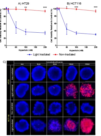
Hypericin treated cells stimulated with light exhibited a significant dose
dependant reduction in cell viability as compared to untreated controls (100nM
Hypericin: HT29 38% cell viability, p=0.0001 and HCT116 34% cell viability,
p<0.0001), whilst control cultures kept in the dark failed to show any response to
treatment (100nM Hypericin: HT29 99% cell viability, p=0.33 and HCT116 99%
cell viability, p=0.18) (Figure 1A and 1B). A similar result was observed when
HT29 and HCT116 spheroids were subjected to Hypericin-mediated PDT, as
indicated by increasing propidium iodide fluorescence, which was not apparent in
control spheroids kept in the dark (Figure 1C).
2D vs. 3D response to Hypericin-PDT
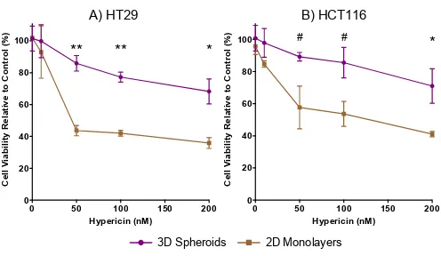
Both HT29 and HCT116 spheroids were significantly more resistant to
Hypericin mediated PDT induced cell death as compared to their respective
monolayer cell cultures. (100nM Hypericin: HT29 spheroids 35% more viable,
p<0.0001 and HCT116 spheroids are 32% more viable, p=0.01) (Figure 2A &
2B).
Expression of the ABCG2 protein
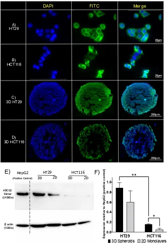
The ABCG2 transmembrane associated protein was observed in both
HT29 and HCT116 cell lines in 2D cultures, as assessed by immunofluorescence
(Figures 3A & 3B). Figures 3C and 3D illustrate the distribution of ABCG2 protein
of ABCG2 was most prominent in the outer layers of cells, whereas the core of
the spheroid had lower levels of expression. On western blot analysis, the protein
expression of ABCG2 was lower in HCT116 as compared to HT29 2D cultures
(Figure 3C). There was a significant reduction in ABCG2 protein expression in
2D as compared to spheroid cultures for both HT29 and HCT116 (Figure 3E &
3F). Similar to 2D cultures, HCT116 demonstrated lower expression of ABCG2
protein as compared to HT29 in spheroid cultures.
ABCG2 mediated resistance to Hypericin-PDT
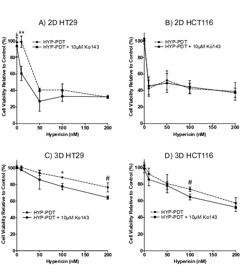
Figures 4A and 4B show 2D cultures of HT29 and HCT116 cells co-treated
with 10µM Ko143 (ABCG2 inhibitor) and Hypericin-PDT or Hypericin-PDT alone.
A significant difference in cell viability was observed in 2D HT29 cells co-treated
with Ko143 and Hypericin-PDT compared to Hypericin-PDT only treated cells
(10nM Hypericin: 38% decrease in cell viability, p=0.02). With increasing
concentrations of Hypericin, the Ko143 co-treated 2D HT29 cells showed a dose
dependent decrease in cell viability. However, at the higher concentrations of
Hypericin, there was no difference between the co-treated and Hypericin-PDT
only cultures (Figure 4A). In 2D HCT116 cultures, no significant difference in cell
viability was observed between Ko143 treated and untreated samples subjected
to Hypericin-PDT (p=0.94) (Figure 4B). In spheroid models of both HT29 and
HCT116 cell lines, a significant difference in cell viability was observed between
Ko143 and Hypericin-PDT co-treated and Hypericin-PDT alone cell cultures
(100nM Hypericin: HT29 spheroids 11% less viable, p=0.01 and HCT116
effect of Ko143 on both HT29 and HCT116 spheroid viability was still apparent at
higher doses of Hypericin-PDT.
Penetration of Hypericin in CRC spheroids
Hypericin penetrated through to the central core of both HT29 and HCT116
spheroids (Figure 5A and 5B). The penetration of Hypericin through the
spheroids corresponds with the expression of ABCG2 protein. A higher ABCG2
protein expression in the thicker peripheral layers of proliferating cells in HT29
spheroids was observed, as compared to HCT116 spheroids (Figure 3C and 3D),
and may have amounted to the lower retention of Hypericin in the peripheral cell
layers of HT29 spheroids as compared to HCT116 spheroids (Figure 5).
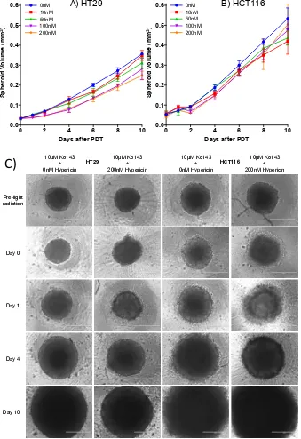
Re-growth of spheroids following ABCG2 inhibition and Hypericin-PDT
Twenty-four hours following ABCG2 inhibition and Hypericin-PDT, HT29 and
HCT116 spheroids had lost their compact spheroidal integrity as indicated by the
loose cellular debris and loss of structure (Figure 6C). By day 4, spheroids had
begun to re-form their shapes and continued to increase in volume. By day 10,
Ko143 and 200nM Hypericin co-treated spheroids were significantly larger as
compared to the sizes of spheroids on day 0. (HT29 spheroids: 0.22mm3
increased volume, p<0.005 and HCT116 spheroids: 0.44mm3 increased volume,
p<0.02) (Figure 6A & 6B).
DISCUSSION
Photodynamic therapy is an attractive treatment for solid cancers, which
can be combined with other therapies and serve as an adjunct to surgical
was hampered by unwanted adverse effects, notably photohypersensitivity
reactions and non-light toxicities [31]. In comparison, Hypericin possesses
negligible dark toxicity, yet retains a potent phototoxicity, giving it a beneficial
therapeutic index.
Many anti-cancer drugs that have shown promise in vitro have
subsequently failed to achieve their potential in clinical studies. Some of this is
attributable to the methods used in pre-clinical evaluation. Traditional 2D culture
models are simplistic representations of cancers in vivo and can give misleading
results about drug efficacy. In comparison, 3D spheroids are recognised to be
better models of cancers in vivo and have previously been shown to be useful in
PDT related studies [32]. Generally, 3D spheroids are more resistant to
anti-cancer treatments, including PDT, as compared to 2D cultures [23,33]. These
findings have been corroborated by our results as well as other studies
assessing Hypericin-PDT in 2D and 3D cell cultures [34]. Yang et al. also
reported an increased resistant to PDT in 3D cultures as compared to 2D models
of breast cancer when challenged with 5-ALA mediated PDT [35]. Unlike 2D cell
cultures, where the exposure to PS and light is uniform, the spherical structure of
spheroids influences the diffusion of the PS and penetration of radiating light.
This produces a diminishing gradient of PS and light towards the core of the 3D
structure, simulating in vivo conditions, which, in combination with decreasing
oxygen tensions, limits the PDT effect in the centre of the spheroid. 3D in vitro
cancer models therefore fill an essential gap between simple monolayer cell
cultures and resource intensive and expensive animal models for pre-clinical
drug evaluation [35].
The breast cancer resistance protein also known as ABCG2, is a
well-known member of the ATP-binding cassette (ABC) transporter superfamily [36]. It
plays a vital role in the uptake, distribution and elimination of xenobiotics and
other metabolites. ABCG2 has been documented to be over-expressed in
various cancer cell lines, and can confer resistance to various chemotherapeutics
by mediating the ATP-dependent efflux of compounds from cells [37,38]. We have demonstrated the expression of ABCG2 protein in CRC cell models with an
upregulation in spheroids, suggesting that adaptive cell signalling pathways exist
in cancer cells to promote survival [39]. Additionally, we have shown that the
protein expression of ABCG2 is higher in the outer layers of spheroids as
compared to the inner and central areas (Figure 3C and 3D). The non-uniformed
expression of ABCG2 protein in spheroids highlights the physiological advantage
they possess in providing a better in vitro platform for anti-cancer evaluations.
The relevance of our in vitro findings to the clinical scenario is evidenced in the
work of Liu et al. who found ABCG2 to be highly expressed in human colorectal
cancers as compared to low expression in non-cancer tissue [40].
Previous studies have shown a correlation between high expression levels
of ABCG2, low intracellular accumulation of PS and limited PDT effect [41–43].
Jendzelovsky et al. identified Hypericin as a preferential substrate for ABCG2
[44]. When HT29 cells were treated with Hypericin, they showed an increased
expression in ABCG2 compared to untreated cells, whilst inhibition of ABCG2
increased the intracellular Hypericin levels. The effect of ABCG2 inhibition on
intracellular Hypericin accumulation has also been confirmed by others [45].
Furthermore, ABCG2 has been implicated in playing a protective role against
mediator of the efficacy of Hypericin-PDT. Our studies have confirmed this in
CRC models, demonstrating an additive cytotoxic effect when Hypericin-PDT is
combined with Ko143 inhibition of ABCG2. Similar findings have also been
reported by others in 2D cultures of oesophageal, bladder, breast, glioblastoma
and colorectal cancers cell lines [41,48–50].
We have observed the diffusion of Hypericin through to the core of the
spheroid models. However, only ~40-50% spheroid cellular death was achieved
at the highest concentration of Hypericin, when both HT29 and HCT116
spheroids were co-treated with Hypericin-PDT and Ko143. Oxygen tensions are
known to vary in spheroids, with decreasing gradients from the outer layers to the
hypoxic core [51,52]. It is possible that the lack of oxygen in the core could have
been a limiting factor to the overall cytotoxic effects of PDT in spheroids.
To further highlight the clinical relevance of evaluating PDT in spheroids,
we observed that both HT29 and HCT116 spheroids had begun to regrow and
reform their shapes by day 4 after Hypericin-PDT. This highlights the need for
repeat application of PDT in order to control malignant proliferation and achieve
effective cancer killing. Such strategies have been developed through the use of
low dose fractionated PDT, which appears to be more efficacious than single
dose PDT application [53,54].
In summary, our studies add to the evidence base about the potential
application of Hypericin-PDT as an anti-cancer strategy in CRC. We have shown
that 3D spheroid models of CRC are more resistant to Hypericin-PDT as
inhibition of the ABCG2 transporter. Further research is required to confirm these
findings in pre-clinical small animal models, but our initial findings offer an
exciting insight into new strategies for enhancing PDT efficacy in CRC.
DECLARATIONS OF INTEREST: NONE
FUNDING
University of Leeds 110 Anniversary Research Scholarships
REFERENCES
[1] D.E.J.G.J. Dolmans, D. Fukumura, R.K. Jain, Photodynamic therapy for cancer., Nat. Rev. Cancer. 3
(2003) 380–7. doi:10.1038/nrc1071.
[2] T.J. Dougherty, C.J. Gomer, B.W. Henderson, G. Jori, D. Kessel, M. Korbelik, J. Moan, Q. Peng,
Photodynamic therapy., J. Natl. Cancer Inst. 90 (1998) 889–905.
http://www.ncbi.nlm.nih.gov/pubmed/9637138.
[3] P. Agostinis, K. Berg, K.. Cengel, T.. Foster, A.. Girotti, S.. Gollnick, S.. Hahn, M.. Hamblin, A.
Juzeniene, D. Kessel, M. Koberlik, J. Moan, P. Mroz, D. Nowis, J. Piette, B. Wilson, J. Golab,
Photodynamic Terapy of cancer: an update, CA Cancer J Clin. 61 (2011) 250–281.
doi:10.3322/caac.20114.PHOTODYNAMIC.
[4] S.L. Crockett, N.K.B. Robson, Taxonomy and Chemotaxonomy of the Genus Hypericum., Med.
Aromat. Plant Sci. Biotechnol. 5 (2011) 1–13. http://www.ncbi.nlm.nih.gov/pubmed/22662019.
[5] P. Agostinis, A. Vantieghem, W. Merlevede, P.A.M. De Witte, Hypericin in cancer treatment: More light
on the way, Int. J. Biochem. Cell Biol. 34 (2002) 221–241. doi:10.1016/S1357-2725(01)00126-1.
[6] K. Maduray, L. Davids, The Anticancer Activity of Hypericin in Photodynamic Therapy, J. Bioanal.
[7] A. Kubin, F. Wierrani, U. Burner, G. Alth, W. Grunberger, Hypericin - The Facts About a Controversial
Agent, Curr. Pharm. Des. 11 (2005) 233–253. doi:10.2174/1381612053382287.
[8] Y. Xu, D. Wang, Z. Zhuang, K. Jin, L. Zheng, Q. Yang, K. Guo, Hypericin-mediated photodynamic
therapy induces apoptosis in K562 human leukemia cells through JNK pathway modulation, Mol. Med.
Rep. 12 (2015) 6475–6482. doi:10.3892/mmr.2015.4258.
[9] V. Sacková, P. Fedorocko, B. Szilárdiová, J. Mikes, J. Kleban, Hypericin-induced photocytotoxicity is
connected with G2/M arrest in HT-29 and S-phase arrest in U937 cells., Photochem. Photobiol. 82
(2006) 1285–91. doi:10.1562/2006-02-22-RA-806.
[10] J. Mikes, J. Kleban, V. Sacková, V. Horváth, E. Jamborová, A. Vaculová, A. Kozubík, J. Hofmanová,
P. Fedorocko, Necrosis predominates in the cell death of human colon adenocarcinoma HT-29 cells
treated under variable conditions of photodynamic therapy with hypericin., Photochem. Photobiol. Sci.
6 (2007) 758–66. doi:10.1039/b700350a.
[11] B. Kleemann, B. Loos, T.J. Scriba, D. Lang, L.M. Davids, St John’s Wort (Hypericum perforatum L.)
photomedicine: Hypericin-photodynamic therapy induces metastatic melanoma cell death, PLoS One.
9 (2014). doi:10.1371/journal.pone.0103762.
[12] L. Balogová, M. Maslaňáková, L. Dzurová, P. Miškovský, K. Stroffeková, Bcl-2 proapoptotic proteins
distribution in U-87 MG glioma cells before and after hypericin photodynamic action., Gen. Physiol.
Biophys. 32 (2013) 179–87. doi:10.4149/gpb_2013021.
[13] A.M. Lima, C.D. Pizzol, F.B.F. Monteiro, T.B. Creczynski-Pasa, G.P. Andrade, A.O. Ribeiro, J.R.
Perussi, Hypericin encapsulated in solid lipid nanoparticles: Phototoxicity and photodynamic efficiency,
J. Photochem. Photobiol. B Biol. 125 (2013) 146–154. doi:10.1016/j.jphotobiol.2013.05.010.
[14] B. Ehrenberg, J.L. Anderson, C.S. Foote, Kinetics and Yield of Singlet Oxygen Photosensitized by
Hypericin in Organic and Biological Media, Photochem. Photobiol. 68 (1998) 135–140.
doi:10.1111/j.1751-1097.1998.tb02479.x.
[15] R. Ritz, C. Scheidle, S. Noell, F. Roser, M. Schenk, K. Dietz, W.S.L. Strauss, In Vitro Comparison of
Hypericin and 5-Aminolevulinic Acid-Derived Protoporphyrin IX for Photodynamic Inactivation of
Medulloblastoma Cells, PLoS One. 7 (2012) 1–11. doi:10.1371/journal.pone.0051974.
215–216. doi:10.1038/nm.3818.
[17] M. Vinci, S. Gowan, F. Boxall, L. Patterson, M. Zimmermann, W. Court, C. Lomas, M. Mendiola, D.
Hardisson, S.A. Eccles, Advances in establishment and analysis of three-dimensional tumor
spheroid-based functional assays for target validation and drug evaluation, BMC Biol. 10 (2012) 29.
doi:10.1186/1741-7007-10-29.
[18] L.-B. Weiswald, S. Richon, P. Validire, M. Briffod, R. Lai-Kuen, F.P. Cordelières, F. Bertrand, D.
Dargere, G. Massonnet, E. Marangoni, B. Gayet, M. Pocard, I. Bieche, M.-F. Poupon, D. Bellet, V.
Dangles-Marie, Newly characterised ex vivo colospheres as a three-dimensional colon cancer cell
model of tumour aggressiveness, Br. J. Cancer. 101 (2009) 473–482. doi:10.1038/sj.bjc.6605173.
[19] L.B. Weiswald, D. Bellet, V. Dangles-Marie, Spherical cancer models in tumor biology, Neoplasia. 17
(2015) 1–15. doi:10.1016/j.neo.2014.12.004.
[20] F. Pampaloni, E.G. Reynaud, E.H.K. Stelzer, The third dimension bridges the gap between cell culture
and live tissue, Nat. Rev. Mol. Cell Biol. 8 (2007) 839–845. doi:10.1038/nrm2236.
[21] Y.-C. Chen, X. Lou, Z. Zhang, P. Ingram, E. Yoon, High-Throughput Cancer Cell Sphere Formation for
Characterizing the Efficacy of Photo Dynamic Therapy in 3D Cell Cultures, Sci. Rep. 5 (2015) 12175.
doi:10.1038/srep12175.
[22] F. Hirschhaeuser, H. Menne, C. Dittfeld, J. West, W. Mueller-Klieser, L.A. Kunz-Schughart,
Multicellular tumor spheroids: An underestimated tool is catching up again, J. Biotechnol. 148 (2010)
3–15. doi:10.1016/j.jbiotec.2010.01.012.
[23] S. Breslin, L. O’Driscoll, Three-dimensional cell culture: The missing link in drug discovery, Drug
Discov. Today. 18 (2013) 240–249. doi:10.1016/j.drudis.2012.10.003.
[24] O.J. Klein, B. Bhayana, Y.J. Park, C.L. Evans, In vitro optimization of EtNBS-PDT against hypoxic
tumor environments with a tiered, high-content, 3D model optical screening platform, Mol. Pharm. 9
(2012) 3171–3182. doi:10.1021/mp300262x.
[25] B. Gustavsson, G. Carlsson, D. MacHover, N. Petrelli, A. Roth, H.J. Schmoll, K.M. Tveit, F. Gibson, A
review of the evolution of systemic chemotherapy in the management of colorectal cancer, Clin.
Colorectal Cancer. 14 (2015) 1–10. doi:10.1016/j.clcc.2014.11.002.
guidelines for metastatic colorectal cancer, Color. Dis. 14 (2012) 31–47.
doi:10.1111/j.1463-1318.2011.02765.x.
[27] K.C. Chen, Y.S. Hsieh, Y.F. Tseng, M.J. Shieh, J.S. Chen, H.S. Lai, J.M. Lee, Pleural photodynamic
therapy and surgery in lung cancer and thymoma patients with pleural spread, PLoS One. 10 (2015)
1–7. doi:10.1371/journal.pone.0133230.
[28] E. V Filonenko, A.D. Kaprin, B.Y. a. Alekseev, O.I. Apolikhin, E.K. Slovokhodov, V.I.
Ivanova-Radkevich, A.N. Urlova, 5-Aminolevulinic acid in intraoperative photodynamic therapy of bladder
cancer (results of multicenter trial), Photodiagnosis Photodyn. Ther. 16 (2016) 106–109.
doi:http://dx.doi.org/10.1016/j.pdpdt.2016.09.009.
[29] K. Moghissi, K. Dixon, S. Gibbins, A Surgical View of Photodynamic Therapy in Oncology: A Review,
Surg. J. 01 (2015) e1–e15. doi:10.1055/s-0035-1565246.
[30] D. van Straten, V. Mashayekhi, H. de Bruijn, S. Oliveira, D. Robinson, Oncologic Photodynamic
Therapy: Basic Principles, Current Clinical Status and Future Directions, Cancers (Basel). 9 (2017) 19.
doi:10.3390/cancers9020019.
[31] S.I. Moriwaki, J. Misawa, Y. Yoshinari, I. Yamada, M. Takigawa, Y. Tokura, Analysis of photosensitivity
in Japanese cancer-bearing patients receiving photodynamic therapy with porfimer sodium
(Photofrin)., Photodermatol. Photoimmunol. Photomed. 17 (2001) 241–3. doi:DOI
10.1034/j.1600-0781.2001.170507.x.
[32] S.J. Madsen, C.H. Sim, B.J. Tromberg, V. Cristini, N. De Magalhães, H. Hirschberg, Multicell tumor
spheroids in photodynamic therapy, Lasers Surg. Med. 38 (2006) 555–564. doi:10.1002/lsm.20350.
[33] G. Mehta, A.Y. Hsiao, M. Ingram, G.D. Luker, S. Takayama, Opportunities and challenges for use of
tumor spheroids as models to test drug delivery and efficacy, J. Control. Release. 164 (2012) 192–
204. doi:10.1016/j.jconrel.2012.04.045.
[34] A.A.R. Kamuhabwa, A. Huygens, P.A.M. De Witte, Photodynamic therapy of transitional cell
carcinoma multicellular tumor spheroids with hypericin., Int. J. Oncol. 23 (2003) 1445–1450.
[35] Y. Yang, X. Yang, J. Zou, C. Jia, Y. Hu, H. Du, H. Wang, Evaluation of photodynamic therapy
efficiency using an in vitro three-dimensional microfluidic breast cancer tissue model, Lab a Chip -
[36] W. Mo, J. Zhang, Review Article Human ABCG2 : structure , function , and its role in multidrug
resistance, Int. J. Biochem. Mol. Biol. 3 (2012) 1–27.
[37] M. Galetti, P.G. Petronini, C. Fumarola, D. Cretella, S. La Monica, M. Bonelli, A. Cavazzoni, F.
Saccani, C. Caffarra, R. Andreoli, A. Mutti, M. Tiseo, A. Ardizzoni, R.R. Alfieri, Effect of ABCG2/BCRP
expression on efflux and uptake of Gefitinib in NSCLC cell lines, PLoS One. 10 (2015) 1–18.
doi:10.1371/journal.pone.0141795.
[38] K. Noguchi, K. Katayama, Y. Sugimoto, Human ABC transporter ABCG2/BCRP expression in
chemoresistance: basic and clinical perspectives for molecular cancer therapeutics., Pharmgenomics.
Pers. Med. 7 (2014) 53–64. doi:10.2147/PGPM.S38295.
[39] J. Liao, F. Qian, N. Tchabo, P. Mhawech-Fauceglia, A. Beck, Z. Qian, X. Wang, W.J. Huss, S.B. Lele,
C.D. Morrison, K. Odunsi, Ovarian cancer spheroid cells with stem cell-like properties contribute to
tumor generation, metastasis and chemotherapy resistance through hypoxia-resistant metabolism,
PLoS One. 9 (2014) 1–13. doi:10.1371/journal.pone.0084941.
[40] H.G. Liu, Y.F. Pan, J. You, O.C. Wang, K. Te Huang, X.H. Zhang, Expression of ABCG2 and its
significance in colorectal cancer, Asian Pacific J. Cancer Prev. 11 (2010) 845–848.
[41] J.H. Kim, J.M. Park, Y.J. Roh, I.-W. Kim, T. Hasan, M.-G. Choi, Enhanced efficacy of photodynamic
therapy by inhibiting ABCG2 in colon cancers, BMC Cancer. 15 (2015) 504.
doi:10.1186/s12885-015-1514-4.
[42] J. Morgan, J.D. Jackson, X. Zheng, S.K. Pandey, R.K. Pandey, Substrate Affinity of Photosensitizers
Derived from Chlorophyll-a: The ABCG2 Transporter Affects the Phototoxic Response of Side
Population Stem Cell-like Cancer Cells to Photodynamic Therapy, Mol. Pharm. 7 (2010) 1789–1804.
doi:10.1021/mp100154j.
[43] T. Ishikawa, Y. Kajimoto, W. Sun, H. Nakagawa, Y. Inoue, Y. Ikegami, S.I. Miyatake, T. Kuroiwa, Role
of Nrf2 in cancer photodynamic therapy: Regulation of human ABC transporter ABCG2, J. Pharm. Sci.
102 (2013) 3058–3069. doi:10.1002/jps.23563.
[44] R. Jendzelovsky, J. Mikes, J. Koval, K. Soucek, J. Prochazkova, M. Kello, V. Sackova, J. Hofmanova,
A. Kozubik, P. Fedorocko, R. Jendzelovský, J. Mikes, J. Koval’, K. Soucek, J. Procházková, M. Kello,
affect the outcome of hypericin-mediated photodynamic therapy in HT-29 adenocarcinoma cells,
Photochem. Photobiol. Sci. 8 (2009) 1716–1723. doi:10.1039/b9pp00086k.
[45] M. Šemeláková, R. Jendželovský, P. Fedorocko, Drug membrane transporters and CYP3A4 are
affected by hypericin, hyperforin or aristoforin in colon adenocarcinoma cells, Biomed. Pharmacother.
81 (2016) 38–47. doi:10.1016/j.biopha.2016.03.045.
[46] S. Shen, D. Callaghan, C. Juzwik, H. Xiong, P. Huang, W. Zhang, ABCG2 reduces ROS-mediated
toxicity and inflammation: A potential role in Alzheimer’s disease, J. Neurochem. 114 (2010) 1590–
1604. doi:10.1111/j.1471-4159.2010.06887.x.
[47] A.G. Poleshko, I.D. Volotovski, The role of ABCG2 in maintaining the viability and proliferative activity
of bone marrow mesenchymal stem cells in hypoxia, Biophysics (Oxf). 61 (2016) 271–276.
doi:10.1134/S0006350916020159.
[48] G.A. Barron, H. Moseley, J.A. Woods, Differential sensitivity in cell lines to photodynamic therapy in
combination with ABCG2 inhibition, J. Photochem. Photobiol. B Biol. 126 (2013) 87–96.
doi:10.1016/j.jphotobiol.2013.07.003.
[49] P. Palasuberniam, X. Yang, D. Kraus, P. Jones, K.A. Myers, B. Chen, ABCG2 transporter inhibitor
restores the sensitivity of triple negative breast cancer cells to aminolevulinic acid-mediated
photodynamic therapy., Sci. Rep. 5 (2015) 13298. doi:10.1038/srep13298.
[50] S.A. Abdel Gaber, P. Müller, W. Zimmermann, D. Hüttenberger, R. Wittig, M.H. Abdel Kader, H. Stepp,
ABCG2-mediated suppression of chlorin e6 accumulation and photodynamic therapy efficiency in
glioblastoma cell lines can be reversed by KO143, J. Photochem. Photobiol. B Biol. 178 (2018) 182–
191. doi:10.1016/j.jphotobiol.2017.10.035.
[51] L.C. Kimlin, G. Casagrande, V.M. Virador, In vitro three-dimensional (3D) models in cancer research:
An update, Mol. Carcinog. 52 (2013) 167–182. doi:10.1002/mc.21844.
[52] L.M. Langan, N.J.F. Dodd, S.F. Owen, W.M. Purcell, S.K. Jackson, A.N. Jha, Correction: Direct
measurements of oxygen gradients in spheroid culture system using electron paramagnetic resonance
oximetry, PLoS One. 11 (2016) 1–13. doi:10.1371/journal.pone.0160795.
[53] H.S. De Bruijn, S. Brooks, A. Van Der Ploeg-Van Den Heuvel, T.L.M. Ten Hagen, E.R.M. De Haas,
BF-200 ALA in normal mouse skin, PLoS One. 11 (2016) 1–20. doi:10.1371/journal.pone.0148850.
[54] H.S. De Bruijn, A.G. Casas, G. Di Venosa, L. Gandara, H.J.C.M. Sterenborg, A. Batlle, D.J. Robinson,
Light fractionated ALA-PDT enhances therapeutic efficacy in vitro; The influence of PpIX concentration
and illumination parameters, Photochem. Photobiol. Sci. 12 (2013) 241–245.
FIGURES
Figure 1. Light dependant cytotoxicity of Hypericin A) HT29 and B) HCT116 2D cultures were incubated with varying concentrations of Hypericin and then irradiated with light or kept in the dark.
After 24 hours, cell viability was assessed by staining cultures with Propidium Iodide and quantifying
fluorescence. C) HT29 and HCT116 spheroids were treated with varying concentrations of Hypericin +/- light. Spheroids were then stained with Hoechst 33342 (blue fluorescence) and Propidium Iodide
(red fluorescence) and then imaged. Scalebar = 400μm. Data are shown relative to control treated
cells and represent means with SD of 3 independent experiments. *p<0.01, **p<0.001, ***p<0.0001. C) L ig h t ir ra d ia te d N o n i rr a d ia te d
0nM 10nM 50nM 100nM 200nM
Concentration of Hypericin
H T 2 9 H C T 1 1 6 L ig h t ir ra d ia te d N o n i rr a d ia te d
0 50 100 150 200
0 20 40 60 80 100 Hypericin (nM) C e ll V ia b il ity R e la ti v e to C o n tr o l (% ) * ** ***
0 50 100 150 200
0 20 40 60 80 100 Hypericin (nM) C e ll V ia b il ity R e la ti v e to C o n tr o l (% ) * *** ***
A) HT29 B) HCT116
Figure 2. Comparison between 2D and spheroid CRC cell models in response to Hypericin-PDT. A) HT29 and B) HCT116 2D and spheroid models were incubated with varying concentrations of Hypericin and then irradiated with light. After 24 hours, cultures were stained with Propidium
Iodide and fluorescence was quantified. Data are shown relative to control treated cultures and
represent means with SD of 3 independent experiments. #p<0.05, *p<0.01, **p<0.001.
0 50 100 150 200
0 20 40 60 80 100 Hypericin (nM) C e ll V ia b il ity R e la ti v e to C o n tr o l (% )
*
**
**
0 50 100 150 200
0 20 40 60 80 100 Hypericin (nM) C e ll V ia b il ity R e la ti v e to C o n tr o l (% )
*
# #A) HT29
B) HCT116
[image:26.595.42.538.75.363.2]Figure 3.Expression of ABCG2 protein in 2D and spheroid CRC cell models. A) 2D HT29 B)
2D HCT116 C) HT29 spheroid sections and D) HCT116 spheroid sections were fixed and incubated with a primary anti-BCRP (ABCG2) and secondary Alexa Fluor 488 antibody (Green) and mounted
with DAPI (Blue). E) Western blot analysis of ABCG2 in protein extracts from 3D spheroid and 2D models of HT29 and HCT116 (HepG2 cell lysates served as positive control). F) Quantitative analysis of western blots. Data are shown relative to the protein expression of ABCG2 in HepG2
lysates and represent means with SD of 3 independent experiments. *p<0.01, **p<0.001.
F)
200μm 200μm 20μm 20μm
DAPI FITC Merge
A) HT29 B) HCT116 C) 3D HT29 D) 3D HCT116 HT29 HCT116 0.0 0.2 0.4 0.6 0.8 1.0 Ex p re s s io n r e la ti v e to H e p G 2 (p o s iti v e c o n tr o l)
3D Spheroids 2D Monolayers
* ** ABCG2 Dimer (140kDa) β actin (42kDa) HT29 HepG2
(Positive Control) 3D 2D
HCT116
3D 2D
Figure 4.Co-treating 2D and spheroid CRC cell models with Ko143 and Hypericin-PDT (HYP-PDT) A) 2D HT29 B) 2D HCT116 C) HT29 spheroid and D) HCT116 spheroid cultures were co-treated with varying concentrations of Hypericin and 10μM Ko143 or Hypericin alone and then
irradiated with light. After 24 hours, cultures were stained with Propidium Iodide and fluorescence
was quantified. Data are shown relative to control treated cultures and represent means with SD of
3 independent experiments. #p<0.05, *p<0.01, **p<0.005.
0 50 100 150 200
0 20 40 60 80 100 Hypericin (nM) C e ll V ia b il ity R e la ti v e to C o n tr o l (% )
**
HYP-PDTHYP-PDT + 10µM Ko143
0 50 100 150 200
0 20 40 60 80 100 Hypericin (nM) C e ll V ia b il ity R e la ti v e to C o n tr o l (% ) HYP-PDT
HYP-PDT + 10µM Ko143
#
*
0 50 100 150 200
0 20 40 60 80 100 Hypericin (nM) C e ll V ia b il ity R e la ti v e to C o n tr o l (% )
HYP-PDT + 10µM Ko143 HYP-PDT
0 50 100 150 200
0 20 40 60 80 100 Hypericin (nM) C e ll V ia b il ity R e la ti v e to C o n tr o l (% ) HYP-PDT
HYP-PDT + 10µM Ko143
#
A) 2D HT29
B) 2D HCT116
Figure 5. Penetration of Hypericin through CRC spheroid cultures. A) HT29 and B) HCT116 spheroids were incubated with Hypericin (HYP, Red) for 16 hours. Spheroids were then sectioned,
mounted with DAPI (Blue) and fluorescently imaged. Scalebar = 100μm.
DAPI
DAPI HYP
HYP
Merge
A) HT29
Figure 6. Long-term culture of spheroids following Ko143 and Hypericin-PDT co-treatment A) HT29 and B) HCT116 spheroids were incubated with varying concentrations of Hypericin and 10μM Ko143 and then irradiated with light. Volumes of spheroids were measured for 10 days
following light radiation. C) Images of 10μM Ko143 treated only and 10μM Ko143 and 200nM Hypericin co-treated HT29 and HCT116 spheroids pre-light radiation and at days 0, 1, 4 and 10
following light irradiation. Scalebar = 400µm. Data shown represents means with SD of 3
independent experiments.
C)
10μM Ko143+ 0nM Hypericin Pre-light radiation HT29 HCT116 Day 0 Day 1 Day 4 Day 10 10μM Ko143 + 200nM Hypericin 10μM Ko143 + 0nM Hypericin 10μM Ko143 + 200nM Hypericin0 2 4 6 8 10
0.0 0.1 0.2 0.3 0.4 0.5 0.6
Days after PDT
Sp h e ro id V o lu m e (m m 3) 0nM 10nM 50nM 100nM 200nM
0 2 4 6 8 10
0.0 0.1 0.2 0.3 0.4 0.5 0.6
Days after PDT
Sp h e ro id V o lu m e (m m 3) 0nM 10nM 50nM 100nM 200nM