AFTER SINGLE TOOTH REPLACEMENT WITH PLATFORM
SWITCHED IMPLANT: A CLINICO-RADIOGRAPHIC STUDY
A Dissertation submitted in partial fulfillment of the
requirements for the degree of
MASTER OF DENTAL SURGERY
BRANCH
–
II
PERIODONTOLOGY
THE TAMILNADU Dr. M.G.R. MEDICAL UNIVERSITY
CHENNAI
–
600032
In all your ways acknowledge Him, And He will make your paths straight.
- Proverbs 3:6
I offer my sincere accolades and thanks to God Almighty, for giving me the strength,
endurance, Knowledge, ability and opportunity to undertake this research study.
This thesis represents not only my work on implants but is also a milestone in my career.
This thesis is the result of many experiences I have encountered from remarkable individuals
who I wish to acknowledge.
It is with immense gratitude, that I acknowledge the support and help from the Chairman
Prof. K.R. Arumugam, M.Pharm., and Vice Chairman Prof. Dr. A.Babu Thandapani, M.Pharm., PhD for providing me the facilities to carry out my work.
It is my pleasant duty to acknowledge my profound thanks to the Principal Prof. Dr. K.S.
Prem Kumar, M.D.S., for all kinds of precious guidance, encouragement and generous help.
I cannot find words to express my gratitude to my respected and beloved teacher and guide
Dr. C.S. Prabhahar, M.D.S., Professor and Head of the Department of Periodontology who during my tenure contributed a rewarding experience by giving me intellectual freedom in my
work, engaging me in new ideas and demanding a high quality of work in all my endeavours.
My deep sense of thanks to Dr. M. Navarasu, M.D.S., and Dr. M.Umayal, M.D.S., for
their passionate participation, stimulating discussions and input that helped me in completion of
this thesis.
I extend my sincere thanks to Mithra Scans for providing me with the CBCT on time. I owe
my special thanks to Mr. Rafi, Dental Creations for his exemplary work. I am grateful to
Dr.A.Deepta Ramarao for helping me with the statistical analysis of the study. I gained a lot from her keen scientific insight.
I am indebted to my co-pg Dr. T. Suganya Harshini and fortunate to have colleagues like
Dr. B. Karthiga, Dr. R.P. Gomathi, Dr. K.B.R. Ramya Kumari, M.D.S., and Dr. R. Nivetha, M.D.S., who provided moral support, valuable suggestions and amiable co-operation that I
needed to move on.
I thank the Administrative Officer Mr. G. Babu, Librarian Mr. P. Shankar, B.A.,
M.L.I.Sc., and the attender Mrs. M. Ayammal who helped me during my thesis work.
I earnestly thank the participants in this study for their immense patience and trust.
I share the credit of my work with Dr. Anusuya, Dr. Jamuna, Ms. Jenifer and Mr.
Arumugam. Their untiring efforts and unfailing support has driven me tirelessly all the time.
from my wife Sangeetha and daughters Kiruba and Samantha has propelled me every day
during my course of work. They have always stood by me, cherished with me every moment and
This agreement herein after the “Agreement” is entered into on this day Dec 2018, between the Best Dental Science College represented by its Principal having address at Best Dental Science College, Madurai– 625104, (hereafter referred to as, ‘the College’)
And
Mr.Dr.C.S.PRABHAHAR, M.D.S., aged 45 years working as Professor and HOD in Department of Periodontology at the College, having residence address at 9/3 Vallalar Street, Alagappapuram, Karaikudi–630 002, Sivagangai district.
(herein after referred to as the ‘Principal Investigator’) And
Mr. Dr.V.BENEDICT aged 39 years currently studying as Post Graduate student in Department of Periodontology, Best Dental College, Madurai- 625104 (herein after referred to as the ‘PG/Research student and co-investigator’)
Whereas the PG/Research student as part of his curriculum undertakes to research on
“EVALUATION OF PERI-IMPLANT SOFT & HARD TISSUE RESPONSE AFTER SINGLE TOOTH REPLACEMENT WITH PLATFORM SWITCHED IMPLANT: A CLINICO-RADIOGRAPHIC STUDY” for which purpose PG/Principal Investigator shall act as Principal Investigator and the college shall provide the requisite infrastructure based on availability and also provide facility to the PG/Research student as to the extent possible as a Co-investigator.
Whereas the parties, by this agreement have mutually agreed to the various issues including in particular the copyright and confidentiality issues that arise in this regard.
Now this agreement witnessed as follows
1. The parties agree that all the Research material and ownership therein shall become the vested right of the college, including in particular all the copyright in the literature including the study, research and all other related papers.
2. To the extent that the college has legal right to do so, shall grant to license or assign the copyright so vested with it for medical and/or commercial usage of interested persons/entities subject to a reasonable terms/conditions including royalty as deemed by the College.
3. The royalty so received by the college shall be shared equally by all the parties.
AB Alumina oxide Blasted
AE Acid Etched Surface
ANSYS Analysis System
BBL Buccal Bone Level
BII Bone Implant Interface
BOP Bleeding on probing
CBCT Cone Beam Computed Tomography
CT Computed Tomography
DBL Distal Bone Level
Di Bone level on the implant side
Dt Bone level on the tooth side
FEA Finite Element Analysis
FP Fixed Prosthesis
HCl Hydro Chloric acid
IAJ Implant Abutment Junction
IS Implant Survival
ISQ Implant Stability Quotient
JE Junctional Epithelium
MPo Myelo Peroxidase
MT Mucosal Thickness
MM Mobility Measuring Device
Ncm Newton centimeter
NO Nitric Oxide
PD Probing Depth
PH Papilla Height
PISF Peri Implant Sulcus Fluid
PRISMA Preferred Reporting Items for Systematic reviews and Meta Analysis
PS Platform Switched implant
RA Regular platform and Angulated abutment
RCT Randomized Controlled Trial
REC Buccal peri-implant mucosal level
RP Removable Prosthesis
RVG Radio Visuo Graphy
SA Platform Switching and Angulated abutment
SBI Sulcus Bleeding Index
SD Standard Deviation
SP Standard Platform implants
T1DM Type I Diabetes Mellitus
TABLE NO
TABLES PAGE NO
1 DESCRIPTIVE STATISTICS OF CLINICAL PARAMETERS AT BASELINE
58
2 DESCRIPTIVE STATISTICS OF CLINICAL PARAMETERS AT 3 MONTHS
58
3 DESCRIPTIVE STATISTICS OF CLINICAL PARAMETERS AT 6 MONTHS
58
4 DESCRIPTIVE STATISTICS OF CLINICAL PARAMETERS AT 9 MONTHS
59
5 DESCRIPTIVE STATISTICS OF CLINICAL PARAMETERS AT 12 MONTHS
59
6(A) COMPARISON OF GINGIVAL INDEX ACROSS THE TIMELINE
60
6(B) PAIRWISE COMPARISON ACROSS THE TIME LINE 60
7(A) COMPARISON OF PLAQUE INDEX ACROSS THE TIMELINE
61
7(B) PAIRWISE COMPARISON ACROSS THE TIME LINE 61
8(A) COMPARISON OF SULCUS BLEEDING INDEX ACROSS THE TIMELINE
62
8(B) PAIRWISE COMPARISON ACROSS THE TIME LINE 62
10(A) COMPARISON OF PROBING POCKET DEPTH ACROSS THE TIMELINE
64
10(B) PAIRWISE COMPARISON ACROSS THE TIME LINE 64
11 DESCRIPTIVE STATISTICS OF RADIOLOGICAL PARAMETERS AT BASELINE
65
12 DESCRIPTIVE STATISTICS OF RADIOLOGICAL PARAMETERS AT 3 MONTHS
65
13 DESCRIPTIVE STATISTICS OF RADIOLOGICAL PARAMETERS AT 12 MONTHS
65
14(A) COMPARISON OF BONE LOSS ACROSS THE TIMELINE 66
GRAPHS NO
GRAPHS PAGE NO
1 COMPARISON OF GINGIVAL INDEX ACROSS THE
TIMELINE
60
2 COMPARISON OF PLAQUE INDEX ACROSS THE
TIMELINE
61
3 COMPARISON OF SULCUS BLEEDING INDEX ACROSS
THE TIMELINE
62
4 COMPARISON OF THICKNESS OF KERATINIZED
MUCOSA ACROSS THE TIMELINE
63
5 COMPARISON OF PROBING POCKET DEPTH ACROSS
THE TIMELINE
64
FIGURE
NO FIGURES PAGE NO
1 ARMAMENTARIUM FOR THE SURGERY 38
2 NSK SG20 S-MAX 20:1 IMPLANT CONTRA ANGLE HANDPIECE
38
3 NSK PHYSIODISPENSER UNIT 38
4 ADIN IMPLANT KIT 39
5 DRILLS 39
6 IMPLANT DRIVES & HEX DRIVES 40
7 TORQUE RATCHET 40
8 PARALLELING PIN 40
9 CBCT UNIT 41
10 PRE-OPERATIVE CBCT 41
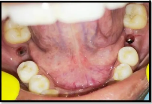
11 EDENTULOUS SITE AT 36,46 42
12 PRE OPERATIVE MODEL 42
13 MEASUREMENT OF WIDTH OF ATTACHED GINGIVA
BYWILLIAM’S PROBE AT IMPLANT SITE
42
14 THICKNESS OF KERATINIZED MUCOSA AT IMPLANT
SITE
17 PREFABRICATED CAST WITH SURGICAL STENT 44
18 SURGICAL TEMPLATE PLACED AT IMPLANT SITE 44
19 LOCAL ANAESTHESIA GIVEN(INFERIOR ALVEOLAR
NERVE BLOCK)
44
20 MID-CRESTAL INCISION AT IMPLANT SITE 45
21 ELEVATION OF FULL - THICKNESS MUCOPERIOSTEAL FLAP
45
22 OSTEOTOMY BY PILOT DRILL AT IMPLANT SITE 45
23 IMPLANT PLACEMENT 46
24 COVER SCREW PLACED AFTER INSERTION OF THE
IMPLANT
46
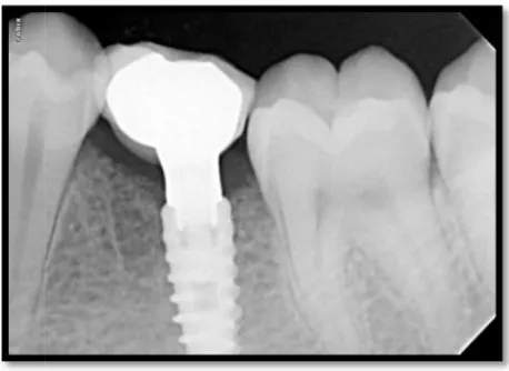
25 RVG AFTER IMPLANT PLACEMENT 46
26 SUTURING AFTER IMPLANT PLACEMENT 47
27 SECOND STAGE SURGERY (COVER SCREW EXPOSED) 47
28 GINGIVAL FORMER PLACED 47
29 CUFF FORMATION AFTER 1 WEEK 48
30 CASTABLE PLASTIC ABUTMENT PLACED 48
31 ELASTOMERIC IMPRESSION & SCREW RETAINED PLATFORM SWITCHED CERAMIC PROSTHESIS
48
32 CHECKING PROBING POCKET DEPTH IMMEDIATELY
AFTER PROSTHESIS PLACEMENT
34 POST OPERATIVE MODEL 49
35 CBCT AFTER 3 MONTHS OF IMPLANT PLACEMENT 50
SL NO CONTENTS PAGE NO
1 INTRODUCTION 1 - 5
2 AIM AND OBJECTIVES 6
3 REVIEW OF LITERATURE 7 - 25
4 MATERIALS AND METHODS 26 - 50
5 STATISTICAL ANALYSIS 51
6 RESULTS 52 - 66
7 DISCUSSION 67 - 69
8 SUMMARY AND CONCLUSION 70
9 BIBLIOGRAPHY
INTRODUCTION
Replacement of missing tooth and lost tissues is an integral part of dentistry. Over the years replacement of teeth were carried out using either a removable or a fixed dental prosthesis.
Although these replacements were satisfactory in most of the cases, patient’s compliance was
found to be less particularly in long span edentulous and completely edentulous patients.Dentists put forth considerable clinical skills in an effort to cope with the consequences of partial and/or
complete edentulism1,2. Unfortunately under the conventional partial or complete dentures, the
residual alveolar ridge resorption is unavoidable3. Dental implant emerged as an alternative option after the accidental observation of integration of titanium screws into bone was observed by Bothe et al in 1940 and later described by Gottlieb Leventhal in 1951. The use of
osseointegrated endosseous implant to support the fixed or removable prosthetic treatment have
residual alveolar bone preservation3.
The reactions described by Leventhal and Bothe et al. were later coined into the term
"osseointegration" by Per-Ingvar Branemark4. Osseointegration is defined as direct bone
anchorage to an implant body, which provides support for a prosthesis; it allows the transmission
The Branemark osseointegrated screw, has been universally accepted as a recognized treatment
for edentulous jaws1. Dental implants from then on have made a huge change in treatment modalities in dentistry.
Osseointegrated dental implants have offered additional treatment options for edentulous
and partially edentulous patients7.Implant is a biocompatible alloplastic material or device that is
surgically placed into orofacial tissues and used for anchorage, functional, therapeutic, and/or
esthetic purposes8.
Primary implant stability plays main role in the success of implants which depends on
biocompatibility of the implant material, macroscopic and microscopic nature of the implant
surface, the status of the implant bed in both health and morphologic context, the surgical
technique, the health of the person receiving the treatment, gender, occlusion and the health of the tissues in the mouth, jaw type the subsequent prosthetic design and long-term loading phase1,5,9,10,11. Length and diameter of the implant and quantity and quality of bone are also found to play an important role in the success of implant therapy10,12,13.
Replacement of a single missing tooth with an implant -supported crown is a conservative approach than preparing two adjacent teeth for a tooth supported fixed partial denture6.Since the introduction of dental implants for the replacement of missing teeth, various modifications in implant designs & surgical techniques have been developed to improve the
prognosis of the implant supported prosthesis. Bone remodeling occurs during the first year in
response to occlusal forces and establishment of normal dimensions of the peri- implant soft
tissues. Thus an implant with marked bone loss may be judged as surviving rather than
Crestal bone loss has been documented as one of the important factors that affect the long
term prognosis of an implant and its success. The use of a smaller diameter abutment on a larger
diameter implant collar, is believed to shift the Implant Abutment Junction (IAJ) horizontally
inward. This phenomenon is called platform switching16,17.
Excessive dynamic loading leads to stress/strain concentrations which induce marginal
bone loss18,19.The biomechanical theory proposed that connecting the implant to a
smaller-diameter abutment may limit bone resorption by shifting the stress-concentration zone away
from the crestal bone–implant interface and directing the forces of occlusal loading along the
axis of the implant20. The physical repositioning of the IAJ away from the external outer edge of
the implant and neighboring bone forms microgap which limit bone resorption by containing the
inflammatory cell infiltrate within the angle formed at the interface away from the adjacent
crestal bone 8,18. In this way peri-implant bone will be shielded from the inflammatory
connective tissue infiltrate which further reduces crestal bone resorption17,21.
Soft tissue around natural teeth consists of approximately 1mm of connective tissue,
1mm of epithelium22. This natural seal is also present around implants, and its development is
the major cause of bone resorption after implant exposure leading to formation of supra crestal
attached tissues (biological width) 21,23,24.The average biological width around the implant is
3mm that consist of JE (1.8mm) which is longer than natural teeth JE and 1.05mm connective
tissue component25.There should be attached keratinized mucosa on the palatal and buccal aspect
around all implants. It forms a strong seal around the implant with a cuff of circular fibres26.
In a two-stage surgical approach, the delayed formation of the biological width is
Bone resorption of 1.5- 2.0mm may occur at the implant- abutment junction after second
stage surgery16,28. A vertical bone loss ranging from 0.9 -1.6mm in the first year and mean
annual crestal bone loss ranging from 0.05-0.13mm in the maintenance period implies that it
shows successful implant osseointegration1,29,30,32.
Platform switching also provides the clinician with additional surgical and prosthetic
treatment options for use with wide-diameter implants. Moreover, the implant design
modifications involved in platform switching offer multiple advantages and potential
applications.
The primary criterion for defining a successful implant is the crestal bone level around
dental implant following restoration31.The bone levels are usually more or less stable, but small
changes such as 0.2 mm per annum are impossible to measure with conventional
radiographs14.Hence there arises a need for CBCT.
CBCT scanning has opened up new dimension to dentistry in the aspect of diagnosis,
treatment planning, surgical preparation and execution, follow-up, and management of
complications33. Traditional dental imaging techniques provide diagnosis related to implant
planning only partially. The marginal bone on the buccal and the lingual/palatal surface of
implant, the proximity of the implant to the buccal and lingual/palatal plates and possible
perforation of the plates cannot be assessed with periapical radiographs. Though CT provides the
same with accuracy, the radiation exposure to the patient is comparatively high. The drawbacks
of these modalities were improved with the introduction of Cone Beam Computed
Tomography(CBCT) for imaging orofacial structures. Upon acquiring the image, CBCT
manufacturers use advanced mathematical algorithms so that as the data are projected in screen
they are already corrected for magnification. The measurements provided are very precise for
Although there are numerous studies to support the concept of platform switching, we
require further study to thoroughly elucidate the benefits it provide in random Indian population
AIM & OBJECTIVES
AIM:
The purpose of this study is to clinically evaluate the peri-implant soft tissue health
and hard tissue response by CBCT after single tooth replacement with platform switched
implants for a period of 1 year at regular 3 months time interval.
OBJECTIVES OF THE STUDY:
To clinically evaluate the peri-implant soft tissue health ( Gingival Index, Plaque Index,
Sulcus Bleeding Index ,keratinized mucosa thickness) before and after placement of implant and
prosthesis at regular 3 months interval.
To clinically evaluate Probing Depth, around implant after placing prosthesis at regular 3
months time interval.
To radiographically evaluate the peri-implant crestal bone alteration by taking CBCT
REVIEW OF LITERATURE
Surgical cobalt-chromium-molybdenum alloy was introduced in 1938 by Strock (Boston,
MA) when he replaced a single maxillary left incisor tooth with a root form, l-piece implant that
lasted more than 15 years. In 1946, Strock designed the first titanium, 2-piece screw implant that
was initially inserted without the permucosal post36.
The success of implant depends on the connection of bone to implant surface which is
known as "Osseointegration", a term coined by Branemark in 1952. Achieving proper
osseointegration is the main goal to have proper anchorage under normal loading conditions.
The osseointegration depends on some factors such as its surface characteristics, its design,
coating of the surface, technique of placement, laser treatment of implant surface, bone source to
augment the socket to have a proper primary stability, otherwise it may negatively influence the
osseointegration by formation of fibrous tissue at the bone implant interface leading to bone
resorption36.
Classification of Implants:
Dental implants may be classified under four categories
A - Depending on the placement within the tissues
B - Depending on the materials used
C - Depending on their reaction with bone
Misch bone density classification:
Misch described four bone densities found in the anterior and posterior edentulous regions of the maxilla and mandible.
Bone density can be determined by tomographic radiographs, especially CT(computer
tomography). Each CT axial image has 260,000 pixels, and each pixel has a CT number
(Hounsfield unit) related to the density of the tissues within the pixel. In general, the higher the
CT number, the denser the tissue.
D1 bone is primarily dense cortical bone (> 1250 Hounsfield units)
D2 bone has dense to thick porous cortical bone on the crest and coarse trabecular bone
underneath (850-1250 Hounsfield units)
D3 bone has a thinner porous cortical crest and fine trabecular bone within (: 350--850
Hounsfield units)
D4 bone has almost no crestal cortical bone (150--350 Hounsfield units)
D5: <150 Hounsfield units36
Components of dental implants:
The most common root form design has a separate abutment and implant body, They are
PLATFORM SWITCHED IMPLANTS
Platform switching is a current concept in implant dentistry which was introduced by
Richard J Lazzara, Stephen S. Porter in 2006 to minimize crestal bone loss around implants17. In platform switched implants, the diameter of the abutment is smaller than the implant collar
diameter which increases the distance between the implant – abutment interface, in association
with inflammatory cell infiltrate and the marginal bone, which obviously leads to decrease in the
crestal bone resorption38. The increasing degree of mismatch between the implant body and the
abutment makes more favourable marginal bone level around implants39. The success of implant
is assessed by crestal bone level changes from baseline to the period of study. In two piece
implant system, bone remodeling takes place from uncovering of submerged implant both
horizontally and vertically at the crestal margin and biologic width was re-established. Biological
N. Enkling et al (2006) aimed to evaluate the crestal bone level changes on platform switched implants after 3 years whereas most of the studies were limited to 12 months. He
conducted a 3 year randomized clinical trial with 25 patients. Two implants with a diameter of 4
mm were inserted crestally in the posterior mandible. The intraindividual allocation of platform
switched (3.3 mm platform) and standard implant (4mm platform) was randomized. Single tooth
crown was cemented after 3 months of submerged healing. Patients were monitored at short
intervals for healing and oral hygiene. Brunner – langer model was employed for the statistical
analysis of bone levels. The mean radiographic peri – implant bone loss was 0.69 ± 43mm
(platform switching) and 0.74 ± 0.57 mm (standard platform) at the end of 3 years. The mean
intraindividual difference was 0.05 ± 0.58 mm. The crestal bone level alterations depended on
time (p <0.001) regardless of platform type (p= 0.363). This three year randomized clinical trial
using identical implants with different platforms limited vertical bone loss regardless of platform
type. Thus, the hypothesis of a bone resorption preventing effect of platform switching could not
be confirmed41.
Capiello et al (2008) aimed on clinical and radiographic examination of two piece implants that were restored according to platform – switching protocol. This prospective study
evaluated the bone loss around implants. A total of 131 implants were placed in 45 patients
requiring oral rehabilitation, following a non-submerged surgical protocol. 75 test implants, with
a healing abutment 1mm narrower than the implant platform were placed at the time of surgery.
A healing abutment of the same diameter as the implant were inserted in the remaining control
implants. All implants were positioned at the crestal level. Clinical and radiographic examination
were performed prior to surgery, at the end of surgery, 8 weeks after placing implants, at the time
of provisional prosthesis insertion, at the time of definitive prosthesis insertion and 12 months
0.6mm and 1.2mm (mean 0.95 ± 0.32mm), while for the control cases, bone loss was between
1.3mm and 2.1mm (mean 1.67 ± 0.37mm). Thus the data confirmed that the use of a narrower
abutment increased the distance between the implant – abutment microgap and the crestal bone
reducing bone resorption. Thus, platform switching seems to reduce peri-implant crestal bone
resorption and increase the long-term predictability of implant therapy23.
Luigi Cannullo et al (2008) aimed to evaluate the soft tissue response on post-extraction immediately restored implants using platform switching concept. 22 patients were included in
the study. 22 implants of 5.5mm platform diameter were placed immediately into fresh
extraction sockets in maxillae without compromised bone tissue. The Post-extraction bone
defects were filled eventually with bovine bone matrix mixed with collagen. Implants were
randomly divided into two groups. 11 implants were connected with 3.8mm diameter abutment
(test group) and remaining 11 implants were connected with 5.5mm diameter abutment (control
group). A provisional crown with non-functional immediate positioning was adapted. Definitive
prostheses were placed after two months. Periodontal parameter, buccal peri-implant mucosal
changes (REC), mesial and distal papilla height (PH) and vertical height of jumping distance
(VHG) were measured at the time of implant placement, after definitive prosthesis insertion and
every 6 months thereafter. All implants were clinically osseointegrated. The mean follow up was
25 months. The test group showed a +0.18mm REC gain. The mean values were statistically
significant (P ≤ 0.005) compared with the control group. (PH = -0.88mm; REC = -0.45mm).
Periodontal parameters showed on difference between the two groups. The mean value of bone
filling was 7.51mm in the test group (97.4% of VHG) and 8.57mm in the control group (95.2%
Roberto Lungo et al (2008) conducted a study to examine biopsy specimens to explain the biologic processes occuring around a platform switched implant. A 65 year old women
presented to the university to obtain a prosthesis supported by three implants (Prevail, 3i/Implant
innovations) that had previously been inserted in a dental office. The implants had been placed
before 2 months in a one-step surgical procedure. They featured a full osseotite surface and were
10mm long with an extended platform of 4.8mm, body of 4mm and an internal attachment of
4.1mm in diameter. The transition from the extended part measuring 4.8mm to the 4.1mm
attachment was fabricated with a 16 degree chamfer about 0.35mm long. The implants were
placed in the anterior mandible to support removable overdenture. The implant was perfectly
integrated both clinically and radiographically. With the patient’s consent, it was decided to
remove the implant because of prosthetic rehabilitation difficulties. After removal, the implant
was sectioned and subjected to histologic and histomorphometric analysis. An inflammatory
connective tissue infiltrate was localized over the entire surface of the implant platform and
approximately 0.35mm coronal to the implant-abutment junction along the healing abutment.
Platform switching appeared to be a valid method of reducing crestal bone loss. The biological
processes responsible for this occurrence seem to be linked to distancing of the inflammatory
connective tissue infiltrate from the alveolar crest. This in turn results from a more inward
displacement of the microgap on the implant platform switching enables preservation of
peri-implant hard and soft tissue over time43.
Paolo vigolo et al (2008) conducted a 5 year study to clinically assess and compare crestal bone changes around platform switched restorations on wider diameter implants. They
included 5mm diameter implant with an external hexagon in patients from the year 2000 to 2002
in a private office. 182 implants were placed in 144 patients. They were placed in posterior areas
scheduled under Group A and the 97 implants restored with platform switched prosthetic
components were under Group B. All implants survived. Marginal bone resorption was measured
using intra-oral radiographs each year after abutment and crown insertion. Statistically
significant difference was observed after 1year. The mean marginal bone resorption was 0.9mm
(SD= 0.3mm) for GROUP A implants and 0.6 mm (SD= O.2 mm) for Group B implants after 1
year. There was no significant change in marginal bone resorption at the second, third, fourth and
fifth year of follow up20.
Jason Schrotenboer et al (2008) studied the effect of microthreads and platform switching on crestal bone stress levels. Two-dimensional finite elemant imaging was used. Cross
sectional model of an implant with 5mm platform and 13mm length placed in premolar region of
the mandible was created with finite imaging. The test models consisted of microthreads at the
crestal portion and the control model consisted of smooth neck. The implant model was reverse
engineered to resemble a commercially available microthread implant. A force of 100N at 90º
vertical and 15ºoblique angles were created over abutments of different diameters (4.0mm: 20%
platform switching; 4.5mm: 10% platform switching; 5.0mm: standard) upon loading, the
microthread implant model had 29% greater stress (31.61MPa in oblique and 9.31MPa in
vertical) than the smooth neck implant (24.51 and 7.20MPa, respectively) at the crestal bone
adjacent to the implant. The microthread model and smooth neck model showed reduced stress at
the crestal level, when the abutment diameter decreased from 5.0 to 4.0mm. They concluded that
microthreads increased crestal stress upon loading. Less stress were translated to the microthread
and smooth neck group implants by using platform switched technique44.
patients. They were divided into four groups according to platform diameter 3.8mm (control),
4.3 (test group 1), 4.8mm (test group 2) and 5.5mm (test group 3). After 3 months, implants were
connected to 3.8mm diameter abutment and final restorations was performed. Radiographic bone
height was measured by two independent examiners at the time of implant placement (baseline)
and after 9, 15, 21 and 33 months. After 21 months, all implants were clinically osseointegrated.
69 implants were taken into analysis, as 11 implants had to be excluded due to early
unintentional coverscrew exposure. Radiographic examination showed a mean bone loss of
0.99m (SD = 0.42mm) for test Group 1, 0.82mm (SD = 0.36mm) for test Group 2, 0.56 (SD =
0.31mm) for test Group 3. These values were statistically lower when compared with control
group (1.49mm, SD = 0.54mm). Remaining 60 implants were evaluated after 33 months because
five patients were lost during follow-up. This data showed no difference compared with 21
months data except for test Group 2 (0.87mm) and test Group 3 (0.64mm). There was an inverse
correlation between the extent of mismatching and amount of bone loss. This study suggested
that marginal bone level alterations could be related to extent of implant/abutment mismatching.
Marginal bone levels were better maintained with implants restored with platform-switching
concept26.
Roberto crespi et al (2009) conducted a study to assess marginal bone around platform switched implants and conventional implants after 24 months. A total of 45 patients were
selected and divided into two groups. The first group comprised of 34 implants placed with
external hexagon junction with the abutment and the second group of 34 implants placed with
platform switched abutments. All implants were positioned immediately after tooth extraction
and were loaded immediately. After 24 months, a cumulative survival rate of 100% was reported
for all implants. The platform switched showed a mean bone loss of 0.78 ± 0.49mm and the
significant difference between the groups. They concluded that no differences were found in
bone level after 24 months between platform switched and conventional implants used in an
immediate loading protocol45.
Roberto cocchetto et al (2009) aimed at a study to evaluate the hard tissue response around wider platform-switched implants. The study aimed to examine whether shifting the
microgap further inward by increasing the discrepancy between the abutment diameter and
implant platform would result in a reduced crestal bone loss. 10 patients requiring mandibular or
maxillary implant restorations and having alveolar crest thickness of atleast 8.0mm at the implant
placement site were included in the study. Fifteen certain PREVAIL implants with a body
diameter of 5.0mm, an expanded platform feature with maximum diameter of 5.8mm at the
collar and a prosthetic seating surface of 5.0mm were used. The lengths used were 8.5, 10.0, 11.5
or 13.0mm. The healing abutment of 4.1mm were connected in a single stage protocol. Periapical
radiographs taken before and immediately after surgery, 8 weeks after implant placement,
immediately after definitive prosthesis insertion and at 12 and 18 months after loading, revealed
an average peri-implant bone loss of 0.3mm. The results of the study showed that patients
receiving wide platform-switched implants may experience less crestal bone loss than the use of
regular platform switching or traditional non-platform switched implants31.
Jui-Ting Hsu et al (2009) did an experimental and three dimensional finite analyses to estimate the bone strain and micromovement at the bone-implant interface (BII) for platform
switching and different diameters of single, immediately loaded mandibular implant. Four
models were created and implants assembled were 5mm in diameter with abutments of 5 or 4mm
Analysis (FEA), bone strains were found higher in immediately loaded implants than in
delayed-loaded implants. Platform switching slightly reduces strain (<10%) in crestal bone. However,
bone strain decreases on increasing the implant diameter in immediately and delayed loaded
implants. Micromotion at BII does not differ between implants with and without platform
switching46.
Hee-Jun kwon (2009) aimed to study the influence of tooth and implant-side marginal bone level on the interproximal papilla dimension in a single implant with a microthread, conical
seal and platform-switched design implant. 17 patients were treated with single implants.
Periapical radiographs were taken. The bone levels on the implant (Di) and tooth sides (Dt) were
recorded. The dimension of the papilla (Ph) was measured as the shortest distance from the top
of the papilla to the crestal bone. The marginal bone levels of the implants were also measured.
The pearson correlation coefficient was used to correlate the variables. Regression analysis was
used to determine whether Di or Dt had a significant (P<0.05) influence on ph. The result
showed that there is positive correlation between Ph and Di (r = 0.413; p = 0.023) and between
Ph and Dt (r = 0.830; p<0.0001). However, only Dt had a significant influence on Ph. They
finally concluded that Dt is the dominant factor that influences the interproximal soft tissue
dimension between a natural tooth and a single implant with a micro-thread, conical seal and
platform-switched design47.
Atieh MA and Ibrahim HM et al (2010) aimed at systemic review and meta-analysis of marginal bone preservation around platform switched implants. A literature search of electronic
databases was performed up to March 15, 2010. 10 studies with 1,239 implants were included.
The review and meta analysis were done according to the guidelines of PRISMA. The marginal
presented that an implant-abutment diameter difference > or = 0.4 was associated with a more
favourable bone response. They concluded that platform switching may preserve inter implant
bone height and soft tissue levels. The degree of marginal bone resorption is inversely related to
the extent of the implant-abutment mismatch. However, further studies are needed to confirm
this concept48.
Tomas Linkevicius et al (2010) studied about the influence of thin mucosal tissues on crestal bone stability around platform switched implants. The study included 4 patients with
twelve two-piece implants. Among 12 implants, 6 were traditional matching implants and 6 were
platform switched implants. The mean age of the patients was 43 years (range 37 to 56 years).
Mucosal tissue thickness was measured to be 2mm or less at implant sites. Implants were
restored with 5 splinted crowns and single 3-unit fixed partial denture after 2 months of healing.
Intra-oral radiographs were taken at implant placement and after 1 year follow-up. The statistical
significance level was set to p < 0.05. Bone loss around test implants was 1.81 ± 0.39mm on the
mesial site and 1.70 ± 0.35mm on the distal aspect. Bone loss around control implants was 1.60
± 0.46mm on the mesial site and 1.76 ± 0.45mm on the distal site. No statistically significant
difference was found. Finally they concluded that platform switched implants did not preserve
crestal bone better when compared with traditional implants, if thin mucosal tissues were present
at the time of implant placement49.
Luigi Canullo et al (2010) aimed to compare the composition of peri-implant microbiotas associated with platform switched implants and traditional implants. 48 implants
placed in 18 subjects were included in the study. 33 implants were restored with platform
loading. Samples were individually analyzed for their content of 40 bacterial species using
checkerboard DNA-DNA hybridization. Microbiologic parameters were compared between
platform switching and control implant group and also between implants and teeth. There was no
significant difference between groups for any of the species. The platform switched implants
showed a small trend for lower levels of early colonizer members of the Actinomyces, purple
and yellow complexes, campylobacter species, Trannerella forsythia (previously T. forsythensis)
and Porphyromonas gingivalis. Teeth and implants presented similar microbial profiles. The
study finally suggested a lack of association between peri-implant microbiota and the marginal
bone loss of platform-switched or traditional implants50.
Annibali S et al (2012) did a systemic review and meta analysis of studies comparing platform switching and conventionally restored implants. They reviewed literature to compare
implant survival (IS) and marginal bone loss (MBL) around implants. They conducted the study
through randomized controlled human clinical trials (RCTs) comparing IS and MBL in PS and
conventionally restored implants with 12 months of follow-up. Review and meta-analysis were
performed according to PRISMA statement. 10 RCTs involving 435 subjects and 993 implants
contributed to this review. The cumulative estimated implant success rate revealed no significant
difference between two groups. A smaller amount of MBL [MD 0.55mm, 95% CI (0.86;
-0.24), P = 0.0006] was noted around PS implants at patient level. Subgroup analyses revealed
loss MBL when platform switching showed a larger mismatching at implant level. Thus, they
concluded that PS technique appeared to be useful in limiting bone resorption. They suggested
that further research is needed to identify the factors most associated with successful outcomes51.
created. Models were supported by external hexagon implant (13mm × 5mm) varying platforms
[R1 regular or S1 switching] and abutments [S1straight or A1 angulated 15 º]. The models were
created using Mimics 13 and solid works 2010 software programme. ANSYS workbench 10.0
was used for numerical analysis. Oblique forces (100N) were applied to the palatal surface of
the Central Incisior. The bone / Implant interface was considered perfectly integrated. Minimum
(ϭmin) and maximum (ϭmax) stress values were obtained. The highest stress values (ϭmax) were
observed in the RA [Regular Platform and Angulated abutment, 51 MPa], followed by SA
(Platform switching and Angulated abutment, 44.8 MPa), RS (regular platform and straight
abutment, 36.5 MPa) for cortical bone. The highest stress values (ϭmax) were observed in the RA
(6.55 MPa), followed by RS (5.88 MPa) SA (5.60 MPa) and SS (4.82 MPa). The result of the
study showed that regular platform generated higher stress in the cervical peri implant region on
the cortical and trabecular bone than platform switched implants, irrespective of the abutment
used (straight or angulated)52.
Georgios E Romanos et al (2014) aimed at a study to evaluate the long term success of immediately occlusal loaded implants with a progressive thread design and platform shifting in
the edentulous mandible. 78 implants were connected with their abutments in 13 patients
immediately after surgery. The implants were splinted using a fixed temporary restoration. The
patients were advised to take soft /liquid diet for first 6 to 8 weeks of healing to reduce excessive
loading in the bone-to-implant interface. Final prostheses were fabricated by taking abutment
level impressions without removing abutments. Final restorations were delivered 4 to 8 weeks
after surgery. They were cemented temporarily to evaluate the peri-implant soft tissue condition
had values in normal levels. The periotest values showed continuous reduction, representing high
stability. The crestal bone level was relatively stable and only minimal crestal bone loss was
observed in some implants53.
Brent A.Wenzel et al (2014) conducted a study using dog model to evaluate the effect of platform shift on crestal bone levels and mucosal profile following flap surgery and subcrestal
implant placement in presence/absence of gap defects. Four dental implants were placed into the
left/right edentulated posterior mandible in five adult male hound labrador mongrel dogs using
flap surgery. Implants were placed subcrestally with/without a 1 X5mm (width × depth) gap
defect and using platform shift/switch and standard abutments. Block biopsies were collected for
histological/histometric analysis following an 8 weeks healing interval. No significant
differences were observed in crestal resorption and peri-implant mucosal profile and apical
extension of epithelial attachment among different groups. They concluded that clinical
strategies including flap surgery and subcrestal implant placement, implant with platform switch
and standard abutments, surgical approach and abutment selection seem to have limited impact
on crestal remodelling, associated bone loss and mucosal profile54.
Erhan Dursun et al (2014) did a study to compare the marginal bone level alterations, stability measures, volume of Myelo Peroxidase (MPO) and Nitric Oxide (NO) of Peri-Implant
Sulcus Fluid (PISF) between Platform Switched (PS) and Standard Platform (SP) implants. 32
implants were inserted into mandibular premolar / molar regions with a single stage protocol and
final restorations made after 3 months of osseointegration. Periapical radiographs were used to
measure marginal bone loss. Implant stability was measured by mobility measuring device and
resonance frequency analysis, PISF MPO and nitric oxide level analysis were done
and 12 months follow up. There was no healing problem for any implant at the end of the study
period. Mean bone loss measures were 0.84 and 0.76mm, and mean Implant Stability Quotient
(ISQ) values were 74.04 and 76, and mean mm values were found as -4.82 and -6.26 for PS and
SP implants respectively. There were no significant differences between implant types on
clinical peri-implant indices, PISF volume and laboratory measures and the study showed that
platform switching seemed not to affect marginal bone level, clinical peri-implant parameters,
MPO and NO (Nitric Oxide) metabolism around implants38.
Tomas Linkevicius et al (2014) conducted a study to compare how laser-microtextured implants and platform switched implants maintain crestal bone stability in thin peri-implant
tissues. 30 patients with thin mucosal tissues were included in the study. 30 laser microtextured
implants of 4.6mm diameter were under Group I (Tapered, Internal laser-10K, Bio Horizons,
Brimingham, AL, USA). 30 platform switched implants were under Group II (Certain Prevail;
Biomet/3i, Palm Beach Gardens, FL, USA). Implants were placed in posterior mandible and
restored with screw-retained metal-ceramic restorations. Radiographs were taken after implant
placement, 2 months after healing, at prosthetic restoration delivery and after 1 year follow-up.
Mean crestal bone loss was calculated, Mann-Whitney U-test was applied and the significance
was set to 0.05. The crestal bone loss was 0.71 ± 0.25mm SD and 1.02 ± 0.25mm SD after 2
months of healing in groups 1 and 2 respectively. At restorations delivery the crestal bone loss
was 1.10 ± 0.30mm SD and 1.37 ± 0.27mm SD in groups 1 and 2 respectively. The crestal bone
loss was 1.41 ± 0.42mm SD and 1.43 ± 0.23mm SD in groups 1 and 2 respectively after 1 year
follow up. They finally concluded that both the implant type did not eliminate crestal bone loss.
Maarten Glibert et al (2014) conducted a study to analyze the clinical and radiographic outcome of implant with and without platform shift. 115 implants were placed in 48
consecutively treated patients. 39.1% were of diameter 5.0mm enabling platform shifting with a
4.0mm wide prosthetic component. 60.9% were of diameter 4.0mm with a 4.0mm component.
Radiographic crestal bone levels and other multivariate statistical analysis were performed to
evaluate crestal bone loss at baseline and 1 year. All implants survived and mean marginal bone
loss was 0.73mm. There was statistically significant difference in bone loss between
platform-shifted (0.63mm) and non-platform-platform-shifted (1.02mm) implants. Both implants yielded high
survival and limited crestal bone loss after 1 year of loading. Crestal bone loss was minimized
using platform shifted implants placed in sufficiently voluminous bone. This study concluded
that implant diameter with platform shifting can be considered to limit the crestal bone loss56.
Tomas Linkevicius et al (2014) explained the influence of soft tissue thickness on crestal bone changes around platform switched implants. 80 patients received 80 bone-level
implants of 4.1mm in diameter with platform switching. Tissue thickness was measured and the
cases were distributed between Group 1 (with thin soft tissue 2mm or less, n=40) and Group 2
(with thick soft tissue more than 2mm,n=40). Implants were placed with single stage approach
and restored with screw-retained restorations. Radiographic examination was performed after
implant placement, 2 months after healing, after restoration and at 1 year follow up. Crestal bone
loss was calculated. The Mann-Whitney U-test was performed and significance was set to
p≤0.05. Implants in Group 1 (thin tissue) showed 0.79mm of bone loss after 2 months, and
1.17mm after 1 year follow up. Implants in Group 2 (thick tissue) showed bone loss of 0.17mm
after 2 months and 0.21mm after 1 year follow-up. The differences were significant (P<0.001)
switched implants if mucosal tissue is thick and also suggested that crestal bone loss cannot be
prevented if mucosal tissue is thin57.
Mohammed D. Al Amri et al (2015) did a longitudinal study to compare the clinical and radiographic status of platform switched implants placed in patients with and without type 2
diabetes mellitus (T2DM). A total of 45 male non-smokers were included in the study. 23
patients with T2DM were under Group-I and 22 self reported non-diabetic controls were under
Group-II. The platform switched implants that were used had a diameter of 3.5mm and length
ranging from 10mm to 14mm (straumann Dental Implant system). All implants were placed at
the level of the alveolar crest in the posterior mandible using an insertion torque of 35Ncm.
Healing abutments were connected to the implants using hand torque. Peri-implant Bleeding On
Probing (BOP), Probing Depth (PD), Marginal Bone Loss (MBL) and Hemoglobin A1C
(HbA1C) levels were measured at 12 and 24 months of follow-up. The mean age of participants
in groups 1 and 2 were 42.4 years (40-46 years) and 41.8 years (39-44 years), respectively. The
mean duration of T2DM was 14.5 ± 0.7 months in Group-I. There was no significant difference
in the mean HbA1C levels, peri-implant BOP, PD and MBL in both groups. The study concluded
that platform-switched implants can remain clinically and radiographically stable in patients with
T2DM in a manner similar to non-diabetic individuals. However, bone loss around implants is
influenced by several factors (such as oral hygiene status, glycemic control and tobacco
smoking) and not merely platform switching is emphasized58.
Luigi Cannullo et al (2016) conducted a study to evaluate the 10 year post-loading radiological and esthetic outcomes of immediately loaded post extractive implants with or
Padua, Italy). The test group received definitive restorations using platform-switching concept
(abutment 3.8mm in diameter). The control group received standard restoration (abutment
5.5mm in diameter). Outcome measures were survival rates of implants and prosthesis,
peri-implant marginal bone loss and periodontal indices 10 years after prosthetic loading. Also,
esthetic parameters including soft tissue buccal peri-implant mucosal levels (REC) and mesial
and distal papilla height (PH) were taken at definitive restoration, 2 and 10 years thereafter.
Nineteen implants were analyzed after 10 years of follow-up. Neither implants nor prostheses
failed. The post-operative radiographs demonstrated an overall mean bone loss of 0.18 ± 0.14mm
in the test group and of 0.80 ± 0.40mm in the control group (P = 0.00108). Test group presented
0.23 ± 0.51mm of REC gain and PH was of 0.21 ± 0.3mm on average. The control group
presented a REC of -0.59 ± 0.80mm with PH of -1.12 ± 0.55mm, demonstrating a slight
continuous soft tissue shrinkage during the entire follow-up. The mean values were statistically
significant and different between test and control group for both REC gain (P = 0.01174) and PH
(P = 0.0009). This study concluded that immediate single implant restorations rehabilitated with
platform switching protocol may provide peri-implant alveolar bone-level stability and avoid
continuous soft tissue shrinkage after 10 years of prosthetic loading compared to a platform
matching restoration. However, studies involving larger sample sizes are required to confirm
these preliminary results59.
Eisner Salamance et al (2017) conducted a study to observe the changes in vertical and horizontal marginal bone levels in platform switched and platform matched dental implants. 51
patients aged between 28 and 80 years who received 60 dental implants were included in the
study of 1 year period. Dental implants were placed 6 weeks after tooth extraction. Periapical
radiographs were used to measure peri-implant alveolar bone changes before and 12 months after
and the most apical and horizontal marginal defect using periapical radiographs. The marginal
bone measurements showed a bone gain of 0.23 ± 0.58mm in the vertical gap and 0.22 ±
0.53mm in the horizontal gap of platform matching. Platform switching showed a bone gain of
0.93 ± 1mm in the vertical gap and 0.50 ± 0.56mm in the horizontal gap was found. The average
vertical gap reduction from the baseline until 12 months was 0.92 ± 1.11mm in platform
switching and 0.29 ± 0.85mm in platform matching. They concluded that platform switching
seemed to be more effective for a better peri-implant alveolar bone vertical and horizontal gap
MATERIALS AND METHODS
STUDY PATTERNThis prospective clinical trial was conducted on 14 subjects. The study was approved by
the institutional ethical committee of Best Dental Science College and Hospital, Ultra nagar,
Madurai, Tamil Nadu, India (Annexure-I) and informed consent was obtained from all the
participants (Annexure-II) according to WORLD MEDICAL ASSOCIATION DECLARATION
OF HELSINKI61.
SOURCE OF DATA
The study was conducted in the Department of Periodontology, Best Dental Science
College & Hospital during the period November 2016–November 2018.
SUBJECT AND SITE SELECTION
14 subjects (age group 18-60), attending the outpatient section, Department of
Periodontology, Best Dental Science College & Hospital were selected for the study according to
the following inclusion & exclusion criteria.
Inclusion criteria:
Subjects with atleast one missing maxillary/mandibular posterior tooth
Subjects should be periodontally and systemically healthy ( not taking medications
known to interfere with periodontal tissue health or healing)
Subjects with stable occlusion relationship
Presence of opposing teeth
Exclusion criteria:
Subjects with compromised psychologic and mental conditions
Smokers
Subjects having parafunctional habits
Subjects with multiple missing teeth
Supraerupted opposing teeth
Subjects having any systemic diseases like diabetes, hyper thyroidism / hypo thyroidism
STUDY DESIGN:
Subjects were selected according to the above mentioned inclusion and exclusion criteria.
The medical and dental history was recorded. Scaling was done 1 week prior to surgery and oral
hygiene instructions were given. Irreversible hydrocolloid (COLTENE COLTOPRINT
chromatic alginate, manufacture by COLTENE WHALEDENT Pvt.Ltd., India) impression of
the surgical site and the opposite arch were taken using standard trays. Acrylic surgical template
was fabricated and used to maintain the precision of the osteotomy. Preoperative CBCT was
done to estimate the bone quality and width of the bone at the alveolar crest of the edentulous
area. It also measures the distance of crest from the inferior alveolar canal (width & length),
Maxillary sinus and Mental foramen, so as to maintain a 2mm clearance. Accordingly the
ARMAMENTARIUM FOR SURGERY:
Mouth mirror
UNC -15 probe
William's probe
Explorer
B.P handle no.3 and no-15 B.P blade
Metal suction tip
Austin’s retractor
Straight/curved artery forceps
Toothed tissue forceps
Periosteal elevator
Surgical scissors
Needle holder
2ml syringe loaded with 2% lignocaine HCL with 1: 80,000 adrenaline
NSK physiodispenser
ADIN TOUAREGTM-S implant kit
Normal saline (NS-eurolife)
Cotton swab and gauge
Dappen dish
Bite block
Disposable gloves, face masks and head cap
Suture material (3-0 black silk suture)
ADIN IMPLANT KIT:
This kit contains all the surgical and prosthetic instruments required for placement of all designs
of ADIN dental implants, that includes:
Tip drill, Drills, Torque ratchet, Depth gauge ,Drill extender ,Implant drive,Hand hex
driver and torque hex driver
Drills:
The kit contains 5 universal drills of diameters D2.0mm, D2.8mm. D3.2mm, D3.65mm and
D4.2mm. All drills are having easily identifiable depth markings at 8mm, 10mm, ll.5mm, 13mm,
15mm, 16mm and 18mm.
Torque Ratchet:
The ADIN torque ratchet is an all titanium two piece construction, which makes it simple to
dismantle and clean. It can be used for surgery and prosthetics with indicated torque values
ranging from 0-50 Ncm. The indicated torque value for primary stabilization of implant is 35
Ncm and for tightening of screw retained prosthesis is 25Ncm.
Depth Gauge:
The depth gauge is designed to fit the 2mm pilot hole as well as the 3.3mm osteotomy sites. The
depth markings on the gauge correspond to the lengths of the ADIN Implants.
Hand hex Driver and Torque hex Driver:
The hand driver and torque drivers along with torque ratchet, handpiece torque drivers are used
ADIN TOUAREGTM–S IMPLANT:
The TouaregTM-S spiral implants are tapered implants with a spiral tap that condenses
bone during placement for enhanced immediate stability. It has two large variable threads and a
tapered core for accurate implant placement, self-drilling, improved esthetics and better load
distribution.
The Touareg TM-S implants features a special round shaped apex that pushes the bone
graft with minimal harm to anatomic structures.AB /AE- Alumina Oxide Blasted / Acid Etched
surface treatment involves blasting with sulfuric acid and hydrochloric acid to remove embedded
particles. Built in platform switching specifies, the prosthetic connection of this implant system
in a standard internal hex 3.5mmD for all implants regardless of the diameter.
Implants are available in four diameters D3.5mm, D3.75mm, D4.2mm D5.0mm and
D6.0mm and in lengths of L6mm, L8mm, L10mm, L11.5mm, L13mm L16mm and L18mm.
We used D4.2mm and L10mm,11.5mm and 13 mm for our subjects and the horizontal diameter
difference between implant seating surface and the abutment is 0.7mm. The package of ADIN
Cover Screw:
At the time of insertion of a two-stage implant body, a first-stage cover screw is placed
into the top of the implant to prevent bone, soft tissue, or debris from invading the abutment
connection area during healing36.
Healing Abutment / Gingival Former:
After a prescribed healing period sufficient to allow a supporting bone interface to
develop, a second-stage procedure may be performed to expose the two-stage implant or to
attach a transepithelial portion. This transepithelial portion is termed a “permucosal extension” because it extends the implant above the soft tissue and results in the development of a
permucosal seal around the implant. This implant component has also been called a “healing abutment” because stage II uncover surgery often uses this device for initial soft tissue healing36.
Abutment:
An abutment for screw retention uses a hygiene cover screw placed over the abutment to
prevent debris and calculus from invading the internally threaded portion of the abutment
retention during prosthesis fabrication between prosthetic appointments. An abutment for screw
retention uses a screw to retain the prosthesis or superstructure36.We used screw retained
engaging castable abutment & prosthesis for our study.
Analog:
An analog is defined as something that is analogous or similar to something else. An
NSK PHYSIODISPENSER:
It is the reliable surgical micromotor that follows exact command. Maximum output /
Maximum torque is 230 W /5-80 Ncm and Maximum Pump Output is 75mL/min. The surgic
NSK SG20 S-Max 20:1 non-optic implant contra angle handpiece is used for prolonged surgical
procedures.
Features:
Dual irrigation system
20:1 reduction
Non-optic
Ergonomic comfort grip
Ceramic bearings are 25% harder than conventional steel bearings but are only half the
weight. Thus efficiency and durability is increased. Ergonomic design with stainless steel body
gives best possible access to the operating site, excellent balance and tactile feel. The NSK
Quattro water spray (M95L/M95) effectively cools the entire operating field. All programmes
can be performed by hand free mode due to foot control availability. It can control some modes
like speed control, coolant flow and direction of rotation. Though the speed range is
200-40,000rpm min-1noise production and vibration are very less.
SURGICAL PROCEDURE:
All surgical procedures were performed by one clinician and under standard conditions.
The subjects were adviced to rinse with chlorhexidine gluconate for 30 seconds immediately
before the procedure. The surgical site was anesthetized using 2% Lignocaine HCL with
adrenaline(LIGNOX 2%A,1: 80,000). Open flap technique was done in all cases. The osteotomy
A crestal incision was made along the crest of the ridge, bisecting the existing zone of
keratinized mucosa. A periosteal elevator was used then to reflect the mucoperiosteal
(full-thickness) flap both bucally and lingually to the level of mucogingival junction, exposing the
alveolar ridge of the implant surgical sites. The bone at the implant site was thoroughly debrided
of the granulation tissue and the knife-edge ridges if any were flattened. Sequential drilling was
initiated with the pilot drill of 2mm size through the indentation on bone made by the tip drill,
according to the implant size. In between guide pin was inserted to check the prepared depth and
its orientation. Periapical radiographs with guide pin were taken in between to analyse the
relationship to neighboring vital structures.
The final osteotomy diameter of the implant determines the final drill used. For D2 bone
the width of the final drill used is one size lesser than the actual width of the implant. For D3
bone the number of steps of drilling and time of preparation are reduced because the less dense
D3 bone easily expands and often permits larger - diameter implants to be inserted.
After the final drilling the implant was inserted with the help of an insertion tool and a torque
ratchet. Minimum 35 to 40 Ncm of torque was achieved to ensure primary stability. After
complete insertion of the implant into the bone, cover screw placed and the surgical site was
thoroughly irrigated with sterile saline. Proper closure of flaps were achieved by suturing, with
3-0 black silk suture.
Subjects were prescribed antibiotics (amoxicillin 500 mg+clavulanic acid 125mg thrice
daily), analgesics (aceclofenac 100mg+paracetamol 500mg twice daily ) and antacid
(pantaprazole 40mg twice daily) for five days. Povidone iodine 2% oral rinse was also prescribed
to facilitate plaque control. Subjects were adviced to apply ice pack to the area intermittently for
After 8-10 weeks for mandibular arch, 12-14 weeks for maxillary arch 2nd stage of surgery was
done by confirming the osseointegration of the implant. Cover screw was removed and gingival
former was placed. Simple interrupted sutures were placed. After one week gingival former was
removed and castable plastic abutment was placed and rubber base impression material
(FLEXCEED vinyl polysiloxane impression material, manufactured by GC DENTAL
PRODUCTS CORP,JAPAN) was used to record the details by closed tray method. Gingival
former was placed again, after removing plastic abutment. Impression, castable abutment and
implant analog were given to lab, Screw retained platform switched ceramic prosthesis was
fabricated and delivered to the subjects by tightening the screw around 25Ncm. The hole was
filled with light-cured universal flowable composite resin (G - Aenial manufactured by GC
DENTAL CORP,JAPAN). The parameters to be checked are given in the followed table.
OUTCOME VARIABLES:
SULCUS BLEEDING INDEX (SBI)- 1971 MUHLEMANN AND SON
An early sign of gingivitis is bleeding on probing and, in 1971, Muhlemann and Son described
the Sulcus Bleeding Index (SBI).
The Criteria For Scoring Are As Follows:
0–Healthy Looking papillary and marginal gingiva no bleeding on probing
1–Healthy Looking Gingiva, Bleeding On Probing
2–Bleeding On Probing, Change In Color, No Edema
3–Bleeding On Probing, Change In Color, Slight Edema
4–Bleeding On Probing, Change In Color, Obvious Edema
Four gingival units are scored systematically for each tooth: The Labial, Lingual,
Marginal Gingival (M Units), Mesial and Distal Papillary Gingival (P Units). Scores for these
units are added and divided by four. Adding the scores of the undivided teeth and dividing them
by the number of teeth can determine the sulcus bleeding index.
GINGIVAL INDEX–APSE ET AL
Score
0 Normal mucosa
1 Minimal inflammation with colour change and minor edema
2 Moderate inflammation with redness, edema and glazing
3
Severe inflammation with redness, edema, ulceration and spontaneous
bleeding without probing
PLAQUE INDEX–MOMBELLI ET AL
Score
0 No detection of plaque
1
Plaque only recognized by running a probe across the smooth marginal
surface of the implant
2 Plaque can be seen by the naked eye
Mucosal thickness (MT):
For measuring the mucosal thickness endodontic file (Densply) number 15 was used.
The file was inserted 1 mm apically under the sulcus and the distance between the tip of file and
rubber stop was recorded as the mucosal thickness using an absolute digital caliper.
Width of keratinized tissue (KT):
The keratinized tissue width was measured by using William's probe mid-facially from
gingival margin to mucogingival junction of the implant.
Sulcus probing depth ( PD):
Depth of sulcus was measured using a plastic probe (Colorvue manufactured by Hu-Friedy
Mfg.co, USA) at the mesial (PD1), distal (PD2) and middle of buccal (PD3) and lingual (PD4)
areas.
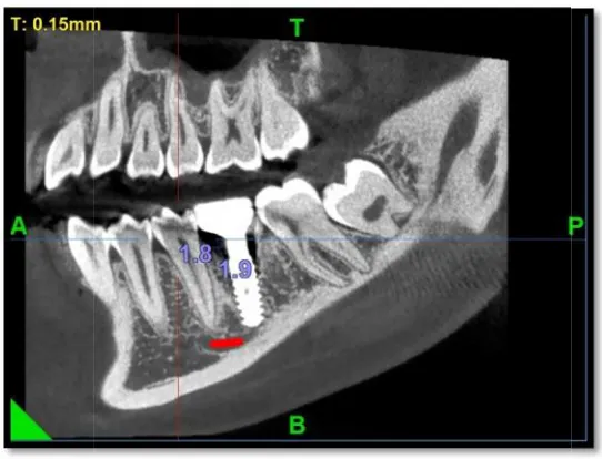
The distance between implant shoulder and alveolar crest:
The level of bone around the implant was calculated by Cone Beam Computed Tomograph
CBCT. The distance between implant shoulder and observed alveolar crest will be recorded at