EFFICACY OF HOME BASED PARTICLE REPOSITIONING MANEUVER IN TREATMENT OF POSTERIOR CANAL BENIGN PAROXYSMAL
POSITIONAL VERTIGO
A dissertation submitted to the Tamil Nadu Dr. M. G. R. Medical University, Chennai in
partial fulfilment of the requirement for the MS Otorhinolaryngology (Branch IV) degree
EFFICACY OF HOME BASED PARTICLE REPOSITIONING MANEUVER IN TREATMENT OF POSTERIOR CANAL BENIGN PAROXYSMAL
POSITIONAL VERTIGO
Dissertation submitted to the
THE TAMIL NADU DR. MGR MEDICAL UNIVERSITY, CHENNAI
In partial fulfillment of the requirements for the degree of
MASTER OF SURGERY
IN
OTORHINOLARYNGOLOGY
By
RANJU R L
Register number: 221514354
DEPARTMENT OF OTORHINOLARYNGOLOGY
CHRISTIAN MEDICAL COLLEGE
VELLORE
CERTIFICATE
This is to certify that “EFFICACY OF HOME BASED PARTICLE
REPOSITIONING MANEUVER IN TREATMENT OF POSTERIOR CANAL BENIGN PAROXYSMAL POSITIONAL VERTIGO” is the bonafide work of Dr. Ranju. R. L under my supervision in the Department of Otorhinolaryngology, Christian
Medical College Vellore in partial fulfillment of the requirements for the M.S ENT
Examination Branch IV of the Tamil Nadu Dr. M.G.R Medial University to be held in
May 2019 and no part thereof has been submitted for any other degree.
Dr. Anjali Lepcha
Professor and Head
Department of ENT Unit 4
Christian Medical College & Hospital
Vellore - 632004.
CERTIFICATE BY THE HEAD OF THE DEPARTMENT/ PRINCIPAL
This to certify that “EFFICACY OF HOME BASED PARTICLE REPOSITIONING
MANEUVER IN TREATMENT OF POSTERIOR CANAL BENIGN
PAROXYSMAL POSITIONAL VERTIGO” is the bonafide work of Dr.Ranju.R .L under the supervision of Dr.Anjali Lepcha, Professor and Head of ENT unit 4 in the
Department of ENT, Christian Medical College Vellore.
Dr. Rita Ruby Anbuselvi Dr. Anna B Pulimood
Professor and Head, Principal
Department of Otorhinolaryngology, Christian Medical College
Christian Medical College, Vellore. Vellore.
DECLARATION
I, Ranju R L, do hereby declare that the dissertation titled “EFFICACY OF HOME
BASED PARTICLE REPOSITIONING MANEUVER IN TREATMENT OF POSTERIOR CANAL BENIGN PAROXYSMAL POSITIONAL VERTIGO” is a genuine record of research done by me under the supervision and guidance of Dr Anjali
Lepcha, Professor and head, Department of ENT-Unit 4, Christian Medical College,
Vellore and has not previously formed the basis of award of any degree, diploma,
fellowship or other similar title of any university or institution.
Vellore Ranju R L
Date Post graduate student
MS Otorhinolaryngology
Acknowledgements
I would like to thank God for giving me this opportunity to study and His divine
providence in helping me to finish this dissertation.
I would like to express my deep gratitude to my esteemed guide and teacher, Professor
and head, Dr. Anjali Lepcha for designing the study, for her constant support, meticulous
guidance without which this dissertation would not have been possible.
I am extremely thankful to Dr. Ann Mary Augustine for all the suggestions in completing
the work, Mr.Lenny for his suggestions and kind words and Ms.Tunny Sebastian, for all
the help in analyzing the data and results.
I would like to thank Dr. Rita Ruby, Professor and Head of ENT for her constant support
and words of encouragement.
I would like to thank Dr. Ajay Philip and all my colleagues for helping me to recruit
their patients in my study and encouraging me to be persistent in my effort.
I would like to thank Dr. Ramanathan for helping me in the initial steps of my thesis.
I am very grateful to Dr. Roshna, Dr. Lalee, Dr.Suma and Dr. Ajoy Varghese for all the
moral support.
I would like to express my thankfulness to Mrs.Premlatha for coordinating the work and
I would like to thank all my patients for being a part of my study.
I would like to thank Rev. Subir Lal Nath for helping me with the translation of consent
forms. I thank Dr. Kiran Babu for his kind permission to take his photographs
demonstrating home based particle repositioning maneuver.
Lastly, I am thankful to God, my husband, my parents, my parents in law and my
brothers for all the moral support, technical help and fine tuning my work and
PLAGARISM CERTIFICATE
CONTENTS
1. ABSTRACT………...1
2. INTRODUCTION………..2
3. AIMS AND OBJECTIVES………6
4. LITERATURE REVIEW………...7
A. ANATOMY………7
a. Parts of ear………7
b. Vestibular system……….8
Bony labyrinth Membranous labyrinth Vestibular sensory system Sensory cells Saccule and utricle Otoconial layer Semicircular canal B. PHYSIOLOGY……….17
a. Motion decomposition and orientation in the head………17
b. Movement detection………18
c. Role of semicircular canals………..19
d. Role of otolith organs………...20
e. Role of vestibular nerve………...22
f. Central processing………22
g. Motor output of vestibular system………24
Vestibular reflexes Cervical reflexes h. Nystagmus ……….………..27
C. BENIGN PAROXYSMAL POSITIONAL VERTIGO……….28
b. Epidemiology ……….29
c. Causes ……….…29
d. Pathophysiology ……….……30
e. Treatment ………31
Treatment maneuvers Comparison of different treatment maneuvers in treatments Surgical treatment modalities Chair treatment for BPPV 5. MATERIALS AND METHODS……….………..46
6. DATA ANALYSIS AND RESULTS………57
7. DISCUSSION……….77
8. CONCLUSION……….………..82
9. BIBLIOGRAPHY………...83
10. ANNEXURE……….91
a. Figures & tables
b. Information sheet
c. Consent form
d. Proforma for patients
e. Data sheet
f. Reference number
1 ABSTRACT
BACKGROUND: Benign paroxysmal positional vertigo (BPPV) is one of the most
common causes of vertigo in patients visiting the outpatient department (OPD). Many
patients find it difficult to visit the hospital numerous times for a standard Epley’s
maneuver which has to be performed only by a specialist.
OBJECTIVE: Our aim is to compare the efficacy of a home based particle repositioning
procedure (HBPRP) with the standard Epley’s maneuver in treating patients with
posterior canal BPPV.
METHODS: This was a prospective non blinded randomized controlled study comparing
two groups, where one group received the standard treatment and other received a new
HBPRP. The vertigo scale, nystagmus duration during Hallpike test and frequency of
vertigo, were documented on first, second and third visits. Complications if any were
also noted during second and third visit. The parameters were compared in both the
groups following the treatment in all visits.
RESULTS: Thirty patients were randomized into 2 groups. There were 15 patients in
each arm. Group 1 received Epley and group 2 received HBPRP. There was no
significant difference in the baseline characteristics of patients like age, gender, co
morbid illness in both groups. Statistical analysis showed that there was no difference in
the reduction in vertigo scale, duration of nystagmus following Hallpike test, frequency
2
CONCLUSIONS: This study showed that HBPRP is a safe and effective procedure and
can be taught as a home based treatment for patients diagnosed with posterior canal
BPPV.
Key words: BPPV, Epley maneuver, Dix – Hallpike test, home based particle
3 INTRODUCTION
Benign paroxysmal positional vertigo (BPPV) is one of the most common vestibular
disorders. It accounts for about 17 % to 20% of all vertigo cases(1–3) . The prevalence
of disease is 11 -64 / 10000. The life time prevalence is 2.4%(4) . There is a 1 year
prevalence of 1.6% and 1 year incidence of 0.6% (4). BPPV presents with short
episodes of vertigo lasting for a few seconds, usually precipitated by changing head
positions with respect to gravity and associated with nausea, vomiting and nystagmus(3) .
The disease is benign and usually self limiting but can be troublesome for the patient. It
usually lasts for about 2 weeks. It can recover spontaneously in 20% by 1 month and in
50% by 3 months(5,6). The mean age of incidence is fourth and fifth decades, but it also
can occur in childhood(7). The most commonly affected canal is the posterior
semicircular canal(60-90%), second most common is the horizontal canal (5-30%) and
anterior canal is rare(3,8,9). BPPV is most commonly found in elderly and in
women(4,10), though there are studies which report the contrary. (11)
‘Romeo and Juliet’ by Shakespeare gives the earliest reference of BPPV. In medical
literature, Adler first described the positionally induced vertigo. (12) Later in 1921
Barany described BPPV for the first time. He described it as an otolith disease .(12) The
diagnosis is by a simple test which can be performed in the outpatient clinic itself and
treatment is cheap and effective. In 1952, Dix and Hallpike described the classic
positioning which induced the characteristic nystagmus and it is now used as the
4
The most common cause of BPPV is that following head injury(14,15). It is also seen
following an episode of vestibular neuronitis(16) and following prolonged bed rest(17).
Such patients usually undergo multitude of investigations instead of a simple test for
BPPV.
Earlier in 1992, it was Epley who first reported the canalolith repositioning procedure
which is used as treatment for BPPV(2,18). It is both cheap and effective. There are other
treatment maneuvers used for BPPV like Semonts maneuver and Brandt Daroff’s
exercise (19,20). Both Epley’s manueuver and Semonts maneuver have been shown to be
superior to Brant Daroff’s exercise. But both these manoevres have to be performed by
specialists, as there could be complications if performed by the patient him/herself or by
bystanders.
There are surgical treatments for patients with recurrent BPPV. The surgical treatment is
considered in patients with refractory symptoms, even after undergoing multiple particle
repositioning maneuvers (21). The various surgical treatments used are laser assisted
partitioning, posterior semicircular canal occlusion, utricular ablation and singular nerve
section (22–26).
Another recent technique for diagnosis and management of BPPV is the mechanical
assistance chairs. Epley Omniax System developed by Dr. John M. Epley and TRV chairs
5
The conventional Epley’s maneuver is difficult in patients with neck pain and cervical
spine diseases. It also needs a physician to perform the maneuver. The patient has to visit
the hospital multiple times for repeating the maneuver.
Home based particle repositioning procedure is a potential alternative to the Epley's
maneuver since it circumvents the inconveniences related to the latter. The patient can
perform the exercise by themselves without the need of a physician or health personnel.
6
AIMS AND OBJECTIVES
AIMS
To compare the efficacy of a Home based particle repositioning procedure (HBPRP)
with the standard Epley’s maneuver in treating patients with posterior canal BPPV.
OBJECTIVES
To assess the decrease in intensity of vertigo and nystagmus in posterior canal BPPV
in patients undergoing HBPRP
To assess the decrease in frequency of symptoms in posterior canal BPPV in patients
undergoing HBPRP
To note any difficulties or complications associated with the use of the HBPRP
7 LITERATURE REVIEW
ANATOMY
PARTS OF EAR
The ear is a multifaceted organ that connects the central nervous system to the external
head and neck. This structure as a whole can be thought of as 3 separate organs that work
in a collective to coordinate certain functions, such as hearing and balance. Ear has 3
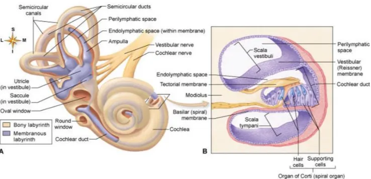
[image:22.612.77.330.329.503.2]parts- external ear middle ear and inner ear (fig 1).
Fig 1: Anatomy of ear(29)
The three parts of ear helps in the detection, conduction and transformation of auditory
signals to electrical stimuli. The signals are transmitted to the central nervous system by
the afferent auditory nerve fibers
The external ear consists of the pinna, which is situated in the lateral part of temporal
8
the tympanic membrane. That canal is skin lined and ends at the tympanic membrane, or
eardrum, the outer surface of which is skin lined. The tympanic membrane has a middle
portion, which is fibrous tissue and an inner layer which is mucous membrane.
The middle ear consists of the inner surface of the tympanic membrane, the middle ear
mucous membrane in which the ossicles of hearing, the malleus, incus and stapes is
situated. The stapes is the smallest ossicle. It is found at the junction of the middle ear
with the inner ear. The middle ear is an air containing compartment. The middle ear
together with Eustachian tube,aditus, antrum and mastoid air cells is called middle ear
cleft.
The inner ear lies deep within the petrous temporal bone, in a chamber of communicating
ducts and cavities known as the bony labyrinth. The inner ear, or membranous labyrinth,
a fluid-filled membranous structure with a shape similar to that of the bony labyrinth, is
suspended within the bony labyrinth by a supportive network of connective tissue.
The membranous labyrinth may be functionally and anatomically divided into two main
portions: the peripheral auditory apparatus, or cochlea, and the peripheral vestibular
apparatus. The peripheral vestibular apparatus incorporates five structures: the three
semicircular canals (SCCs), and two otolith organs, the utricle and saccule
VESTIBULAR SYSTEM
Our vestibular system ia a very complex system. It helps in the maintenance of our
9
Peripheral sensory apparatus – helps to detect and to transmit information to central
processing system about head position, angular and linear velocity(30)
Central processing system - all informations from sensory inputs are processed, helps to
decide body and head orientation in space (30)
Motor output system – helps in the eye movements and body movements which happens
during head & postural adjustments
The peripheral system involves membraneous labyrinth and bony labyrinth. It lies within
the inner ear .Its lateral, medial and anterior borders are formed by middle ear, temporal
bone and cochlea respectively
Bony labyrinth
The parts of bony labyrinth are the three semicircular canal, the cochlea and vestibule.
The bony labyrinths is filled with fluid called perilymph . it was found to be having
similar composition of cerebrospinal fluid and also is in continuation with CSF.(30)
Perilymph communicates with cerebrospinal fluid through a duct called cochlear
aqueduct. It was found that the function of inner ear was affected by the disorders
affecting the pressure of the spinal fluid. The fluid has high concentration of sodium than
10
Fig 2: Anatomy of bony and membranous labyrinth(32)
Membranous labyrinth
The membranous labyrinth is situated within the bony labyrinth (fig 2). It is surrounded
by perilymph. Within the membranous labyrinth, there is endolymph. In terms of
electrolyte composition, endolymph is very much similar to intravascular fluid. There is
high concentration of potassium than sodium. Three semicircular canals and otolith
organs are parts of membranous labyrinth. Each semicircular canal end in a dialated end
called ampulla. Utricle and saccule are the two otolith organs. The sensory
neuroepithelium is present in ampulla and in macula with otolith organs. It is supplied by
the vestibular part of vestibulocochlear nerve, the 8th cranial nerve.
11
Vestibular system has a resting vestibular tonus. It is due to the constant continuous
influx of impulses from the vestibular nerve to CNS. It is because of this that any
disturbance in one of the two vestibular organs will cause imbalance
Vestibular nerve
Approximately 18000 afferent nerve fibres are present in the human vestibular nerve. It is
found that in scarpa’s ganglion, with increasing age, there is a reduction in number of
hair cells and nerve cells(33,34). Bipolar neurons are present in scarpa’s ganglion. They
are located lateral part of internal auditory canal. There is superior and inferior vestibular
nerve and associated with it, there is superior and inferior group of cells. Cristae of
superior and lateral canals, anteroposterior part of macula of saccule and macula of
utricle are innervated by superior vestibular nerve. Main position of macula of saccule
and crista of posterior canal are supplied by the inferior vestibular nerve. Large and small
fibres are seen in the nerve. Type 1 hair cells seen in the summit of crista or the striola are
innervated by large fibres. The periphery of macula and slope of crista has more of small
fibres. Bimodal innervation is seen in auditory system. Type II cells, the phylogenitically
older cells make direct contact with efferents. Efferent terminals contact the terminal or
afferent nerve fibre in type 1 cells. Effect of efferent system is weak and inhibitory in
nature on the afferent system. Hence as a response to stimulation, the primary it may
allow afferent receptors to decrease or increase their activity. There are evidence to
12
involved in initiating eye or body movement cause efferent activity preceding
movements(35).
Sensory cells
There two types of sensory cells type I and type II (36).
The afferent nerve fibre form a nerve chalice at the terminal end and it surrounds the
flask shaped type I cells (fig 3). There are collateral extensions found in many calices
which end in type II cells(37). It is found that apart from collaterals synapsing with the
type II cells, they synapse directly with the outer membrane of nerve calyces which
surrounds the type I cells. It was also observed that efferent nerve endings also terminate
on type II hair cells and on the afferent nerve calyce. Microvilli are present in the apical
surface of hair cells. These microvilli forms the stereocilia. One of the stereocilia is
elongated to form kinocilia. It is the true cilia with 9+2 microtubule arrangement. The
steriocilia are arranged in increasing height towards the kinocilia. The stereocilia, which
are nonmotile and rigid, are not true cilia but instead consist of actin filaments in a
paracrystalline array with other cytoskeletal proteins. The kinicilia is the longest
stereocilia and is located eccentrically which will impart a polarization of the hair cell.
Approximately 70 stereocilia and one kinocilium are found in the upper surface of hair
cells(34). The stereocilia extend their rootlets into the cuticular plate which is the thicker
region on the upper surface of hair cells. The stereocilia in macula and crista are slightly
different in length. They a few microns of length in macula whereas in crista they are
13
Fig 3:Anatomy of type I and II hair cells(39)
The phylogenitically older cells are type II cells. They are cylindrical in shape. The
arrangement of steriocilia and kinocilia are same as type 1 cells.
Saccule and utricle
The utricle lies superior to saccule and it slopes anteriorly upwards at an angle of
approximately 30 degree. It is oblong and irregular. In the superior dialated part of
utricle, lies the macula utriculi. It lies mostly in the horizontal plane. Macula of both the
sides are in the same plane. It was found to have almost 33000 hair cells(40).
The saccule is hook shaped. It is found in the vertical plane. It lies in the medial wall of
the vestibule in a spherical recess. The mean area was found to be 2.4meter square(41).
Approximately 18000 hair cells are found in saccular macula. A curved line, named
14
called pars 'externa' and the part which is on the concave side is called pars 'interna'. It is
found that type I cells are seen in high density in the striola(44). Based on the location of
the kinocilium facing the striola each hair cell is structurally polarized. The kinocilium is
found to be polarized away from the striola in saccule. In utricle it is toward the striola.
The striola of the utricle have a thinner otoconial layer compared to that of the one found
in saccule.
There are a number of small hexagonal and cylindrical shaped bodies with pointed ends,
which are called otoconia or statoconia overlying the neuroepithelium (fig 4). They are
calcium carbonate particles. They are embedded in the otoconial membrane which is a
gelatinous substance. The hair cells project into the gelatinous membrane. They are
displaced by the otoconial mass relative to sensory epithelium.
Otoconial layer
A gelatinous layer, a subgelatinous space and otoconia are present in otoconial layer. The
length of each otoconia is 3 -19 micrometers. It is made of organic protein matrix along
with inorganic calcium carbonate in crystallized form of calcite and has specific gravity
of 2.71. The atypical cytoplasm of supporting cells produce the organic material which
form the core on which inorganic material is seeded. The enzyme carbonic anhydrase
present over the epithelium helps in the reaction which lead on to the formation of
calcium carbonate crystals. They are in turn trapped in the matrix which will result in
formation of otoconia. It was found that the otoconia undergoes chemical change with
15
were found to undergo turnover. The dark cells found in utricle help in this.
Fig 4: Anatomy of otoconial membrane with neuroepithelium(47)
Semicircular canals
• The openings of semicircular canals are widened to form ampulla. The ampulla is
supplied by Eighth cranial nerve (vestibular division). The ampulla has crista
ampullaris which is the specialized sensory epithelium(48). It has specialized hair
cells, cupula, supporting cells other than connective tissue, nerve fibres and blood
vessels. The crista forms a raised section of the wall and is found to extend across
floor of ampulla. It is saddle shaped. The crista has a apex which is the central part
and peripheral sloping part. The shape helps in packing maximum number of hair
cells (fig 5).
• It is found that the lateral semicircular canal is polarized towards the utricle,
16
The hair cells are not in direct contact with endolymph. They are embedded into cup7ula,
a gelatinous membrane. The investigations has shown that the structure of cupula is
extremely hydrous structure. It results in distortion during fixation which contains
proteoglycans arranged in a filamentous network(49–51). Studies have given evidence
that it is secreted by supporting cells(52,53). It has same specific gravity as the
endolymph surrounding it-prevents from floating in it. The transferring of endolymph
fluid movement stimuli to the hair cells is by the help of cupulla in the ampulla of
SCC(54). This will in turn produce kinetic reflexes.
The cupula is attached to the ampulla wall firmly and it is considered as a physiological
necessity. The cupula studies has shown that it has a diaphragm like displacement in the
central section and at the base (55). It was also found that the endolymph pass through
the subcupular space (56). It was found in studies using ultrafine particles of dextran
magnet which was injected into the membranous canal , that the fluid was able to pass
through the subcupular space under very low pressure. There was no evidence of obvious
increase in ampullary pressure. In the midpoint of cupula there was a displacement of as
17
Fig 5: Anatomy of crista ampullaris(58)
Supporting cells are the cells which surrounds the sensory cells. They are secretory in
nature. The sensory cells are provided insulation by them. Precursor cells which give rise
to sensory cells are also formed by them. The dark cells are found in membranous
labyrinth in sensory epithelia, except saccule(59). They are believed to produce
endolymph. Associated with dark cells, there are pigmented cells or melanocytes. They
help in development and maintenance of unique composition of endolymph.
PHYSIOLOGY
Motion decomposition and orientation in the head
The inner ear has an anatomical design in which the peripheral system reflects six degress
of freedom. The rotational motion is detected by semicircular canals whereas the macules
detect the translations. The orientation of semicircular canals are in such a way that left
and right canals act as parallel systems (fig 6). The right anterior canal is parallel to left
18
In the lateral plane both horizontal canals are parallel. It has to be noted that in upright
position, with respect to horizontal axis, the horizontal canal make an angle of 30 degree.
With the sagittal plane of the head, vertical canals make an angle of 45 degree.
Fig 6: Orientation of semicircular canals(30)
Movement detection
The movement detection and orientation of vestibular system follows the laws of physics.
Whenever the head and body movement occurs, they are related to accelerations. These
are sensed by the vestibular organ. It is due to the rigid coupling of sensory epithelium
and bony structure. The inertial forces drive the fluid filled system, which is attached to
the skull. There is a fluid lag during any motion of head. This relative displacement is
trigger for the detection of movement. In order to limit the hair cell movement during
head movements, the design of the canal system in a way that defection of hair cells and
19 Role of semicircular canals
The detection of movement in head is due to the trigger produced by movement of
endolymph in the SCC. The centripetal force is the only force acting on object which is
rotating in a constant angular speed. During constant rotatory motion , the sensory
epithelium is not triggered by any force in a way it stimulates the cupula. It detects only
changes in rotation that is they are rate sensors (fig 7a).
Through evolution, SCC is also adapted to detect all natural human movements including
transient rotations such as back and forth movements. The generation of eye movement
that matches the head velocity is with the help of sensory input by the SCC. It helps to
maintain the eye position during head movements.
The SCC which has a coplanar pairing of the canals results in push pull change in quality
of SCC output. In a shared plane when angular motion occurs, the endolymph is
displaced in opposite directions. The neural firing increases in one vestibular and
decreases on the other (fig 7b).
There are certain advantages of push pull arrangement. The first advantage is sensory
redundancy, that is if one SCC is affected by any disease, the input to the CNS will not be
stopped because of the other other pair. The second is common mode rejection. Because
of this pairing, any simultaneous changes in the neural firing ( for example the change
which can occur due to raised body temperature ) will be ignored by the brain. The third
20
a) b)
Fig 7: a)Direction of cupular deflection ; b) direction of head movement with endolymph
movement(30)
Role of Otolith organs
The otolith organs are oriented in such a way that the utricle is horizontal whereas the
saccule is in vertical plane. Most of the movements are detected by both the organs
because of the curved structure.
Thus the translational movements causing linear acceleration along with static tilts of
head are detected by both utricle and saccule. The ototlith membrane has a density higher
than the endolymph surrounding it. Any movement of the membrane causes the defection
of hair follicles, which are embedded at its base. They produces a signal which is sent to
21
According to Equivalence principle of Einstein, no single physical device can distinguish
gravity from linear acceleration. Since otolith cannot distinguish between linear
acceleration and tilt, this gives difficulty to CNS. The sum of all accelerations are sensed
by the otolith organs during natural movements. By interpreting these signals, they are
able to initiate postural changes and eye reflexes which are mediated by vestibular nuclei.
They enable in diverting the appropriate signals either to trunk, limb and neck muscles
with the help of vestibulospinal tract or to the eye muscles with the help of
vestibuloocular reflex. The central nervous system of humans distinguish between tilt and
linear accelerations since vestibular, visual and proprioceptive senses functions together.
In short, during both active or passive movements, the gravitational acceleration that is
the sum total of all accelerations acting on head are detected by the otolith organs. The
direction of gravity is detected by otolith organs when there is no movement. The
gravitational acceleration due to gravitational force is is a linear acceleration. The otolith
organs detect the gravitational acceleration as tilt (fig 8). The head tilt laterally causes
excitation of utricle due to the shear force exerted on utricle, this force is lessened on
saccule. The sensory ambiguity problem arises since two sources contribute to
gravitational acceleration- gravitational field and linear motion. The signals to the brain
22
Fig 8: Direction of movement of stereocilia with accelaration(60)
Similar to that of semicircular canal, otoliths also has redundancy. Push pull mechanism
is also involved in otoliths.
Role of Vestibular nerve
From bipolar neurons of vestibular ganglion, afferent fibres passes through vestibular
nerve fibres. There are two different types of vestibular afferent neurons. The regular
afferents have a tonic rate and also there is little variability between spike intervals. The
irregular afferents has no firing at rest and they have variable interspike intervals. For
VOR, regular afferents play an important role. Irregular afferents are important for VSR.
Central processing
The vestibular inputs from afferents goes to vestibular nuclear complex and cerebellum.
The primary processor is vestibular nuclear complex. The adaptive processor is
23
Vestibular nucleus
There are four major nuclei in the complex. Superior, medial, lateral and descending
along with seven minor nuclei present. VOR relays in superior and medial nuclei. VSR
relays on lateral nuclei. Descending nuclei has no primary outflow of its own but is
connected to all other nuclei. Between the two vestibular nuclei on either sides, there is a
system of mutual inhibition.
Blood supply
Posterior inferior cerebellar arteries are the important supplying arteries for central
vestibular arteries. Anteroinferior cerebellar arteries are the important supply for
peripheral vestibular system.
Cerebellum
From the vestibular nucleus complex, major outflow is towards the cerebellum.
Vestibulocerebllum is the term given to parts of cerebellum which has direct input from
vestibular afferents. The projections from cerebellum to the nuclear complex is
inhibitory. Cerebellar flocculus is the part which maintain the gain of VOR. The
cerebellar nodulus is involved in adjusting the duration of VOR and processing of otolith
input. The vermis is involved in VSR.
24
Neural integrator is a brain stem structure which transform the velocity to position. For
the horizontal occuomotor system, the nucleus prepositus hypoglossi which is situated
below medial vestibular nucleus provide this function. For vestibulospinal system, though
a neural integrator must exist, the location is unknown.
Motor output of vestibular system
VOR output
The motor neurons of occulomotor nuclei serves as output neurons for VOR. The
extraoccular muscles are paired with semicircular canals. This helps in the conjugate
movements of eyes in the plane of head motion.
The tracts involved are ascending tract of Deiters and medial longitudinal fasciculus.
Ascending tract of Deiters carry information from vestibular nucleus to ipsilateral
abducens nucleus. MLF carry output to the occulomotor nuclei.
VSR output
Anterior horn cells of spinal cord are the output neurons for VSR. The connection
between mototr neurons and VSR are more complex. The pathways involved are lateral
vestibulospinal tract, medial vestibulospinal tract and reticulospinal tract. The lateral
vestibular tract is from the ipsilateral vestibulat nucleus that receives informations from
otolith and cerebellum. The medial vestibulat tract is originating from contralateral
25
respect to sensory output from SCC. The reticulospinal tract has input from all vestibular
nuclei, all sensory and motor systems involved in maintenance of balance
Vestibular reflexes
Vestibulo-ocular reflex
It has two components, angular and linear VOR. Rotation is compensated by angular
VOR which is mediated by SCC. Translation is compensated by linear VOR which is
mediated by otolith.
Vestibulospinal reflex
Stabilizing the body is the purpose of VSR. It has different reflexes as its component
including dynamic, static or tonic and sensory input from canal and otolith.
Vestbulocolic reflex
Its action is mainly on neck muscles for the stabilization of head. The movement detected
by semicircular canals and otoliths are counteracted by reflex neck movements because
of the pathways mediated by VCR.
Cervical reflexes
26
This reflex mostly interacts with VOR. In certain situations, COR helps in the eye
movements which are driven by proprioceptors in neck. The clinical significance of this
reflex is less. It is facilitated in situations where the vestibular apparatus gets injured.
Cervicospinal reflex
This reflex is mediated by neck afferent activity which bring about limb position changes.
By means of altering motor tone of body, CSR supplement VSR. CSR is also an
assembly of several reflexes. It mediate certain reflex signals, from medullary reticular
formation- an inhibitory pathway and from vestibular nucleus – an excitatory pathway.
Cervicocolic reflex
The head on the body is stabilized by this reflex. There is reflexive contractions of
neck muscles in response to the afferent sensory changes that are caused by changes in
neck position. Mostly in vertical plane, CCR works to stabilize head movements. After
labyrinthine loss, it may also be facilitated.
Visual reflexes
Visual system influences the vestibular circuit, drives the postural reactions and visual
after response. Visual responses occur at a longer latency due to the delays in the
multisynaptic visual mechanisms. After vestibular loss these visual tracking reflexes may
be facilitated.
27
They play a role in maintaining postural stability, especially in people who has bilateral
vestibular loss. Compared to normal subjects, they use these reflexes to a greater
extent(61).
Nystagmus
When head rotates, the eye response has a slow phase which is a drift till the eyes reaches
the outer canthus and fast phase where the eyes returns to its initial position. The fast
phase determines the direction. The slow phase is the one which represent the vestibular
output. It follows Ewald’s law and Alexander’s law
Ewald’s law
• Ewald's first law: "The axis of nystagmus parallels the anatomic axis of
the semicircular canal that generated it".
• Ewald's second law: "Ampullopetal endolymphatic flow produces a stronger
response than ampullofugal flow in the horizontal canal”
• Ewald's third law: "Ampullofugal flow produces a stronger response than
ampullopetal flow in the vertical canals (anterior and posterior semicircular
canals)
Alexander’s law
Alexander’s law refers to nystagmus that occurs after acute unilateral vestibular loss.
28
• The first element says that nystagmus after an acute vestibular impairment has the
fast phase directed toward the healthy ear.
• The second element says nystagmus is greatest when gaze is directed toward the
healthy ear, is attentuated at central gaze and may be absent when gaze is directed
toward the impaired ear.
• The third element says that nystagmus with central gaze is augmented when vision
is denied. This became apparent with the implementation of electrographic testing
BENIGN PAROXYSMAL POSITIONAL VERTIGO
BPPV is said to be one of the most common cause of peripheral vertigo with a life time
prevalence of 2.4%. It is more common seen in elderly.
SYMPTOMS
The clinical symptoms are the patient has a rotational vertigo feeling when the position of
body is shifted especially when there is a change in head position. Hence the patients
usually complain that they had sudden onset of vertigo while turning in the bed or
extending the neck. There can be associated lightheadedness or nausea. There is usually a
latency of a few seconds before the vertigo starts, after any change in head position.
Associated auditory symptoms like hearing loss or tinnitus are no usually present. The
vertigo last for a few seconds to a minute and then disappears. It is a self limiting disease.
29
to months. It is also noted that in a very minority of patients, the disease lasts for a
longer time.
EPIDEMIOLOGY
The prevalence of BPPV in general population is found to be varying in different studies.
It varies from 11 to 64 per ten thousand population in different studies(1). It is found to
be more common in elderly and in women(4,10). It is also noted in studies that it affect
the right side more than the left(62). Posterior canal BPPV is the most commonly
encountered BPPV. Second most common is horizontal canal BPPV. Anterior canal is
rarely involved(63). BPPV can also arise from multiple canals(8,64).
CAUSES
Mostly the cause of BPPV is unknown(idiopathic). There are studies which showed the
role of hormonal factors in BPPV since it is common in middle ages women. It was also
shown that there is a relation between bone mineral density and incidence of BPPV. In
both women and men with BPV, the rates of osteopenia and osteoporosis were found to
be in a higher side. Impaired calcium metabolism is also proposed as a cause for BPPV. It
is calcium which is present in otoconia in the form of calcite crystals. Because of change
in estrogen levels calcium metabolism can be affected. In turn leading to disturbance in
internal otoconia structure, interconnections and also the attachments to gelatinous
membrane. The capacity of endolymph to dissolve dislodged otoconia may also decrease
30
damage which causes the otolith detachment from macula can cause BPPV(65,66).
Patients who engage in a persistent head –tilt position or after mastoid surgery rarely
develop BPPV.
Mechanical damage due to head trauma is a common cause for BPPV(67,68). Traumatic
BPPV is mostly bilateral and with involvement of multiple canals on same side. It is
usually seen in younger age group with equal incidence among men and women. There is
more chance of recurrent episodes and is also more difficult to treat. It is also found to be
seen in patients with inner ear diseases like vestibular neuritis, Meniere’s disease and
labyrinthitis that cause degeneration and detachment of otoconia. The incidence is also
found to be higher in patients with migraine. Diseases like Giant cell arteritis, diabetes,
and hyperuricemia have also been found to have association with BPPV.
PATHOPHYSIOLOGY
For BPPV, a pathophysiological concept was first given by Schucknecht. The theory of
cupulolithiasis was proposed by him in 1962. He proposed that the disease might be due
to the detached otoconia from utricle which is acting on the cupula of posterior SCC. He
called it theory of cupulolithiasis. At the time proposal of this theory there was no
confirming studies, it was mainly from a theoretical point of view. In 1969 Schuknecht
found confirmatory findings. He found basophilic staining masses which were attached to
cupula in patients with BPPV symptoms. It was assumed that they were utricular otoliths
which got detached by decalcification. Further support for this theory was given by
31
BPPV symptoms in five patients. Thus Cupulolithiasis became the main theory for
almost thirty years, though it could not explain the latency and fatiguability of
nystagmus. Later Epley suggested the theory of canalolithiasis in the attempt of
explaining latency and fatiguability. He suggested that the free-floating particles in canal
were the cause for BPPV. The latency in the nystagmus caused because of BPPV was
studied. It was found that movement of detached otoconia through ampulla is the cause
for latency. It was also found that until otoconia enter narrow duct of semicircular canals,
the pressure caused by moving otoconia is negligible(69). Another observation was that
particle – wall interactions account for the variability in latency and duration of BPPV. A
neural component for BBPV was suggested by Brandt and Citron(70)
and Hallpike(71). Degenerative changes in inferior vestibular nerve were demonstrated
in temporal bones of patients with BPPV by Gacek and Gacek(72). Inflammatory
changes and focal degeneration of inferior vestibular ganglion and nerve was
demonstrated in the temporal bones of patients with BPPV. These findings were similar
to the features found in infection of ganglion cells by neurotropic virus(73,74). This
probably explains the concurrence of BPPV along with other diseases like vestibular
neuronitis and Meniere’s(75–77).
TREATMENT
TREATMENT MANEUVERS
The treatment of BPPV started almost 50 years back dating back to the time of
32
patients, mainly to decrease the symptoms. These exercises were mostly based on
habituation than concentrating on reposition or dislodgement of debris from semicircular
canals. Cawthorne did not consider whether the symptoms are due to canalolithiasis or
cupulolithiasis. Cawthorne treated the patients by instructing the patients to repeat the
movements which caused vertigo. This was based on the concept of central adaptation.
Brandt & Daroff instructed the patients to lie down on the provocative side, sit up for
thirty seconds and then to lie down on opposite side ever three hours. It was found that
more than 60% of the patients were free of symptoms after repeating the same for seven
to ten days. The treatment was aimed at detaching the particles from posterior canal
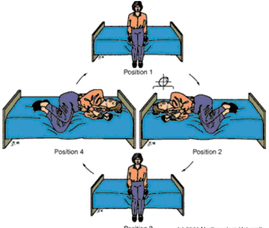
cupula. Semont, a physiotherapist in France and Sterkers where the ones who modified it
to a more acceptable physician controlled treatment called Liberatory maneuver or
Semont maneuver. It is done as follows (fig 9):
33
1. The patient is asked to sit on the edge of the bed. Turn the patient’s head 45 degrees
to the unaffected side.
2. Then patient is asked to quickly lie down on the affected side. Ask them to maintain
the position for 30 seconds.
3. Then quickly ask the patient to move and lie down on the opposite end. The patient is
instructed not to change the direction of the head. They are instructed to keep it at
45-degree angle and lie down for 30 seconds,looking at the floor.
4. The patient is returned slowly to the sitting position and wait a few minutes.
In 1992, Epley suggested repositioning procedure following the proposal of
canalolithiasis theory. After one year, Herdman along with Tusa and Zee modified the
Epley’s maneuver to a one treatment method which had high success. There has been
many modification of the classical Epley’s maneuver since then. Today most commonly
it is done as follows (fig 10):
1. The patient is asked to sit upright on a table or bed and is positioned in a way that
the patient’s shoulder should meet the edge of table or bed when the patient lie
down
2. The examiner takes a position close to the bed to prevent falls
3. Then the patient is asked to turn the head towards the affected side about 45°
a. The patient also asked to keep the eyes open so that the examiner can observe
nystagmus
34
5. The examiner holds this position for 30 seconds
6. Then the patient’s head is rotated 90° to opposite side while keeping the patient still
lying back flat and head hanging over edge of table or bed
7. Then that position is held for 30 seconds
8. The patient is asked to turn or the assistant is made to turn the patient’s body and
head so that the body is facing to the side and head is facing towards the ground at a
45° angle
9. Then that position is held for 30 seconds
10.The patient is returned to upright sitting position
We have to ensure that the patient should not lean head backwards but should
maintain a forward and downward position of head
The patient is asked to sit upright with head still for a time period of
35
Fig 10: Epley maneuver(81)
Another hybrid maneuver is Gans repositioning maneuver which was developed in 2000.
It was specially designed for people who had vertibrobasilar insufficiency, cervical
spondylosis, hip disease, obesity etc. It is also found to be effective. The steps include
(fig 11):
1. In first position, the patient’s head is turned 45 degree to the unaffected side and
patient is made to lay down on the side of vertigo. This position is expected to
move the otolith debris to center of posterior canal
2. Then the patient is made to roll over to unaffected side, maintaining the same
position of head – 45 degree to the unaffected side. This will move otolith debris
move to common crus.
3. The patient is made to shake the head side to side 3 to 4 times in that position.
This helps the otolith debris to traverse the common crus.
4. The patient is returned back to initial position, then head turned forward to centre
36
Fig 11: Gans repositioning maneuver(82)
There are home based treatment maneuvers used in the treatment of BPPV like Brandt-
Daroff exercise was developed as a habitual physical therapy which has repetitive
movements. It did not concentrate on detachment or replacement of otoconia.
The Brandt – Daroff exercise is done as follows (fig 12):
1. The patient sit upright in a bed
2. The patient move to lying down position on one side with nose pointing up about
45 degree
3. The patient maintain the position for 30 seconds
4. Then the patient sit upright in a bed
37
Fig 12: Brandt – Daroff exercise(83)
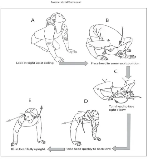
Half somersault exercise or foster maneuver
Another home based exercise is Half somersault exercise or Foster maneuver. It is a
comparatively newer maneuver. It was devised by Dr.Carol A Foster who herself was
suffering from vertigo. The maneuver intends to reposition the displaced otoconia.
It is done as follows (fig 13):
1. The maneuver starts with the patient in a kneeling down position, then patient
should sit back on his/her calves. The patient should keep their palms on floor just
ahead of knees at shoulder width.
2. Then patient should bend the head down in such a way that the top of head touches
the floor in front of the knees. The patient can bend the elbows. This the usual
38
3. Then the patient should turn the head 45 degree towards that direction where
he/she have worst vertigo. The patient should be looking at the elbow on that side
4. Then the patient should raise the head and body so that they are parallel to the
floor at the level of shoulder. The head should be kept turned towards the side
during this entire time
5. Then the patient should lift the head above the level of the body by the neck
6. After that patient can return to the starting position
7. Each position should be maintained for 15 sec or till the vertigo subsides
8. Then the patient should rest for 15 min
[image:53.612.87.324.349.605.2]
Fig 13 : Half somersault exercise or foster maneuver(84)
39
As told earlier majority of patients present with posterior canal BPPV and majority come
with canalolithiasis. Different clinicians give different treatment modalities based on
whether the patient is having canalolithiais or cupulolithiais. Usually canalolithiasis is
treated by Canalolith repositioning maneuver and cupulolithiais is treated by Semont’s
maneuver. However the choice of treatment depends on the clinician and also depend
upon the comfort of patient.
For most of the patients, canalolith repositioning maneuver is more comforatable. The
debris are adherent to cupula according to Schuknecht’s theory of cupulolithiais. Based
on this theory, the Semont’s Liberatory maneuver takes into consideration the fact that
rolling the head is not enough for repositioning the particles
There have been different studies comparing different treatment modalities. Ultimately
the treatment depends on the condition of patient, his comorbid illness and the choice of
the physician. There have been a lot of studies conducted comparing the efficacy of
different treatment modalities.
In a randomized study by Dispenza et al, he compared Epley repositioning maneuver,
Semont repositioning maneuver and a Hybrid maneuver. The study also showed no
statistical difference in efficacy of treatment(85).
A randomized controlled trial conducted by Sushil Gaur et al in the department of ENT in
a tertiary care centre compared Epley maneuver and medical treatment. It showed that
40
low recurrence rate with the group getting Epley maneuver as treatment. The patients
with medical treatment needed more hospital visits compared to the other group(7).
In a study by Devangi S Desai et al, which was conducted in the physiotherapy
department of a pioneer physiotherapy college included 35 patients in a prospective
longitudinal followup study. The study involved patients randomly divided into two
groups. One group was treated with Epley maneuver only and the other group was treated
with Epley and Brandt Daroff exercises. Both the groups gave significant improvement in
the DHI score and Dix Halpike test. It was concluded in the study that both the treatment
approaches give good results, but combined approaches could give better results(86).
Abir Omara et al conducted a randomized controlled study on 30 patients diagnosed with
posterior canal BPPV. One group received Epley repositioning maneuver and other group
received Gans repositioning maneuver. In their study there was no statistical difference
between two groups in terms of treatment efficancy(87).
A study by Soto et al included 106 BPPV patients each undergoing a different treatment
maneuver for BPPV. This was a prospective study whch compared three different
physical treatments for BPPV. At the 1st week of follow up Epley and Semont maneuver
showed better results than brandt daroff exercises. However on the 3rd month of follow
up Epley maneuver proved to be more effective. From their findings they suggested a
41
A comparative study conducted by Abdel Kader et al compared rolling-over maneuver,
Epley and Brandt-Daroff maneuver. The study demonstrated a success rate of 90% for
Epley, 85% for rolling-over maneuver and 80% for Brandt-Daroff maneuver. The
recurrence rate was noted to be high after treatment with Brandt-Daroff, but there was no
statistically significant difference(83).
A systemic review was done comparing the efficacy of Semont maneuver and Epley
maneuver by Bonnie and Melissa. It included six studies of which four studies did not
show any statistically significant difference between the two. However the study
concluded Semont maneuver as an alternative for patients with cervical, lumbar, cardiac,
or respiratory pathologies
Zhang et al conducted a meta-analysis of ten studies based on predefined criteria,
evaluated by the Cochrane evaluation system comparing the effect of Semonts maneuver
with other methods for BPPV treatment. High recovery rate and low recurrence was
noted compared to controls. Over all Semont maneuver displayed a similar outcome
compared to Epley and Brandt Daroff exercises.
SURGICAL TREATMENT MODALITIES
It was Richard R Gacek who first proposed a surgical treatment for BPPV. He first
conducted several animals and then he performed posterior ampullary nerve transection
42
Gacek originally described the procedure as a transcanal procedure under local
anaesthesia. Later many surgeons preferred general anaesthesia.
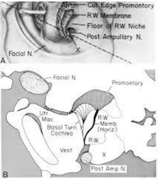
Singular nerve through the canal of Morgagni in temporal bone from posterior ampulla to
saccular nerve. It is inverted J shape which has 3 segments: the part joining internal
auditory canal (canalicular), the curve of J (intermediate), the part entering ampulla
(cribriform). The nerve lies close to round window in the intermediate segment. It can be
[image:57.612.75.234.300.482.2]located 1 to 2 mm deep to posteroinferior margin of round window (fig 14).
Fig 14: Landmark of posterior ampullary nerve(89)
Parnes and McClure in 1990 introduced posterior semicircular canal occlusion for BPPV.
This was based on a study by Money and Scott which showed preserved function of other
canals after ablation of one semicircular canal. It preserved the hearing unless
membranous labyrinth was damaged. The principle of surgery was to prevent endolymph
movement and to prevent movement of cupula in response to angular acceleration and
43
under general anesthesia. First the osseous part of posterior semicircular canal is exposed
via a mastoidectomy. To prevent any damage, occlution of canal is made at a point far
from ampulla and vestibule. The area bisected by lateral SCC, 3mm posterior to facial
nerve is the target area. Then the canal is skeletoniszed 180 degree for a length of 3 to 4
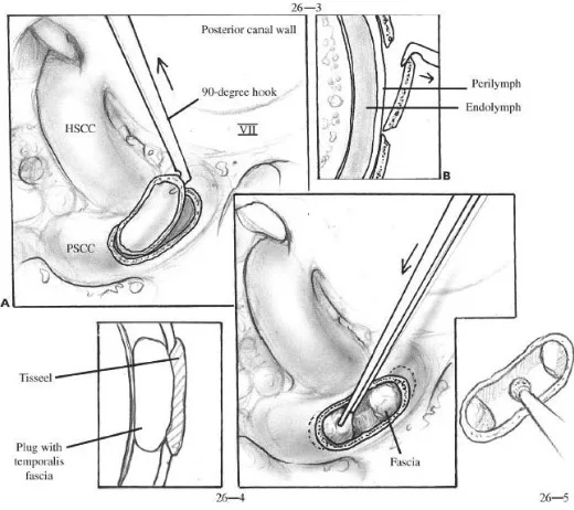
mm. The membraneous canal is exposed by removal of endosteal bone (fig 15). The
membraneous labyrinth is compressed completely with dry bone chips and fibrin glue is
gently inserted to fill the canal. Temporalis fascia is to cover the area in order to prevent
[image:58.612.78.338.323.554.2]perilymph leak. The occlusion is completed by secondary fibrosis.
Fig 15: Posterior semicircular canal occlusion(90)
After 1990s, there was a drastic decrease in the surgical treatment of BPPV. The singular
nerve section have been almost abandoned by surgeons. Studies have shown that there
44
different studies. There have been cases where the singular nerve could not be identified
intraoperatively. The studies show the success rates from 79% to 94%, probably due to
the variant anatomy. The review of studies also show that success of SNS surgery
compared to posterior semicircular canal occlution was low, probably because of the
difficult anatomy. The SCO is more straight forward than SNS because of this reason.
Most of the studies show 100% success rate with SCO. When different studies were
compared, the incidence of post operative hearing loss was almost same in both the
groups, almost 5%.
Literature show a decrease in number of surgical treatment after 1990’s. The surgical
techniques are mostly confines to the centres where it was developed. This may be due to
the difficulty in procedure or the difficulty in training. The use of videonystagmography
and improvement in accuracy of diagnosis has reduced the intractable BPPV.
CHAIR TREATMENT FOR BPPV
In addition to the multiple treatment maneuvers and the surgical treatments which exists
for BPPV, it was found that 10 to 20% of the patients could not be treatment adequately
with the conventional methods. There was lot of patients with cervical disc and hip joint
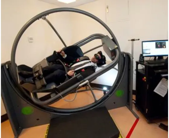
problems. This led to the development of mechanical and rotational chairs. Dr.Epley
developed the Epley Omniax rotator (fig 16) at Portland and Dr.Richard Vitton
developed the TRV chairs in Marseille. It is possible to have a 360 degree circular
movement in all planes along with simultaneous monitoring of nystagmus or any eye
45
Fig 16: Epley Omniax repositioning system(91)
West et al conducted a retrospective cohort study of 150 patients diagnosed with BPPV in
a tertiary care centre. Their study concluded that Epley Omniax rotator and TRV chair
treatments are valuable in management of refractory and complex cases where
conventional treatment is difficult(28).
Tan et al conducted a prospective study on 165 patients with a six month follow up,
comparing the canalolith repositioning maneuver with chair treatment. The study
concluded that there is no statistical difference between the chair treatment and
46 MATERIALS AND METHODS
This study was a prospective, non-blinded randomized control trial on the effect of home based
particle repositioning maneuver in the treatment of posterior canal BPPV
The study was based in the Ear, Nose Throat (ENT) department of a tertiary care academic
hospital in South India after obtaining institutional ethical committee clearance and institutional
research board clearance ( IRB No 9642). The patients were recruited from the outpatient
department (OPD). Patients who were diagnosed with posterior canal BPPV who fulfilled the
inclusion criteria and who gave written consent for this study were recruited for the study.
Inclusion criteria :
a. Adults above 18 years of either sex with typical history of BPPV
b. Dix Hallpike test positive for posterior canal BPPV
c. Willing to come for follow up at 2nd week and 4th week
d. Patients willing to give informed consent
Exclusion criteria:
a. History of cardiac diseases like aortic aneurysm or symptoms suggestive of
acute coronary syndrome.
b. History of cervical spine diseases like fracture spine/ unstable spine/
craniocervical anomalies
c. Any clinical suspicion of other coexistent vestibular disorders like
47
The patients who presented to the OPD with clinical features suggestive of BPPV underwent a
Hallpike test. All patients who were diagnosed with posterior canal BPPV after
Dix-Hallpike’s test and fulfilling the inclusion criteria were randomized by the principal investigator
(PI) into 2 groups using a block randomization table. A detailed history was taken and proforma
was filled regarding baseline characteristics like age, gender, affected side, duration of illness,
frequency of vertigo per day, duration of each vertigo episode, change with physical activities,
any previous episodes, co-morbid illness including hypertension, diabetes mellitus,
hypothyroidism, dyslipidemia and vitamin D deficiency.
We have elaborated the methodology as follows:
Those who had Dix-Hallpike positive with typical history of Posterior canal BPPV, nystagmus duration was noted
Underwent randomization with help of block randomization table to group 1 and 2. In all patients vertigo scale was noted along with nystagmus duration
48 Group 1 was given Epley in the OPD
on day 1 (first visit)
Group 2 was taught HBPRP in OPD in the presence of a bystander on day 1 (first visit) to be done daily at home once daily
On second visit after 1 week, Dix Hallpike repeated and nystagmus time if present, vertigo scale noted, patient reassessed
On second visit after 1 week, Dix Hallpike repeated and nystagmus time if present, vertigo scale noted, patient reassessed
If improving, continued the same treatment, review after 4 weeks
If improving, continued the same treatment, review after 4 weeks
If worsened, checked for involvement of other canals or other associated diagnosis. If same diagnosis, given Epley and is noted
If worsened, checked for involvement of other canals or other associated diagnosis If any other associated diagnosis in the exclusion criteria, patient was taken out of the study
If any other associated diagnosis in the exclusion criteria, patient was taken out of the study
49
DIX HALLPIKE TEST
Dix Hallpike test was done in OPD for the patients who had history suggestive of BPPV. It was
done as follows:
The patient was made to sit on a couch. Then the examiner held the head of the patient, turned it
to 45 degree to the right side. Then the patient was placed in supine position with head hanging
down the edge of table, 30 degree with horizontal. The patient’s eyes were observed for any
nystagmus. Then the test was repeated on the left side. The eyes were observed for nystagmus,
type, duration and latency and presence of vertigo.
NYSTAGMUS DURATION
On third visit after 4 weeks, Dix Hallpike repeated and nystagmus time if present, vertigo scale noted, patient reassessed
On third visit after 4 weeks, Dix Hallpike repeated and nystagmus time if present, vertigo scale noted, patient reassessed
If worsens, check for involvement of other canals or other associated diagnosis. If same diagnosis, given Epley and same noted
If worsens, check for involvement of other canals or other associated diagnosis
If any other associated diagnosis in the exclusion criteria, patient is taken out of the study
50
During Dix-Hallpike test the duration of nystagmus was measured using a stop watch and noted
in seconds. The nystagmus was looked for latency, duration, direction and fatiguability. In BPPV
nystagmus usually appeared after a latency of 2-5 seconds, lasting for less than a minute and
always in one direction that is towards the undermost ear.
VERTIGO SCALE
The patients were asked to score the vertigo on a scale of 0 to 5. It was a subjective scale where
the patients gave a score from 0 to 5 depending upon the perceived intensity during each visit.
They were asked to compare the intensity of vertigo with the previous visits during 2nd and 3rd
visit.
SAMPLE SIZE
A pilot study was conducted and sample size was calculated. The main outcome measures taken
were vertigo scale and nystagmus duration during Dix Hallpike test. The sample size was
calculated using formula:
N= 4S2/D2
S= 1.73 (standard deviation) (MEAN=8)
D=1.0 (Precision of mean value)
N = 11
Minimum of 11 in each arm
51 Epley maneuver
Group 1 included patients who underwent a standard Epley’s manuever at the clinic by
the PI. The PI performed all Epley’s maneuver in the following manner after explaining
the procedure to the patient:
1. The patient was seated upright on an examination table, with their legs fully extended
in front of them.
2. The PI held the patient’s head at a 45-degree angle towards the side they experienced
the worst vertigo.
3. The patient was rapidly brought down on the bed so that they were lying down with
their shoulders touching the table and head hanging down over the edge of the table. The patient’s head was kept facing the side worst affected by vertigo, head hanging down the
table, 30 degree from the horizontal.
4. The PI held the patient’s head in this position for 30 seconds to 2 minutes, until their
dizziness stopped. The patient’s head was rotated to the opposite side by 90 degrees
maintaining the 30 degree angle with the horizontal. Again, the PI held the head in this
position for 30 seconds to 2 minutes, until their dizziness stopped.
5. Next, the patients’s body was rolled 90 degree in the same direction that they faced till
52 The PI held the patient in this position for 30 seconds to 2 minutes, until their dizziness
stopped.
6. Finally, patient was brought back up to a sitting position with chin tucked down to the
chest. The patient was given post procedure instructions which included a) Avoid
driving home after Epley. b) Patient should avoid lying on the side of the BPPV for 2
nights after the procedure. c)The patient was also asked to avoid looking up or down for
8 hours after the procedure. Epley’s procedure was done after Dix-Hallpikes test at 1st
visit, second visit and third visit
Home based particle repositioning maneuver
In Group 2- the patients were taught the home based particle repositioning procedure (HBPRP)
by demonstration in the presence of a bystander by the PI. The steps included
1- The patient was seated upright on an examination table, with their legs fully extended in
front of them.
2- The patient rotated their head at a 45-degree angle towards the side they experienced the
worst vertigo.
3- The patient then lay rapidly down on the bed with the head extended but not beyond the
edge of the bed. The chin was tilted 30 degree above the head and was kept facing the
53
outer edge of the ceiling . The patient was asked to maintain this position for a period of
30 seconds to 2 minutes, until their dizziness stopped (fig 17)
4- The patient rotated his/her head 90 degrees in the opposite direction, maintaining the 30
degree tilt angle of chin with the horizontal, looking at an imaginary point at the outer
edge of the ceiling on that side. Again, the patient was asked to maintain the position for
a time of 30 seconds to 2 minutes, until their dizziness stopped (fig 18)
5- Next, the patient rolled 90 degree in the same direction that they were facing, onto their
side with the chin almost touching the bed. Again, the patient was asked to maintain the
position for a time of 30 seconds to 2 minutes, until their dizziness stopped (fig 19).
6- Finally, the patient sat up to a sitting position with the chin tucked to the chest (fig 20)
The same post procedure instructions were followed out as Group 1.