Akt Finds its New Path to Regulate
Cell Cycle Through Modulating Skp2
Activity and its Destruction by APC/Cdh1
The Harvard community has made this
article openly available. Please share how
this access benefits you. Your story matters
Citation
Gao, Daming, Hiroyuki Inuzuka, Alan Tseng, and Wenyi Wei. 2009.
Akt finds its new path to regulate cell cycle through modulating
Skp2 activity and its destruction by APC/Cdh1. Cell Division 4:11.
Published Version
doi:10.1186/1747-1028-4-11
Citable link
http://nrs.harvard.edu/urn-3:HUL.InstRepos:4621705
Terms of Use
This article was downloaded from Harvard University’s DASH
repository, and is made available under the terms and conditions
applicable to Other Posted Material, as set forth at
http://
Open Access
Review
Akt finds its new path to regulate cell cycle through modulating
Skp2 activity and its destruction by APC/Cdh1
Daming Gao, Hiroyuki Inuzuka, Alan Tseng and Wenyi Wei*
Address: Department of Pathology, Beth Israel Deaconess Medical Center, Harvard Medical School, Boston, MA 02215, USA
Email: Daming Gao - [email protected]; Hiroyuki Inuzuka - [email protected]; Alan Tseng - [email protected]; Wenyi Wei* - [email protected]
* Corresponding author
Abstract
Skp2 over-expression has been observed in many human cancers. However, the mechanisms underlying elevated Skp2 expression have remained elusive. We recently reported that Akt1, but not Akt2, directly controls Skp2 stability by interfering with its association with APC/Cdh1. As a result, Skp2 degradation is protected in cancer cells with elevated Akt activity. This finding expands our knowledge of how specific kinase cascades influence proteolysis governed by APC/Cdh1 complexes. However, it awaits further investigation to elucidate whether the PI3K/Akt circuit affects other APC/Cdh1 substrates. Our results further strengthen the argument that different Akt isoforms might have distinct, even opposing functions in the regulation of cell growth or migration. In addition, we noticed that Ser72 is localized in a putative Nuclear Localization Sequence (NLS), and that phosphorylation of Ser72 disrupts the NLS and thus promotes Skp2 cytoplasmic translocation. This finding links elevated Akt activity with the observed cytoplasmic Skp2 staining in aggressive breast and prostate cancer patients. Furthermore, it provides the rationale for the development of specific Akt1 inhibitors as efficient anti-cancer therapeutic agents.
Introduction
In dividing cells, the cell cycle is tightly controlled by mul-tiple regulatory mechanisms to ensure that DNA is faith-fully replicated only once in the S phase and then distributed equally between two daughter cells in the M phase. Defective cell cycle regulation can lead to genomic instability, which ultimately facilitates cancer develop-ment. Many key regulators governing the cell cycle pro-gression are short-lived proteins, and selective degradation of these regulators by the ubiquitin-proteas-ome system has recently been shown to be a major mech-anism for ensuring ordered and coordinated cell cycle progression [1,2]. Moreover, the irreversible nature of proteolysis guarantees the uni-directional execution of the cell cycle program, driving the cell cycle from one stage to
the next. There are two related, multi-subunit E3 ubiqui-tin ligase enzymes, the Anaphase Promoubiqui-ting Complex (APC) and the Skp1-Cullin1-F-box complex (SCF) that are considered to be the major driving forces governing proper cell cycle progression [3]. SCF is active from the late G1 phase until the G2 phase and mediates the ubiq-uitination of G1 cyclins and Cdk inhibitors. SCF consists of the invariable components Skp1, Cul1, and Rbx1, as well as a variable component known as an F-box protein that is responsible for substrate recognition. There are 68 putative F-box proteins encoded in the human genome which can form individual SCF complexes, each with dif-ferent F-box proteins incorporated into the core Skp1/ Cullin-1-Rbx1 complex [4]. The diversity of these SCF complexes ultimately provides the high stringency
neces-Published: 23 June 2009
Cell Division 2009, 4:11 doi:10.1186/1747-1028-4-11
Received: 1 June 2009 Accepted: 23 June 2009
This article is available from: http://www.celldiv.com/content/4/1/11 © 2009 Gao et al; licensee BioMed Central Ltd.
Cell Division 2009, 4:11 http://www.celldiv.com/content/4/1/11
sary for substrate specificity. The well-characterized F-box proteins Skp2, Cdc4/Fbw7, and β-Trcp1 target p27 [5], cyclin E [6,7], and Cdc25A [8,9], respectively, for ubiqui-tination and degradation. In all cases, proper phosphor-ylation of the substrate is required for interaction with the F-box proteins. Unlike SCF, APC is active from the late G2 phase to the mid-G1 phase, and is responsible for the deg-radation of mitotic cyclins, securin, and geminin. Although APC is composed of 11 subunits, the general structure is very similar to SCF. The substrate adaptors Cdc20 and Cdh1 are equivalent to the F-box proteins, but both Cdh1 and Cdc20 do not require post-translational modification of their respective substrates for recognition, instead, binding to their substrates via Destruction Boxes (D-Box) or KEN Boxes.
Skp2 was originally identified as an S-phase Kinase Cdk2/ Cyclin A-associated protein [10]. Subsequently, the iden-tification of an F-box domain within its coding sequence suggested the presence of E3 ubiquitin ligase activity [5,11]. Besides its major downstream target p27, recent studies have demonstrated that the Skp2/SCF complex also targets numerous other substrates for degradation, many of which are negative cell cycle regulators. These include p21, p57, p130 and FOXO1 [1]. p27 functions as a tumor suppressor such that its inactivation predisposes mice to cancer development [12]. However, in contrast with known tumor suppressor genes such as p53 or Rb, homozygous loss or silencing of the p27 gene is rarely found in human cancers. Instead, it is reduced p27 pro-tein expression which is often linked to human malig-nancy, suggesting that regulation occurs mainly at the post-translational level [13]. Indeed, elevated Skp2 expression is frequently observed in many tumors includ-ing breast and prostate carcinomas [14,15]. It has been proposed that enhanced Skp2 expression leads to the accelerated degradation of targets such as p27 and other cell cycle regulators, thus promoting cell cycle progression and favoring transformation. Furthermore, overexpres-sion of Skp2 facilitates transformation of Rat1 cells in soft agar and in nude mouse xenografts [14]. The oncogenic potential of Skp2 is further illustrated in transgenic mice. In one report, overexpression of Skp2 in the mouse pros-tate induced hyperplasia, dysplasia and low-grade carci-noma [16], while others have reported that Skp2 transgenic mice co-expressing N-Ras develop lymphomas [17]. These findings support the contention that Skp2 overexpression inversely correlates with low p27 expres-sion, and positively correlates with tumor malignancy and poor diagnosis.
However, the molecular mechanisms underlying elevated Skp2 expression have not been fully explored. We and others have previously demonstrated that Cdh1 is the upstream E3 ubiquitin ligase which promotes Skp2
destruction [18,19]. In contrast to the frequency of Skp2 overexpression, loss of Cdh1 is not a frequent event in human cancer. Thus, loss of Cdh1 cannot explain the observation of elevated Skp2 levels in carcinomas. On the other hand, hyperactivation of the Akt pathway through various means of genetic alterations is considered a hall-mark of many cancers. Furthermore, it has been reported that activation of the PI 3-K (phosphoinositide 3-kinase)/ Akt pathway enhances p27 destruction [20]. This suggests that sustained Akt activity can influence Skp2 activity. Consistent with this, studies have also demonstrated that Akt can contribute to Skp2 overexpression, although the mechanism has not been explored [21,22].
manner. However, recent studies have begun to suggest isoform-specific functions for Akt. This was first high-lighted by distinct phenotypes born out from the Akt1 and Akt2 knockout mice [37]. At the level of signaling and cell cycle progression, Akt1 has been shown to promote cell cycle progression, whereas Akt2 promotes cell cycle exit in myoblasts [38]. The PHLPP1 and PHLPP2 isoforms differentially dephosphorylate Akt1 and Akt2 leading to distinct accessibility of each Akt isoform to substrates such as p27, FOXO3a and GSK-3 [39,40]. Finally, Akt1 and Akt2 have been shown to function in an opposing manner in the regulation of breast cancer cell invasive migration. (Irie et al., 2005).
The Skp2 Ser72 Akt phosphorylation site is
conserved in large mammals
Following these clues, we and others recently demon-strated that elevated Akt activity could positively influence Skp2 activity, and impair its destruction by the APC/Cdh1 E3 ubiquitin ligase complex [41,42] (Figure 1). At first
glance, this offers a molecular mechanism for the fre-quently observed Skp2 overexpression in many human cancers and links this phenomenon to the abnormal acti-vation of the PI 3-K and Akt pathway, which is well-docu-mented as being aberrantly activated in the majority of human cancers. The Ser72 site is conserved in most large mammals we examined, including primates, dogs, horses, pigs, cows, and even rats. However, this putative Ser72 Akt phosphorylation site is not present in the mouse sequence. It is known that similar inter-species differences also exist for other Akt substrates including p27 [36] and caspase-9 [43]. We further found that the Ser72 site is not conserved in Xenopus or Zebrafish, thus indicating that the Akt/Skp2 regulatory pathway might have evolved as a gain-of-function event later in evolution. On the other hand, it is well-established that tumorigenesis differs dra-matically between humans and mice [44]. Hence, it is plausible that for large animals with a longer life span than mice, which requires more cell division events, an additional layer of cell cycle control is developed. It is also
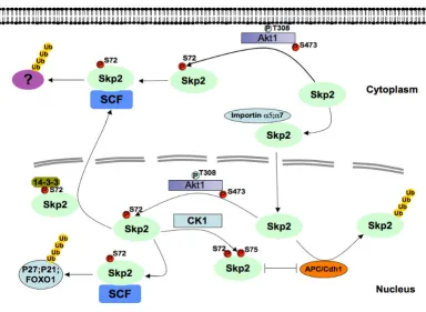
[image:4.612.120.505.347.628.2]Schematic model for how Akt1-dependent phosphorylation of Skp2 at the Ser72 site promotes Skp2 cytoplasmic localization and stabilizes Skp2 by impairing its association with the APC/Cdh1 E3 ubiquitin ligase complex
Figure 1
Cell Division 2009, 4:11 http://www.celldiv.com/content/4/1/11
possible that there is another universal molecular mecha-nism shared by most species to control Skp2 stability/ activity, which is not identified yet.
The potential role for Casein Kinase I in Skp2
stability control
Our results further demonstrate that phosphorylation of human Skp2 at Ser72 creates a priming site, and that CKI may be one of the kinases which phosphorylates Ser75, a process that would lead to subsequent dissociation from Cdh1 and stabilization of Skp2. Sequence analysis also reveals that the Ser75 site is not conserved in all mammals we examined; although most large mammals contain Ser72, they do not contain Ser75, which is replaced by an Asn. This indicates that CKI or other Ser75 kinases are not likely to efficiently phosphorylate Skp2 in these species. Clearly additional studies are required to examine in more detail the regulation of Ser75 phosphorylation in human Skp2, and to determine if CKI is the physiologically-rele-vant kinase, and similarly whether Akt regulates Skp2 sta-bility in species that do not harbor Ser75.
Regardless, our data clearly points to an important dis-tinction between human and mouse Skp2 regulation by the Akt pathway. First, phosphorylation of Ser72 is critical for the ability of Akt to regulate Skp2 stability. Since mouse Skp2 does not contain this site, it explains why mouse Skp2 expression is not affected by the Akt pathway. Secondly, additional experiments using human Skp2 have revealed distinct contributions of phosphorylation events to Skp2 stability. Phosphorylation of Ser72 by Akt is suffi-cient to disrupt the association between importin and Skp2, leading to Skp2 cytoplasmic translocation. Moreo-ver, phosphorylation of both Ser72 and Ser75 by Akt1 and CKI, respectively, is required to disrupt the association between Cdh1 and Skp2, thus stabilizing Skp2. However, since Cdh1 is primarily localized in the nucleus, phospho-rylation of Skp2 by Akt itself might be sufficient to stabi-lize a portion of Skp2 by cytoplasmic translocation.
Akt1 regulates both Skp2 stability and Skp2
transcription
Our data argues that the function of Akt at regulating Skp2 levels is primarily through the regulation of Skp2 protein stability. Substitution of the Ser72 residue to a non-phos-phorylatable Ala created a much more unstable Skp2 pro-tein. Conversely, the phospho-mimetic S72D/S75D Skp2 mutant is much more stable than the wild-type protein. This is likely due to the ability of Akt to affect Cdh1-medi-ated Skp2 degradation since the S72D/S75D mutant resists degradation, and because Akt failed to protect the S72A mutant from degradation. Moreover, in cells where Akt1 is depleted, reduced phosphorylation of Ser72 in Skp2 correlates with a marked decrease in Skp2 levels. This process is likely due to enhanced Skp2 degradation
by Cdh1, since inactivation of Cdh1 resulted in restora-tion of Skp2 to a level comparable to control siRNA-treated samples. Our results do not necessarily disagree with a recent report which showed that activation of PI 3-K and Akt also influences Skp2 mRNA levels [45]. It has been shown that Skp2 is a downstream target of E2F-1 [46], and thus the regulation of its expression at the level of transcription is higher in cells in which Rb is defective. It has also been reported that activation of Akt promotes the binding of E2F-1 to the proximal Skp2 promoter [47]. Therefore, Skp2 upregulation in most human cancers might be due to a synergistic action of upregulated Skp2 mRNA levels with a concomitant evasion of Cdh1-medi-ated degradation.
Does Akt regulate other APC/Cdh1 substrates
other than Skp2?
For most SCF/F-box complexes, the regulation of sub-strate recognition occurs at the level of the subsub-strate, such that the F-box protein will usually not recognize its down-stream substrates without a specific combination of phos-phorylation events. On the other hand, the interaction of Cdh1 and Cdc20 with their substrates usually does not require any post-translational modifications [48]. In this case, the regulation of APC activity occurs primarily on the APC complex itself. Phosphorylation of Cdc20 by Plk and Cdc2/Cyclin B is required for the activation of the APC/ Cdc20 complex while phosphorylation of Cdh1 by the Cdk2/Cyclin A complex terminates Cdh1 activity by dis-sociating Cdh1 from the APC core subunits [49]. Recently, it was shown that APC/Cdh1 ubiquitinates and degrades its substrates with different kinetics, and the preference of degradation order depends on the relative processivity of substrate multiubiquitination by APC/Cdh1 [50].
Akt upon serum stimulation allows for the early induction of Skp2, which correlates with p27 disappearance. This protective mechanism mediated by the Akt pathway is very similar to the Cdk2/cyclin E complex, which protects Cdc6 from Cdh1-mediated destruction [53]. Thus, our finding expands our knowledge of how specific kinase cascades could influence Cdh1-governed proteolysis. In contrast to the SCF complex, where phosphorylation trig-gers substrate recognition, we propose that a specific phosphorylation event by Akt, in addition to CKI, disa-bles the degradation mediated by APC/Cdh1. Upon Cdk2/cyclin E phosphorylation, the interaction between Cdh1 and Cdc6 is reduced [53]. Similar to their finding, we found that the interaction between Skp2 and Cdh1 is also affected by Akt phosphorylation.
The next outstanding question is whether Akt could affect the stability of other APC/Cdh1 substrates. We found that manipulation of Akt activity does not affect the expression of other known Cdh1 substrates we examined, including Cdc20, Plk-1, Geminin, Cdc6, Securin, cyclin A and cyclin B [3,49]. These results indicate that Akt might only specif-ically affects the destruction of a subgroup of Cdh1 sub-strates including Skp2 and others that are still unidentified. We used the Scansite program to screen all known Cdh1 downstream targets, and found that except for Cdc25A and DNMT1, none of the other Cdh1 sub-strates contains the canonical Akt site. Therefore, it is interesting to further investigate whether depletion of Akt1 results in decreased expression of both Cdc25A and DNMT1. Furthermore, similar to what Akt does to Skp2, whether elevated Akt activity protects Cdc25A and DNMT1 from Cdh1-mediated destruction.
Akt promotes Skp2 cytoplasmic translocation
The Akt pathway functions to promote both cell survival and cell growth by inactivating many of its downstream
substrates [23]. Interestingly, by the same token, phos-phorylation of Akt substrates usually results in their cyto-plasmic translocation. In the case of p27, p21 and FOXO proteins, the Akt phosphorylation site is proximal to the Nuclear Localization Sequence (NLS) and when phospho-rylated creates a binding site that can be recognized by the 14-3-3 proteins. Recruitment of 14-3-3 results in the masking of the NLS and subsequent cytoplasmic translo-cation [54]. We also observed an interaction of 14-3-3 with Skp2 in cells expressing activated Akt. However, Ser72 phosphorylation per se is not sufficient for recruit-ment of 14-3-3. Thus it is possible that 14-3-3 is recruited to Skp2 after additional Akt phosphorylation events, or indirectly through interaction with Akt. The NLS of Skp2 resembles that of SV40 T antigen, which is recognized by the importin complex, the only difference being that Skp2 harbors the Ser72 Akt site. We also demonstrated that deletion of the NLS results in cytoplasmic localization, indicating the requirement of this sequence for nuclear import. Moreover, in vitro biochemical analyses demon-strated that deletion of the NLS disrupts the association between Skp2 and importin, providing further evidence that the specific association between Skp2 and the impor-tin complex requires the NLS. Alternatively, the phospho-rylation of serine or threonine residues by specific kinases within the NLS could impair the interaction between the importin complex and the NLS [55,56]. In keeping with this notion, we also demonstrated that phosphorylation of human Skp2 by Akt at Ser72 greatly reduces the inter-action between Skp2 and importin. It is possible that both of these mechanisms contribute to the cytoplasmic trans-location of Skp2 subsequent to Akt phosphorylation. This process is likely to be important for Skp2 function because Akt phosphorylation of the Skp2 downstream targets p27, p21 and FOXO1 also promotes their cytoplasmic localiza-tion, thus terminating their function. Our findings add a new dimension to this model by suggesting that
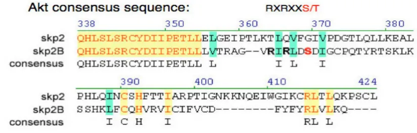
[image:6.612.97.510.95.227.2]Akt-Sequence alignment of the Skp2 and Skp2B proteins reveals that there is an additional potential Akt phosphorylation site at the C-terminus of the Skp2B protein
Figure 2
Cell Division 2009, 4:11 http://www.celldiv.com/content/4/1/11
induced cytoplasmic translocation of Skp2 may lead to elevated degradation of its downstream targets by the cytoplasmic SCF/Skp2 complex. Lin et al suggested that cytoplasmic Skp2 plays an important role in promoting cellular motility [42]. It is well documented that cytoplas-mic Skp2 is frequently observed in more advanced breast and prostate cancer. Therefore, it is plausible that cyto-plasmic Skp2 activity promotes metastasis. It is important to further elucidate the novel substrates for cytoplasmic Skp2, which will provide important insight for developing new anti-cancer treatments.
Akt isoform specificity in the regulation of Skp2
protein stability
Interestingly, our data points to Akt isoform specificity in the regulation of Skp2 protein stability. Using siRNA's, we found that Akt1, but not Akt2, is responsible for phospho-rylation of Skp2 at Ser72, and in turn, modulation of its protein stability. Furthermore, we demonstrated that when overexpressed in 293T cells, human Skp2 specifi-cally interacts with endogenous Akt1, but not Akt2. Although the precise mechanism by which Akt1 can, whereas Akt2 cannot, signal to Skp2 has yet to be defined, and likely mechanisms include the localization of distinct Akt isoforms in cells and tissues. It is plausible that the nuclear localization of Akt1 which has been observed in some cell lines may allow it to interact with nuclear Skp2 and promote nuclear export, and that the more cytoplas-mic localization of Akt2 may restrict its accessibility to Skp2. Although these and other possibilities have yet to be tested, our data is consistent with the recent finding that only Akt1 promotes G1 progression, DNA synthesis and proliferation of C2 myoblasts, whereas Akt2 is primarily required for exit from mitosis [38].
Is Skp2B a better substrate for Akt?
Skp2 cytoplasmic localization has been observed in many clinical tumor samples and is correlated with aggressive malignancy and poor diagnosis [15,57-60]. Our results offer a molecular mechanism for the cytoplasmic localiza-tion of Skp2. Furthermore, since elevated Akt also inacti-vates the Bad, Caspase-9 and FOXO proteins to allow tumor cells to evade the apoptosis pathway, cancer cells with cytoplasmic Skp2 localization tend to be more advanced. Recently, another novel Skp2 splicing isoform (Skp2B) was identified, which possesses many distinct molecular properties because it differs from the Skp2 pro-tein at the carboxyl-terminus [61]. One major difference is that in contrast with the nuclear localization of Skp2, Skp2B localizes to the cytoplasm [58]. Comparing the Skp2 and Skp2B sequences showed that Skp2B contains the identified NLS, and does not have an obvious nucleus export signal (NES) sequence at its unique carboxyl-termi-nus. However, Skp2B contains an additional high proba-bility Akt site in this region (Figure 2). This might make
Skp2B an extremely effective strong substrate for Akt, which could result in cytoplasmic translocation by unknown mechanism.
Concluding remarks
Collectively, we and others provide evidence that Akt directly controls Skp2 stability and oncogenic activity. We identified one major Akt phosphorylation site on Skp2 at Ser72 that is located within a putative Nuclear Localiza-tion Sequence (NLS). We demonstrated that phosphoryla-tion of Ser72 by Akt1, but not Akt2, promotes Skp2 cytoplasmic translocation, likely due to a disruption of the NLS. This finding provides an explanation for previ-ous observations whereby cytoplasmic Skp2 staining is detected in tissues from advanced breast and prostate can-cer. Thus in cells released from serum-starvation, elevated Akt activity in the early G1 phase protects Skp2 from con-stitutive degradation by Cdh1. Hence, our finding expands our knowledge of how specific kinase cascades influence proteolysis governed by the APC/Cdh1 com-plex. In addition, these findings provide insight into how the activated PI3-K/Akt pathway leads to elevated Skp2 expression and subsequent enhanced p27 destruction in human cancers, providing further evidence that elevated Akt activity and cytoplasmic Skp2 expression may be caus-ative for breast and prostate cancer progression. These results may provide a rationale to develop specific Akt1 inhibitors as efficient anti-cancer drugs.
Competing interests
The authors declare that they have no competing interests.
Authors' contributions
WW drafted the manuscript. DG designed the figures. All authors read and approved the final manuscript.
Acknowledgements
We thank Alex Toker, Lixin Wan and Shavali Shaik for critical reading of the manuscript. We also thank Qing Zhang, Haifeng Yang, Alex Toker and Pier Paolo Pandolfi for helpful discussion. Wenyi Wei is a V Scholar, Kimmel Scholar, MLSC New Investigator and Karin Grunebaum Cancer Research Foundation Faculty Research Fellow. Research in our lab is supported in part by the Emerald Foundation, DOD Prostate Program New Investigator Award and the start-up package provided by the Beth Israel Deaconess Medical Center to Wenyi Wei.
References
1. Cardozo T, Pagano M: The SCF ubiquitin ligase: insights into a molecular machine. Nat Rev Mol Cell Biol 2004, 5(9):739-751. 2. Peschiaroli A, Dorrello NV, Guardavaccaro D, Venere M, Halazonetis
T, Sherman NE, Pagano M: SCFbetaTrCP-mediated degrada-tion of Claspin regulates recovery from the DNA replicadegrada-tion checkpoint response. Mol Cell 2006, 23(3):319-329.
3. Nakayama KI, Hatakeyama S, Nakayama K: Regulation of the cell cycle at the G1-S transition by proteolysis of cyclin E and p27Kip1. Biochem Biophys Res Commun 2001, 282(4):853-860. 4. Jin J, Cardozo T, Lovering RC, Elledge SJ, Pagano M, Harper JW:
5. Carrano AC, Eytan E, Hershko A, Pagano M: SKP2 is required for ubiquitin-mediated degradation of the CDK inhibitor p27. Nat Cell Biol 1999, 1(4):193-199.
6. Koepp DM, Schaefer LK, Ye X, Keyomarsi K, Chu C, Harper JW, Elledge SJ: Phosphorylation-dependent ubiquitination of cyclin E by the SCFFbw7 ubiquitin ligase. Science 2001,
294(5540):173-177.
7. Strohmaier H, Spruck CH, Kaiser P, Won KA, Sangfelt O, Reed SI:
Human F-box protein hCdc4 targets cyclin E for proteolysis and is mutated in a breast cancer cell line. Nature 2001,
413(6853):316-322.
8. Busino L, Donzelli M, Chiesa M, Guardavaccaro D, Ganoth D, Dor-rello NV, Hershko A, Pagano M, Draetta GF: Degradation of Cdc25A by beta-TrCP during S phase and in response to DNA damage. Nature 2003, 426(6962):87-91.
9. Jin J, Shirogane T, Xu L, Nalepa G, Qin J, Elledge SJ, Harper JW: SCF-beta-TRCP links Chk1 signaling to degradation of the
Cdc25A protein phosphatase. Genes Dev 2003,
17(24):3062-3074.
10. Zhang H, Kobayashi R, Galaktionov K, Beach D: p19Skp1 and p45Skp2 are essential elements of the cyclin A-CDK2 S phase kinase. Cell 1995, 82(6):915-925.
11. Sutterluty H, Chatelain E, Marti A, Wirbelauer C, Senften M, Muller U, Krek W: p45SKP2 promotes p27Kip1 degradation and induces S phase in quiescent cells. Nat Cell Biol 1999,
1(4):207-214.
12. Nakayama K, Ishida N, Shirane M, Inomata A, Inoue T, Shishido N, Horii I, Loh DY, Nakayama K: Mice lacking p27(Kip1) display increased body size, multiple organ hyperplasia, retinal dys-plasia, and pituitary tumors. Cell 1996, 85(5):707-720. 13. Blain SW, Massague J: Breast cancer banishes p27 from nucleus.
Nat Med 2002, 8(10):1076-1078.
14. Gstaiger M, Jordan R, Lim M, Catzavelos C, Mestan J, Slingerland J, Krek W: Skp2 is oncogenic and overexpressed in human can-cers. Proc Natl Acad Sci USA 2001, 98(9):5043-5048.
15. Signoretti S, Di Marcotullio L, Richardson A, Ramaswamy S, Isaac B, Rue M, Monti F, Loda M, Pagano M: Oncogenic role of the ubiq-uitin ligase subunit Skp2 in human breast cancer. J Clin Invest
2002, 110(5):633-641.
16. Shim EH, Johnson L, Noh HL, Kim YJ, Sun H, Zeiss C, Zhang H:
Expression of the F-box protein SKP2 induces hyperplasia, dysplasia, and low-grade carcinoma in the mouse prostate. Cancer Res 2003, 63(7):1583-1588.
17. Latres E, Chiarle R, Schulman BA, Pavletich NP, Pellicer A, Inghirami G, Pagano M: Role of the F-box protein Skp2 in lymphomagen-esis. Proc Natl Acad Sci USA 2001, 98(5):2515-2520.
18. Bashir T, Dorrello NV, Amador V, Guardavaccaro D, Pagano M:
Control of the SCF(Skp2-Cks1) ubiquitin ligase by the APC/ C(Cdh1) ubiquitin ligase. Nature 2004, 428(6979):190-193. 19. Wei W, Ayad NG, Wan Y, Zhang GJ, Kirschner MW, Kaelin WG Jr:
Degradation of the SCF component Skp2 in cell-cycle phase G1 by the anaphase-promoting complex. Nature 2004,
428(6979):194-198.
20. van Duijn PW, Trapman J: PI3K/Akt signaling regulates p27(kip1) expression via Skp2 in PC3 and DU145 prostate cancer cells, but is not a major factor in p27(kip1) regulation in LNCaP and PC346 cells. Prostate 2006, 66(7):749-760. 21. Andreu EJ, Lledo E, Poch E, Ivorra C, Albero MP, Martinez-Climent
JA, Montiel-Duarte C, Rifon J, Perez-Calvo J, Arbona C, et al.: BCR-ABL induces the expression of Skp2 through the PI3K path-way to promote p27Kip1 degradation and proliferation of chronic myelogenous leukemia cells. Cancer Res 2005,
65(8):3264-3272.
22. Mamillapalli R, Gavrilova N, Mihaylova VT, Tsvetkov LM, Wu H, Zhang H, Sun H: PTEN regulates the ubiquitin-dependent deg-radation of the CDK inhibitor p27(KIP1) through the ubiqui-tin E3 ligase SCF(SKP2). Curr Biol 2001, 11(4):263-267. 23. Woodgett JR: Recent advances in the protein kinase B
signal-ing pathway. Curr Opin Cell Biol 2005, 17(2):150-157.
24. Coffer PJ, Jin J, Woodgett JR: Protein kinase B (c-Akt): a multi-functional mediator of phosphatidylinositol 3-kinase activa-tion. Biochem J 1998, 335(Pt 1):1-13.
25. Parsons R: Human cancer, PTEN and the PI-3 kinase pathway. Seminars in cell & developmental biology 2004, 15(2):171-176.
26. Gao T, Furnari F, Newton AC: PHLPP: a phosphatase that directly dephosphorylates Akt, promotes apoptosis, and suppresses tumor growth. Molecular cell 2005, 18(1):13-24. 27. Testa JR, Bellacosa A: AKT plays a central role in
tumorigene-sis. Proc Natl Acad Sci USA 2001, 98(20):10983-10985.
28. Majumder PK, Sellers WR: Akt-regulated pathways in prostate cancer. Oncogene 2005, 24(50):7465-7474.
29. Samuels Y, Wang Z, Bardelli A, Silliman N, Ptak J, Szabo S, Yan H, Gazdar A, Powell SM, Riggins GJ, et al.: High frequency of muta-tions of the PIK3CA gene in human cancers. Science 2004,
304(5670):554.
30. Datta SR, Dudek H, Tao X, Masters S, Fu H, Gotoh Y, Greenberg ME:
Akt phosphorylation of BAD couples survival signals to the cell-intrinsic death machinery. Cell 1997, 91(2):231-241. 31. Zhao X, Gan L, Pan H, Kan D, Majeski M, Adam SA, Unterman TG:
Multiple elements regulate nuclear/cytoplasmic shuttling of FOXO1: characterization of phosphorylation- and 14-3-3-dependent and -in14-3-3-dependent mechanisms. Biochem J 2004,
378(Pt 3):839-849.
32. Brunet A, Bonni A, Zigmond MJ, Lin MZ, Juo P, Hu LS, Anderson MJ, Arden KC, Blenis J, Greenberg ME: Akt promotes cell survival by phosphorylating and inhibiting a Forkhead transcription fac-tor. Cell 1999, 96(6):857-868.
33. Zhou BP, Liao Y, Xia W, Spohn B, Lee MH, Hung MC: Cytoplasmic localization of p21Cip1/WAF1 by Akt-induced phosphoryla-tion in HER-2/neu-overexpressing cells. Nat Cell Biol 2001,
3(3):245-252.
34. Viglietto G, Motti ML, Bruni P, Melillo RM, D'Alessio A, Califano D, Vinci F, Chiappetta G, Tsichlis P, Bellacosa A, et al.: Cytoplasmic relocalization and inhibition of the cyclin-dependent kinase inhibitor p27(Kip1) by PKB/Akt-mediated phosphorylation in breast cancer. Nat Med 2002, 8(10):1136-1144.
35. Liang J, Zubovitz J, Petrocelli T, Kotchetkov R, Connor MK, Han K, Lee JH, Ciarallo S, Catzavelos C, Beniston R, et al.: PKB/Akt phos-phorylates p27, impairs nuclear import of p27 and opposes p27-mediated G1 arrest. Nat Med 2002, 8(10):1153-1160. 36. Shin I, Yakes FM, Rojo F, Shin NY, Bakin AV, Baselga J, Arteaga CL:
PKB/Akt mediates cell-cycle progression by phosphorylation of p27(Kip1) at threonine 157 and modulation of its cellular localization. Nat Med 2002, 8(10):1145-1152.
37. Whiteman EL, Cho H, Birnbaum MJ: Role of Akt/protein kinase B in metabolism. Trends Endocrinol Metab 2002, 13(10):444-451. 38. Heron-Milhavet L, Franckhauser C, Rana V, Berthenet C, Fisher D,
Hemmings BA, Fernandez A, Lamb NJ: Only Akt1 is required for proliferation, while Akt2 promotes cell cycle exit through p21 binding. Mol Cell Biol 2006, 26(22):8267-8280.
39. Brognard J, Sierecki E, Gao T, Newton AC: PHLPP and a second isoform, PHLPP2, differentially attenuate the amplitude of Akt signaling by regulating distinct Akt isoforms. Mol Cell
2007, 25(6):917-931.
40. Yoeli-Lerner M, Yiu GK, Rabinovitz I, Erhardt P, Jauliac S, Toker A:
Akt blocks breast cancer cell motility and invasion through the transcription factor NFAT. Mol Cell 2005, 20(4):539-550. 41. Gao D, Inuzuka H, Tseng A, Chin RY, Toker A, Wei W:
Phosphor-ylation by Akt1 promotes cytoplasmic localization of Skp2 and impairs APCCdh1-mediated Skp2 destruction. Nat Cell Biol 2009, 11(4):397-408.
42. Lin HK, Wang G, Chen Z, Teruya-Feldstein J, Liu Y, Chan CH, Yang WL, Erdjument-Bromage H, Nakayama KI, Nimer S, et al.: Phospho-rylation-dependent regulation of cytosolic localization and oncogenic function of Skp2 by Akt/PKB. Nat Cell Biol 2009,
11(4):420-432.
43. Fujita E, Jinbo A, Matuzaki H, Konishi H, Kikkawa U, Momoi T: Akt phosphorylation site found in human caspase-9 is absent in mouse caspase-9. Biochem Biophys Res Commun 1999,
264(2):550-555.
44. Rangarajan A, Hong SJ, Gifford A, Weinberg RA: Species- and cell type-specific requirements for cellular transformation. Can-cer Cell 2004, 6(2):171-183.
45. Auld CA, Hopkins RG, Fernandes KM, Morrison RF: Novel effect of helenalin on Akt signaling and Skp2 expression in 3T3-L1 preadipocytes. Biochem Biophys Res Commun 2006,
346(1):314-320.
Publish with BioMed Central and every scientist can read your work free of charge
"BioMed Central will be the most significant development for disseminating the results of biomedical researc h in our lifetime."
Sir Paul Nurse, Cancer Research UK
Your research papers will be:
available free of charge to the entire biomedical community
peer reviewed and published immediately upon acceptance
cited in PubMed and archived on PubMed Central
yours — you keep the copyright
Submit your manuscript here:
http://www.biomedcentral.com/info/publishing_adv.asp
BioMedcentral
Cell Division 2009, 4:11 http://www.celldiv.com/content/4/1/11
47. Reichert M, Saur D, Hamacher R, Schmid RM, Schneider G: Phosph-oinositide-3-kinase signaling controls S-phase kinase-associ-ated protein 2 transcription via E2F1 in pancreatic ductal adenocarcinoma cells. Cancer Res 2007, 67(9):4149-4156. 48. Harper JW, Burton JL, Solomon MJ: The anaphase-promoting
complex: it's not just for mitosis any more. Genes Dev 2002,
16(17):2179-2206.
49. Peters JM: The anaphase-promoting complex: proteolysis in mitosis and beyond. Mol Cell 2002, 9(5):931-943.
50. Rape M, Reddy SK, Kirschner MW: The processivity of multiubiq-uitination by the APC determines the order of substrate degradation. Cell 2006, 124(1):89-103.
51. Gieffers C, Peters BH, Kramer ER, Dotti CG, Peters JM: Expression of the CDH1-associated form of the anaphase-promoting complex in postmitotic neurons. Proc Natl Acad Sci USA 1999,
96(20):11317-11322.
52. Kamura T, Hara T, Matsumoto M, Ishida N, Okumura F, Hatakeyama S, Yoshida M, Nakayama K, Nakayama KI: Cytoplasmic ubiquitin ligase KPC regulates proteolysis of p27(Kip1) at G1 phase. Nat Cell Biol 2004, 6(12):1229-1235.
53. Mailand N, Diffley JF: CDKs promote DNA replication origin licensing in human cells by protecting Cdc6 from APC/C-dependent proteolysis. Cell 2005, 122(6):915-926.
54. Poon IK, Jans DA: Regulation of nuclear transport: central role in development and transformation? Traffic 2005,
6(3):173-186.
55. Hennekes H, Peter M, Weber K, Nigg EA: Phosphorylation on protein kinase C sites inhibits nuclear import of lamin B2. J Cell Biol 1993, 120(6):1293-1304.
56. Zhang F, White RL, Neufeld KL: Cell density and phosphoryla-tion control the subcellular localizaphosphoryla-tion of adenomatous polyposis coli protein. Mol Cell Biol 2001, 21(23):8143-8156. 57. Drobnjak M, Melamed J, Taneja S, Melzer K, Wieczorek R, Levinson
B, Zeleniuch-Jacquotte A, Polsky D, Ferrara J, Perez-Soler R, et al.:
Altered expression of p27 and Skp2 proteins in prostate can-cer of African-American patients. Clin Cancer Res 2003,
9(7):2613-2619.
58. Radke S, Pirkmaier A, Germain D: Differential expression of the F-box proteins Skp2 and Skp2B in breast cancer. Oncogene
2005, 24(21):3448-3458.
59. Dowen SE, Scott A, Mukherjee G, Stanley MA: Overexpression of Skp2 in carcinoma of the cervix does not correlate inversely with p27 expression. Int J Cancer 2003, 105(3):326-330. 60. Penin RM, Fernandez-Figueras MT, Puig L, Rex J, Ferrandiz C, Ariza A:
Over-expression of p45(SKP2) in Kaposi's sarcoma corre-lates with higher tumor stage and extracutaneous involve-ment but is not directly related to p27(KIP1) down-regulation. Mod Pathol 2002, 15(11):1227-1235.