This is a repository copy of
VEGF-A isoforms differentially regulate ATF-2-dependent
VCAM-1 gene expression and endothelial-leukocyte interactions
.
White Rose Research Online URL for this paper:
http://eprints.whiterose.ac.uk/80658/
Version: Published Version
Article:
Fearnley, GW, Odell, AF, Latham, AM et al. (8 more authors) (2014) VEGF-A isoforms
differentially regulate ATF-2-dependent VCAM-1 gene expression and
endothelial-leukocyte interactions. Molecular Biology of the Cell, 25 (16). 2509 - 2521.
ISSN 1059-1524
https://doi.org/10.1091/mbc.E14-05-0962
[email protected]
https://eprints.whiterose.ac.uk/
Reuse
Unless indicated otherwise, fulltext items are protected by copyright with all rights reserved. The copyright
exception in section 29 of the Copyright, Designs and Patents Act 1988 allows the making of a single copy
solely for the purpose of non-commercial research or private study within the limits of fair dealing. The
publisher or other rights-holder may allow further reproduction and re-use of this version - refer to the White
Rose Research Online record for this item. Where records identify the publisher as the copyright holder,
users can verify any specific terms of use on the publisher’s website.
Takedown
If you consider content in White Rose Research Online to be in breach of UK law, please notify us by
MBoC
|
ARTICLE
VEGF-A isoforms differentially regulate
ATF-2–dependent VCAM-1 gene expression
and endothelial–leukocyte interactions
Gareth W. Fearnleya, Adam F. Odella, Antony M. Lathama, Nadeem A. Mughala,b,
Alexander F. Brunsc, Nicholas J. Burgoyned, Shervanthi Homer-Vanniasinkamb, Ian C. Zacharye,
Monica C. Hollsteinf, Stephen B. Wheatcroftc, and Sreenivasan Ponnambalama
aEndothelial Cell Biology Unit, School of Molecular and Cellular Biology, and cDivision of Cardiovascular and Diabetes
Research, Faculty of Medicine and Health, LIGHT Laboratories, University of Leeds, Leeds LS2 9JT, United Kingdom;
bLeeds Vascular Institute, Leeds General Infirmary, Leeds LS1 3EX, United Kingdom; dFios Genomics, Edinburgh EH16
4UX, United Kingdom; eDivision of Cardiovascular Biology and Medicine, Rayne Institute, University College London,
London, United Kingdom; fGerman Cancer Research Center (DKFZ), 69120 Heidelberg, Germany
ABSTRACT Vascular endothelial growth factor A (VEGF-A) regulates many aspects of vascu-lar physiology. VEGF-A stimulates signal transduction pathways that modulate endothelial outputs such as cell migration, proliferation, tubulogenesis, and cell–cell interactions. Multi-ple VEGF-A isoforms exist, but the biological significance of this is unclear. Here we analyzed VEGF-A isoform–specific stimulation of VCAM-1 gene expression, which controls endotheli-al–leukocyte interactions, and show that this is dependent on both ERK1/2 and activating transcription factor-2 (ATF-2). VEGF-A isoforms showed differential ERK1/2 and p38 MAPK phosphorylation kinetics. A key feature of VEGF-A isoform–specific ERK1/2 activation and nuclear translocation was increased phosphorylation of ATF-2 on threonine residue 71 (T71). Using reverse genetics, we showed ATF-2 to be functionally required for VEGF-A–stimulated endothelial 1 gene expression. ATF-2 knockdown blocked VEGF-A–stimulated VCAM-1 expression and endothelial–leukocyte interactions. ATF-2 was also required for other endothelial cell outputs, such as cell migration and tubulogenesis. In contrast, VCAM-1 was essential only for promoting endothelial–leukocyte interactions. This work presents a new paradigm for understanding how soluble growth factor isoforms program complex cellular outputs and responses by modulating signal transduction pathways.
INTRODUCTION
The 58 human receptor tyrosine kinases (RTKs) can be classified into 20 subfamilies, which regulate animal development, health, and dis-ease (Lemmon and Schlessinger, 2010). These type I membrane proteins contain an extracellular ligand-binding domain that “trans-mits” information through a transmembrane domain to a cytoplas-mic tyrosine kinase domain. Ligand binding triggers tyrosine kinase domain activation, with subsequent transautophosphorylation within RTK dimers, recruitment, and phosphorylation of different signal transduction enzymes and substrates. Targeting RTK activity for therapeutic gain is complicated due to the increasing number of soluble and membrane-bound ligands.
Such complexity is exemplified by the vascular endothelial growth factor (VEGF) family. These ligands bind class III RTKs (VEGF receptors 1–3 [VEGFR1–3]) and coreceptors such as neuropilins
Monitoring Editor
Carl-Henrik Heldin Ludwig Institute for Cancer Research
Received: May 7, 2014 Revised: Jun 13, 2014 Accepted: Jun 17, 2014
This article was published online ahead of print in MBoC in Press (http://www .molbiolcell.org/cgi/doi/10.1091/mbc.E14-05-0962) on June 25, 2014. The authors declare no competing financial interests.
Address correspondence to: Sreenivasan Ponnambalam (s.ponnambalam@leeds .ac.uk).
© 2014 Fearnley et al. This article is distributed by The American Society for Cell
Biology under license from the author(s). Two months after publication it is avail-able to the public under an Attribution–Noncommercial–Share Alike 3.0 Unported Creative Commons License (http://creativecommons.org/licenses/by-nc-sa/3.0). “ASCB®,” “The American Society for Cell Biology®,” and “Molecular Biology of
the Cell®” are registered trademarks of The American Society of Cell Biology.
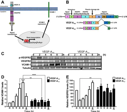
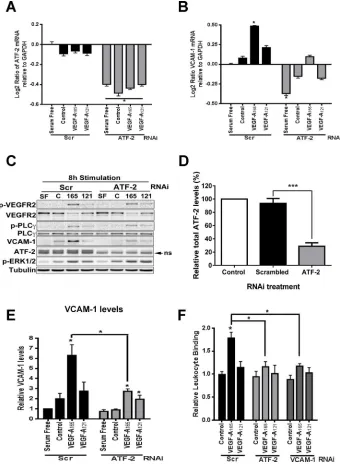
human endothelial cells (Figure 1C). We first compared VCAM-1 and VEGFR2 levels in endothelial cells stimulated with a maximal stimulatory dose (0.25 nM) of either VEGF-A165 or VEGF-A121 for 2,
4, 6, 8, and 24 h (Figure 1C). We used endothelial tubulin levels as a control, and such comparisons were used to evaluate varying pro-tein expression in response to VEGF-A stimulation (Figure 1C). Quantification of endothelial VCAM-1 levels revealed a peak of VCAM-1 expression after VEGF-A165 stimulation for 8 h
correspond-ing to ∼6.5-fold increase compared with the 0 h time point (Figure 1D). This peak in VCAM-1 levels was transient and dropped to ∼2.5-fold rise after VEGF-A165 stimulation for 24 h (Figure 1D). In
comparison, VEGF-A121 stimulation failed to significantly elevate
VCAM-1 levels (Figure 1C).
VEGFR2 activation leads to transautophosphorylation of multiple tyrosine residues: Y1175 is a key site that undergoes such phospho-rylation (Takahashi et al., 2001; Holmqvist et al., 2004; Koch et al., 2011). Monitoring VEGFR2-pY1175 appearance showed that VEGF-A165 stimulates rapid and transient phosphorylation of this site,
whereas VEGF-A121 treatment did not produce significant Y1175
phosphorylation (Figure 1C). VEGF-A-stimulation promotes VEGFR2 ubiquitination, endocytosis, and proteolysis (Ewan et al., 2006). We then asked whether VEGFR2 turnover and synthesis were different upon treatment with either VEGF-A165 or VEGF-A121 isoform (Figure
1E). VEGF-A165 stimulation promoted VEGFR2 degradation over a
short time period (2–4 h), with VEGFR2 levels reduced by ∼60% after 2 h (Figure 1E). However, VEGFR2 levels returned to baseline after VEGF-A165 stimulation for 8 h (Figure 1E). VEGFR2 levels continued
on an upward trajectory, with ∼50% increase after VEGF-A165
stimu-lation for 24 h (Figure 1E). In contrast, VEGF-A121 stimulation
ap-peared to have little effect on VEGFR2 protein levels (Figure 1E). These findings show that two different VEGF-A isoforms have differ-ent capabilities in stimulating the turnover and synthesis of not only VEGFR2, but also those of another membrane receptor, VCAM-1.
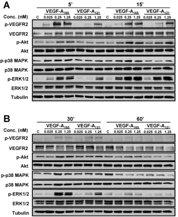
VEGF-A isoforms differentially regulate multiple signal transduction pathways
VEGF-A stimulates multiple MAPK signal transduction pathways in endothelial cells (Horowitz and Seerapu, 2012; Koch and Claesson-Welsh, 2012), which regulate multiple cellular outcomes (Nakatsu et al., 2003; Karihaloo et al., 2005; Zhang et al., 2008; Xu et al., 2011). In this context, we asked whether the increase in endothelial VCAM-1 levels (Figure 1D) was linked to altered signal transduction pathways activated by the two VEGF-A isoforms, using ligand titra-tion followed by signal transductitra-tion pathway analysis (Figure 2). Ac-tivation of VEGFR2 and downstream signaling events was first as-sessed by monitoring transautophosphorylation at cytoplasmic residue Y1175 (Figure 2, A and B). Phosphorylation of Y1175 could be detected within 5 min of stimulation with either VEGF-A165 or
VEGF-A121, but there were concentration-dependent effects (Figure
2A). Quantification showed that VEGF-A121–stimulated
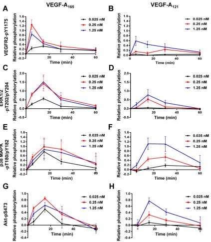
VEGFR2-Y1175 phosphorylation at 0.025 and 0.25 nM was significantly re-duced (Figure 3B) in comparison to VEGF-A165 (Figure 3A).
How-ever, under saturating conditions of VEGF-A (1.25 nM), the peak level of VEGFR2 activation in response to VEGF-A165 (Figure 3A)
was similar to that induced by VEGF-A121 (Figure 3B).
VEGF-A activates ERK1/2, p38 MAPK, and serine/threonine pro-tein kinase c-Akt (Akt; also known as propro-tein kinase B) pathways in endothelial cells (Koch et al., 2011; Koch and Claesson-Welsh, 2012). Both VEGF-A165 and VEGF-A121 stimulation promoted a
rapid and transient peak in ERK1/2 activation within 15 min, with differing magnitudes (Figure 2). Quantification showed that VEGF-A121 stimulation resulted in a generally lower level of peak activation
(NRPs) NRP1 and NRP2 (Koch et al., 2011). The VEGF-A gene alone encodes seven or more different isoforms that bind VEGFR1 (Flt-1), VEGFR2 (KDR), and neuropilins (Harper and Bates, 2008). VEGF-A gene dose is critical, as heterozygous VEGF-A+/− knockout mouse
embryos die during embryogenesis (Carmeliet et al., 1996; Keyt et al., 1996); VEGFR2−/− knockout mice also exhibit embryonic
le-thality (Shalaby et al., 1995). The most-studied VEGF-A ligand is a mature 165-residue processed polypeptide (VEGF-A165), which
pro-motes endothelial cell survival, proliferation, migration, and angiogenesis. VEGF-A–regulated endothelial responses are espe-cially associated with pathological conditions such as tumor pro-gression (Chung and Ferrara, 2011; Meadows and Hurwitz, 2012).
The binding of VEGF-A to VEGFR2 triggers sustained signal transduction, increased trafficking, and proteolysis (Bruns et al., 2009; Horowitz and Seerapu, 2012; Koch and Claesson-Welsh, 2012; Nakayama and Berger, 2013). A key aspect of VEGF-A–stimu-lated reprogramming of endothelial cell function is elevated expres-sion of 100–200 genes (Schweighofer et al., 2009; Rivera et al., 2011). VEGF-A–regulated target genes are implicated in a multitude of cellular functions, including cell adhesion, signal transduction, and transcriptional control. A major question concerns the nature of the mechanism(s) that control VEGF-A–stimulated gene expression. Although VEGF-A–stimulated signal transduction via MEK1–extra-cellular signal-regulated kinase 1 and 2 (ERK1/2), 38-kDa stress- and mitogen-activated protein kinase (p38 MAPK), and JNK pathways could potentially provide multiple means of elevating gene expres-sion, the exact mechanism by which such signal transduction is inte-grated with nuclear transcriptional control is unclear. One well-known target is the membrane receptor vascular cell adhesion molecule 1 (VCAM-1), whose expression on endothelial cells pro-motes binding to leukocyte integrin α4β1 (VLA-4), thus promoting endothelial–leukocyte interactions (Jain et al., 1996; Melder et al., 1996). The mechanism underlying this VEGF-A–stimulated gene ex-pression is unclear, with studies suggesting roles for NF-κB (Kim et al., 2001a) and forkhead (Abid et al., 2008) transcription factors in regulating VCAM-1 gene transcription.
A major question is the role of the increasing number of VEGF splice isoforms in regulating vascular and animal function. The hu-man VEGF-A gene alone expresses eight isoforms ranging from 121 to 206 residues in length. One idea is that the VEGF-A gene en-codes both proangiogenic and antiangiogenic isoforms that are ex-pressed in different tissues to modulate the vascular response dur-ing health and disease (Harper and Bates, 2008). To evaluate the link between VEGF-A–stimulated gene expression and isoform func-tionality, we investigated VCAM-1 expression. Studies on VEGF-A165 and the VEGF-A isoform 121 residues in length (VEGF-A121)
showed that these growth factor isoforms differentially activated signal transduction pathways linked to a novel event in the nucleus that regulates VCAM-1 expression.
RESULTS
VEGF-A isoforms differentially regulate VCAM-1 and VEGFR2 turnover and synthesis
The VEGF-A165 isoform promotes increased endothelial VCAM-1
with either VEGF-A165 or VEGF-A121 (Supplemental Figure S1).
These data suggest that these two different VEGF-A isoforms have differential capabilities not only in stimulating VEGFR2 activation, but also in other downstream signal transduction pathways.
VEGF-A isoform–specific stimulation of activating transcription factor 2
Exactly how short-term RTK signal transduction integrates with long-term cellular responses is not well understood (Lemmon and Schlessinger, 2010). The VEGFR–VEGF-A axis stimulates intracellu-lar signaling over a short time frame (0–1 h) and regulates long-term endothelial responses such as leukocyte recruitment, cell migration (>24 h), and tubulogenesis (5–7 d; Chung and Ferrara, 2011; Koch et al., 2011). To identify a nuclear switch that was responsive to VEGF-A isoform–specific MAPK signal transduction and could influ-ence VCAM-1 expression, we focused on activating transcription factor 2 (ATF-2), which is known to undergo VEGF-A–stimulated phosphorylation in cardiac myocytes and endothelial cells (Seko et al., 1998; Salameh et al., 2010). ATF-2 belongs to the basic region (Figure 3D) than VEGF-A165 (Figure 3C). Of interest, saturating
con-ditions of VEGF-A, which resulted in similar levels of VEGFR2 peak activation (Figure 3, A and B), exhibited approximately twofold dif-ference in ERK1/2 peak activation between the two isoforms (Figure 3, C and D). VEGF-A165 and VEGF-A121 also triggered sustained and
pronounced p38 MAPK activation (Figure 2). Quantification showed that VEGF-A121 –stimulated p38 MAPK activation was more
pro-nounced (Figure 3F) than for VEGF-A165 (Figure 3E) under saturating
ligand conditions. VEGF-A165 and VEGF-A121 also caused
differen-tial Akt activation (Figure 2, A and B). Quantification showed that both VEGF-A165 (Figure 3G) and VEGF-A121 (Figure 3H) promoted a
rapid peak in Akt activation within 15 min. However, VEGF-A165
(Figure 3G) had greater efficacy than VEGF-A121 (Figure 3H), as a
[image:4.585.84.502.46.416.2]much lower concentration of ligand was required to achieve a sig-nificant response. Of interest, at saturating ligand conditions (1.25 nM), the peak in Akt activation was comparable between the two VEGF-A isoforms (Figure 3, G and H). Further analysis of these data sets presented as histograms shows the statistical significance of the changes in signal transduction events detected upon stimulation
FIGURE 1: VEGF-A isoform–specific regulation of VCAM-1 gene expression. (A) Schematic depicting VEGF-A isoform– specific regulation of VCAM-1 gene expression through modulation of gene transcription in endothelial cells.
(B) Schematic depicting human VEGF-A–coding mRNA with exons 1–8 and splice variants VEGF-A165 and VEGF-A121.
(C) Endothelial cells subjected to 0.25 nM VEGF-A165 or VEGF-A121 for the specified times indicated (hours), lysed, and
probed by immunoblotting to assess phospho-VEGFR2 (VEGFR2-pY1175), VEGFR2, VCAM-1, or tubulin protein levels.
(D, E) Quantification of (D) VCAM-1 and (E) VEGFR2 protein levels from immunoblotting studies of VEGF-A165–and
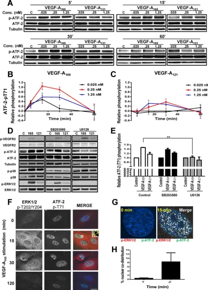
MAPK is significantly reduced by addition of SB203580, which inhibits both the α and β forms of p38 MAPK (Figure 4D). Quantifica-tion of VEGF-A–stimulated phospho–ATF-2 levels showed that that U0126 (MEK1 inhibi-tor) but not SB203580 (p38 MAPK inhibiinhibi-tor) completely blocked ligand-stimulated phos-phorylation of ATF-2 (Figure 4E).
A key aspect of growth factor–stimulated MAPK activation is nuclear translocation of activated protein kinases and phosphoryla-tion of key substrates, which in turn regulate gene transcription (Plotnikov et al., 2011). We previously showed that VEGF-A stimula-tion causes translocastimula-tion of activated ERK1/2 into the nucleus of endothelial cells (Jopling et al., 2009). To correlate ERK1/2 transloca-tion with ATF-2 activatransloca-tion, we monitored the intracellular distribution of phospho-ERK1/2 and ATF-2–pT71 using microscopy (Figure 4F). Activated phospho-ERK1/2 was de-tected in both the cytoplasm and nucleus within 15 min; this also correlated with a peak of phospho-ATF-2 in the nucleus (Figure 4F). Microscopy analysis of phospho-ERK1/2 and phospho-ATF-2 in the nucleus revealed substantial codistribution of both proteins at ∼15 min post–VEGF-A165
stimu-lation (Figure 4G). Quantification of nuclear phospho-ERK1/2 and phospho–ATF-2 showed a >10-fold rise in codistribution after VEGF-A165 stimulation (Figure 4H). Such
findings show a close link of VEGF-A165–
stimulated MAPK signal transduction lead-ing to ERK1/2 activation, nuclear transloca-tion, and downstream activation of ATF-2.
The VEGF-A coreceptor NRP1 has been shown to attenuate VEGF-A–stimulated sig-nal transduction (Pan et al., 2007; Herzog et al., 2011). Therefore we evaluated whether NRP1 was essential for optimal VEGF-A–stimulated ATF-2 activation. Using immunoblotting, we monitored ATF-2–pT71 levels after 15 min of stimulation with either VEGF-A165 or VEGF-A121 in NRP1-depleted and control endothelial
cells (Supplemental Figure S2A). Quantification revealed that both VEGF-A165– and VEGF-A121–stimulated ATF-2 activation was
re-duced in NRP1-depleted endothelial cells (Supplemental Figure S2B). One consequence of a reduction in NRP1 levels was a con-comitant reduction in VEGF-A–stimulated ERK1/2 activation (Sup-plemental Figure S2C). These data suggest that NRP1 influences the ability of the VEGFR2–VEGF-A165 complex to effectively activate
downstream ERK1/2 and thus ATF-2.
VEGF-A and ATF-2 are required for VCAM-1 expression and endothelial–leukocyte interactions
VEGF-A–stimulated VCAM-1 gene expression has implicated both the NF-κB pathway and forkhead transcription factors (Kim et al., 2001a,b; Abid et al., 2006; Dejana et al., 2007). On the basis of our data, we hypothesized that ATF-2 acts as a nuclear “switch” for converting VEGF-A isoform–specific short-term cytosol-to-nucleus signaling (via the MEK1-ERK1/2 pathway) into VCAM-1 gene subdomain/leucine zipper (bZIP) family of DNA-binding
transcrip-tion factors and undergoes activatranscrip-tion upon cellular stress or plasma membrane receptor activation (Lau and Ronai, 2012). To determine whether VEGF-A isoforms differentially regulate ATF-2 activation, we monitored ATF-2–pT71 levels (Figure 4A). Endothelial cells con-tain basal phospho-ATF-2, which is further elevated upon VEGF-A stimulation (Figure 4A). Of note, maximal ATF-2–pT71 levels de-tected upon VEGF-A165 addition were approximately twofold to
fivefold higher (Figure 4B) than comparable VEGF-A121 (Figure 4C)
ligand concentrations.
[image:5.585.35.383.45.469.2]Multiple signal transduction pathways, including JNK, p38 MAPK, ERK1/2, and ATM, stimulate phosphorylation at different sites on ATF-2 (Lau and Ronai, 2012). To assess whether the increase in phospho–ATF-2 was dependent on MEK1-ERK1/2 or p38 MAPK pathways, we used cell-permeable small-molecule inhibitors spe-cific for either pathway (Figure 4D). VEGF-A–stimulated activation of ERK1/2 is significantly reduced by addition of U0126, a MEK1 inhibi-tor (Figure 4D). In contrast, VEGF-A–stimulated activation of p38
FIGURE 2: VEGF-A isoform–specific activation of signal transduction. (A, B) Endothelial cells
subjected to different VEGF-A165 or VEGF-A121 concentrations (0, 0.025, 0.25, or 1.25 nM) for
(A) 5 and 15 min or (B) 30 and 60 min were lysed and probed for VEGFR2,
observed for VEGF-A165 (Figure 5B). ATF-2 knockdown
substan-tially reduced the VEGF-A–stimulated increase in VCAM-1 mRNA levels by ∼25% (Figure 5B). There is thus a functional requirement for the presence of ATF-2 in VEGF-A–stimulated VCAM-1 expres-sion in endothelial cells.
One question is the link between the requirement for ATF-2 in endothelial signal transduction and protein expression. To address this, we used immunoblotting to monitor protein levels and phos-phorylation events 8 h after VEGF-A stimulation in control or ATF-2–depleted endothelial cells (Figure 5C). This time point of post– VEGF-A stimulation was used as the point of maximal ligand-stimulated VCAM-1 expression (Figure 1D). ATF-2 knock-down caused ∼75% reduction in ATF-2 protein levels (Figure 5D). Activated VEGFR2-pY1175 levels were significantly elevated at this transcription, thus modulating VEGF-A isoform–specific long-term
endothelial responses (including leukocyte recruitment). To evalu-ate ATF-2 requirement in VEGF-A–stimulevalu-ated gene transcription, we first used specific small interfering RNA (siRNA) duplexes to deplete endothelial ATF-2. As expected, ATF-2 mRNA levels were depleted only in endothelial cells treated with ATF-2–specific siRNA duplexes (ATF-2 knockdown) in comparison to scrambled siRNA duplex treatment (control), under both nonstimulated and VEGF-A–stimulated conditions (Figure 5A). We then analyzed whether ATF-2 depletion affected VCAM-1 mRNA levels (Figure 5B). On VEGF-A165 stimulation, we detected ∼1.4-fold increase in
VCAM-1 mRNA levels compared with controls (Figure 5B). VEGF-A121–stimulated endothelial cells produced ∼1.2-fold increase in
[image:6.585.84.507.43.528.2]VCAM-1 mRNA levels, but this was substantially less than that
FIGURE 3: Quantification of VEGF-A isoform–specific signal transduction. Quantification of (A, B) VEGFR2-pY1175,
(C, D) ERK1/2-pT202/pY204, (E, F) p38-pT180/pY182, and (G, H) Akt-pS473 levels upon (A, C, E, G) VEGF-A165 or
FIGURE 4: VEGF-A isoform–specific intracellular signaling regulates ATF-2 phosphorylation. (A) Immunoblotting of VEGF-A–stimulated endothelial cells for ATF-2-pT71, total ATF-2, and tubulin upon ligand titration. (B, C) Quantification
of phosphorylated ATF-2-pT71 levels upon (B) VEGF-A165 and (C) VEGF-A121 titration. Error bars indicate ±SEM (n= 4).
(D) Immunoblotting VEGFR2, ATF-2, ERK1/2, and p38 MAPK total and phosphorylated levels after preincubation with MEK1 inhibitor (U0126) or p38 MAPK inhibitor (SB203580), followed by VEGF-A isoform (1.25 nM) stimulation for
15 min. (E) Quantification of ATF-2-pT71 levels upon activation by VEGF-A165 or VEGF-A121 in the presence of MEK1
inhibitor (U0126) or p38 MAPK inhibitor (SB203580). Error bars indicate ±SEM (n= 3). (F) Endothelial cells stimulated
time point but were mirrored by a large de-crease in VEGFR2 levels (Figure 5C). Differ-ential phosphorylation in ERK1/2 and phos-pholipase Cγ1 was also evident (Figure 5C). These findings showed that ATF-2 knock-down did not significantly affect VEGFR2 turnover and downstream signal transduc-tion in response to VEGF-A stimulatransduc-tion.
We probed for VCAM-1 expression in control or ATF-2–depleted cells 8 h after VEGF-A isoform stimulation (Figure 5C). VCAM-1 expression was clearly ATF-2 de-pendent (Figure 5C), and quantification highlighted a relatively large (greater than threefold) increase in VCAM-1 levels ob-served upon VEGF-A165 stimulation
com-pared with cells incubated in serum-free or complete medium (Figure 5E). Depletion of ATF-2 reduced VCAM-1 to baseline levels in A–stimulated cells (Figure 5E). VEGF-A121 also stimulated a smaller, 30–40%
in-crease in VCAM-1 protein levels, which was also ATF-2 dependent (Figure 5, C and E). These data showed that increased VCAM-1 expression caused by VEGF-A not only was ATF-2 dependent but also influenced VCAM-1 mRNA levels.
Endothelial VCAM-1 binds leukocyte α4β1 integrin (VLA-4) and promotes leuko-cyte recruitment onto the endothelium be-fore transendothelial migration (Sixt et al., 2006; Nourshargh et al., 2010; Reglero-Real et al., 2012). A major question is whether VEGF-A–stimulated and ATF-2–dependent VCAM-1 expression can influence leukocyte binding to endothelial cells. To test this idea, we used a binding assay that monitored the binding of fluorescent-labeled human leuko-cyte HL-60 cells to an endothelial cell mono-layer (Figure 5F). VEGF-A165 stimulated
∼75% increase in leukocyte binding to the endothelial monolayer (Figure 5F). However, VEGF-A121 caused only a small, ∼15%
[image:8.585.37.377.47.519.2]in-crease in leukocyte binding to endothelial cells (Figure 5F). ATF-2 knockdown com-pletely ablated VEGF-A–stimulated binding of leukocytes to endothelial cells (Figure 5F). This phenomenon was VCAM-1 dependent, as VCAM-1 knockdown also completely in-hibited endothelial–leukocyte interactions (Figure 5F). These data confirm that the VEGF-A–stimulated expression of endothe-lial VCAM-1 not only is ATF-2 dependent but also is sufficient to enable recruitment of leu-kocytes and enhance cell–cell interactions.
FIGURE 5: ATF-2 requirement for VEGF-A isoform–specific control of VCAM-1 expression in
endothelial cells. (A, B) Endothelial cells stimulated with 0.25 nM VEGF-A165 or VEGF-A121 in
different growth conditions for 4 h were analyzed by qRT-PCR for (A) ATF-2 or (B) VCAM-1
mRNA levels. GAPDH mRNA was used as an internal control. Error bars denote ±SEM (n= 3).
(C) Endothelial cells subjected to RNA interference and knockdown with either scrambled (Scr)
or ATF-2–specific (ATF-2) siRNA duplexes were then stimulated with 0.25 nM VEGF-A165 or
VEGF-A121 for 8 h, lysed, and subjected to immunoblot analysis for a variety of proteins,
including ATF-2, VEGFR2, and VCAM-1. (D) Quantification of ATF-2 knockdown in endothelial
cells. Error bars indicate ±SEM (n= 3). (E) Quantification of VCAM-1 levels after 8 h of VEGF-A
stimulation. Error bars denote ±SEM (n≥ 3). (F) Endothelial cells treated with scrambled (Scr),
ATF-2, or VCAM-1 siRNA duplexes were stimulated with 0.25 nM VEGF-A165 or VEGF-A121 (7 h)
before binding of calcein-labeled, activated HL-60 leukocytes and lysis and measurement (see
Materials and Methods). Error bars denote ±SEM (n≥ 3). *p < 0.05, ***p < 0.005.
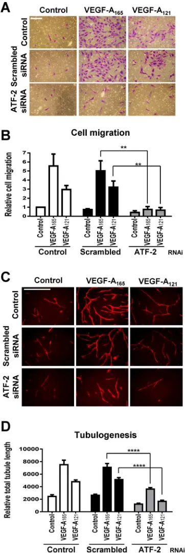
question as to the importance of ATF-2 in endothelial cell function and responses such as cell migration and tubule formation (tubulo-genesis). To address this, we first compared the roles of these two VEGF-A isoforms in promoting endothelial cell migration, tubulo-genesis, and ex vivo angiogenesis. VEGF-A165 produced a marked
dose-dependent stimulation in endothelial cell migration (Supple-mental Figure S3, A and B) and tubulogenesis (Supple(Supple-mental Figure S3, C and D). However, VEGF-A121 showed a much reduced or
mod-est stimulation in such endothelial cell responses, and such effects were especially evident at intermediate or substoichiometric con-centrations of VEGF-A (Supplemental Figure S3, B and D). Ex vivo angiogenesis assays using mouse aortic slices (Supplemental Figure S3E) showed VEGF-A165 had approximately threefold higher
capac-ity to stimulate vascular sprouting (Supplemental Figure S3F). Thus each VEGF-A isoform had a distinct capacity to promote differential endothelial cell outputs; VEGF-A165 was generally more biologically
active at low or substoichiometric concentrations.
This raised the question as to whether ATF-2 was equally impor-tant for such differential programming of these endothelial cell re-sponses. ATF-2 knockdown completely abolished either VEGF-A165–
or VEGF-A121–stimulated cell migration (Figure 6, A and B). ATF-2
knockdown also inhibited (∼50%) both VEGF-A165– and VEGF-A121–
stimulated tubulogenesis (Figure 6, C and D). ATF-2 was also re-quired for endothelial cell proliferation (Supplemental Figure S4). Depletion of ATF-2 resulted in a approximately twofold decrease in VEGF-A165–stimulated cell proliferation (Supplemental Figure S4).
Of interest, depletion of ATF-2 also significantly reduced endothelial cell proliferation in complete media (Supplemental Figure S4).
Although VCAM-1 was required for endothelial–leukocyte adhe-sion (Figure 5F), this raised the possibility that it may also be re-quired for other endothelial responses. To test this idea, we treated endothelial cells with scrambled or VCAM-1–specific siRNA du-plexes before assessing VEGF-A isoform–specific endothelial tubu-logenesis (Supplemental Figure S5, A and B). There was no signifi-cant difference in tubulogenesis between control and VCAM-1–depleted endothelial cells (Supplemental Figure S5B).
DISCUSSION
In this study, we show that different VEGF-A isoforms have differen-tial capacities to regulate VCAM-1 gene expression and modulate endothelial–leukocyte binding via a novel mechanism (Figure 7). In this model, two VEGF-A isoforms with similar binding affinities dif-ferentially program VEGFR2 activation and downstream signal trans-duction to act on a common nuclear “switch” that regulates VCAM-1 expression. This “switch” comprised nuclear ATF-2, a transcription factor that is regulated by increased signal transduction from the MEK1-ERK1/2 pathway. Our findings show that ATF-2 is an impor-tant factor that regulates both VEGF-A–regulated responses and other essential pathways.
A key feature in VEGF-A–stimulated VCAM-1 expression is the requirement for ERK1/2 activation and ATF-2 expression. Maximal VCAM-1 expression is dependent on endothelial cell stimulation by a specific VEGF-A165 isoform. This isoform greatly increased VEGFR2
phosphorylation at residue Y1175 in comparison to the VEGF-A121
isoform. This correlated with an increased ability to promote ERK1/2 activation and nuclear translocation (Figure 7). Translocation of acti-vated ERK1/2 into the nucleus revealed close proximity to actiacti-vated phospho–ATF-2. It is feasible that the T71 residue on ATF-2 is di-rectly phosphorylated by ERK1/2. Alternatively, another target of ERK1/2, such as the p90 ribosomal S6 kinase (p90rsk or MAPKAP-K1), can also translocate into the nucleus and phosphorylate key transcription factors (Arthur, 2008; Gerits et al., 2008).
ATF-2 is required for VEGF-A–stimulated endothelial cell responses
[image:9.585.70.251.46.589.2]VEGF-A–stimulated signal transduction regulates diverse long-term responses in endothelial cells, such as cell migration and tubulogen-esis (Chung and Ferrara, 2011; Koch et al., 2011). This raises the
FIGURE 6: VEGF-A and ATF-2 regulation of endothelial cell responses. (A–D) Control, scrambled, or ATF-2–specific siRNA duplex–treated endothelial cells were seeded into different assays
and stimulated with 0.25 nM VEGF-A165 or VEGF-A121 and assessed
for endothelial cell (A, B) migration or (C, D) tubulogenesis. Bar,
200 µm (A, B), 400 µm (C, D). Error bars indicate ±SEM (n= 3).
ing affinity to VEGFR2 (Keyt et al., 1996; Delcombel et al., 2013), but unique recep-tor–ligand complexes can produce different functional outputs. Of interest, it has been noted that the murine orthologues of VEGF-A121 and VEGF-A165 show capacity to
dif-ferentially elevate expression of another cell adhesion molecule, ICAM-1, in the mouse ocular endothelium (Usui et al., 2004). The underlying mechanism regulating such dif-ferential VEGF-A–regulated ICAM-1 expres-sion is unknown but raises the speculation that ATF-2 may be involved in this phenom-enon as well. In this context, it is well known that different VEGF-A isoforms have the ca-pacity to trigger differential VEGFR2 activa-tion and downstream signal transducactiva-tion (Zhang et al., 2000, 2008; Bates et al., 2002; Nakatsu et al., 2003; Herve et al., 2005; Chen et al., 2010). An important question concerns the mechanism underlying VEGF-A–stimulated gene transcription (Goddard and Iruela-Arispe, 2013). STAT3 (Bartoli et al., 2003), Egr3 (Liu et al., 2003), forkhead-like transcription factors (Abid et al., 2006), FoxO- and Ets-related factors (Dejana et al., 2007), and HLX (Testori et al., 2011) have all been implicated in regulating VEGF-A–de-pendent gene transcription.
How ATF-2 regulates VCAM-1 gene transcription is intriguing. ATF-2 was origi-nally identified as a nuclear transcriptional switch that was activated upon DNA dam-age or stress, thus enabling gene expression linked to an antiapoptotic response or cell proliferation (Lau and Ronai, 2012). The ATF-2 polypeptide can undergo phosphory-lation at different Ser/Thr residues at the N-terminus (T52, S62, T69, T71, S73, S121) or C-terminus (S490, S498; Lau and Ronai, 2012). One possibility is that phospho– ATF-2 directly binds to the VCAM-1 pro-moter to stimulate early gene transcription: this may occur via the formation of ATF-2 homodimers or heterodimers with factors such as c-jun. In addition, ATF-2 may recruit other factors such as p300 (Kawasaki et al., 1998) or IRF3 (Panne et al., 2004) to pro-moter loci, which further modulate target gene expression. It has also been proposed that ATF-2 has intrinsic histone acetyltransferase activity (HAT) such that it acts to derepress gene transcription upon recruitment (Kawasaki et al., 2000), and other transcription factors such as NF-κB or forkhead could directly stimulate VCAM-1 gene transcription.
Our study shows evidence that different VEGF-A isoforms regu-late both angiogenesis and inflammation via an ATF-2–dependent mechanism. The increased recruitment of activated leukocytes to VEGF-A165–stimulated cells, in contrast to VEGF-A121-stimulation,
argues that this has a functional role in vivo. This could be useful in the VEGF-A isoform–specific recruitment of leukocytes into a devel-oping blood vessel during angiogenesis. This would be useful in controlling vascular development and endothelial–leukocyte VEGF-A–stimulated intracellular signaling over a short time
frame (0–2 h) caused early VCAM-1 gene transcription and increased mRNA levels (2–8 h) with concomitant peak in VCAM-1 expression at the cell surface after 8 h. In this way, short-range signal transduc-tion is translated into intermediate and long-range effects such as membrane protein expression and subsequent interactions that modulate endothelial interactions with the environment. A key fea-ture is that two different VEGF-A isoforms of either 165 or 121 resi-dues in length show significantly altered ability to promote VCAM-1 expression. This is largely due to decreased ERK1/2 activation by the shorter VEGF-A121 isoform. The different VEGF-A isoforms have
a conserved N-proximal region (residues 1–111) and variable C-ter-minus (112–206). Of note, all VEGF-A isoforms display similar
bind-FIGURE 7: A mechanism for VEGF-A isoform–specific regulation of endothelial–leukocyte interactions. Schematic describing VEGF-A isoform–specific stimulation of intracellular signaling and ATF-2–regulated VCAM-1 gene expression. Numbered steps denote the following:
1) VEGFR2 activation by either VEGF-A165 or VEGF-A121 programs differential phosphorylation
of residue Y1175; 2) VEGF-A165 stimulates elevated ERK1/2 phosphorylation and activation
compared with VEGF-A121; 3) phosphorylated ERK1/2 translocates to the nucleus and regulates
ATF-2 phosphorylation; 4) VEGF-A165 is more potent than VEGF-A121 in promoting ATF-2
phosphorylation as a result of increased ERK1/2 activity; 5) VEGF-A165–stimulated ATF-2 activity
[image:10.585.37.374.48.456.2]0.25, or 1.25 nM VEGF-A isoform for the desired time period. Cells were then washed three times with ice-cold PBS and lysed in buffer with 2% (wt/vol) SDS, Tris-buffered saline, 1 mM phenylmethylsulfo-nyl fluoride, and protease inhibitor cocktail (Sigma-Aldrich, Poole, United Kingdom). Protein concentration was determined using the bicinchoninic acid assay (ThermoFisher, Loughborough, United Kingdom). A 25 µg amount of protein lysate was subjected to SDS– PAGE before analysis by immunoblotting. For immunofluorescence analysis, cells were serum starved for 3 h before being stimulated with VEGF-A165. Cells were fixed and processed as previously
de-scribed (Bruns et al., 2010). Images were acquired using a Delta-Vision wide-field deconvolution microscope (Applied Precision, Issaquah, WA). Relative colocalization was quantified using ImageJ (National Institutes of Health, Bethesda, MD) as previously described (Bruns et al., 2010; Jopling et al., 2011).
Pharmacological inhibition of signal transduction
Cells were seeded and starved as stated previously, then pretreated with 10 µM SB203580 or U0126 (LC Labs, Boston, MA) for 30 min before stimulation with 1.25 nM VEGF-A isoform in MCDB131 plus 0.2% (wt/vol) BSA for 15 min. Cells were then processed via SDS– PAGE before immunoblot analysis.
Lipid-based transfection of siRNA duplexes
Cells were reversed transfected with siRNA duplexes using Lipo-fectamine RNAiMAX (Invitrogen, Paisley, United Kingdom). Per well of a six-well plate, 15 µl of 2 µM siRNA duplexes was added to 481 µl of serum/antibiotic-free OptiMEM (Invitrogen) and allowed to settle at room temperature for 5 min. Then 4 µl of Lipofectamine was added, and the mixture was inverted briefly and incubated at room temperature for 20 min. HUVECs were seeded at 2.5 × 10⁵ cells/ml in a 1 ml volume of OptiMEM, followed by immediate dropwise addition of the siRNA/Lipofectamine mixture. Cells were left at room temperature for 30 min before being returned to the incuba-tor. After 6 h total of incubation, medium was replaced for ECGM. Cells were allowed to recover for 72 h before treatment or process-ing for analysis.
Quantitative reverse transcription PCR
HUVECs were treated with siRNA duplexes specific for either ATF-2 or a scrambled (Scr) sequence for 72 h before growth arrest induced by overnight serum starvation (serum free) and 2 mM thy-midine supplementation. Control cells were released for 4 h in full growth medium together with either 0.25 nM VEGF-A165 or
VEGF-A121 before extraction of total RNA with the RNeasy Plus Mini Kit
(Qiagen, Manchester, United Kingdom). A 1 µg total amount of RNA was reverse transcribed using the GoScript Reverse Transcription System (Promega, Southampton, United Kingdom). Real-time quantitative reverse transcription PCR (qRT-PCR) was per-formed using Power SYBR Green master mix (Applied Biosystems, Warrington, United Kingdom) with the following primer sets: glyc-eraldehyde-3-phosphate dehydrogenase (GAPDH; endogenous control), forward primer 5′-GTC TCC TCT GAC TTC AAC AGC G-3′, reverse primer 5′-ACC ACC CTG TTG CTG TAG CCA A-3’; ATF-2, forward primer 5′-GGT AGC GGA TTG GTT AGG ACT C-3′, reverse primer 5′TGC TCT TCT CCG ACG ACC ACT T-3′; and VCAM-1, forward primer 5′-GAT TCT GTG CCC ACA GTA AGG C-3′, reverse primer 5′TGG TCA CAG AGC CAC CTT CTT G-3′. qRT-PCR was carried out in multiwell plates run on an ABI 7900HT Fast Real-Time PCR System (Applied Biosystems) and gene ex-pression analyzed using the ΔΔCT method standardized against an endogenous control, GAPDH.
balance within a vascular niche. Alternatively, such a phenomenon could be extremely useful during pathogenic infection or injury: the release of specific VEGF-A isoforms into the damaged vasculature not only could promote leukocyte recruitment but also could attune leukocyte recruitment to the extent of infection or injury. VEGF-A165
has been shown to stimulate VCAM-1 expression (Kim et al., 2001a; Abid et al., 2006), but this has also been contradicted (Stannard et al., 2007). In this context, it has been shown that proinflammatory cytokines such as interleukin 1β or tumor necrosis factor α (TNFα) promote NF-κB, SP1, AP-1, and IRF recruitment to the VCAM-1 lo-cus to stimulate VCAM-1 expression (Collins et al., 1995; Weber, 1996; Hordijk, 2006; Sixt et al., 2006). A link between ATF-2 and NF-κB has also been proposed in regulating VCAM-1 expression during shear stress (Cuhlmann et al., 2011). Expression of a mutant ATF-2 in a mouse model has been shown to inhibit VCAM-1 expression (Reimold et al., 2001).
The interactions between endothelial cells and leukocytes can be subverted in major disease states ranging from atherosclero-sis, rheumatoid arthritis, and pathogenic infection to cancer. This study now provides a mechanism to explain how different VEGF-A isoforms regulate not only angiogenesis but also inflammation in such disease states. Immune cells are well known to secrete proangiogenic cytokines such as TNFα and vascular endothelial growth factor A (VEGF-A; Griffioen and Molema, 2000; Naldini and Carraro, 2005). The angiocrine model postulated by Rafii and colleagues suggests that the endothelium secretes soluble and membrane-bound factors that act in a paracrine manner on neighboring cells to influence vascular development in tissues such as liver (Butler et al., 2010; Ding et al., 2010). Our work shows that VEGF-A isoforms have unique abilities to instruct the endothelium and influence leukocyte recruitment at local sites through cell–cell interactions. It will be a challenge to decipher the myriad of biological properties of the VEGF family with func-tional roles in both angiogenesis and inflammation in healthy and diseased states.
MATERIALS AND METHODS Antibodies and growth factors
Antibodies were goat anti-VEGFR2 (R&D Systems, Minneapolis, MN), rabbit anti-ERK1/2, mouse anti–phospho-ERK1/2 (Thr202/ Tyr204), rabbit anti-p38, rabbit anti–phospho-p38 (Thr180/Tyr182), rab-bit anti–phospho-VEGFR2 (Tyr1175), rabbit anti–ATF-2, rabbit anti– phospho-ATF-2 (Thr71), rabbit anti-NRP1 (Cell Signaling Technology, Danvers, MA), mouse anti–α-tubulin, mouse anti–PECAM-1 (CD31; Santa Cruz Biotechnology, Santa Cruz, CA), and mouse anti–VCAM-1 (DAKO, Glostrup, Denmark).
Reagents were as follows. Endothelial cell growth medium (ECGM) was from PromoCell (Heidelberg, Germany). Scrambled, ATF-2, NRP1, and VCAM1 siRNA duplexes were purchased as siGENOME SMARTpools from Dharmacon (Thermo Scientific, Lafayette, CO) unless otherwise stated in the figure legends. Re-combinant human VEGF-A165 was from Genentech (San Francisco,
CA), and VEGF-A121 was from Promocell.
Cell culture, immunoblotting, and immunofluorescence studies
diaphragm and flushed with 500 µl of Hank’s balanced salt solution to remove blood products. Fat and fascia were cleaned from the aorta by sharp dissection and the vessel sliced into 0.5-mm rings with a scalpel. Rings were serum starved overnight at 37°C in 5 ml OptiMEM supplemented with penicillin-streptomycin. On ice, purified type 1 rat-tail collagen (Millipore, Watford, United King-dom) was diluted to 1 mg/ml with DMEM before adding 2 µl/ml of 5 M NaOH. A 55 µl amount of this embedding matrix was pipetted per well into a 96-well plate and aortic ring submerged within. Plates were left for 15 min at room temperature before incubation at 60 min at 37°C. A 150 µl amount of OptiMEM containing 2.5% (vol/vol) FCS and penicillin-streptomycin was added per well with appropriate VEGF-A. Aortic rings were incubated at 37°C for 5 d with a medium change on day 3. Wells were washed with 150 µl of PBS containing 2 mM CaCl2 and 2 mM MgCl2 and fixed in 4% (vol/vol) Formalin for
30 min. The collagen was permeabilized with three 15 min washes with PBS buffer containing 2 mM MgCl2, 2 mM CaCl2, and 0.25%
(vol/vol) Triton X-100. Rings were blocked in 30 µl of 1% (wt/vol) BSA in PBLEC (PBS containing 100 µM MnCl2, 1% [vol/vol] Tween-20,
2 mM CaCl2, 2 mM MgCl2) for 30 min at 37°C. A 2.5 µg amount of
BS1 lectin–fluorescein isothiocyanate (Sigma-Aldrich) in PBLEC was added per well, followed by overnight incubation at 4°C. Wells were washed three times with 100 µl of PBS containing 2 mM MgCl2,
2 mM CaCl2, and 0.25% (vol/vol) Triton X-100 and incubated for 2 h
with 1 µg/ml 4′,6-diamidino-2-phenylindole (in PBLEC). Wells were washed three times with 100 µl PBS containing 0.1% (vol/vol) Triton X-100 and then with 100 µl of sterile water. Aortic sprouts were imaged using an EVOS-fl inverted digital microscope (Life Technolo-gies). The number of initial sprouts (vascular sprouts emanating di-rectly from the aortic ring) was counted, and sprout intensity (total image intensity − aortic ring intensity) was determined using ImageJ software.
Endothelial cell proliferation assay
Two thousand endothelial cells were seeded per well of a 96-well plate and left to acclimatize in complete growth medium overnight. On the next day, medium was changed and cells starved in MCDB131 medium plus 0.2% BSA (wt/vol) for 3 h. Cells were then stimulated with the desired concentration of VEGF-A isoforms in a final 100 µl volume for 24 h. Bromodeoxyuridine, 10 µM, was added per well after 20 h. A cell proliferation enzyme-linked immunosor-bent assay was then used according to manufacturer’s instructions (Roche Diagnostics, Mannheim, Germany). The color change was developed using 3,3′,5,5′-tetramethylbenzidine solution and the re-action quenched with 1 M H2SO 4. Absorbance was measured at
450 nm using a variable-wavelength 96-well plate reader (Tecan, Mannedorf, Switzerland).
Statistical analysis
This was performed using a one-way analysis of variance (ANOVA), followed by Tukey’s post hoc test or two-way ANOVA followed by Bonferroni multiple comparison test, using Prism software (Graph-Pad, La Jolla, CA). Significant differences between control and test groups were evaluated with *p < 0.05, **p < 0.01, ***p < 0.005, and ****p < 0.001 indicated on the graphs. Error bars in graphs and histograms denote ±SEM.
Leukocyte-binding assay
We labeled 2 × 10⁵ HL-60 leukocytes/well with 0.5 µg/ml calcein (Invitrogen) for 30 min at 37°C. Cells were then pelleted and washed twice in 5 ml RPMI plus 10% (vol/vol) fetal calf serum (Invitrogen). Cells were left for 30 min at 37°C to allow deesterification of calcein agent. Then 100 nM phorbol 12-myristate 13-acetate (PMA; Sigma-Aldrich, Poole, United Kingdom) was added to the cells and left to incubate for 30 min at 37°C. Cells were again pelleted and washed twice in 5 ml RPMI. Then 2 × 10⁵ HL-60 leukocytes/well were added onto a confluent layer of HUVECs that had been previously stimu-lated with full growth medium (±VEGF-A165 or VEGF-A121 for 7 h) and
left to adhere for 1 h at 37°C. Nonadhered leukocytes were removed by gentle rinsing with PBS. Cells were then lysed in 200 µl of RIPA buffer. Then 50 µl was analyzed by fluorescence excitation at 488 nm and emission at 520 nm in a multiwall plate format using a 96-well FLUOstar OPTIMA florescence plate reader (BMG LABTECH, Ayles-bury, United Kingdom). Values were compared with controls where VEGF-A was absent.
Cell migration assay
HUVECs were seeded at 3 × 10⁴ cells/well into a 8 µm pore size Transwell filter inserted into a 24-well companion plate (BD Biosci-ences, Oxford, United Kingdom) in MCDB131 plus 0.2% (wt/vol) BSA. ECGM or MCDB131 plus 0.2% (wt/vol) BSA containing the desired concentration of VEGF-A was added to the lower chambers to stimulate cell migration. Cells were allowed to migrate for 24 h before being fixed and stained with 0.2% (wt/vol) crystal violet in 20% (vol/vol) methanol. Nonmigrated cells were then removed from the upper chamber using a moist cotton bud. Three to five random fields were imaged per Transwell filter and the average number of migratory cells calculated.
Tubulogenesis assay
Primary human foreskin fibroblasts (Promocell) were cultured to confluency in 48-well plates in Q333 fibroblast growth medium (PAA Laboratories, Pasching, Austria). Then 6500 HUVECs were seeded onto the fibroblast monolayer in a 1:1 mixture of Q333 and ECGM and left for 24 h. Medium was then removed and replaced with fresh ECGM ± VEGF-A as desired; medium was replaced ev-ery 2–3 d for 7 d. Cocultures were then fixed in 200 µl for 20 min and blocked in 1% (wt/vol) BSA for 30 min at room temperature. Cocultures were then incubated with 1 µg/ml mouse anti-human PECAM-1 (CD31; Santa Cruz Biotechnology, Santa Cruz, CA) over-night at room temperature. Cells were washed three times with PBS before incubation with donkey secondary antibody anti-mouse Alexa Fluor 594 conjugate (Invitrogen) for 2–3 h at room tempera-ture. Wells were then washed three times with PBS. Endothelial tubules were then visualized by immunofluorescence microscopy using an EVOS-fl inverted digital microscope (Life Technologies). Five random fields were imaged per well. Both the number of branch points and the total tubule length were then quantified from each photographic field using the open source software AngioQuant (www.cs.tut.fi/sgn/csb/angioquant) and values aver-aged. For a more detailed method see Fearnley et al. (2014).
Ex vivo aortic sprouting assay
This protocol was adapted from previous studies (Baker et al., 2012). All animal and tissue procedures were carried out in accordance with United Kingdom Home Office regulations and guidance at room temperature unless otherwise stated. Briefly, male wild-type C57Bl/6 mice were killed in accordance with United Kingdom Home Office regulations. Thoracic aorta was harvested from aortic arch to
ACKNOWLEDGMENTS
Harper SJ, Bates DO (2008). VEGF-A splicing: the key to anti-angiogenic therapeutics? Nat Rev Cancer 8, 880–887.
Herve MA, Buteau-Lozano H, Mourah S, Calvo F, Perrot-Applanat M (2005). VEGF189 stimulates endothelial cells proliferation and migration in vitro and up-regulates the expression of Flk-1/KDR mRNA. Exp Cell Res 309, 24–31.
Herzog B, Pellet-Many C, Britton G, Hartzoulakis B, Zachary IC (2011). VEGF binding to NRP1 is essential for VEGF stimulation of endothelial cell mi-gration, complex formation between NRP1 and VEGFR2, and signaling via FAK Tyr407 phosphorylation. Mol Biol Cell 22, 2766–2776. Holmqvist K, Cross MJ, Rolny C, Hagerkvist R, Rahimi N, Matsumoto T,
Claesson-Welsh L, Welsh M (2004). The adaptor protein shb binds to tyrosine 1175 in vascular endothelial growth factor (VEGF) receptor-2 and regulates VEGF-dependent cellular migration. J Biol Chem 279, 22267–22275.
Hordijk PL (2006). Endothelial signalling events during leukocyte transmigra-tion. FEBS J 273, 4408–4415.
Horowitz A, Seerapu HR (2012). Regulation of VEGF signaling by membrane traffic. Cell Signal 24, 1810–1820.
Howell GJ et al. (2004). Endothelial cell confluence regulates Weibel-Palade body formation. Mol Membr Biol 21, 413–421.
Jain RK, Koenig GC, Dellian M, Fukumura D, Munn LL, Melder RJ (1996). Leukocyte-endothelial adhesion and angiogenesis in tumors. Cancer Metastasis Rev 15, 195–204.
Jopling HM, Howell GJ, Gamper N, Ponnambalam S (2011). The VEGFR2 receptor tyrosine kinase undergoes constitutive endosome-to-plasma membrane recycling. Biochem Biophys Res Commun 410, 170–176. Jopling HM, Odell A, Hooper NM, Zachary IC, Walker JH, Ponnambalam S
(2009). Rab GTPase Regulation of VEGFR2 trafficking and signaling in endothelial cells. Arterioscler Thromb Vasc Biol 29, 1119–1124. Karihaloo A, Karumanchi SA, Cantley WL, Venkatesha S, Cantley LG, Kale S
(2005). Vascular endothelial growth factor induces branching morpho-genesis/tubulogenesis in renal epithelial cells in a neuropilin-dependent fashion. Mol Cell Biol 25, 7441–7448.
Kawasaki H, Schiltz L, Chiu R, Itakura K, Taira K, Nakatani Y, Yokoyama KK (2000). ATF-2 has intrinsic histone acetyltransferase activity which is modulated by phosphorylation. Nature 405, 195–200.
Kawasaki H, Song J, Eckner R, Ugai H, Chiu R, Taira K, Shi Y, Jones N, Yokoyama KK (1998). p300 and ATF-2 are components of the DRF complex, which regulates retinoic acid- and E1A-mediated transcription of the c-jun gene in F9 cells. Genes Dev 12, 233–245.
Keyt BA, Berleau LT, Nguyen HV, Chen H, Heinsohn H, Vandlen R, Ferrara N (1996). The carboxyl-terminal domain (111–165) of vascular endothe-lial growth factor is critical for its mitogenic potency. J Biol Chem 271, 7788–7795.
Kim I, Moon SO, Kim SH, Kim HJ, Koh YS, Koh GY (2001a). Vascular endothelial growth factor expression of intercellular adhesion molecule 1 (ICAM-1), vascular cell adhesion molecule 1 (VCAM-1), and E-selectin through nuclear factor-kappa B activation in endothelial cells. J Biol Chem 276, 7614–7620.
Kim I, Moon SO, Park SK, Chae SW, Koh GY (2001b). Angiopoietin-1 reduces VEGF-stimulated leukocyte adhesion to endothelial cells by reducing ICAM-1, VCAM-1, and E-selectin expression. Circ Res 89, 477–479.
Koch S, Claesson-Welsh L (2012). Signal transduction by vascular endothe-lial growth factor receptors. Cold Spring Harb Perspect Med 2, a006502. Koch S, Tugues S, Li X, Gualandi L, Claesson-Welsh L (2011). Signal
trans-duction by vascular endothelial growth factor receptors. Biochem J 437, 169–183.
Lau E, Ronai ZA (2012). ATF2—at the crossroad of nuclear and cytosolic functions. J Cell Sci 125, 2815–2824.
Lemmon MA, Schlessinger J (2010). Cell signaling by receptor tyrosine kinases. Cell 141, 1117–1134.
Liu D, Jia H, Holmes DI, Stannard A, Zachary I (2003). Vascular endothelial growth factor-regulated gene expression in endothelial cells: KDR-medi-ated induction of Egr3 and the relKDR-medi-ated nuclear receptors Nur77, Nurr1, and Nor1. Arterioscler Thromb Vasc Biol 23, 2002–2007.
Meadows KL, Hurwitz HI (2012). Anti-VEGF therapies in the clinic. Cold Spring Harb Perspect Med 2, a006577.
Melder RJ, Koenig GC, Witwer BP, Safabakhsh N, Munn LL, Jain RK (1996). During angiogenesis, vascular endothelial growth factor and basic fibroblast growth factor regulate natural killer cell adhesion to tumor endothelium. Nat Med 2, 992–997.
Nakatsu MN, Sainson RC, Perez-del-Pulgar S, Aoto JN, Aitkenhead M, Taylor KL, Carpenter PM, Hughes CC (2003). VEGF(121) and VEGF(165) regulate blood vessel diameter through vascular endothelial growth REFERENCES
Abid MR, Nadeau RJ, Spokes KC, Minami T, Li D, Shih SC, Aird WC (2008). Hepatocyte growth factor inhibits VEGF-forkhead-dependent gene expression in endothelial cells. Arterioscler Thromb Vasc Biol 28, 2042–2048.
Abid MR, Shih SC, Otu HH, Spokes KC, Okada Y, Curiel DT, Minami T, Aird WC (2006). A novel class of vascular endothelial growth factor-respon-sive genes that require forkhead activity for expression. J Biol Chem 281, 35544–35553.
Arthur JS (2008). MSK activation and physiological roles. Front Biosci 13, 5866–5879.
Baker M, Robinson SD, Lechertier T, Barber PR, Tavora B, D’Amico G, Jones DT, Vojnovic B, Hodivala-Dilke K (2012). Use of the mouse aortic ring assay to study angiogenesis. Nature Prot 7, 89–104.
Bartoli M, Platt D, Lemtalsi T, Gu X, Brooks SE, Marrero MB, Caldwell RB (2003). VEGF differentially activates STAT3 in microvascular endothelial cells. FASEB J 17, 1562–1564.
Bates DO, Cui TG, Doughty JM, Winkler M, Sugiono M, Shields JD, Peat D, Gillatt D, Harper SJ (2002). VEGF165b, an inhibitory splice variant of vascular endothelial growth factor, is down-regulated in renal cell carcinoma. Cancer Res 62, 4123–4131.
Bruns AF, Bao L, Walker JH, Ponnambalam S (2009). VEGF-A-stimulated signalling in endothelial cells via a dual receptor tyrosine kinase system is dependent on co-ordinated trafficking and proteolysis. Biochem Soc Trans 37, 1193–1197.
Bruns AF, Herbert SP, Odell AF, Jopling HM, Hooper NM, Zachary IC, Walker JH, Ponnambalam S (2010). Ligand-stimulated VEGFR2 signaling is regu-lated by co-ordinated trafficking and proteolysis. Traffic 11, 161–174. Butler JM, Kobayashi H, Rafii S (2010). Instructive role of the vascular niche
in promoting tumour growth and tissue repair by angiocrine factors. Nat Rev Cancer 10, 138–146.
Carmeliet P et al. (1996). Abnormal blood vessel development and lethality in embryos lacking a single VEGF allele. Nature 380, 435–439. Chen TT, Luque A, Lee S, Anderson SM, Segura T, Iruela-Arispe ML (2010).
Anchorage of VEGF to the extracellular matrix conveys differential sig-naling responses to endothelial cells. J Cell Biol 188, 595–609.
Chung AS, Ferrara N (2011). Developmental and pathological angiogenesis. Annu Rev Cell Dev Biol 27, 563–584.
Collins T, Read MA, Neish AS, Whitley MZ, Thanos D, Maniatis T (1995). Transcriptional regulation of endothelial cell adhesion molecules: NF-kappa B and cytokine-inducible enhancers. FASEB J 9, 899–909. Cuhlmann S et al. (2011). Disturbed blood flow induces RelA expression
via c-Jun N-terminal kinase 1: a novel mode of NF-KB regulation that promotes arterial inflammation. Circ Res 108, 950–959.
Dejana E, Taddei A, Randi AM (2007). Foxs and Ets in the transcriptional regulation of endothelial cell differentiation and angiogenesis. Biochim Biophys Acta 1775, 298–312.
Delcombel R et al. (2013). New prospects in the roles of the C-terminal domains of VEGF-A and their cooperation for ligand binding, cellular signaling and vessels formation. Angiogenesis 16, 353–371.
Ding BS et al. (2010). Inductive angiocrine signals from sinusoidal endothe-lium are required for liver regeneration. Nature 468, 310–315. Ewan LC, Jopling HM, Jia H, Mittar S, Bagherzadeh A, Howell GJ, Walker
JH, Zachary IC, Ponnambalam S (2006). Intrinsic tyrosine kinase activity is required for vascular endothelial growth factor receptor 2 ubiquitina-tion, sorting and degradation in endothelial cells. Traffic 7, 1270–1282. Fearnley GW, Smith GA, Odell AF, Latham AM, Wheatcroft SB, Harrison
MA, Tomlinson DC, Ponnambalam S (2014). Vascular endothelial growth factor a-stimulated signaling from endosomes in primary endothelial cells. Methods Enzymol 535, 265–292.
Gerits N, Kostenko S, Shiryaev A, Johannessen M, Moens U (2008). Rela-tions between the mitogen-activated protein kinase and the cAMP-de-pendent protein kinase pathways: comradeship and hostility. Cell Signal 20, 1592–1607.
Goddard LM, Iruela-Arispe ML (2013). Cellular and molecular regulation of vascular permeability. Thromb Haemost 109, 407–415.
Griffioen AW, Molema G (2000). Angiogenesis: potentials for pharmaco-logic intervention in the treatment of cancer, cardiovascular diseases, and chronic inflammation. Pharm Rev 52, 237–268.
protein (MAP) kinases, and S6 kinase (p90rsk) in cultured rat cardiac myocytes. J Cell Physiol 175, 239–246.
Shalaby F, Rossant J, Yamaguchi TP, Gertsenstein M, Wu XF, Breitman ML, Schuh AC (1995). Failure of blood-island formation and vasculogenesis in Flk-1-deficient mice. Nature 376, 62–66.
Sixt M, Bauer M, Lammermann T, Fassler R (2006). Beta1 integrins: zip codes and signaling relay for blood cells. Curr Opin Cell Biol 18, 482–490.
Stannard AK, Khurana R, Evans IM, Sofra V, Holmes DI, Zachary I (2007). Vascular endothelial growth factor synergistically enhances induction of E-selectin by tumor necrosis factor-alpha. Arterioscler Thromb Vasc Biol 27, 494–502.
Takahashi T, Yamaguchi S, Chida K, Shibuya M (2001). A single autophos-phorylation site on KDR/Flk-1 is essential for VEGF-A-dependent activa-tion of PLC-gamma and DNA synthesis in vascular endothelial cells. EMBO J 20, 2768–2778.
Testori J et al. (2011). The VEGF-regulated transcription factor HLX controls the expression of guidance cues and negatively regulates sprouting of endothelial cells. Blood 117, 2735–2744.
Usui T et al. (2004). VEGF164(165) as the pathological isoform: differential leukocyte and endothelial responses through VEGFR1 and VEGFR2. Invest Ophthalmol Vis Sci 45, 368–374.
Weber C (1996). Involvement of tyrosine phosphorylation in endothelial adhesion molecule induction. Immunol Res 15, 30–37.
Xu D, Fuster MM, Lawrence R, Esko JD (2011). Heparan sulfate regulates VEGF165- and VEGF121-mediated vascular hyperpermeability. J Biol Chem 286, 737–745.
Zhang Y, Furumura M, Morita E (2008). Distinct signaling pathways confer different vascular responses to VEGF 121 and VEGF 165. Growth Fac-tors 26, 125–131.
Zhang HT, Scott PA, Morbidelli L, Peak S, Moore J, Turley H, Harris AL, Ziche M, Bicknell R (2000). The 121 amino acid isoform of vascular endothelial growth factor is more strongly tumorigenic than other splice variants in vivo. Br J Cancer 83, 63–68.
factor receptor 2 in an in vitro angiogenesis model. Lab Invest 83, 1873–1885.
Nakayama M, Berger P (2013). Coordination of VEGF receptor trafficking and signaling by coreceptors. Exp Cell Res 319, 1340–1347.
Naldini A, Carraro F (2005). Role of inflammatory mediators in angiogenesis. Curr Drug Targets 4, 3–8.
Nourshargh S, Hordijk PL, Sixt M (2010). Breaching multiple barriers: leuko-cyte motility through venular walls and the interstitium. Nat Rev Mol Cell Biol 11, 366–378.
Pan Q et al. (2007). Blocking neuropilin-1 function has an additive effect with anti-VEGF to inhibit tumor growth. Cancer Cell 11, 53–67.
Panne D, Maniatis T, Harrison SC (2004). Crystal structure of ATF-2/c-Jun and IRF-3 bound to the interferon-beta enhancer. EMBO J 23, 4384–4393. Plotnikov A, Zehorai E, Procaccia S, Seger R (2011). The MAPK cascades:
signaling components, nuclear roles and mechanisms of nuclear translo-cation. Biochim Biophys Acta 1813, 1619–1633.
Reglero-Real N, Marcos-Ramiro B, Millan J (2012). Endothelial membrane reorganization during leukocyte extravasation. Cell Mol Life Sci 69, 3079–3099.
Reimold AM, Kim J, Finberg R, Glimcher LH (2001). Decreased immediate inflammatory gene induction in activating transcription factor-2 mutant mice. Int Immunol 13, 241–248.
Rivera CG, Mellberg S, Claesson-Welsh L, Bader JS, Popel AS (2011). Analy-sis of VEGF–a regulated gene expression in endothelial cells to identify genes linked to angiogenesis. PLoS One 6, e24887.
Salameh A, Galvagni F, Anselmi F, De Clemente C, Orlandini M, Oliviero S (2010). Growth factor stimulation induces cell survival by c-Jun. ATF2-dependent activation of Bcl-XL. J Biol Chem 285, 23096–23104. Schweighofer B, Testori J, Sturtzel C, Sattler S, Mayer H, Wagner O, Bilban
M, Hofer E (2009). The VEGF-induced transcriptional response com-prises gene clusters at the crossroad of angiogenesis and inflammation. Thromb Haemost 102, 544–554.