O R I G I N A L P A P E R
Serum estrogen levels and prostate cancer risk in the prostate
cancer prevention trial: a nested case–control study
Song Yao•Cathee Till• Alan R. Kristal•Phyllis J. Goodman•Ann W. Hsing •
Catherine M. Tangen•Elizabeth A. Platz•Frank Z. Stanczyk•Juergen K. V. Reichardt•
Li Tang•Marian L. Neuhouser•Regina M. Santella •William D. Figg•
Douglas K. Price•Howard L. Parnes• Scott M. Lippman•Ian M. Thompson•
Christine B. Ambrosone•Ashraful Hoque
Received: 17 January 2011 / Accepted: 26 May 2011 / Published online: 11 June 2011 ÓThe Author(s) 2011. This article is published with open access at Springerlink.com
Abstract
Objective Finasteride reduces prostate cancer risk by blocking the conversion of testosterone to dihydrotestos-terone. However, whether finasteride affects estrogens levels or change in estrogens affects prostate cancer risk is unknown.
Methods These questions were investigated in a case– control study nested within the prostate cancer prevention trial (PCPT) with 1,798 biopsy-proven prostate cancer cases and 1,798 matched controls.
Results Among men on placebo, no relationship of serum estrogens with risk of prostate cancer was found. Among those on finasteride, those in the highest quartile of base-line estrogen levels had a moderately increased risk of Gleason score\7 prostate cancer (for estrone, odds ratio [OR]=1.51, 95% confidence interval [CI]=1.06–2.15; for estradiol, OR=1.50, 95% CI=1.03–2.18). Finaste-ride treatment increased serum estrogen concentrations; however, these changes were not associated with prostate cancer risk.
S. YaoL. TangC. B. Ambrosone
Department of Cancer Prevention and Control, Roswell Park Cancer Institute, Buffalo, NY, USA
C. TillA. R. KristalP. J. Goodman C. M. TangenM. L. Neuhouser
Cancer Prevention Program, Fred Hutchinson Cancer Research Center, Seattle, WA, USA
A. W. Hsing
Division of Cancer Epidemiology and Genetics, National Cancer Institute, Bethesda, MA, USA
E. A. Platz
Department of Epidemiology, Johns Hopkins Bloomberg School of Public Health, Baltimore, MA, USA
F. Z. Stanczyk
Departments of Obstetrics and Gynecology, and Preventive Medicine, University of Southern California,
Los Angeles, CA, USA
J. K. V. Reichardt
School of Pharmacy and Molecular Sciences, James Cook University, Townsville, QLD, Australia
R. M. Santella
Department of Environmental Health Sciences, Columbia University Mailman School of Public Health,
New York, NY, USA
W. D. FiggD. K. Price
Medical Oncology Branch and Affiliates, National Cancer Institute, Bethesda, MA, USA
H. L. Parnes
Prostate and Urologic Cancer Research Group,
Division of Cancer Prevention, National Cancer Institute, Bethesda, MA, USA
S. M. Lippman
Department of Thoracic Head and Neck Medical Oncology, University of Texas M.D. Anderson Cancer Center, Houston, TX, USA
I. M. Thompson
Department of Urology, University of Texas Health Science Center at San Antonio, San Antonio, TX, USA
A. Hoque (&)
Department of Clinical Cancer Prevention, The University of Texas M. D. Anderson Cancer Center, 1155 Herman P. Pressler, CPB6.3494, Unit 1360, Houston, TX 77030-3721, USA e-mail: ahoque@mdanderson.org
Conclusion Our findings confirm those from previous studies that there are no associations of serum estrogen with prostate cancer risk in untreated men. In addition, finasteride results in a modest increase in serum estrogen levels, which are not related to prostate cancer risk. Whe-ther finasteride is less effective in men with high serum estrogens, or finasteride interacts with estrogen to increase cancer risk, is uncertain and warrants further investigation.
Keywords Prostate cancerEtiologyEstrogen
EstradiolNested case–control study
Introduction
Prostate cancer is the most common non-skin malignancy among men in the United States, with an estimated 217,730 incident cases and 32,050 deaths in 2010 [1]. Relatively few etiologic factors for prostate cancer have been con-clusively identified. The difficulty in identification of these risk factors may be best illustrated by studies on the rela-tionship between sex steroid hormones and prostate cancer risk. Prostate cancer is generally deemed androgen-related, and almost always responds initially to androgen-depriva-tion therapy [2]. However, numerous epidemiologic studies on circulating androgen levels and prostate cancer risk have been inconclusive, and a recent pooled-analysis of 18 prospective studies found no association between andro-gens and prostate cancer risk [3]. Nevertheless, the prostate cancer prevention trial’s (PCPT) finding that finasteride, a reductase inhibitor suppressing production of active 5a-dihydrotestosterone (DHT), significantly reduced the risk of prostate cancer, provides compelling evidence for the importance of androgens in prostate carcinogenesis [4].
In comparison to androgens, the role of estrogens in prostate carcinogenesis is more elusive. As men age and their risk of prostate cancer increases, testosterone con-centrations decline while estradiol remains stable, resulting in an elevating ratio of estradiol to testosterone [5–7]. The temporal coincidence of this shifting hormonal relationship and prostate cancer risk may suggest a possible relationship between estrogens and carcinogenesis [8]. This hypothesis is further supported by findings in the Noble rat [9] and the aromatase knockout mice models [10] that androgen plus estrogen, but not androgen alone, induces prostate malig-nancy. However, previous observational studies on circu-lating estrogens and prostate cancer risk have not reported positive correlations as summarized in pooled- and meta-analyses [3,11,12]. In fact, estrogens have been inversely linked to prostate cancer risk in at least three prospective studies [13–15]. More well-designed epidemiologic studies are warranted to clarify the relationships between estrogens and prostate cancer risk.
The relationships between estrogens and prostate cancer risk after finasteride treatment may differ from those among men not on the drug, because the intervention alters the course of prostate carcinogenesis. Moreover, finasteride has a direct influence on estrogen concentrations by sup-pressing the conversion of testosterone (T) to DHT, increasing the amount of T available for aromatization to estrogens [16]. It is unknown whether finasteride-associ-ated increase in estrogen levels has any effect on prostate cancer risk.
Latent prostate cancer is common among older men, and this undiagnosed disease will cause misclassification of disease status in studies on prostate cancer etiology. This bias is minimized in the PCPT, because participants were screened annually and those not diagnosed with cancer during the trial were recommended for biopsy at the end of the study. Therefore, the PCPT provides an ideal study population to refine etiologic risk factors for early stage prostate cancer. In this nested case–control study, we investigate the relationships between serum estrone and estradiol and risk of prostate cancer overall and by Gleason grade, the effects of finasteride treatment on serum estro-gens concentrations, and associations of the changes in estrogen concentrations due to finasteride treatment with prostate cancer risk.
Methods and materials
Study design and population
with prostate cancer, were offered an end-of-study biopsy. All biopsies were performed under transrectal ultrasono-graphic guidance and included a minimum of six cores. Biopsies were reviewed by both the pathologist at the local study site and at a central PCPT pathology laboratory to confirm the diagnosis of adenocarcinoma. Discordant pathology diagnoses were reviewed by a referee patholo-gist, and concordance was achieved in all cases [4]. The Gleason scoring system was used centrally to grade the tumor. Low-grade prostate cancer was defined as tumors with Gleason score\7 and high-grade prostate cancer with Gleason scoreC7.
At the termination of the PCPT after 7 years, a total of 1,809 men were biopsy-proven to develop prostate cancer, and consisted the case pool for this nested case–control study. Biopsy-negative controls were frequency matched to cases on age in 5-year increments, treatment arm (finaste-ride vs. placebo) and positive family history (first degree relative with prostate cancer). Controls were oversampled to include all non-whites to increase power for analyses by race/ethnicity. The final sample size for this study, after accounting for missing estrogen data, was 1,798 cases and 1,798 controls.
Data and biospecimen collection
Following informed consent and enrollment, data on socio-demographic characteristics, including age, race, educa-tion, physical activity, smoking, alcohol consumpeduca-tion, and family history of prostate cancer were collected. Height and weight were measured at the baseline clinic visit, and weight was measured annually thereafter. Body mass index (BMI) was calculated as weight (kg) divided by the square of height (m2) and categorized as\25 kg/m2 (normal), 25–29.9 kg/m2 (overweight), andC 30 kg/m2 (obese). Non-fasting blood samples were collected from all partic-ipants at the baseline visit and annually thereafter. Detailed procedures for blood collection, processing, and storage have been described previously [17]. A portion of the serum was used for PSA testing and the reminder was aliquoted and stored at-70°C until analysis.
Measurement of serum estrone and estradiol concentrations
For men in the finasteride arm, baseline levels were mea-sured based on serum samples collected approximately 3 months prior to randomization. For the majority of the men in the placebo arm, to reduce intra-individual vari-ability and to conserve limited pre-randomization samples, 0.5 ml serum samples collected at baseline and at Year 3 (n=1,667) was pooled before estrogen analysis. For a small subset of men without Year 3 samples available,
samples collected at an alternate year were used (n=180 cases and n =56 controls at Year 1–7). We tested the intra-class correlation for serum levels of estrone and estradiol based on 150 men on the placebo with separate measurement at baseline and at Year 3, which were 0.62 and 0.74, respectively. For those men, the mean of the two assays were used in the analysis. There was another subset of 162 men who were measured based on a single pre-randomization sample. When prostate cancer was diag-nosed within 3 years from randomization (n =201 from both treatment arms), post-randomization samples col-lected at an earlier time point before diagnosis was used. Serum concentrations of estrone and estradiol were deter-mined by radioimmunoassay (RIA). Before RIA, purifica-tion steps including an organic solvent extracpurifica-tion and Celite column partition chromatography were performed to increase assay sensitivity and specificity, which are essential for measurement of the usually low concentra-tions of estrogens in men [13]. The coefficients of variation (CV) for estrone and estradiol assays were calculated based on blind pools of control samples included in the analysis. Across different pools, the intra-batch CV% ranged from 8.7–19.1% for estrone and 8.6–20.6% for estradiol. The inter-batch CV% ranged from 9.1–14.6% for estrone and 10.4–13.7% for estradiol. The sensitivities of the estrone and estradiol RIAs were 10 and 5 pg/ml, respectively. Statistical analysis
Standard univariate approaches including chi-square test for categorical variables andttest for continuous variables were used for comparisons of descriptive characteristics between cases and controls. Pearson correlation coeffi-cients were used to examine associations between contin-uous variables. To examine the effects of finasteride treatment on circulating estrogen concentrations, the absolute change as a linear outcome was modeled by treatment arm (finasteride vs. placebo) with control for baseline value and age. The linear regression coefficient and correspondingp-value were used as indicators for age-and baseline-adjusted treatment effects. Similar regression models were used with log-transformed ratio of concen-trations at follow-up to these at baseline, which after back transformation are reported as percentage change.
distribution of change. In addition, change was also cate-gorized into quartiles based on the distribution among controls; however, results were similar and are thus not presented. Removing potential outliers that fell off three times of the interquartile range from the median, or were determined to be physiologically inappropriate, did not substantially alter the results. Therefore, results based on all measures including these potential outliers are presented.
Unconditional logistic regression models were used to derive odds ratios (ORs) and 95% confidence intervals (CIs) for overall prostate cancer risk, and polytomous logistic regression models were used for low-grade (Gleason score\7) and high-grade (Gleason scoreC7) prostate cancer compared to controls. All of these analyses were performed separately by treatment groups. Covariates included in the models were age (continuous), BMI (continuous), race (white vs. non-white), and sex hormone-binding globulin (SHBG) (continuous). Additional adjust-ment for testosterone concentrations was performed for all models. Potential effect modification by testosterone levels, BMI, race, family history of prostate cancer, diabetes and cause of cancer diagnosis, i.e., whether cancer was diag-nosed after an elevated PSA or abnormal DRE (for-cause) or at the end of the trial without cause (not-for-cause), were tested by including a multiplicative term in the models. Analyses were performed using SAS 9.0 (SAS Institute, Cary, NC). Allp-values were 2-sided with a significance level of 0.05.
Results
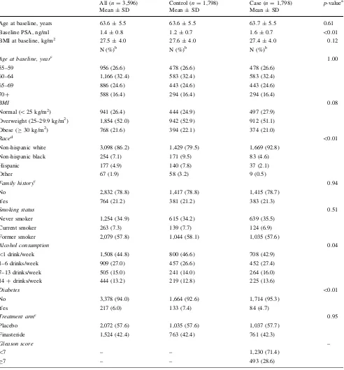
Demographic and lifestyle characteristics of the study population are given in Table1. PSA levels at baseline were significantly higher in cases than in controls. The majority of men were overweight or obese. The proportion of non-white men in controls was higher than in cases due to sampling strategy. Case and controls did not differ by smoking status; but as reported previously [18], cases were slightly more likely to consume alcohol than controls. Only a small proportion of men had diabetes at baseline, with a higher proportion in controls than in cases.
Among men in the placebo group, there were no sig-nificant associations between baseline estrone or estradiol levels and overall prostate cancer risk (Table2). Results remained similar when stratified by Gleason grade, with the exception of a reduced risk of high-grade prostate cancer in the second quartile of estrone level (OR=0.57, 95% CI=0.36–0.89). When Gleason grade of 8–10 was used as a definition of high-grade cancer, the results were similar to those of Gleason grade of 7–10 (data not shown). There were no significant effect modifications by testosterone
levels, BMI, race, diabetes, family history of prostate cancer, or cause of cancer diagnosis (data not shown).
Associations between baseline levels of estrone and estradiol among men in the finasteride group are shown in Table3. Compared to men in the lowest quartile of estrone levels, those in the highest quartiles had 43% increased risk of low-grade prostate cancer risk. Similarly, men in the highest quartile of estradiol levels had 34% increased risk of low-grade cancer risk. Revising the definition of high-grade cancer by Gleason high-grade 8–10 did not substantially change the results (data not shown). The increased risk became slightly stronger after control for testosterone, and were strongest among those without a positive family history of prostate cancer (for estrone, OR =1.77, 95% CI=1.18–2.66, P for trend\0.01; for estradiol, OR=1.79, 95% CI=1.17–2.75, P for trend=0.01). Nevertheless, interaction testings for testosterone, BMI, race, diabetes, family history of prostate cancer or cause of cancer diagnosis showed no effect modification (data not shown).
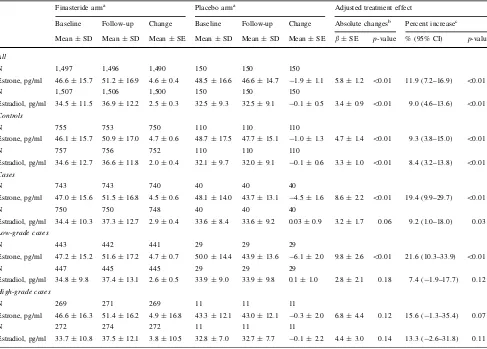
Table4 summarizes the effects of finasteride treatment on serum estrogen concentrations. Overall, concentrations of estrogens at year 3 of the trial were increased signifi-cantly in the finasteride but not in the placebo group. These effects were similar after stratification for case–control status or tumor grade, with the exception of a significant decrease in placebo men who developed low-grade cancer. After adjustment for age and baseline estrogen concentra-tions, overall treatment effects were ?11.9% for estrone and?9% for estradiol, and did not differ by case–control status or grade; although not all tests reached statistical significance among low-grade and high-grade cases due to limited sample size.
There were no significant associations of cancer risk in the finasteride group with either the absolute or the percent change in estrone or estradiol following treatment, with an exception of an inverse trend between percentage increase in estradiol concentrations and risk of low-grade cancer (Table5). However, the same inverse association was not found with the absolute change and the odds ratio con-trasting the highest to lowest quartile was not statistically significant.
Discussion
of both estrone and estradiol increased modestly following finasteride treatment; however, this increase was not associated with prostate cancer risk.
[image:5.595.53.545.76.606.2]Previous epidemiologic studies provide inconsistent results on the relationships between circulating levels of estrogens and prostate cancer risk. While most studies
Table 1 Demographic and lifestyle characteristics of cases and controls in the PCPT
All (n=3,596) Control (n=1,798) Case (n=1,798) p-valuea
Mean±SD Mean±SD Mean±SD
Age at baseline, years 63.6±5.5 63.6±5.5 63.7±5.5 0.61
Baseline PSA, ng/ml 1.4±0.8 1.2±0.7 1.6±0.7 \0.01
BMI at baseline, kg/m2 27.5±4.0 27.6±4.0 27.4±4.0 0.12
N (%)b N (%)b N (%)b
Age at baseline, yearc 1.00
55–59 956 (26.6) 478 (26.6) 478 (26.6)
60–64 1,166 (32.4) 583 (32.4) 583 (32.4)
65–69 886 (24.6) 443 (24.6) 443 (24.6)
70? 588 (16.4) 294 (16.4) 294 (16.4)
BMI 0.08
Normal (\25 kg/m2) 941 (26.4) 444 (24.9) 497 (27.9)
Overweight (25–29.9 kg/m2) 1,854 (52.0) 942 (52.9) 912 (51.1)
Obese (C30 kg/m2) 768 (21.6) 394 (22.1) 374 (21.0)
Raced \0.01
Non-hispanic white 3,098 (86.2) 1,429 (79.5) 1,669 (92.8)
Non-hispanic black 254 (7.1) 171 (9.5) 83 (4.6)
Hispanic 177 (4.9) 140 (7.8) 37 (2.1)
Other 67 (1.9) 58 (3.2) 9 (0.5)
Family historyc 0.94
No 2,832 (78.8) 1,417 (78.8) 1,415 (78.7)
Yes 764 (21.2) 381 (21.2) 383 (21.3)
Smoking status 0.51
Never smoker 1,254 (34.9) 615 (34.2) 639 (35.5)
Current smoker 263 (7.3) 139 (7.7) 124 (6.9)
Former smoker 2,079 (57.8) 1,044 (58.1) 1,035 (57.6)
Alcohol consumption 0.04
\1 drink/week 1,508 (44.8) 800 (46.6) 708 (42.9)
1–6 drinks/week 909 (27.0) 457 (26.6) 452 (27.4)
7–13 drinks/week 505 (15.0) 241 (14.0) 264 (16.0)
14?drinks/week 444 (13.2) 219 (12.8) 225 (13.6)
Diabetes \0.01
No 3,378 (94.0) 1,664 (92.6) 1,714 (95.3)
Yes 217 (6.0) 133 (7.4) 84 (4.7)
Treatment armc 0.95
Placebo 2,072 (57.6) 1,035 (57.6) 1,037 (57.7)
Finasteride 1,524 (42.4) 763 (42.4) 761 (42.3)
Gleason score –
\7 – – 1,230 (71.4)
C7 – – 493 (28.6)
a p-values are based on studentttest for continuous variables and chi-square test for unordered categorical variables between cases and controls.
For ordered categorical variables, ordinal numbers were assigned to levels and then treated as continuous variables to give trendp-values
b For specific variables, men with missing data are not shown. The count and percentage are based on those with known values
c Cases and controls are matched on age, family history, and treatment arms
found null associations [19–26], which may have driven a same conclusion in several meta- and pooled-analysis [3,
11, 12], three studies showed reduced risk among those with high estrogen levels [13–15]. In contrast, a recent case-cohort study with 275 prostate cancer cases reported an increased risk among those in the highest 3 quartiles of estrone levels, but there were no associations with estradiol levels [27]. This inconsistency may be due to the balance of the opposing effects of estrogens in the prostate, and the lack of assessment of estrogen receptor activity in the prostate gland. It has been hypothesized that estrogens may play dual and opposing roles in prostatic homeostasis and carcinogenesis [28]. Estrogens can cause abnormal prolif-eration, inflammation, and prostate malignancy, mediated by estrogen receptor a (ER-a), but they may also confer important beneficial effects, including anti-proliferation,
anti-inflammation, and anti-carcinogenesis, mediated by ER-b. The balance between activities of the two ER sub-types may dictate prostatic responses to estrogens [29]. The exact mechanisms for coordination between these opposing effects of estrogens and disruption during prostate carci-nogenesis are unclear.
[image:8.595.54.541.72.421.2]Among men in the finasteride group, high estrogen levels at baseline were associated with an increased risk of low-grade prostate cancer. It is possible that finasteride could modify the relationships between estrogens and prostate cancer risk, although a mechanism for this is not obvious. Alternatively, estrogen may modify the efficacy of finasteride for prevention of low-grade prostate cancer. Because of the nested case–control design, we could not distinguish these possibilities. This issue needs to be investigated in the entire PCPT cohort and would require
Table 4 Changes of serum estrone and estradiol concentrations between baseline and follow-up in the PCPT
Finasteride arma Placebo arma Adjusted treatment effect
Baseline Follow-up Change Baseline Follow-up Change Absolute changesb Percent increasec
Mean±SD Mean±SD Mean±SE Mean±SD Mean±SD Mean±SE b±SE p-value % (95% CI) p-value
All
N 1,497 1,496 1,490 150 150 150
Estrone, pg/ml 46.6±15.7 51.2±16.9 4.6±0.4 48.5±16.6 46.6±14.7 -1.9±1.1 5.8±1.2 \0.01 11.9 (7.2–16.9) \0.01
N 1,507 1,506 1,500 150 150 150
Estradiol, pg/ml 34.5±11.5 36.9±12.2 2.5±0.3 32.5±9.3 32.5±9.1 -0.1±0.5 3.4±0.9 \0.01 9.0 (4.6–13.6) \0.01
Controls
N 755 753 750 110 110 110
Estrone, pg/ml 46.1±15.7 50.9±17.0 4.7±0.6 48.7±17.5 47.7±15.1 -1.0±1.3 4.7±1.4 \0.01 9.3 (3.8–15.0) \0.01
N 757 756 752 110 110 110
Estradiol, pg/ml 34.6±12.7 36.6±11.8 2.0±0.4 32.1±9.7 32.0±9.1 -0.1±0.6 3.3±1.0 \0.01 8.4 (3.2–13.8) \0.01
Cases
N 743 743 740 40 40 40
Estrone, pg/ml 47.0±15.6 51.5±16.8 4.5±0.6 48.1±14.0 43.7±13.1 -4.5±1.6 8.6±2.2 \0.01 19.4 (9.9–29.7) \0.01
N 750 750 748 40 40 40
Estradiol, pg/ml 34.4±10.3 37.3±12.7 2.9±0.4 33.6±8.4 33.6±9.2 0.03±0.9 3.2±1.7 0.06 9.2 (1.0–18.0) 0.03
Low-grade cases
N 443 442 441 29 29 29
Estrone, pg/ml 47.2±15.2 51.6±17.2 4.7±0.7 50.0±14.4 43.9±13.6 -6.1±2.0 9.8±2.6 \0.01 21.6 (10.3–33.9) \0.01
N 447 445 445 29 29 29
Estradiol, pg/ml 34.8±9.8 37.4±13.1 2.6±0.5 33.9±9.0 33.9±9.8 0.1±1.0 2.8±2.1 0.18 7.4 (-1.9–17.7) 0.12
High-grade cases
N 269 271 269 11 11 11
Estrone, pg/ml 46.6±16.3 51.4±16.2 4.9±16.8 43.3±12.1 43.0±12.1 -0.3±2.0 6.8±4.4 0.12 15.6 (-1.3–35.4) 0.07
N 272 274 272 11 11 11
Estradiol, pg/ml 33.7±10.8 37.5±12.1 3.8±10.5 32.8±7.0 32.7±7.7 -0.1±2.2 4.4±3.0 0.14 13.3 (-2.6–31.8) 0.11
aEstrogen concentrations at baseline and at follow-up of approximately three years after on the study are presented in raw values. The changes are calculated as values at follow-up minus values at baseline
bTreatment effect is calculated as regression coefficient (b) of treatment arm (finasteride vs. placebo) in the linear regression model with absolute changes of estrogen concentrations as the dependent variable. Thep-values compare the mean changes in estrogen concentrations between the treatment arms, with adjustment for baseline concentrations and age
c
measurement of serum estrogen levels from all partici-pants, which is currently available only from cases and controls included in this nested study. We examined the linear correlations of baseline concentrations of estrone and estradiol with the change of 5a-androstane-3a,17b-diol glucuronide (3a-diol G), a metabolite of DHT that reflects concentrations of intra-prostatic DHT and thus indirectly measures the finasteride treatment effect, following finas-teride treatment. There were significant inverse associa-tions between baseline estrogens and change in 3a–dG (r= -0.09 for estrone andr= -0.11 for estradiol, both P\0.001). However, because the strength of these cor-relations was weak, it does not support a strong or direct effect of estrogens on finasteride treatment efficacy. It is also possible that the observed increased risk was biased by factors potentially related to estrogen. Estrogen concen-trations are positively associated with obesity; however, the effects of finasteride on cancer risk did not differ by obesity [30]. The possibility that whether other factors such as diet or physical activity modify the effects of finasteride treat-ment have not yet been evaluated.
Baseline estrogens may have been associated with high PSA and therefore bias in cancer detection; but there were no associations of estrogen concentrations with baseline PSA (Spearman correlation coefficient ranges from-0.08 to 0.05 in cases or controls from the placebo or finasteride arm, with or without adjustment for age and BMI). Lastly, we could not rule out the possibility that these findings were due to chance alone. However, the hypotheses were all a priori and the associations were observed consistently for estrone and estradiol with significant trends, which lower the likelihood of chance findings. Overall, we cannot explain the findings of a positive association of baseline estrogens with cancer risk in finasteride-treated men, which warrants further research.
We had hypothesized that the magnitudes of increases in estrogens following finasteride treatment may have increased prostate cancer risk. However, with only a single exception, neither the changes in estrogens nor the post-treatment estrogen concentrations were associated with cancer risk. There was a suggestive trend of increased risk of low-grade cancer with percentage increase in estrone; however, because the odds ratio contrasting extreme quartiles was not statistically significant and there was no association of absolute change with risk, we deem this as a chance finding. Moreover, the magnitude of changes in estrogens in the finasteride arm following treatment were modest (mean change, 4.6 pg/ml for estrone and 2.5 pg/ml for estradiol), compared to the inter-quartile differences in at baseline (for estrone, Q1B35.5 pg/ml, Q4[53.2 pg/ ml; for estradiol, Q1B26.7 pg/ml, Q4[39.4 pg/ml). Thus, it is unlikely that changes in estrogens of these small magnitudes have physiological or clinical significance.
The study benefits from vigorous annual screening by DRE and PSA test, and an end-of-study biopsy offered to all men who were cancer-free at the exit of the study. These measures greatly reduced the likelihood of undiagnosed prostate cancer in the control group and minimize mis-classification. Moreover, the PCPT adapted a centralized and standardized approach for cancer grading, minimizing misclassification. Other strengths include purification steps before RIA for estrogen measurement and a large sample size nested within a completed multi-center trial. However, several limitations should also be considered in this study. First, non-fasting blood samples were collected at different daily times and thus variations in estrogen levels throughout the day could not be controlled for; however, these variations were not likely to be systematically dif-ferent between cases and controls [17]. Second, circulating estrogens may not reflect intraprostatic levels [31]. Local expression of aromatase and production of estrogen in the prostate further complicates the relevance of circulating levels to prostate cancer etiology [32]. However, this issue is not specific to our study but common to all studies examining circulating biomarkers, and measurement of intra-prostatic hormone levels is currently not feasible for large-scale epidemiologic studies.
In summary, we found no evidence for an association of estrogen levels with prostate cancer risk among men not on finasteride. Among those taking finasteride, there may be a positive association of pretreatment estrogen concentra-tions and risk of low-grade prostate cancer; however, we know of no mechanisms that could explain such an asso-ciation. These results add to a body of previous published studies finding no associations of circulating estrogen concentrations with prostate cancer risk. The moderate increase in risk of low-grade prostate cancer associated with high baseline levels of estrogen among men taking finasteride warrants further investigation.
Acknowledgments Funded in part by grants P01 CA108964, R03
CA117490, CA054174 and CA37429 from the National Cancer Institute.
Open Access This article is distributed under the terms of the
Creative Commons Attribution Noncommercial License which per-mits any noncommercial use, distribution, and reproduction in any medium, provided the original author(s) and source are credited.
References
1. Jemal A, Siegel R, Xu J, Ward E (2010) Cancer statistics, 2010. CA Cancer J Clin 60:277–300
2. Sharifi N, Gulley JL, Dahut WL (2005) Androgen deprivation therapy for prostate cancer. JAMA 294:238–244
4. Thompson IM, Goodman PJ, Tangen CM, Lucia MS, Miller GJ et al (2003) The influence of finasteride on the development of prostate cancer. N Engl J Med 349:215–224
5. Vermeulen A, Kaufman JM, Goemaere S, van Pottelberg I (2002) Estradiol in elderly men. Aging Male 5:98–102
6. Baulieu EE (2002) Androgens and aging men. Mol Cell Endo-crinol 198:41–49
7. Krieg M, Nass R, Tunn S (1993) Effect of aging on endogenous level of 5 alpha-dihydrotestosterone, testosterone, estradiol, and estrone in epithelium and stroma of normal and hyperplastic human prostate. J Clin Endocrinol Metab 77:375–381
8. Ho SM (2004) Estrogens and anti-estrogens: key mediators of prostate carcinogenesis and new therapeutic candidates. J Cell Biochem 91:491–503
9. Bosland MC, Ford H, Horton L (1995) Induction at high inci-dence of ductal prostate adenocarcinomas in NBL/Cr and Spra-gue-Dawley Hsd:SD rats treated with a combination of testosterone and estradiol-17 beta or diethylstilbestrol. Carcino-genesis 16:1311–1317
10. McPherson SJ, Wang H, Jones ME, Pedersen J, Iismaa TP et al (2001) Elevated androgens and prolactin in aromatase-deficient mice cause enlargement, but not malignancy, of the prostate gland. Endocrinology 142:2458–2467
11. Eaton NE, Reeves GK, Appleby PN, Key TJ (1999) Endogenous sex hormones and prostate cancer: a quantitative review of pro-spective studies. Br J Cancer 80:930–934
12. Shaneyfelt T, Husein R, Bubley G, Mantzoros CS (2000) Hor-monal predictors of prostate cancer: a meta-analysis. J Clin Oncol 18:847–853
13. Chen C, Weiss NS, Stanczyk FZ, Lewis SK, DiTommaso D et al (2003) Endogenous sex hormones and prostate cancer risk: a case-control study nested within the carotene and retinol efficacy trial. Cancer Epidemiol Biomark Prev 12:1410–1416
14. Severi G, Morris HA, MacInnis RJ, English DR, Tilley W et al (2006) Circulating steroid hormones and the risk of prostate cancer. Cancer Epidemiol Biomark Prev 15:86–91
15. Gann PH, Hennekens CH, Ma J, Longcope C, Stampfer MJ (1996) Prospective study of sex hormone levels and risk of prostate cancer. J Natl Cancer Inst 88:1118–1126
16. Rittmaster RS (1994) Finasteride. N Engl J Med 330:120–125 17. Kristal AR, Schenk JM, Song Y, Arnold KB, Neuhouser ML et al
(2008) Serum steroid and sex hormone-binding globulin con-centrations and the risk of incident benign prostatic hyperplasia: results from the prostate cancer prevention trial. Am J Epidemiol 168:1416–1424
18. Gong Z, Kristal AR, Schenk JM, Tangen CM, Goodman PJ et al (2009) Alcohol consumption, finasteride, and prostate cancer risk: results from the prostate cancer prevention trial. Cancer 115:3661–3669
19. Nomura A, Heilbrun LK, Stemmermann GN, Judd HL (1988) Prediagnostic serum hormones and the risk of prostate cancer. Cancer Res 48:3515–3517
20. Barrett-Connor E, Garland C, McPhillips JB, Khaw KT, Wingard DL (1990) A prospective, population-based study of androstene-dione, estrogens, nd prostatic cancer. Cancer Res 50:169–173 21. Hsing AW, Comstock GW (1993) Serological precursors of
cancer: serum hormones and risk of subsequent prostate cancer. Cancer Epidemiol Biomark Prev 2:27–32
22. Dorgan JF, Albanes D, Virtamo J, Heinonen OP, Chandler DW et al (1998) Relationships of serum androgens and estrogens to prostate cancer risk: results from a prospective study in Finland. Cancer Epidemiol Biomark Prev 7:1069–1074
23. Mohr BA, Feldman HA, Kalish LA, Longcope C, McKinlay JB (2001) Are serum hormones associated with the risk of prostate cancer? Prospective results from the Massachusetts male aging study. Urology 57:930–935
24. Platz EA, Leitzmann MF, Rifai N, Kantoff PW, Chen YC et al (2005) Sex steroid hormones and the androgen receptor gene CAG repeat and subsequent risk of prostate cancer in the prostate-spe-cific antigen era. Cancer Epidemiol Biomark Prev 14:1262–1269 25. Tsai CJ, Cohn BA, Cirillo PM, Feldman D, Stanczyk FZ et al (2006) Sex steroid hormones in young manhood and the risk of subsequent prostate cancer: a longitudinal study in African-Americans and Caucasians (United States). Cancer Causes Con-trol 17:1237–1244
26. Barba M, Yang L, Schunemann HJ, Sperati F, Grioni S et al (2009) Urinary estrogen metabolites and prostate cancer: a case-control study and meta-analysis. J Exp Clin Cancer Res 28:135 27. Daniels NA, Nielson CM, Hoffman AR, Bauer DC (2010) Sex
hormones and the risk of incident prostate cancer. Urology 28. Ellem SJ, Risbridger GP (2009) The dual, opposing roles of
estrogen in the prostate. Ann N Y Acad Sci 1155:174–186 29. Ellem SJ, Risbridger GP (2010) Aromatase and regulating the
estrogen:androgen ratio in the prostate gland. J Steroid Biochem Mol Biol 118:246–251
30. Gong Z, Neuhouser ML, Goodman PJ, Albanes D, Chi C et al (2006) Obesity, diabetes, and risk of prostate cancer: results from the prostate cancer prevention trial. Cancer Epidemiol Biomark Prev 15:1977–1983
31. van Landeghem AA, Poortman J, Nabuurs M, Thijssen JH (1985) Endogenous concentration and subcellular distribution of estro-gens in normal and malignant human breast tissue. Cancer Res 45:2900–2906