Copyright © 2004, American Society for Microbiology. All Rights Reserved.
Intranasal Immunization with Inactivated Influenza Virus Enhances
Immune Responses to Coadministered Simian-Human
Immunodeficiency Virus-Like Particle Antigens
Sang-Moo Kang,
1† Lizheng Guo,
1† Qizhi Yao,
2Ioanna Skountzou,
1and Richard W. Compans
1*
Department of Microbiology and Immunology, Emory Vaccine Center, Emory University School of Medicine, Atlanta, Georgia,1and Michael E. DeBakey Department of Surgery, Baylor College of Medicine, Houston, Texas2
Received 28 January 2004/Accepted 27 April 2004
Intranasal immunization with inactivated influenza virus vaccine can provide protective immunity, whereas many other antigens are less effective when used for mucosal immunization. To determine whether influenza virus could enhance immune responses to an antigen coadministered to a mucosal surface, we studied the intranasal immunization of mice with a mixture of simian-human immunodeficiency virus (SHIV) virus-like particles (VLPs) and inactivated influenza virus. Compared to mice immunized with SHIV VLPs alone, mice coimmunized with SHIV VLPs and inactivated influenza virus showed significant increases in serum immu-noglobulin G (IgG) and mucosal IgA antibodies specific to the human immunodeficiency virus envelope protein, neutralizing activities, numbers of gamma interferon- and interleukin 4-secreting lymphocytes, and cytotoxic-T-lymphocyte activities. The levels of enhancement of immune response by coimmunization with inactivated influenza virus were equivalent to those induced by inclusion of immunostimulatory CpG oligode-oxynucleotides (CpG DNA). We also observed that SHIV VLPs bind to influenza virus virions, forming mixed aggregates. These results indicate that inactivated influenza virus can play a role as a mucosal adjuvant to coadministered antigens.
Most current vaccines are given parenterally by intramuscu-lar, subcutaneous, or intradermal injection, although the ma-jority of infectious agents enter their host through a mucosal surface. Parenteral delivery of nonreplicating antigen induces relatively strong systemic immune responses but very rarely induces mucosal immune responses. In contrast, mucosal an-tigen delivery can trigger mucosal immunity at both local and distant sites as well as systemic immune responses, which pro-vides an advantageous approach for immunization. Several studies indicate a crucial role for mucosal immune responses in preventing human immunodeficiency virus (HIV), simian im-munodeficiency virus (SIV), or influenza virus infection (1, 13, 18, 29, 46, 47).
A major problem in mucosal immunization is that mucosally administered antigens are frequently not effective in inducing immune responses and may induce immune tolerance. Bacte-rial toxins, such as cholera toxin and heat-labile enterotoxin, are commonly used as potent mucosal adjuvants in animal models. However, these bacterial toxins are probably not ac-ceptable for use in humans because of their toxicity (20, 28). Therefore, it is of prime importance to develop an alternative mucosal adjuvant or a mucosal immunization approach for use in humans.
Influenza virus vaccines approved in the United States are administered parenterally and consist of inactivated whole-virus or split-whole-virus preparations. Immunization of millions of people with different preparations of influenza virus vaccines
has been proven to be safe, and the vaccine is routinely ad-ministered annually, even to HIV-infected patients. Also, mu-cosal immunization, such as intranasal delivery of inactivated influenza virus in mice, provides protection against the homol-ogous virus as well as heterolhomol-ogous strains (2, 41, 42, 44, 46). Importantly, a recent study demonstrated the potential of influenza virus as an antiviral immune stimulator. Influenza virus vaccination in HIV-infected individuals and healthy con-trol subjects induced enhanced production of interleukin 2 (IL-2) and gamma interferon (IFN-␥) (38). In a different ap-proach utilizing the properties of influenza virus, our labora-tory has recently demonstrated that incorporation of influenza virus hemagglutinin glycoprotein (HA) into simian human im-munodeficiency virus-like particles (SHIV VLPs) enhanced the mucosal immune responses against the HIV antigen (16). However, it is not known whether inactivated influenza virus could play a role as an adjuvant with a coadministered antigen. In this study, we have determined the immune responses in mice after intranasal coimmunization with inactivated influ-enza virus and SHIV VLPs and compared the results with chimeric influenza virus HA-SHIV VLPs and VLPs coadmin-istered with CpG DNA.
MATERIALS AND METHODS
Cells, plasmids, adjuvants, protein, and antibodies.Spodoptera frugiperdaSf9 cells were maintained in suspension in serum-free SF900 II medium (GIBCO-BRL) at 28°C. The cell lines HeLa, EL4, and HUT78 and human MAGI were obtained through the AIDS Research and Reference Reagent Program, Division of AIDS (National Institutes of Health). EL4 and HUT78 cells were maintained in RPMI 1640 with 10% fetal calf serum, and MAGI and HeLa cells maintained in Dulbecco’s modified Eagle medium DMEM with 10% fetal calf serum at 37°C
with 5% CO2.
The HIV 89.6envplasmid was obtained from J. Sodroski (Harvard University,
Cambridge, Mass.). The pc/pS1 transfer vector was obtained from L. K. Miller (University of Georgia, Athens, Ga.).
* Corresponding author. Mailing address: Department of Microbi-ology and ImmunMicrobi-ology, Emory University School of Medicine, 1510 Clifton Rd., Atlanta, GA 30322. Phone: (404) 727-5947. Fax: (404) 727-8250. E-mail: compans@microbio.emory.edu.
† Contributed equally to this study.
9624
on November 8, 2019 by guest
http://jvi.asm.org/
Phosphorothioate oligodeoxynucleotides containing the CpG motif (CpG ODNs) were synthesized and high-performance liquid chromatography purified by Sigma-Genosys (The Woodlands, Tex.). Equal amounts of two different CpG ODNs were suspended in phosphate-buffered saline (PBS) buffer, mixed, and
used to immunize mice: one is 5⬘-TCCATGACGTTCCTGACGTT-3⬘, and the
other is 5⬘-TGACTGTGA ACGTTCGAGATGA-3⬘. Use of these CpG ODNs
as adjuvants was described previously (14, 19, 23, 30, 34).
A recombinant vaccinia virus (rVV) expressing a secreted form of the HIV Env protein was generated previously (17). The HIV gp160 protein was purified from lysates of Hela cells infected with rVV expressing HIV Env protein, using a lectin affinity column (Sigma) (12, 21, 22), and used to coat enzyme-linked immunosorbent assay (ELISA) plates. Rabbit anti-HIV gp120 serum was pro-vided by Robert Doms (University of Pennsylvania).
Production of VLPs and virus.Construction and production of SHIV VLPs and chimeric SHIV-HA VLPs were described previously (16, 48). Briefly, for production of SHIV VLPs, Sf9 insect cells were coinfected with a recombinant
baculovirus (rBV) expressing SIVmac239 gag at a multiplicity of infectivity
(MOI) of 2 and rBV expressing HIV 89.6envat an MOI of 10. For production
of chimeric SHIV-HA VLPs, Sf9 cells were coinfected with rBV SIVgagat an
MOI of 2, rBV HIV 89.6envat an MOI of 10, and rBV HA at an MOI of 5. At
day 3 postinfection, the culture medium was collected and centrifuged at 1,500⫻
gfor 20 min. The supernatant was filtered through a 0.45-m-pore-size filter, and
VLPs were pelleted at 120,000 ⫻g for 1 h at 4°C. After washing VLPs by
suspension in PBS and ultracentrifugation, pelleted VLPs were finally resus-pended in PBS and used for immunization.
Influenza virus A/PR8 (2⫻105PFU per egg) was inoculated into
10-day-embryonated hen’s eggs and incubated for 2 days at 35°C. After 2-day culture, the eggs were kept at 4°C overnight, and allantoic fluid was harvested and
centrifuged at 1,500⫻gfor 20 min to remove cell debris. The cleared
superna-tant was ultracentrifuged at 120,000⫻gfor 1 h to pellet influenza virus particles.
Pelleted influenza virus was suspended in PBS buffer and purified using sucrose gradient centrifugation, and its purity was determined by sodium dodecyl sulfate-polyacrylamide gel electrophoresis. For inactivation, purified virus was treated with 0.01% formalin at 37°C for 3 days and then dialyzed against PBS buffer. Inactivation of the virus was confirmed by inoculation of the virus into 10-day-old embryonated hen’s eggs (41).
Immunization and sample collections.Female C57BL/6J mice (Charles Riv-ers) aged 6 to 8 weeks were used. Each group consisted of six mice. Mice were
immunized intranasally to both nares with SHIV VLPs (50g), chimeric HA/
SHIV VLPs (50g), SHIV VLPs (50g) plus inactivated influenza virus (10g)
(mixed overnight at 4°C before immunization), or CpG DNA (10g) at weeks
0, 2, and 4. All immunizing reagents were suspended in 30l of PBS, and
individual mice received 15l (7.5l each nose) two times with a 2-h resting
interval between doses. Mice were slightly isoflurane-anesthetized and held in-verted with nose down until droplets of vaccine that were applied to both external nares were completely inhaled.
Blood samples were collected by retro-orbital plexus puncture after
anesthe-tizing with isoflurane. Saliva was collected after intraperitoneal injection of 2g
of carbamylcholine chloride to stimulate saliva flow, and vaginal lavages were
collected by washing the vagina with 200l of PBS. Lung tissue was collected at
week 2 after final immunization, washed three times in a large volume of PBS to wash out any contaminating blood, cut into small pieces (2 to 3 mm), suspended
in extraction buffer (2% saponin, 0.1% NaN3in PBS), and rocked overnight (100
mg of lung in 400l of extraction buffer). Five to eight pieces of freshly voided
feces were collected at week 6, weighed, and resuspended in PBS with protease
inhibitors at a ratio of 5l of feces/mg to standardize for variability in the
amount of fecal material collected. The solid feces were resuspended by vortex-ing vigorously until solutions were homogenous. Samples were then spun in a microcentrifuge for 10 min, and supernatants were collected. All samples were
stored at⫺20°C prior to antibody titration.
ELISA.All sera and mucosal secretions were individually collected, and con-centrations of immunoglobulin G (IgG), IgG1, IgG2a, and IgA antibodies to HIV 89.6 Env or influenza virus were determined by ELISA as described
pre-viously (23). Ninety-six-well Microtiter plates (Nunc) were coated with 100l of
purified HIV Env protein (2g/ml) or purified PR8 virus (2g/ml) per well in
borate-buffered saline at 4°C overnight. Plates were blocked with PBS containing 2% bovine serum albumin at 37°C for 1 h. After three washes in PBS containing 0.05% Tween 20, 100-fold-diluted sera or 5-fold diluted mucosal secretion sam-ples were added to the wells and incubated at 4°C overnight. After four washes, the wells were treated with goat anti-mouse IgG, IgG1, IgG2a, or IgA-horserad-ish peroxidase conjugates (SouthernBiotech) for 1 h at room temperature. After removal of the unbound conjugates and washing, horseradish peroxidase
sub-strate, 2,2⬘-azino-bis(3-ethylbenzthiazoline-sulfonic acid containing H2O2
(Sigma) was used to develop color, and the optical density at 405 nm was read by an ELISA reader (Bio-Tek). Antibody concentrations were determined by com-paring the readings for experimental samples with the standard curves for puri-fied mouse IgG, IgG1, IgG2a, and IgA and are represented as the arithmetic
mean⫾1 standard error. Data were analyzed by using the Excel program
(Microsoft). The statistical significance of the difference between groups was
calculated by Student’s two-tailedttest.
Neutralization assays.Neutralization assays were performed using MAGI cells in a 96-well plate and as described previously (8, 16, 23). HIV 89.6 stocks were grown in HUT78 cells obtained from American Type Culture Collection. In
brief, MAGI cells (3.5⫻103cells per well) were added to a 96-well plate 1 day
before infection. Serum samples were heat inactivated (56°C, 30 min), and 25l
of serially twofold-diluted samples in DMEM growth medium was added to an equal volume of virus stocks diluted in DMEM to contain 100 infectious parti-cles. The virus-serum mixtures were incubated at 37°C for 1 h and then added to
MAGI cells with DEAE-Dextran (15g/ml). After 2 h of incubation, an
addi-tional 200l of DMEM was added. Medium was replaced after 1 day of
incu-bation with DMEM containing 10M zidovudine (Sigma). At day 3
postinfec-tion, medium was removed and cells were fixed and stained as described previously (8). The blue cells in the wells without antiserum indicated the total number of infectious virus particles. Neutralization titers were presented as dilutions giving a 50% reduction in number of blue cells.
ELISPOT assays.Spleen obtained from an individual mouse was minced between the frosted ends of glass slides, and spleen cells were isolated as de-scribed previously (23). The live spleen cells were counted by light microscopy after staining with trypan blue and used for enzyme-linked immunosorbent spot (ELISPOT) and cytotoxic-T-lymphocyte (CTL) analysis. All antibodies used in ELISPOT assays were purchased from PharMingen (San Diego). Anti-mouse
IFN-␥(R4-6A2) and IL-4 (BVD4-1D11) antibodies (4g/ml in PBS) were used
to coat Multiscreen 96-well filtration plates (Millipore) at 4°C overnight. After blocking the antibody-coated plates with 10% fetal bovine serum in RPMI,
freshly isolated splenocytes (106cells) were added to each well. For in vitro
stimulation, SIV Gag peptide (amino acids 186 to 205; INQMLNCVGDHQAA
MQIIRD) (24, 27) at a concentration of 5g/ml, inactivated influenza virus (5
g/ml), or influenza virus nucleoprotein peptides (36) (NP1, ASNENMDTM;
NP2, ARSALILRGSVAHKSCLP ACVYGP) at a concentration of 2g/ml was
added and incubated for 36 h at 37°C. For Env-specific stimulation, EL-4 cells
(105cells per well) infected with rVV expressing HIV Env were mixed with
spleen cells and incubated for 36 h at 37°C following a procedure as described previously (16). After six washes with 0.05% Tween 20, biotinylated anti-mouse
IFN-␥(XMG1.2) and IL-4 (BVD6-24G2) antibodies (1g/ml) were added to
the plates. After washing, spots in the plates were developed with stable diami-nobenzidine (Research Genetics) for 3 to 5 min and counted in an ImmunoSpot ELISpot reader (Cellular Technology). Splenocytes from naive mice or wells not containing peptides were used as negative controls.
CTL analysis.EL-4 target cells (H-2b) were prepared by infecting cells with
rVV expressing the HIV Env protein or rVV expressing the influenza virus HA
protein at an MOI of 1. Infected EL-4 cells (104cells) were harvested at 5 to 7 h
postinfection and incubated with fresh splenocytes from immunized or naive mice at different effector-to-target cell ratios. The amount of cell lysis was measured using the CytoTox96 nonradioactive cytotoxicity assay (Promega, Madison, Wis.). This assay measures lactate dehydrogenase, a stable cytosolic
enzyme that is released upon cell lysis, in the same way51Cr is released in a
radioactive assay. The results from these two assays were shown to be almost identical (10, 25), and several studies have used this nonradioactive cytotoxic assay for CTL analysis (3, 6, 33). The percentage of specific target cell lysis was represented based on total cell lysis with 0.1% Triton X-100.
HI by SHIV VLPs.To determine HA titers, inactivated influenza virus was
serially diluted in 100l of PBS deficient in Mg2⫹and Ca2⫹, mixed with an equal
volume of a fresh 0.5% suspension of chicken red blood cells (Lampire Biological Laboratories), and incubated for 2 h at 4°C. The titers were determined as the endpoint dilutions inhibiting the precipitation of red blood cells. Following the procedure as previously described (9), hemagglutination inhibition (HI) activity of SHIV VLPs was determined. SHIV VLPs were serially diluted, mixed with 8 HA units of inactivated influenza virus, incubated for 30 min at room tempera-ture, and then mixed with chicken red blood cells. The HI titers of SHIV VLPs were determined as the end-point dilutions showing inhibition of HA activity.
RESULTS
Serum antibody responses after intranasal coimmunization with SHIV VLPs and inactivated influenza virus.In a previous
on November 8, 2019 by guest
http://jvi.asm.org/
study, we have shown that incorporating the influenza virus HA protein into SHIV VLPs enhanced the mucosal immune responses to the HIV Env protein (16). It was suggested that chimeric SHIV-HA VLP targeting to mucosal cells would be increased due to the binding properties of influenza virus HA protein for neuraminic acid expressed on mucosal epithelial cells. Here, we investigated whether coimmunization with in-activated influenza virus would induce enhanced immune re-sponses to SHIV VLP antigen. Also, as a comparison, we included a group of mice immunized with SHIV VLPs plus CpG oligonucleotides, which have been shown to be a strong mucosal adjuvant with various antigens (14, 19, 30, 31, 34).
Serum samples were collected 2 weeks after each intranasal immunization. After the second immunization, HIV Env-spe-cific antibodies were easily detected in all groups of mice (Fig. 1A). Levels of antibodies were almost twofold higher in mice immunized with chimeric SHIV-HA VLPs than in mice immu-nized with SHIV VLPs alone, as previously shown (16). Inter-estingly, similar twofold-increased levels of antibodies were also observed in mice intranasally coimmunized with SHIV VLPs and inactivated influenza virus, which were statistically significant as determined using Student’sttest (P⬍0.05). A group of mice immunized with SHIV VLPs plus CpG DNA as an adjuvant also showed high levels of HIV Env binding anti-bodies. The difference between mice immunized with chimeric SHIV-HA VLPs or SHIV VLPs plus inactivated influenza virus and mice coimmunized with SHIV VLPs and CpG DNA was not significant (P⬎0.1). These results suggest that
addi-tion of inactivated influenza virus increases inducaddi-tion of anti-body to coadministered SHIV VLP antigen.
Analysis of antibody isotypes was used to determine types of immune responses (Fig. 1B). Serum samples from mice immu-nized with VLPs or chimeric HA/VLPs showed similar levels of HIV Env-binding IgG1 and IgG2a antibodies, while serum samples from mice immunized with VLPs plus inactivated in-fluenza virus showed higher levels of IgG2a than those of IgG1. Overall, mice immunized with VLPs, chimeric HA/ VLPs, or VLPs plus inactivated influenza virus showed mixed T-helper (Th) type 1 and 2 responses. In contrast, mice immu-nized with VLPs plus CpG DNA displayed fourfold-higher levels of IgG2a antibodies, indicating preferential Th1 immune responses.
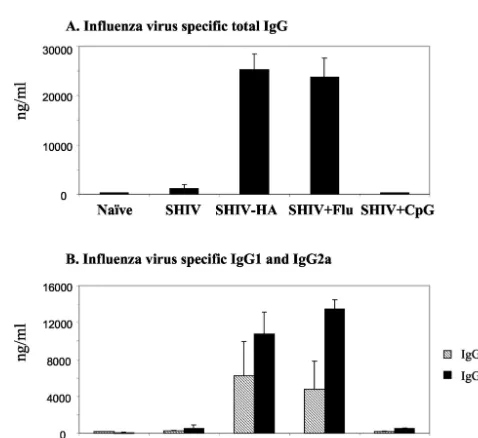
[image:3.603.44.281.69.282.2]Antibody responses against influenza virus. We also ana-lyzed immune responses against influenza virus administered together with SHIV VLPs (Fig. 2). High levels of serum anti-bodies binding to influenza virus were detected in groups of mice immunized with chimeric HA VLPs or VLPs plus inac-tivated influenza virus, and the levels were similar in these two groups (Fig. 2A). Levels of influenza virus binding antibodies in other groups of mice immunized with VLPs or VLPs plus CpG DNA were as low as levels with a naive control, indicating that these antibodies were induced specifically against influ-enza viral antigens. Isotype analysis indicated that higher levels of IgG2a than IgG1 binding to influenza virus were induced, which was more prominent in mice immunized with VLPs plus
FIG. 1. Humoral immune responses against HIV Env after intra-nasal immunization of C57BL/6J mice with SHIV VLPs. Serum sam-ples were collected before immunization (0 wk) and at 2 weeks after the first (2 wk), second (4 wk), and third (6 wk) immunizations, and antibodies were analyzed using ELISA plates coated with purified HIV Env gp160. (A) Anti-HIV Env-specific IgG. (B) Isotypes of IgG1 and IgG2a specific to HIV Env in sera after the third immunization (6 weeks). Error bars are given for six mice in a group. SHIV, SHIV VLPs alone; SHIV-HA, chimeric SHIV-HA VLPs; SHIV⫹Flu, SHIV VLPs coadministered with inactivated influenza virus; SHIV⫹CpG, SHIV VLPs coadministered with CpG DNA. ⴱ, P ⬍ 0.05 (calculated by Student’s two-tailedttest, compared to VLP alone).
FIG. 2. Humoral immune responses against influenza virus after intranasal immunization with chimeric SHIV-HA VLPs or a mixture of SHIV VLPs and inactivated influenza virus. Serum samples were col-lected at week 6 after the final immunizations, and antibodies were analyzed using ELISA plates coated with influenza virus. (A) Serum IgG specific to influenza virus. (B) Isotypes of IgG1 and IgG2a specific to influenza virus in sera after the third immunization (6 weeks). Error bars are given for six mice in a group. Naive, unimmunized mice; SHIV, SHIV VLPs alone; SHIV-HA, chimeric SHIV-HA VLPs; SHIV⫹Flu, SHIV VLPs coadministered with inactivated influenza virus; SHIV⫹CpG, SHIV VLPs coadministered with CpG DNA.
on November 8, 2019 by guest
http://jvi.asm.org/
[image:3.603.300.539.406.625.2]inactivated influenza virus than in mice immunized with chi-meric HA VLPs (Fig. 2B).
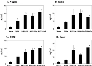
Effect of influenza virus on mucosal IgA responses to HIV Env. Induction of mucosal IgA is one of the key criteria in evaluating mucosal vaccines. Mucosal IgA antibodies binding to the HIV Env protein in vaginal washes, secretory saliva, and lung and fecal extracts were measured after the third immuni-zation as shown in Fig. 3. Intranasal immuniimmuni-zation with VLPs without any adjuvant elicited HIV Env-binding IgA antibodies in most mucosal secretions, although the levels were low. Inclusion of inactivated influenza virus in the intranasal immu-nization with VLPs enhanced the HIV Env-specific IgA anti-bodies approximately twofold in all mucosal secretions ana-lyzed. Interestingly, levels of mucosal IgA antibodies binding to HIV Env in this group of mice were equivalent to those elicited in mice immunized with chimeric HA VLPs or VLPs plus CpG DNA as an adjuvant. These results clearly indicate that inactivated influenza virus can play a role as an adjuvant in enhancing mucosal immune responses to SHIV VLPs.
Effect of influenza virus on induction of neutralizing anti-bodies.One of the important elements for evaluating vaccines is the ability to induce neutralizing antibodies. In mice, non-specific neutralizing activity by nonimmune sera has been pre-viously observed. Using luciferase reporter viruses pseudo-typed with specific HIV Env, normal mouse serum contributed to 20 to 50% neutralization activity of immune sera (11). Other studies using syncytium inhibition (32) or p24 reduction assay
(37) showed very low background activity from preimmune sera, which was less than 10% of immune serum. Our MAGI assay as a measure of neutralization activity showed 25 to 40% of nonspecific background activity compared to those of mune sera, and this difference was consistent with other im-mune sera, validating the neutralizing assay (16) (Fig. 4). Com-pared to that of normal sera, low neutralizing titers could be detected in mice immunized with SHIV VLPs alone. However, the levels of neutralizing activities were enhanced two- to threefold in mice coimmunized with SHIV VLPs and inacti-vated influenza virus compared to normal sera and were sim-ilar to those elicited in groups of mice immunized with chi-meric HA VLPs or VLPs plus CpG DNA as an adjuvant.
The induction of neutralizing antibody activities in mucosal secretions has not been well studied. The pools of vaginal lavages from three mice (two pools in each group) were ana-lyzed to determine the neutralizing activities (Fig. 4B). Weak neutralizing activities were observed in vaginal washes from mice immunized with SHIV VLPs. The groups of mice immu-nized with chimeric HA VLPs, VLPs plus inactivated influenza virus, or VLPs plus CpG DNA showed levels moderately higher than those of the VLP group. These results suggest that inclusion of inactivated influenza virus in SHIV VLP immuni-zation may have an adjuvant effect on induction of HIV-neu-tralizing antibodies.
[image:4.603.99.481.69.345.2]Cytokine production.It is considered to be advantageous for an HIV vaccine to induce two arms of immune responses,
FIG. 3. Mucosal IgA responses to HIV Env after intranasal immunization with SHIV VLPs. Mucosal secretion samples were collected at week 2 after the final immunization (week 6). For vaginal wash and saliva, preimmune samples were used as controls. For lung and fecal extracts, samples from naive (unimmunized) mice were used as controls. (A) IgA in vaginal wash. (B) IgA in saliva. (C) IgA in lung extracts. D) IgA in fecal samples. Standard error bars are given for six mice in a group. SHIV, SHIV VLPs alone; SHIV-HA, chimeric SHIV-HA VLPs; SHIV⫹Flu, SHIV VLPs plus inactivated influenza virus; SHIV⫹CpG, SHIV VLPs plus CpG DNA. The statistical significance of the difference between the SHIV VLP group and other groups was calculated by Student’sttest:ⴱ,P⬍0.05.
on November 8, 2019 by guest
http://jvi.asm.org/
humoral and cellular immune responses. One approach for assessing cellular immune responses is to analyze the pattern of cytokine production. Spleen cells of immunized mice were isolated at 2 weeks after the last immunization, and IFN-␥- and IL-4-secreting lymphocytes in response to stimulation of HIV Env-expressing EL-4 cells or SIV Gag peptide were analyzed by ELISPOT assay (Fig. 5). Immunization with inactivated influenza virus plus SHIV VLPs greatly increased the numbers of IFN-␥-producing cells, by 2.6-fold (Env specific) and 2.7-fold (Gag specific), respectively, over that of the SHIV-alone group (P⬍0.05), and levels were equivalent to those induced by CpG DNA (Fig. 5A).
IL-4 cytokine production is known to be a good indicator for induction of Th2-type immune responses. To determine IL-4 production in immunized mice, a method similar to that used with IFN-␥was employed, using an 4 ELISPOT assay. IL-4-secreting lymphocytes were not significantly increased in groups of mice immunized with chimeric HA VLPs or with VLPs plus CpG DNA compared to mice immunized with SHIV VLPs alone (Fig. 5B). The group of mice immunized with SHIV VLPs plus inactivated influenza virus showed the highest levels of IL-4-secreting lymphocytes, especially in re-sponse to Gag peptide stimulation, increasing 3.2-fold over that of the SHIV alone group.
Since some groups of mice received influenza virus or the HA component in their immunization protocol, we also deter-mined influenza virus-specific IFN-␥- and IL-4-secreting lym-phocytes. Compared to that in mice immunized with SHIV alone, the number of IFN-␥-secreting spleen cells when stim-ulated with inactivated whole virus was increased 2.1- and
2.4-fold in mice immunized with chimeric SHIV-HA VLPs and in mice coimmunized with SHIV VLPs plus inactivated influ-enza virus, respectively, which is statistically significant (Fig. 6A). In response to influenza virus nucleoprotein peptide stim-ulation, the group of mice coimmunized with SHIV VLPs and inactivated influenza virus showed a significantly enhanced level of 3.4-fold in IFN-␥-secreting lymphocytes. The mice which received SHIV-HA VLPs showed a slight increase, al-though they were not exposed to the influenza virus nucleo-protein component. This may be due to bystander activation, since a high frequency of cross-reactive T-cell activation has been reported during virus-induced polyclonal responses (49). Also, mice which were coimmunized with inactivated influenza virus displayed enhanced levels of IL-4-secreting lymphocytes by 3.1-fold (Fig. 6B).
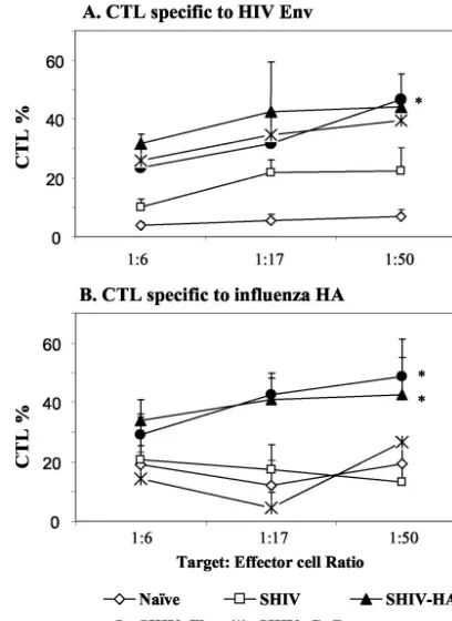
Cellular immune responses.Since effective cytolytic activity is important in controlling HIV infection, we determined di-rectly the CTL activity using a nonradioactive CTL assay. EL-4 cells infected with rVV expressing HIV 89.6 Env were used as target cells, and spleen cells were used as effector cells. Intra-nasal immunization with SHIV VLPs induced a low level of CTL activity in the spleen, although a higher level than that from naive controls (Fig. 7A). Coimmunization with inacti-vated influenza virus and SHIV VLPs resulted in twofold-enhanced CTL activity, at levels similar to or higher than those found after immunization with chimeric HA VLPs or with VLPs plus CpG DNA at an effector-to-target cell ratio of 50 (P⬍0.05).
[image:5.603.57.264.69.253.2]FIG. 4. HIV neutralizing titers. The results are averages from three independent assays. Data are represented as n-fold dilutions giving 50% neutralization activities. (A) Neutralizing titers in serum samples. (B) Neutralizing titers in vaginal washes. For neutralization assays, vaginal wash samples collected at week 6 were pooled together (three pools per group). Sample pools from the naive and SHIV⫹Flu groups did not show variation within the group. Naive, unimmunized control (no neutralizing activity in vaginal wash pools from naive mice⫽1); SHIV, SHIV VLPs alone; SHIV-HA, chimeric SHIV-HA VLPs; SHIV⫹Flu, SHIV VLPs plus inactivated influenza virus; SHIV⫹CpG, SHIV VLPs plus CpG DNA. The statistical significance of the differ-ence between the SHIV VLP group and the other groups was calcu-lated by Student’sttest:ⴱ,P⬍0.05.
FIG. 5. ELISPOT assays of cytokine-producing lymphocytes in re-sponse to stimulation with HIV Env or SIV Gag peptides. Splenocytes were isolated at week 2 after the final immunization (week 6) and stimulated for 1.5 days with EL-4 cells infected with rVV expressing HIV Env or SIV Gag peptides. (A) The spots for IFN-␥-producing cells were expressed based on 106 spleen cells. (B) The spots for
IL-4-producing cells were expressed based on 106spleen cells.
Stan-dard error bars are indicated for six mice in a group. Naive, unimmu-nized control; SHIV, SHIV VLPs alone; HA, chimeric SHIV-HA VLPs; SHIV⫹Flu, SHIV VLPs plus inactivated influenza virus; SHIV⫹CpG, SHIV VLPs plus CpG DNA. The statistical significance of the difference between the SHIV VLP group and the other groups was calculated by Student’sttest:ⴱ,P⬍0.05.
on November 8, 2019 by guest
http://jvi.asm.org/
To determine the CTL activities specific for influenza virus, EL-4 cells infected with rVV expressing influenza virus HA protein were used as target cells (Fig. 7B). Spleen cells from mice immunized with VLPs alone or VLPs plus CpG DNA showed only background levels of CTL activities, similar to those from naive controls. Immunization with chimeric HA VLPs or coimmunization with inactivated influenza virus and VLPs induced significantly higher levels of CTL activities against the influenza virus HA protein (P⬍0.05). In summary, inactivated influenza virus included in the intranasal immuni-zation with SHIV VLPs elicited enhanced cellular immune responses against coadministered SHIV VLPs antigen as well as against influenza virus.
Formation of influenza virus-VLP complexes.In order to further investigate the possible mechanism by which inacti-vated influenza virus particles could enhance the immune re-sponse to VLPs, we determined whether there is a direct as-sociation between influenza virus virions and SHIV VLPs. Previously, influenza virus virions were shown to form mixed aggregates with several other types of enveloped virus parti-cles, and such aggregation resulted in a loss of hemagglutina-tion activity (9). We therefore performed an HI test with SHIV VLPs. As shown in Table 1, we found that SHIV VLPs showed strong inhibition of HA activity of influenza virus, suggesting the possibility of aggregate formation. This was confirmed by direct demonstration of aggregates of influenza virus virions with SHIV VLPs by electron microscopy (Fig. 8).
The formation of complexes between influenza virus virions
[image:6.603.317.521.70.350.2]and other types of enveloped viruses was previously shown to depend on the presence of neuraminic acid residues on virion surfaces, which serve as receptors for binding to the influenza virus HA protein. However, it seemed unlikely that neuraminic acid would be present on surfaces of VLPs grown in insect cells, because these cells are not known to synthesize complex carbohydrate modifications on their surface glycoproteins (26). Consistent with this, we observed that treatment of SHIV
FIG. 6. ELISPOT results of cytokine-producing lymphocytes in re-sponse to stimulation with influenza virus HA protein or nucleoprotein peptide. Splenocytes isolated at week 6 were stimulated for 2 days with EL-4 cells infected with rVV expressing influenza virus HA protein or nucleoprotein peptides. (A) IFN-␥-producing spots. H-2b-restricted
major histocompatibility complex class I peptide NP1 (ASNENMD TM) was used for IFN-␥stimulation as a nucleoprotein peptide. (B) IL-4-producing spots. H-2b-restricted class II peptide NP2 (ARSA
LILRGSVAHKSCLPACVYGP) was used for IL-4 stimulation. Naive, unimmunized control; SHIV, SHIV VLPs alone; SHIV-HA, chimeric SHIV-HA VLPs; SHIV⫹Flu, SHIV VLPs plus inactivated influenza virus. The statistical significance of the difference between the SHIV VLP group and the other groups was calculated by Student’s ttest: ⴱ,P⬍0.05.
[image:6.603.53.274.70.267.2]FIG. 7. CTL activities of spleen cells. EL-4 cells infected with a rVV expressing HIV Env protein or influenza virus HA protein were used as target cells. Target cell lysis was determined using a nonradio-active CTL assay. CTL activities are represented as percentages of target cell lysis. The results are shown with standard error bars for six mice in a group. The ratio on thexaxis is the effector/target cell ratio. (A) CTL lysis of EL-4 cells expressing HIV Env protein. (B) CTL lysis of EL-4 cells expressing influenza virus HA protein. Naive unimmu-nized control; SHIV, SHIV VLPs alone; HA, chimeric SHIV-HA VLPs; SHIV⫹Flu, SHIV VLPs plus inactivated influenza virus; SHIV⫹CpG, SHIV VLPs plus CpG DNA. The statistical significance of the difference between the SHIV VLP group and the other groups was calculated by Student’sttest:ⴱ,P⬍0.05.
TABLE 1. HI activity of SHIV VLPsa
Sample Neuraminidaseb Titer of HI
SHIV VLPs (1 mg/ml) ⫺ 160
SHIV VLPs (1 mg/ml) ⫹ 160
Measles virus (107PFU/ml) ⫺ 192
Measles virus (107PFU/ml) ⫹ 24
aThe inactivated influenza virus (2 mg/ml) had an HA titer of 6,400, and 8 HA
units were added to each well for HI assay. The results are averages from two independent assays. Measles virus (a gift from Richard Plemper, Emory Univer-sity) was used as a positive control for HI activity.
bSHIV VLPs or measles virus was treated with 1.4 U of protease-free
neur-aminidase (Roche, Germany)/ml for 2 h at 37°C.
on November 8, 2019 by guest
http://jvi.asm.org/
[image:6.603.301.541.618.675.2]VLPs with neuraminidase had no effect on their HI activity (Table 1), whereas similar treatment of measles virus caused a reduction in its HI activity. Thus, the formation of complexes between SHIV VLPs and influenza virus virions involves an interaction which does not depend on the presence of sialic acid residues on the VLP surfaces.
DISCUSSION
We have determined the effects of inactivated influenza vi-rus on enhancing humoral and cellular immune responses after intranasal coimmunization with SHIV VLPs. We found that addition of inactivated influenza virus to VLPs resulted in enhanced serum IgG and mucosal secretory IgA antibodies against the HIV Env protein, an increase in neutralizing activ-ity against HIV virus, and increased numbers of IFN-␥- and IL-4-secreting lymphocytes and CTL activities. The results ob-tained in this study clearly indicate that inactivated influenza virus can play a role as an adjuvant to the coadministered VLPs.
It was previously shown that influenza virus particles bind rapidly to other enveloped virus particles, such as vesicular stomatitis virus, Sindbis virus, or murine leukemia virus, form-ing mixed aggregates (9), and that this bindform-ing results from the influenza virus HA protein binding to neuraminic acid residues on the surfaces on these viral particles. There is no evidence for the presence of neuraminic acid on the insect cell surface due to their limited capacity to process N-glycans (26); there-fore, it was surprising that SHIV VLPs were found to inhibit hemagglutination activity of influenza virus. Neither preim-mune nor impreim-mune sera specific to influenza virus or SHIV VLPs blocked the HI activity of SHIV VLPs (data not shown). We also observed that this HI activity was not diminished by treatment of VLPs with neuraminidase, indicating that the inhibition is not mediated via interaction of neuraminic acid on VLPs with the HA of influenza virus, consistent with a recent study showing that cell binding properties of murine pneumo-tropic virus VLPs obtained in an rBV system were
neuramin-idase resistant (45). However, it was found by electron micros-copy that inactive influenza virus is able to form aggregates with SHIV VLPs. It is thus plausible that formation of com-plexes between influenza virus particles and SHIV VLPs could result in enhanced binding of VLPs with mucosal surfaces, which may be a mechanism for enhancing immune responses. It was also previously demonstrated that the influenza virus HA glycoprotein induced vigorous B-cell proliferation and im-munoglobulin synthesis (39, 40). The polyclonal B-cell-activat-ing properties of HA or a combination of this activity with enhanced interaction of VLP aggregates with mucosal surfaces may play roles in enhancing immune responses against the coadministered VLP antigens.
Stimulation of cytokine production or activation of lympho-cytes by inactivated influenza virus may also contribute to enhanced immune responses. We observed an increase in lev-els of IFN-␥, IL-4, and CTL activities produced by spleen cells from mice immunized with a mixture of SHIV VLPs and in-activated influenza virus, which were higher than those from mice immunized with chimeric SHIV-HA VLPs. Previous studies demonstrated that inactivated influenza virus is an ef-fective immunogen for inducing cellular immune responses; dendritic cells pulsed with inactivated influenza virus induced strong CTL responses (4). A recent study demonstrated that vaccination of control subjects and HIV patients with an inac-tivated trivalent influenza virus vaccine formulation induced increases in production of IL-2 and IFN-␥, resulting in anti-HIV activity (38). Also, peripheral blood mononuclear cells from healthy donors, when stimulated in vitro with inactivated or live influenza virus, displayed increases in IFN-␥production and lymphocyte proliferation (5). A combination of these ef-fects of influenza virus may contribute to enhancing immune responses.
The pattern of antibody isotypes is known to be influenced by the type of T-cell immune responses. Live influenza virus injected intraperitoneally resulted in high IFN-␥, low IL-4, high IgG2a, and low IgG1 levels, which is characteristic of a Th1-type response, while the same route of immunization with inactivated influenza virus induced the reversed pattern of immune responses characteristic for Th2-type responses (35). We found that intranasal immunization with SHIV VLPs plus inactivated influenza virus induced slightly higher levels of IgG2a than those of IgG1 and produced both IFN-␥and IL-4 cytokines after in vitro stimulation specific for HIV Env as well as specific to influenza virus, representing both Th1 and Th2 immune responses. In contrast to inactivated influenza virus, CpG DNA coadministered with VLPs induced predominantly Th1-type immune responses. In another study, intranasal in-fection with live influenza virus induced both IgG2a and IgG1 isotypes at similar levels and IgE antibodies as well as IFN-␥
production, indicating a mixed type of immune response (15). These studies indicate that the type of immune response also may be affected by the route of immunization.
[image:7.603.80.242.70.239.2]The mucosal immune compartment is composed of lym-phoid tissues in mucosae and external secretory glands (7). In contrast to systemic immunization, mucosal immunization is capable of stimulating both mucosal and systemic immunity. In a comparative study of intranasal (mucosal) and subcutaneous (systemic) immunization with inactivated influenza virus vac-cines, both induced comparable levels of IgG and neutralizing
FIG. 8. Electron microscopy showing an aggregate of spherical VLPs with densely stained cores (arrows) and spike-covered influenza virus virions, negatively stained with sodium phosphotungstate stain. Magnification,⫻40,000 (two times enlarged from the original picture for clear presentation).
on November 8, 2019 by guest
http://jvi.asm.org/
activity in serum (44). Interestingly, intranasal immunization resulted in better protection against heterosubtypic mucosal virus challenge than subcutaneous immunization, due to the high cross-reactivity of IgA antibody in mucosal secretions (42–44, 46). We found that intranasal coimmunization with inactivated influenza virus and SHIV VLPs significantly in-creased the levels of IgA antibodies in various mucosal sites as well as serum IgG antibodies against HIV and influenza vi-ruses. Therefore, intranasal administration provides an advan-tageous approach for developing mucosal vaccines which may be more broadly cross-reactive with HIV isolates of distinct antigenic subtypes.
To our knowledge, this is the first report demonstrating an adjuvant effect of inactivated influenza virus. Inactivated influ-enza virus has been used for vaccination for more than 60 years since its discovery, proving its safety for human use, which is the most important consideration in developing a human vac-cine. However, the effect of preexisting immunity should be considered in evaluating efficacy of combination vaccines con-taining influenza virus. It is not certain whether the existence of such preexisting immunity would negatively impact the im-mune responses to nonreplicating VLPs combined with inac-tivated influenza virus in contrast to immunization using rep-licating vectors. It is also possible that production of immune complexes could enhance immune responses by targeting to follicular dendritic cells and macrophages or stimulate preex-isting immunity by induction of cytokine production, thus re-sulting in increased immune responses to VLPs. An alternative approach is to utilize influenza virus subtypes to which there is no preexisting immunity in the human population, therefore avoiding potential effects of preexisting anti-HA immunity on induction of immune responses against VLP antigens. De-veloping safe and effective combination vaccines can simplify vaccine delivery, and combination of vaccine antigens against several diseases simultaneously would greatly benefit public health.
ACKNOWLEDGMENTS
S.-M.K is a recipient of AmfAR fellowship award 70587-32-RF, and this work was supported in part by NIH/NIAID grants AI28147, AI30042, and AI057017-01.
REFERENCES
1. Alfsen, A., P. Iniguez, E. Bouguyon, and M. Bomsel.2001. Secretory IgA specific for a conserved epitope on gp41 envelope glycoprotein inhibits
epithelial transcytosis of HIV-1. J. Immunol.166:6257–6265.
2. Armerding, D., H. Rossiter, I. Ghazzouli, and E. Liehl.1982. Evaluation of live and inactivated influenza A virus vaccines in a mouse model. J. Infect.
Dis.145:320–330.
3. Behl, C., J. B. Davis, R. Lesley, and D. Schubert.1994. Hydrogen peroxide
mediates amyloid beta protein toxicity. Cell77:817–827.
4. Bender, A., L. K. Bui, M. A. Feldman, M. Larsson, and N. Bhardwaj.1995. Inactivated influenza virus, when presented on dendritic cells, elicits human
CD8⫹cytolytic T cell responses. J. Exp. Med.182:1663–1671.
5. Blazevic, V., C. M. Trubey, and G. M. Shearer.2000. Comparison of in vitro immunostimulatory potential of live and inactivated influenza viruses. Hum.
Immunol.61:845–849.
6. Brander, C., T. Wyss-Coray, D. Mauri, F. Bettens, and W. J. Pichler.1993. Carrier-mediated uptake and presentation of a major histocompatibility
complex class I-restricted peptide. Eur. J. Immunol.23:3217–3223.
7. Brandtzaeg, P.1989. Overview of the mucosal immune system. Curr. Top.
Microbiol. Immunol.146:13–25.
8. Chackerian, B., D. R. Lowy, and J. T. Schiller.2001. Conjugation of a self-antigen to papillomavirus-like particles allows for efficient induction of
protective autoantibodies. J. Clin. Investig.108:415–423.
9. Compans, R. W.1974. Hemagglutination-inhibition: rapid assay for
neura-minic acid-containing viruses. J. Virol.14:1307–1309.
10. Decker, T., and M. L. Lohmann-Matthes.1988. A quick and simple method for the quantitation of lactate dehydrogenase release in measurements of cellular cytotoxicity and tumor necrosis factor (TNF) activity. J. Immunol.
Methods115:61–69.
11. Dong, M., P. F. Zhang, F. Grieder, J. Lee, G. Krishnamurthy, T. VanCott, C. Broder, V. R. Polonis, X. F. Yu, Y. Shao, D. Faix, P. Valente, and G. V. Quinnan, Jr.2003. Induction of primary virus-cross-reactive human immu-nodeficiency virus type 1-neutralizing antibodies in small animals by using an
alphavirus-derived in vivo expression system. J. Virol.77:3119–3130.
12. Edinger, A. L., M. Ahuja, T. Sung, K. C. Baxter, B. Haggarty, R. W. Doms, and J. A. Hoxie.2000. Characterization and epitope mapping of neutralizing monoclonal antibodies produced by immunization with oligomeric simian
immunodeficiency virus envelope protein. J. Virol.74:7922–7935.
13. Fuller, D. H., P. A. Rajakumar, L. A. Wilson, A. M. Trichel, J. T. Fuller, T. Shipley, M. S. Wu, K. Weis, C. R. Rinaldo, J. R. Haynes, and M. Murphey-Corb.2002. Induction of mucosal protection against primary, heterologous
simian immunodeficiency virus by a DNA vaccine. J. Virol.76:3309–3317.
14. Gallichan, W. S., R. N. Woolstencroft, T. Guarasci, M. J. McCluskie, H. L. Davis, and K. L. Rosenthal.2001. Intranasal immunization with CpG oli-godeoxynucleotides as an adjuvant dramatically increases IgA and protection
against herpes simplex virus-2 in the genital tract. J. Immunol.166:3451–
3457.
15. Grunewald, S. M., C. Hahn, G. Wohlleben, M. Teufel, T. Major, H. Moll, E. B. Brocker, and K. J. Erb.2002. Infection with influenza a virus leads to flu antigen-induced cutaneous anaphylaxis in mice. J. Investig. Dermatol.
118:645–651.
16. Guo, L., X. Lu, S. M. Kang, C. Chen, R. W. Compans, and Q. Yao.2003. Enhancement of mucosal immune responses by chimeric influenza HA/
SHIV virus-like particles. Virology313:502–513.
17. Hallenberger, S., S. P. Tucker, R. J. Owens, H. B. Bernstein, and R. W. Compans.1993. Secretion of a truncated form of the human
immunodefi-ciency virus type 1 envelope glycoprotein. Virology193:510–514.
18. Hocini, H., L. Belec, S. Iscaki, B. Garin, J. Pillot, P. Becquart, and M. Bomsel.1997. High-level ability of secretory IgA to block HIV type 1 trans-cytosis: contrasting secretory IgA and IgG responses to glycoprotein 160.
AIDS Res. Hum. Retrovir.13:1179–1185.
19. Horner, A. A., A. Ronaghy, P. M. Cheng, M. D. Nguyen, H. J. Cho, D. Broide, and E. Raz.1998. Immunostimulatory DNA is a potent mucosal adjuvant.
Cell Immunol.190:77–82.
20. Jertborn, M., A. M. Svennerholm, and J. Holmgren.1992. Safety and im-munogenicity of an oral recombinant cholera B subunit-whole cell vaccine in
Swedish volunteers. Vaccine10:130–132.
21. Jones, D. H., B. W. McBride, M. A. Roff, and G. H. Farrar.1995. Efficient purification and rigorous characterisation of a recombinant gp120 for HIV
vaccine studies. Vaccine13:991–999.
22. Jones, D. H., B. W. McBride, M. A. Roff, V. Maloney, and G. H. Farrar.1994. Purification and characterization of simian immunodeficiency virus
(SIV-mac) envelope glycoprotein gp130 from virus-infected cells. Vaccine12:
250–258.
23. Kang, S. M., and R. W. Compans.2003. Enhancement of mucosal
immuni-zation with virus-like particles of simian immunodeficiency virus. J. Virol.77:
3615–3623.
24. Kaur, A., J. Yang, D. Hempel, L. Gritz, G. P. Mazzara, H. McClure, and R. P. Johnson.2000. Identification of multiple simian immunodeficiency virus (SIV)-specific CTL epitopes in sooty mangabeys with natural and
experimentally acquired SIV infection. J. Immunol.164:934–943.
25. Korzeniewski, C., and D. M. Callewaert.1983. An enzyme-release assay for
natural cytotoxicity. J. Immunol. Methods64:313–320.
26. Kretzchmar, E., R. Geyer, and H. D. Klenk.1994. Baculovirus infection does not alter N-glycosylation in Spodoptera frugiperda cells. Biol. Chem. Hoppe
Seyler375:23–27.
27. Lagranderie, M., N. Winter, A. M. Balazuc, B. Gicquel, and M. Gheorghiu.
1998. A cocktail of Mycobacterium bovis BCG recombinants expressing the SIV Nef, Env, and Gag antigens induces antibody and cytotoxic responses in
mice vaccinated by different mucosal routes. AIDS Res. Hum. Retrovir.14:
1625–1633.
28. Levine, M. M., J. B. Kaper, R. E. Black, and M. L. Clements.1983. New knowledge on pathogenesis of bacterial enteric infections as applied to
vaccine development. Microbiol. Rev.47:510–550.
29. Mazzoli, S., D. Trabattoni, C. S. Lo, S. Piconi, C. Ble, F. Meacci, S. Ruzzante, A. Salvi, F. Semplici, R. Longhi, M. L. Fusi, N. Tofani, M. Biasin, M. L. Villa, F. Mazzotta, and M. Clerici.1997. HIV-specific mucosal and cellular immu-nity in HIV-seronegative partners of HIV-seropositive individuals. Nat.
Med.3:1250–1257.
30. McCluskie, M. J., and H. L. Davis.1999. CpG DNA as mucosal adjuvant.
Vaccine18:231–237.
31. McCluskie, M. J., and H. L. Davis.2001. Oral, intrarectal and intranasal immunizations using CpG and non-CpG oligodeoxynucleotides as adjuvants.
Vaccine19:413–422.
32. McLain, L., Z. Durrani, L. A. Wisniewski, C. Porta, G. P. Lomonossoff, and N. J. Dimmock.1996. Stimulation of neutralizing antibodies to human im-munodeficiency virus type 1 in three strains of mice immunized with a 22
on November 8, 2019 by guest
http://jvi.asm.org/
amino acid peptide of gp41 expressed on the surface of a plant virus. Vaccine
14:799–810.
33. Meyer, D., and J. V. Torres.1999. Induction of cytotoxic and helper T cell responses by modified simian immunodeficiency virus hypervariable epitope
constructs. Viral Immunol.12:117–129.
34. Moldoveanu, Z., A. N. Vzorov, W. Q. Huang, J. Mestecky, and R. W. Com-pans.1999. Induction of immune responses to SIV antigens by mucosally
administered vaccines. AIDS Res. Hum. Retrovir.15:1469–1476.
35. Moran, T. M., H. Park, A. Fernandez-Sesma, and J. L. Schulman.1999. Th2 responses to inactivated influenza virus can be converted to Th1 responses
and facilitate recovery from heterosubtypic virus infection. J. Infect. Dis.180:
579–585.
36. Oran, A. E., and H. L. Robinson.2003. DNA vaccines, combining form of antigen and method of delivery to raise a spectrum of IFN-gamma and
IL-4-producing CD4⫹and CD8⫹T cells. J. Immunol.171:1999–2005.
37. Paoletti, L. C., and R. C. Kennedy.2002. Neutralizing antibody induced in mice by novel glycoconjugates of human immunodeficiency virus type 1
gp120 and env2–3. J. Infect. Dis.186:1597–1602.
38. Pinto, L. A., V. Blazevic, S. A. Anderson, D. J. Venzon, C. M. Trubey, T. Rowe, J. M. Katz, D. Liewehr, M. J. Dolan, and G. M. Shearer.2001. Influenza virus-stimulated generation of anti-human immunodeficiency virus (HIV) activity after influenza vaccination in HIV-infected individuals and
healthy control subjects. J. Infect. Dis.183:1000–1008.
39. Rott, O., J. Charreire, and E. Cash.1996. Influenza A virus hemagglutinin is
a B cell-superstimulatory lectin. Med. Microbiol. Immunol. (Berlin)184:
185–193.
40. Rott, O., J. Charreire, M. Semichon, G. Bismuth, and E. Cash.1995. B cell superstimulatory influenza virus (H2-subtype) induces B cell proliferation by
a PKC-activating, Ca(2⫹)-independent mechanism. J. Immunol.154:2092–
2103.
41. Sha, Z., and R. W. Compans.2000. Induction of CD4(⫹) T-cell-independent
immunoglobulin responses by inactivated influenza virus. J. Virol.74:4999–
5005.
42. Takada, A., S. Matsushita, A. Ninomiya, Y. Kawaoka, and H. Kida.2003. Intranasal immunization with formalin-inactivated virus vaccine induces a broad spectrum of heterosubtypic immunity against influenza A virus
infec-tion in mice. Vaccine21:3212–3218.
43. Tamura, S., Y. Ito, H. Asanuma, Y. Hirabayashi, Y. Suzuki, T. Nagamine, C. Aizawa, and T. Kurata.1992. Cross-protection against influenza virus infec-tion afforded by trivalent inactivated vaccines inoculated intranasally with
cholera toxin B subunit. J. Immunol.149:981–988.
44. Tamura, S. I., H. Asanuma, Y. Ito, Y. Hirabayashi, Y. Suzuki, T. Nagamine, C. Aizawa, T. Kurata, and A. Oya.1992. Superior cross-protective effect of nasal vaccination to subcutaneous inoculation with influenza hemagglutinin
vaccine. Eur. J. Immunol.22:477–481.
45. Tegerstedt, K., K. Andreasson, A. Vlastos, K. O. Hedlund, T. Dalianis, and T. Ramqvist.2003. Murine pneumotropic virus VP1 virus-like particles (VLPs) bind to several cell types independent of sialic acid residues and do not serologically cross react with murine polyomavirus VP1 VLPs. J. Gen.
Virol.84:3443–3452.
46. Tumpey, T. M., M. Renshaw, J. D. Clements, and J. M. Katz.2001. Mucosal delivery of inactivated influenza vaccine induces B-cell-dependent hetero-subtypic cross-protection against lethal influenza A H5N1 virus infection.
J. Virol.75:5141–5150.
47. Wilson, L. A., M. Murphey-Corb, L. N. Martin, R. M. Harrison, M. S. Ratterree, and R. P. Bohm.2000. Identification of SIV env-specific CTL in the jejunal mucosa in vaginally exposed, seronegative rhesus macaques
(Ma-caca mulatta). J. Med. Primatol.29:173–181.
48. Yao, Q., F. M. Kuhlmann, R. Eller, R. W. Compans, and C. Chen.2000. Production and characterization of simian-human immunodeficiency
virus-like particles. AIDS Res. Hum. Retrovir.16:227–236.
49. Zheng, P., and Y. Liu.1997. Costimulation by B7 modulates specificity of cytotoxic T lymphocytes: a missing link that explains some bystander T cell
activation. J. Exp. Med.186:1787–1791.