A Dissertation on
‘’A STUDY OF ROLE OF DIAGNOSTIC LAPAROSCOPY IN CHRONIC NON SPECIFIC ABDOMINAL PAIN WHERE OTHER INVESTIGATIONS
ARE NOT CONCLUSIVE ’’
Dissertation Submitted to
THE TAMIL NADU Dr.M.G.R. MEDICAL UNIVERSITY CHENNAI- 600032
with partial fulfillment of the regulations for the award of the degree of M.S. GENERAL SURGERY
(BRANCH 1)
COIMBATORE MEDICAL COLLEGE COIMBATORE
CERTIFICATE
Certified that this is the bonafide dissertation done by DR. MOHAMED FAYIZ P.T and submitted in partial fulfillment of the requirement for the Degree of M.S. General Surgery, Branch I of the Tamilnadu Dr. M.G.R. Medical University , Chennai.
DATE: UNIT CHIEF
DATE: PROFESSOR & HOD
DEPARTMENT OF GENERAL SURGERY
DATE: DEAN
DECLARATION
I solemnly declare that the dissertation titled “ A STUDY OF ROLE OF
DIAGNOSTIC LAPAROSCOPY IN CHRONIC NON SPECIFIC
ABDOMINAL PAIN WHERE OTHER INVESTIGATIONS ARE NOT CONCLUSIVE” was done by me from 2016 onwards under the guidance and supervision of PROF. DR. D.N. RENGANATHAN M.S
This dissertation is submitted to the Tamilnadu Dr. M.G.R Medical University towards the partial fulfillment of the requirement for the award of M.S Degree in General Surgery (Branch I).
PLACE: DR. MOHAMED FAYIZ P.T
ACKNOWLEDGEMENT
I owe my reverential gratitude and humble thanks to Lord God Almighty for all his mercy , for being with me and showering abundant blessing upon me throughout the course of the study.
I am obliged to record my immense gratitude to DR. B. ASHOKAN Mch , The Dean , Coimbatore Medical College Hospital for providing all the facilities to conduct the studies.
I express my deep sense of gratitude and heart felt thanks to Professor DR. V. ELANGO , M.S, Head of Department of General Surgery for his dynamic
guidance , constant help and encouragement throughout the study.
I express my respectful gratitude and indebtedness to my guide Professor DR. D.N. RENGANATHAN for his valuable guidance and support.
I acknowledge my gratitude to our Registrar Dr. Narayanamoorthy M.S and all my assistant professors of Department of surgery for their encouragement and support.
I acknowledge my colleagues Dr. Vignesh Shankar and Dr. Muhammed Owaise J for their priceless contribution in the process of performing this study.
I am thankful to The ETHICAL COMMITTEE of Coimbatore Medical College for permitting me to proceed with this dissertation.
Lastly I am grateful to all the patients whose cooperation made this work possible.
DATE: SIGNATURE OF THE CANDIDATE
LIST OF ABBREVIATION
MRI - Magnetic Resonance Imaging
CT - Computed tomography
USG - Ultrasonogram
CO2 - Carbon dioxide
GI - Gastrointestinal
TB - Tuberculosis
ATT - Antituberculosis Therapy
ICU - Intensive Care Unit
ABSTRACT BACKGROUND:
The aim of the study is to evaluate the benefits of diagnostic laparoscopy in cases of chronic abdominal conditions where other routine investigations are inconclusive. This study was conducted that it might obviate the need for imaging techniques in establishing the final diagnosis of these conditions.
PATIENTS AND METHODS:
Our study included 50 patients with a history of chronic abdominal pain if 3 months or more duration with unremarkable clinical examination and routine investigations.
RESULTS:
In our study, we achieved definitive diagnosis in 44 (88%) patients. This led to initiation of appropriate treatment in this patient group and pain response in terms of positive outcome (relief/reduction of pain after diagnostic laparoscopy) was seen in 90% of patients.
CONCLUSIONS:
Diagnostic laparoscopy is considered as an effective therapeutic tool and used in diagnosis and management of patients with chronic abdominal pain.
TABLE OF CONTENTS
SL.NO CONTENTS PAGE NO
1. INTRODUCTION 1
2. AIMS AND OBJECTIVES 3
3. REVIEW OF LITERATURE 4
4. METHODOLOGY 57
5. OBSERVATION AND ANALYSIS 60
6. DISCUSSION 73
7. CONCLUSION 76
8. BIBLIOGRAPHY
9. ANNEXURES
1
INTRODUCTION
The success of laparoscopy in making definite and reliable diagnosis of abdominal disorders over the past two decades has firmly established it in the armamentarium of a general surgeon to perform this procedure safely. Despite this fact, general surgeons are still reluctant to use this method of diagnosis as often as they can.
Diagnostic and therapeutic laparoscopy has its most important and ultimate application in the developing world. Less than 20% of the population in the developing world has access to imaging devices like Ultrasound, CT scan, Magnetic resonance imaging (MRI) or Doppler. By happy paradox vast areas of the developing world has access to a laparoscope, thanks largely to its use in wide spread government sponsored family planning campaigns in almost every developing country throughout the world.
2
symptoms. Much is known about the prevalence of social burden and suffering associated with chronic abdominal conditions.
3
AIMS AND OBJECTIVES
AIM OF THE STUDY:
Aim of the study is to study the role of diagnostic laparoscopy in patients with chronic non specific abdominal pain where other clinical symptoms and investigations are not conclusive, whom attending the outpatient department and admitted in CMCH Coimbatore, willing for diagnostic laparoscopy.
OBJECTIVES:
To find out the efficacy of diagnostic laparoscopy in patients with chronic non specific abdominal pain.
4
REVIEW OF LITERATURE
ABDOMINAL PAIN:
The following definitions are often helpful when formulating differential diagnosis.
ACUTE: Continuous or intermittent abdominal discomfort lasting from hours to several hours
SUBACUTE: Continuous or intermittent abdominal discomfort lasting for several days but less than 6 months
CHRONIC: Continuous or intermittent abdominal discomfort lasting for atleast 6 months
ABDOMINAL PAIN
5
CHRONIC ABDOMINAL PAIN
Chronic abdominal pain is typically seen in outpatients. These patients are often worked up by multiple other physicians and there will be Negative workup to date. History of prior surgery is common. Female predominance is common.
Chronic abdominal pain is less likely to reveal underlying organic pathology than acute abdominal pain. A clear relationship with an anatomic structure or underlying process may not always present. Pain may arise from any system, including the genitourinary, gastrointestinal and gynaecological tracts. Often patient experiences referred pain because pain felt to be secondary to adhesion. All non invasive studies have often been performed and negative.
6
CLASSIFICATION OF CHRONIC ABDOMINAL PAIN
ORGANIC
Clear anatomic, physiologic or metabolic cause identified.
FUNCTIONAL
Chronic abdominal pain without any clear source, inspite of thorough diagnostic evaluation
CHRONIC ABDOMINAL PAIN – DIFFERENTIAL DIAGNOSIS
STRUCTURAL OR ORGANIC DISORDERS
INFLAMMATORY DISORDERS
Appendicitis Celiac disease
Eosinophilic gastroenteritis Intestinal tuberculosis Fibrosingmesenteritis
CHRONIC ABDOMINAL
PAIN
STRUCTURAL
INFLAMMATORY VASCULAR METABOLIC NEUROMUSCULA R
FUNCTIONAL
7
Pelvic inflammatory disease Primary sclerosing cholangitis Acalculouscholecystitis
VASCULAR DISORDERS
Mesenteric ischemia Celiac artery syndrome
Superior mesenteric artery syndrome
METABOLIC DISORDERS
Diabetic neuropathy Porphyria
Hereditary angioedema
NEUROMUSCULAR DISORDERS
Myofascial pain syndrome
Anterior cutaneous nerve entrapment syndrome Slipping rib syndrome
Thoracic nerve radiculopathy
OTHERS
Abdominal adhesions Gall stones
Hernias
8 Chronic pancreatitis Endometriosis Ovarian cyst
Intestinal malrotation
FUNCTIONAL GASTROINTESTINAL DISORDERS
Biliary pain (gall bladder or sphincter of oddi) Functional abdominal pain syndrome
Functional dyspepsia Gastroparesis
Irritable bowel syndrome Levatorani syndrome
CHRONIC ABDOMINAL PAIN – DIAGNOSTIC APPROACH :
HISTORY
PHYSICAL EXAMINATION
PSYCHO SOCIAL ASSESSMENT
9
HISTORY
Localisation of pain
Epigastric / upper abdominal pain
Esophageal, stomach, duodenum, gallbladder and pancreas Lower abdominal pain
Large bowel Pelvic pain :
Gynaecologic origin
Chronic pelvic pain syndrome Localised pain
Kidneys, ureters and ovaries Chronic abdominal wall pain
Abdominal cutaneous nerve entrapment syndrome Patient’s perception of the anatomic distribution
Exacerbating and relieving factors Associated symptoms
10 vomiting Diarrhoea Constipation bloody stools Change in appetite Change in bowel habits Weight loss/ gain
PHYSICAL EXAMINATION
Thorough examination Vital signs
Head and neck examination Skin and mouth examination
Thorax (ribs and spine), lower back Peripheral vascular examination Rectal / pelvic examination
PSYCHOSOCIAL ASSESSMENT
Association between chronic abdominal pain History of PTSD, abuse
11
INVESTIGATIONS
Appropriate investigations should be tailored to history and examination findings. Laboratory and imaging tests should be ordered in a conservative and cost effective way. Ask about previous investigations, existing information may be available for review. Investigations are categorised in to
Laboratory investigations endoscopy
Imaging studies
LABORATORY INVESTIGATIONS
CBC with differential count Platelet count
ESR
Serum electrolytes
Glucose, creatinine, BUN Liver function tests Serum lipase / amylase Uric acid
Stool test for ova and cyst Serology for H.pylori
12
ENDOSCOPY
Indicated in greater than 50 year old
UPPER GI ENDOSCOPY
For pain in the upper abdomen Patients with upper GI symptoms
Nausea
Vomiting
Early satiety
COLONOSCOPY
For patients with pain in lower abdomen and / or it is associated with changes in bowel habits
IMAGING STUDIES
USG ABDOMEN AND PELVIS
Upper USG for epigastric and right upper quadrant pain
Pelvic, transvaginal, transrectal USG for lower abdominal pain
CT SCANNING
Dilated intestinal loops
13
Abnormalities in other abdominal organs such as liver, pancreas, kidneys
Inflammatory processes
Retroperitoneal mass
Pelvic mass
FUNCTIONAL GI DISORDERS
All investigations are negative
Irritable bowel syndrome and functional dyspepsia are most common
FUNCTIONAL ABDOMINAL PAIN SYNDROME
Pain located in the abdomen (not pelvis)
Not related to food intake or defecation.
Associated with loss of daily activities and present for more than 6 months.
It cannot be explained by structural / metabolic disorders.
It is believed to be related to altered pain perception and pain modulation circuits.
ROLE OF LAPAROSCOPY :
14
40% patients had no specific etiological diagnosis at the end of their diagnostic workup
After ruling out common diseases many patients are still under diagnosed
Introduction of laparoscopic surgery added a new diagnostic tool
Diagnostic laparoscopy is now one of the diagnostic modality for chronic nonspecific abdominal pain
DIAGNOSTIC LAPAROSCOPY
Diagnostic laparoscopy is a minimally invasive surgical procedure that allows the visual examination of intra abdominal organs to detect the pathology.
The video Image of the liver , stomach, intestine , gallbladder , spleen , peritoneum and pelvic organs can be viewed on the monitor after Insertion of telescope into the abdomen.
Manipulation and biopsy of viscera is possible through abdominal ports.
HISTORY
15
A Swedish internist named Jacobaeuse is credited with performing first diagnostic laparoscopy in human in 1910.He described it’s application in patients with ascites and for early diagnosis of malignant lesions.
Laparoscopy has evolved as an informative important method of diagnosing a wide spectrum of both benign and malignant diseases. Elective diagnostic laparoscopy refers to the use of the procedure in chronic intra abdominal disorders. Emergency diagnostic laparoscopy is performed in patients presenting with acute abdomen.
INDICATIONS FOR DIAGNOSTIC LAPAROSCOPY
NON TRAUMATIC, NON GYNAECOLOGICAL ABDOMEN
LIKE
Appendicitis Diverticulitis Mesenteric adenitis Intestinal adhesion Duodenal perforation Omental necrosis Intestinal infarction
16
GYNAECOLOGICAL ABDOMINAL EMERGENCIES LIKE
Ovarian cyst
Pelvic inflammatory diseases
Acute salpingitis
Ectopic pregnancy
Endometriosis
ABDOMINAL TRAUMA
CONTRAINDICATIONS
Multiple abdominal surgeries (hostile abdomen)
Third trimester pregnancy
Increased intracranial pressure
Massive distension from dilated bowel
ADVANTAGES
Diagnostic laparotomy for abdominal conditions is performed by general surgeon since long , but diagnostic laparoscopy has following advantages
Cosmetically better outcome
Less tissue dissection and disruption of tissue planes
Less pain postoperatively
Low Intra operative and post operative complications
Early return to work
17
INSTRUMENTS
Veress needle
Gas insufflator
Distension media – gas
Light source and cables
Trocar and cannula – 10mm & 5 mm
Laparoscope
Video system
Ancillary instruments: Biopsy forceps, laparoscopic retractors, flushing and suction instruments, scissors, diathermy, clips and staples.
LAPAROSCOPE SETTINGS
Pneumoperitoneum – C02
Intra abdominal pressure – 12 mm hg
Gas flow rate – 1.2 L/min
INSTRUMENTATION
VERESS NEEDLE
18
lineaalba or peritoneum the inner blunt tip is pushed inside the lumen and allowing the outer sharp point to pierce these layers. Once the peritoneum has been pierced, resistance falls and the blunt tip springs out, thereby minimising the risk of damage to underlying intestine. The needle is used for the introduction of gas in to the peritoneal cavity.
VERESS NEEDLE
INSUFFLATION EQUIPMENT
19
INSUFFLATOR
LIGHT SOURCE AND CABLES
20 LIGHT SOURCE
TROCAR AND CANNULA
21
prevent the leakage of gas when the trocar is exchanged for the laparoscope. The most popular being the trumpet valve type.
5MM TROCAR
22
THE LAPAROSCOPE
The laparoscope is an indirect view type of endoscope containing optical elements that provide the surgeon with the wide-angle view under magnification. A fibre optic cable is attached to the scope and transmits light from outside source to the scope which contains fibreglass filaments for further transmission of light to the distal end of the instrument. The scopes vary in diameter upto 10mm.As the size of the scope increases, the amount of light and the size of the image also increase. The laparoscope may have an objective that is directed forward, covering an area of approx.70 degree or it may be directed at a forward and oblique angle , covering an area of 135 degree. The forward- oblique scope (135) results in wider field of vision and does not fog as rapidly as the others. Wider the field, greater the distortion at the edge of the visual and the lesser the magnification.
23
TYPES OF GAS
The gas of choice for pneumoperitoneum is co2 because it is readily available, rapidly absorbed and non explosive. Nitrous oxide has been used but it is less soluble than co2 in blood and it is very slowly absorbed from the peritoneal cavity and supports combustion. Room air can also be used.
24
ANCILLARY INSTRUMENTS
Ancillary instruments are usually used during surgical procedures and are generally passed through a second cannula. They can include an aspiration needle of ovarian cysts, coagulating forceps, biopsy forceps, calibrated probe , scissors for cutting of adhesions and irrigator- aspirator, diathermy for coagulation.
25
LAPAROSCOPIC SCISSOR
PREOPERATIVE PREPARATION OF PATIENT
Optimize the patient
Make the patient medically stable Electrolytes should be normal
Review prior reports if possible
Review prior films / update Imaging
These modalities should be of high quality and with a narrow window (within 6 months)
Bladder catheterization
Decompression of bowel
26
CONSENT
Risk and benefits of open and laparoscopic approaches should be properly explained to patients. Laparoscopy is an approach not a commitment. Laparoscopy may just be an aid in diagnosis, not a manner in which the problem can be entirely cured or fixed. Prepare patient for
Conversion to open
Bowel resection
Possible ostomy Clear goals , realistic expectations
Patient may not improve after laparoscopy
Patient may be on initial placebo effect
Pain may return
OPERATIVE TECHNIQUE
PATIENT POSITION
27
The surgeon stands on the left side of the patient. The first assistant, whose main task is to position the video camera also stands on the patient’s left side. The instrument trolley is placed on the patient’s left side allowing the scrub nurse to assist with placing the appropriate instruments in the operating ports. Television monitors are positioned on either side of the top end of the operating table at a suitable height to surgeon, anaesthetist as well as assistant can see the procedure.
ANAESTHESIA
Two major methods have been advocated. They include,
General anaesthesia
Local anaesthesia
GENERAL ANAESTHESIA
28
It allows the surgeon to perform operative procedures and no discomfort to the patient. With local anaesthesia, anaesthetizing the entire abdominal wall is impossible and the patient is often uncomfortable , operative procedure can be difficult. General anaesthesia is the most popular form of anaesthesia for laparoscopy.
ADVANTAGES
Ability to control ventilation (decreasing the natural tendency to hypercarbia)
Control of voluntary and involuntary patient movements
Relaxation during induction of pneumoperitoneum
Eliminates patient anxiety.
DISADVANTAGES
Cost of equipment
Necessity for backup system
Occasional prolonged recovery time.
29
LOCAL ANAESTHESIA
Proponents of local anaesthesia for laparoscopy are less numerous but no less enthusiastic than those who advocate general anaesthesia. Many authors use local anaesthesia for at least some of their patients.
ADVANTAGES
Low cost
Ability to use in outpatient setting
Avoidance of problems associated with general anaesthesia
Easy adaptability.
Rapid awareness of certain types of complication(i.e Co2 emboli or arrhythmias)
DISADVANTAGES
Minimal to moderate patient discomfort
Anxiety
Delayed treatment of certain complications(hemorrhage , organ perforation)
Necessity to explain procedures during operation
Increased risk from electrical systems if patient moves or breathes deeply during diathermy coagulation
Possible inability to completely block vagal reflexes;
30
COMPLICATIONS OF ANAESTHESIA
Certain hazards of laparoscopy occur as a result of anaesthesia, or its interaction with the gas and electrical systems. These include the following in order of frequency;
Cardiac Arrhythmias
Circulatory Insufficiency
Hypercarbia
Gas embolism
Regurgitation and Aspiration
Pneumothorax.
CREATION OF PNEUMOPERITONEUM:
Pneumoperitoneum on average of 8-10 mm Hg is created using veress needle. Trans umbilical Insertion of the veress needle and optical port should be used. An extra umbilical placement may used when ever surgical periumbilical scars or adhesions suspected.
31
method. Connect co2 tube to needle. Switch off gas when desired pneumoperitoneum is created and remove the veress needle.
The open technique for trocar Insertion is recommended if patients present with severe abdominal distension. Nitrous oxide is used if Diagnostic laparoscopy is performed in a local Anaesthesia because Nitrous oxides had its own analgesic effect. Carbon dioxide is the preferred gas if diagnostic laparoscopy is performed under general anaesthesia. Insufflation should be very slow and with care taken not to exceed 12 mm Hg.
PORT LOCATION
Generally one optical port in umbilicus and one 5mm port in left iliac fossa are referred
A three port approach should be used if there is any difficulty in manipulation
▪10 mm umbilical (optical) port ▪5 mm suprapubic port
▪5 mm Right hypochondrium
32
site on the abdominal wall identified by finger indentation on the parietal peritoneum.
PORT LOCATION
33
The first important step after access to the abdomen has been gained is to check for damage caused by trocar Insertion. A second 5mm port may be then inserted under vision in an appropriate quadrant to take a palpating rod.
A systematic examination of the abdomen must then be performed just as in laparotomy. We begin at the left lobe of liver but any schematic way can be used as long as it is consistent. Next check around the falciform ligament to the right lobe of liver, gallbladder and hiatus. After checking the stomach move on to the caecum and appendix and check the terminal ileum. Follow the colon round to the sigmoid colon and then check the pelvis.
SYSTEMIC PLAN OF INSPECTION OF UPPER ABDOMEN
POSITION: Steep trendelenberg position
START
STRUCTURES JUST
BELOW
CAECUM AND APPENDIX ASCENDING COLON
34
SYSTEMIC PLAN OF INSPECTION IN MID ABDOMEN
POSITION: Reverse the trendelenberg tilt
SYSTEMIC PLAN OF INSPECTION OF PELVIS
POSITION: Steep trendelenberg position
RIGHT LOBE OF THE LIVER AND GALL
BLADDER
TRANSVERSE COLON
LEFT LOBE OF LIVER
SPLEEN
DESCENDING COLON SIGMOID COLON
WALK OVER TO SMALL INTESTINE
FULL LENGTH OF FALLOPIAN TUBE
ROUND LIGAMENT
ANTERIOR CUL DE SAC
35
Abdominal organs are inspected for any pathology. Abdominal cavity is inspected for fluids. Samples are taken if free fluid is present for laboratory tests (biochemistry, microbiology or cytology).Peritoneal lavage and adhesiolysis may need to be performed to improve visualisation of organs. Therapeutic laparoscopy is then undertaken, if indicated and surgeon is experienced enough.
ENDING OF THE OPERATION
Examine the abdomen for any possible bowel injury or haemorrhage. Remove the instruments and then port. Remove telescope leaving gas valve of umbilical port open to let out all the gas. Close the wound with suture. Use vicryl for rectus and non absorbable suture or stapler for skin. Apply adhesive sterile dressing over the wound.
Patient may be discharged on the same day after operation if everything goes well. Patient may have slight pain initially but usually resolves. Diagnostic laparoscopy is a useful method for reducing hospital stay, complications and return to normal activity.
MAJOR COMPLICATIONS OF LAPAROSCOPY
PNEMOPERITONEUM
1.Gas emboli cardiac arrest
36 3.Ventillatory insuffiency
4.Hemorraghage
5.Perforation of viscus 6.Subcutaneous emphysema 7.Hypotension
TROCAR INJURIES
1.Gastrointestinal injuries 2.Major vessel injuries 3.Urinary tract injuries
4.Wound dehiscence,incisional hernia
ANCILLARY INSTRUMENTS;
37
ABDOMINAL ADHESIONS
Abdominal adhesions are band of fibrous tissue that can form between abdominal tissues and organs. Normally internal tissues and organs have slippery surfaces, preventing them from sticking together as the body moves. However abdominal adhesions cause tissues and organs in the abdominal cavity to stick together.
Abdominal adhesions can kink, twist or pull the small or large intestine out of the plane, causing an intestinal obstruction, results in complete or partial blockage of movement of bowel or stool through the intestines.
38
CAUSES
Abdominal surgery is the most frequent cause of abdominal adhesions. Surgery related causes includes cuts involving internal organs, handling of internal organs and tissues, contact of internal organs with foreign materials such as gauze, gloves and stitches, bleed or blood clots that were not rinsed away during surgery.
Abdominal adhesions can also result from inflammation not related to surgery which includes appendix rupture, radiation treatment, gynecological infections and abdominal infections. Rarely abdominal adhesions form from congenital bowel or without apparent cause.
Of patients who undergo abdominal surgery 93% develop abdominal adhesions. Surgery in the lower abdomen and pelvis, including bowel and gynecological operations causes even greater chance of abdominal adhesions. Abdominal adhesions can become larger and tighter as the time passes, sometimes passing years after surgery.
39
COMPLICATIONS
Abdominal adhesions can cause
Intestinal obstruction
Female infertility
Abdominal adhesions can lead to female infertility by preventing fertilized eggs from reaching the uterus where fetal development takes place. Women with abdominal adhesions in or around the fallopian tubes have an increased chance of ectopic pregnancy. Abdominal adhesions inside the uterus may result in repeated miscarriage-a pregnancy failure before 20 weeks.
40
DIAGNOSIS
Abdominal adhesions cannot be detected by tests or seen through imaging technique. Most abdominal adhesions were found during surgery performed to examine the abdomen. However abdominal x-rays, a lower GI series, computed tomography scans can diagnose intestinal obstruction.
XRAYS
Use a small amount of radiation to create an image that is recorded on a film or computer .An x ray does not require anesthesia, the patient will lie on the table or stand during the x-ray, the x-ray machine is positioned over the abdominal area, the person holds his breath as the picture is taken so that the picture is not blurry.
A LOWER GI SERIES
41
CT SCANS
A CT Scan may include the injection of a special dye called contrast media. The person will lie on the table that slicks in to the tunnel shaped device where x-rays are taken.
TREATMENT
Abdominal adhesions that do not cause symptoms generally do not require treatment. Surgery is the only way to treat abdominal adhesions that cause pain, intestinal obstructions or infertility problems. Open or laparoscopic adhesiolysis is performed. Complete intestinal obstruction usually require immediate surgery to clear the blockade
42
Other steps taken during surgery to reduce abdominal adhesions includes using starch and latex free gloves , gentle handling of tissues using moisture drape and swabs and applying saline solution.
43
ABDOMINAL TUBERCULOSIS
Tuberculosis is a disease which has affected maximum of many countries. An early reference to intestinal TB was made in 1643 when autopsy on Louis XIII showed ulcerated intestinal lesions associated with large pulmonary cavity.
ETIOPATHOGENESIS
44
infection in adulthood. It may also be caused by swallowed bacilli which moves through peyers patches of intestinal mucosa and are transported by the macrophages through the lymphatics to the mesenteric nodes which remains dormant.
The most common site of involvement is ileocaecal junction possibly because of increased physiologic stasis, increase of fluid and electrolyte absorption, minimal digestive activity and abundance of lymphoid tissues.
PATHOLOGY
Abdominal TB denotes involvement of GI tract, peritoneum lymphnodes and solid viscera. Ileum and caecum are most common sites involved. In 75% of the cases, both sides of ileocaecal valve are usually involved leading to incompetence of the valve , a finding that differentiate TB from crohn’s disease. Other sites of involvement in the order of descending frequency are ascending colon, jejunum, appendix, duodenum, stomach, esophagus, sigmoid and rectum.
45
involvement may be ascitic or adhesive type. The lymph nodes of small bowel mesentry and retroperitoneum are usually involved.
CLINICAL FEATURES
Site Type Clinical feature
Small Intestine Ulcerative Diarrheoa Stricture Malabsorption Large Intestine Ulcerative Rectal bleeding
Hypertrophic Massive obstruction Peritoneal Ascitic Pain , Distension
Adhesive Obstruction Lymph node
--- Lump, Obstruction
INVESTIGATIONS
Routine laboratory reveals mild anemia and increased sedimentation rate in 50 to 80 percentages of patients. The white blood count is usually normal.
ULTRASONOGRAPHY
46
Intrabdominal fluid which may be free or loculated
Club sandwich sign due to localized fluid collection between bowel loops
Lymphadenopathy may be discrete or conglomerated
Bowel wall thickening in ileocaecal region
Pseudo kidney sign
CT-ABDOMEN
The differential diagnosis usually includes crohns disease, lymphoma and carcinoma. CT is the most helpful imaging to assess intraluminal or extra luminal pathology and disease extent. The most common CT finding is concentric mucosal thickening of ileocaecal region with or without proximal dilatation of intestine.
COLONOSCOPIC FINDINGS
The TB ulcer tends to be circumferential and is usually surrounded by inflamed mucosa. A patulous valve with surrounding heaped up folds with fish mouth opening is more likely to be caused by TB.
USG Guided FNAC
47
HPE
Histopathology of tissue biopsy specimens typically demonstrates granulomatous inflammation which contains epithelioid macrophages, Langerhans giant cells and lymphocytes. The center of granuloma often has caseous necrosis which demonstrates the features of TB but it is not pathognomic.
Other investigations include Ascitic fluid ADA, quantiferon tb gold test, Anti saccharomyces cerevisiae antibody, nucleic acid amplification, TB PCR, Ascitic fluid routine microscopy and culture.
LAPAROSCOPY
48
LAPAROSCOPIC PICTURE OF TB ABDOMEN
MANAGEMENT :
49
month therapy in patients with intestinal TB and may have additional benefits of reduced treatment cost and increased compliance.
Surgical management is usually reserved for patients who have developed complications including free perforations, confined perforations with abscess or fistula, massive bleeding, complete obstruction or obstructions not responding to medical management.
MESENTERIC LYMPHADENITIS :
In the western world Yersinia entercolitica is the main causative organsism. The disease is primarily associated with acute appendicitis, intussusception and lymphoma. In the first decade of life mesenteric lymphadenitis is more common. In second decade the condition is uncommon. Mesenteric lymphnodes can be enlarged because of adenoviral infections. Crohns, appendicitis, gastroenteritis,Yersinia infections, AIDS.
50
MESENETERIC LYMPHADENITIS
CHRONIC APPENDICITIS
51
Generally the 1st symptom is periumbilical pain and then the pain spread towards the right lower abdominal quadrant and finally localized in the right lower abdominal quadrant. On palpation, the signs of RIF tenderness and rebound tenderness may be revealed. Although some patients may benefited from antibiotic treatment in most cases the definitive treatment is surgical removal of appendix.
52
53
OVARIAN CYST
An ovarian cyst is a sac or pouch filled with fluid or other tissue that forms in or on an ovary. Ovarian cyst is very common they can occur during child bearing years or after menopause. Most common ovarian cysts are benign and go away on their own without treatment. Rarely cyst may be malignant.
In most of the cases , cysts do not cause any symptoms many are found during routine pelvic examination or imaging test done for other reasons. Large cysts may cause twisting of the ovary, which usually cause pain in one side that comes and goes or can start suddenly. Cysts that can bleed or burst also may cause sudden severe pain.
54
55
56
GALL BLADDER PATHOLOGY
57
METHODOLOGY
STUDY DESIGN:
Prospective descriptive study
PLACE WHERE STUDY CONDUCTED:
Department of general surgery
Coimbatore medical college and hospital
STUDY PERIOD:
June 2016 to July 2017
STUDY POPULATION:
Patients came to outpatient department and admitted with non-specific abdominal pain whose other clinical symptoms and investigations are not conclusive and they are willing for diagnostic laparoscopy.
SAMPLE SIZE:
58
INCLUSION CRITERIA:
Patients with chronic non-specific abdominal pain which are unexplained by other investigations and clinical symptoms,willing for diagnostic laparoscopy.
Age group-15 to 55 years.
EXCLUSION CRITERIA:
Generalized peritonitis
Inability to tolerate pneumoperitoneum
Uncorrected coagulopathy
Hemodynamic instability
Acute pain abdomen
PRE-OPERATIVE EVALUATION:
Patients with chronic non-specific abdominal pain were admitted in the surgical unit. Thorough clinical examination and review of records and imaging done.
PRE-OPERATIVE PREPARATION:
59
ANAESTHESIA:
General anaesthesia
POSITION:
Supine position.
OPERATIVE PROCEDURE:
After positioning the patient, abdomen painted and draped. Pneumoperitoneum created. 10mm Umbilical (optical) port made and introduces the camera. Thorough inspection of all abdominal quadrants and pelvic viscera done. An attempt was made to treat all surgical pathologies diagnosed at laparoscopy without the need for converting to open. 5mm working ports are created according to the procedure.
POST-OPERATIVE CARE:
Patients were given fluids and ambulated after 12 hours of surgery. Normal diet is started after 24 hours. Patient is discharged after 2 days if everything goes well.
FOLLOW UP:
Follow up were done at 10th day,1 month and 3 months post laparoscopy.
LIMITATIONS:
60
OBSERVATION AND ANALYSIS
OBSERVATION
Patients admitted with chronic non specific abdominal pain in Coimbatore medical college hospital, who are willing for diagnostic laparoscopy were undergone diagnostic laparoscopy, diagnosis made and treated the cause. They were followed up for a period of 3 months and outcome was noted.
ANALYSIS
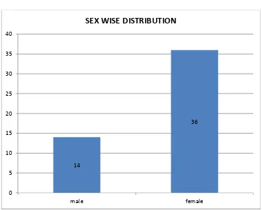
SEX WISE DISTRIBUTION
61
TABLE 1: SEX WISE DISTRIBUTION
14
36
0 5 10 15 20 25 30 35 40
male female
SEX WISE DISTRIBUTION
SEX NO. OF CASES PERCENTAGE
MALE 14 28%
62
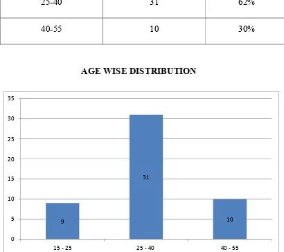
TABLE 2: AGE WISE DISTRIBUTION
Patients in the age group 25 – 40 are commonly affected (62%)
AGE GROUP(YEARS) NO. OF CASES PERCENTAGE
15-25 9 18%
25-40 31 62%
40-55 10 30%
AGE WISE DISTRIBUTION
9
31
10
0 5 10 15 20 25 30 35
63
PREVIOUS HISTORY OF SURGERY :
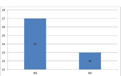
[image:75.612.126.535.420.679.2]54% of the study group was with previous history of surgery compared to the 46% of patients with no history of previous surgery. This may suggest that previous history of surgery may have a positive correlation with chronic nonspecific abdominal pain.
TABLE 3: PREVIOUS HISTORY OF SURGERY
H/O PREVIOUS SURGERY NO. OF CASES PERCENTAGE
PRESENT 27 54%
ABSENT 23 46%
PREVIOUS HISTORY OF SURGERY
27
23
21 22 23 24 25 26 27 28
64
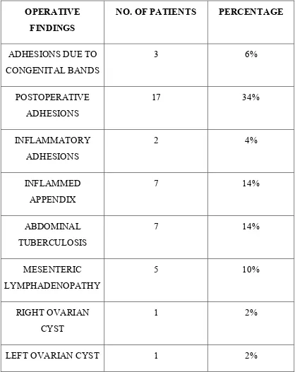
TABLE 4: DISTRIBUTIONS OF OPERATIVE FINDINGS ON
DIAGNOSTIC LAPAROSCOPY
OPERATIVE
FINDINGS
NO. OF PATIENTS PERCENTAGE
ADHESIONS DUE TO CONGENITAL BANDS
3 6%
POSTOPERATIVE ADHESIONS
17 34%
INFLAMMATORY ADHESIONS
2 4%
INFLAMMED APPENDIX
7 14%
ABDOMINAL TUBERCULOSIS
7 14%
MESENTERIC LYMPHADENOPATHY
5 10%
RIGHT OVARIAN CYST
1 2%
65 THICKENED GALL
BLADDER WITH ADHESIONS
1 2%
NO ABNORMALITY DETECTED
5 10%
FREE FLUID 1 2%
TOTAL 50 100%
22 patients (44%) are found to have adhesions, out of them 3 patients (6%)have adhesions due to congenital bands, 17 patients (34%) have Postoperative adhesions and 2 patients (4%) have inflammatory adhesions. Out of 22 patients 19 patients have previous history of surgery.
7 patients (14%)were found to have inflamed appendix and 2 patients (4%) were found to have Ovarian cyst
Abdominal Tuberculosis was noticed in 7 patients (14%) and Mesenteric Lymphadenopathy noted in 5 patients (10%) Thickened gall Bladder with adhesions noted in 1 patient (2%)
66
OPERATIVE FINDINGS IN DIAGNOSTIC LAPAROSCOPY
22
7 7
5 11
2 5
ADHESIONS (44%)
ABDOMINAL TB (14%)
INFLAMMED APPENDIX (14%)
MESENTERIC LYMPHAENITIS (10%)
FREE FLUID (2%)
THICKENED GB WITH ADHESIONS (2%)
OVARIAN CYST (4%)
67
TABLE 5: SHOWING FINAL DIAGNOSIS, TREATMENT GIVEN
AND POSITIVE OUTCOME
DIAGNOSIS OPERATIVE FINDINGS TREATMENT NO. OF PATIENTS POSITIVE OUTCOME
ADHESIONS POSTOP ADHESION
ADHESION DUE TO CONGENITAL BANDS
INFLAMMATORY ADHESIONS
ADHESIOLYSIS 22 (44%) 20 (90.9%)
RECURRENT APPENDICITIS
INFLAMMED APPENDIX
APPENDICECTOMY 7 (14%) 6 (85.7%)
ABDOMINAL TB ABDOMINAL TB
MESENTERIC LYMPHADENITIS (3)
ATT 10 (20%) 9 (90%)
RIGHT / LEFT OVARIAN CYST
RIGHT / LEFT OVARIAN CYST
68 GASTROENTERITIS / COLITIS MESENTERIC LYMPHADENITIS (2)
CONSERVATIVE 2 (4%) 2 (100%)
ACALCULOUS CHOLECYSTITIS
THICKENED GALL BLADDER WITH
ADHESIONS
CHOLECYSTECTOMY 1 (2%) 1 (100%)
IDIOPATHIC CHRONIC ABDOMINAL PAIN NO ABNORMALITY DETECTED FREE FLUID
CONSERVATIVE 6 (12%) 5 (83.5%)
All patients with adhesions (22 patient) undergone Laparoscopic adhesiolysis. Out of them positive outcome seen in 20 patients (90.9%) after 3 months follow up.
All patients with inflamed appendix (7patients) undergone Laparoscopic appendicectomy. Out of them positive outcome seen in 6 patients (89.7%)
Anti Tuberculosis therapy was given to 10 patients. Out of them positive outcome was seen in 9 patients(90%).
69
2 patients with Ovarian cyst undergone Laparoscopic cyst aspiration. Positive outcome was 100%.
1 patient with Acalculus cholecystitis undergone Laparoscopic cholecystectomy positive outcome was 100%.
[image:81.612.122.554.491.712.2]6 patients with idiopathic abdominal pain were given conservative treatment. Positive outcome was seen in 5 patients (83.3%).
TABLE 6: PAIN RESPONSE AFTER DIAGNOSTIC
LAPAROSCOPY
(AFTER 3 MONTHS)
Out of 50 patients, relief of patients was noted in 39 patients (78%). 6 (12%)patients had reduced pain after diagnostic laparoscopy with overall positive response to pain in 90% of the patients in our study. Persistent pain was noted in 5 (10%) patients.
PAIN RESPOPNSE
(FOLLOW UP AFTER 3 MONTHS)
NO. OF
PATIENTS PERCENTAGE
RELIEF 39 78
REDUCED 6 12
PERSISTENT 5 10
70
PAIN RESPONSE AFTER DIAGNOSTIC LAPAROSCOPY
39 6
5
71
TABLE 7: COMPARISON OF DIAGNOSTIC EFFICACY OF
LAPAROSCOPY IN VARIOUS STUDIES
STUDY EFFICACY NO. OF
CASES YEAR OF STUDY OUTCOME (PAIN RESPONSE) MILLER ET
AL 89.8 59 1996 89.3%
SALKY AND
EDGE 76 265
1998
__
RAYMOND
ET AL 85.7 70 2003 71.4%
MAUSSA
AND
MAHFIAZ
78.6 56 2004 80.2%
EL-LABBAN
AND
HOKKAM
83.3 30 2010 80%
TALASKAR
ET AL 82.8 35 2013 81.8%
PRESENT
STUDY
72
73
DISCUSSION
Chronic abdominal pain is most challenging and demanding conditions to treat across the whole age spectrum. Potentially it can be unrewarding for both the patients and the medical team. Abdominal pain was the third most common pain complaint of individuals enrolled in the large health maintenance organic statistics.
All patients included in the study had chronic abdominal pain and they were subjected to laparoscopic evaluation after exclusion of all organic causes of pain by routine radiographic and laboratory tests. The study confirmed that in this difficult patient group, laparoscopy could be safely identified abnormal findings and can improve the outcome in a majority of cases.
74
In this study clear diagnosis obtained in 88% of the cases so the efficiency is 88%. We found a low incidence of recurrent appendicitis as compared to adhesion in this study.
We found that in a selected patient group, laparoscopic evaluation of chronic abdominal pain is usually associated with a positive outcome(90%) in terms of relief or reduced pain, 3 months of laparoscopy.
In our study , among the study population the incidence of chronic abdominal pain is more common in the female population which is 72% of the study population.
In our study , the most common age group affected by chronic abdominal pain is between 25 to 40 years of age which accounts to 62% of the study population.
In our study , the incidence of chronic abdominal pain is more common in patients with previous history of abdominal surgery which accounts to 54% of the study population which explains the increased incidence of adhesions in female patients undergoing abdominal surgeries.
75
adhesions were found to be most common cause of chronic abdominal pain in the study population.
76
CONCLUSION
BIBLIOGRAPHY
1. N.Engl J.Med.1994 Jul 7;331 (1): 55-6 Laparascopic general surgery 2. Soper NJ, Brunt LM Kerbl K
3. Surg Clin North Am. 1993 Apr; 73 (2) : 265 – 89 Complications of laparoscopic surgery Crist DW , Gadacz TR.
4. World J Surg 2006 Apr; 30 (4): 535 – 40 Laparoscopic lysis of adhesions.
5. Szomstein S, Menzo EL, Simpfendorfer C, Zundel N Rosenthal RJ. 6. Open versus closed establishment of Pneumoperitoneum in
laparoscopic surgery. Authors : Bonjer HJ; Hazebroek E.J. Kazemeir Giuffrida M.C. Meijer. W.S; LangeJ.F. British Journal of Surgery. Volume 84, Number 5,May 1997, PP.559. 602 (4).
7. Surg Endosc. 2001 Mar; 15(3) : 275-80. Epub 2000 Dec 12. Trocar and Verees needle injuries during laparoscopy. Schafer M.Lauper M. Karhenbuhl L.
8. Yale J Biol Med. 1998 Nov – Dec; 71 (6) : 551 – 78 Anesthetic complications of laparoscopic surgery. Cunningham AJ.
9. Surg Endosc. 1994 Nov :8 (11) : 1272 – 84 Laparascopic surgery – anesthetic complications Cunningham AJ.
laparoscopy with the Hasson trocar. Mckernan JB , Champion JK. Dept. Of Surgery, Medical college of Georgio, USA.
11. Short practice of Surgery. Bailey and Love’s – 24th edition; Page No. 107.
12. Principles of surgery – Schwartz’s – 8th Edition pp 379-381 Blair A. Jobe and John G.Huntere.
13. Text book of surgery – sabiston – 17th Edition Cralg chang. M.D and Robert V. Rege M.D. Chapter – 18 Page No. 445.
14. Practical Laparascopy alan G Gordon, Patrick J. Taylor.
15. Philips P A Amaral JF : Abdominal access complications in laparoscopic surgery. Jam coll Surg 19 : 525 – 536, 2001.
16. Bobb, R.R.Detection of hepatic cancer – role of peritoneoscopy , Gastrointest.,Endose.,1968.
17. Brugera, M.Bordas, J.M., Mas,P., and Rodes.J. A comparison of the accuracy of peritoneoscopy and liver biopsy in the diagnosis of cirrhosis Gut 1974.
18. Cali RW , Laparoscopy , surg. Clin. North Amer:1980.
19. Coupland GAE Towsend DM: Peritoneoscopy – Use in assessment of intra abdominal malignancy.
PROFORMA
SI No : Opd/Ipd No : Name : DOA : Age : DOD : Sex : Occupation : Address :
Presenting Symptoms:
Abdominal pain
Vomiting/Nausea
Bowel symptoms
Bladder symptoms Past history:
Medical history
Surgical history Personal history:
VITALS :
SYSTEMIC EXAMINATION : Cardiovascular system
Respiratory system Central nervous system Per Abdomen :
Inspection
Palpation
Auscultation
Percussion
CLINICAL IMPRESSION :
INVESTIGATIONS Basic investigations
USG Abdomen and Pelvis CT Abdomen
Endoscopy Colonoscopy
Provisional diagnosis:
Indications for laparoscopy:
Laparoscopic findings:
Laparoscopic intervention done:
Complications-Intra/Postoperative:
Final diagnosis:
CONSENT FORM
xg;g[jy; gotk;
vdf;F ePz;l fhykhf tapw;W typ cs;sJ. gpw
ghpnrhjidfshy; mjw;Fz;lhd fhuzj;ij mwpa ,aytpy;iy.
vdnt mjw;fhf Crp \ykhf tapw;Wg; gFjpapy; JisapLk;
[Laparoscopy]
mWit
rpfpr;irapd;
thapyhf
mjw;fhd
fhuzj;ij
mwpat[k;
[Diagnostic
Laparoscopy],
nkYk;
mt;tHpahfnt mjw;Fz;lhd rpfpr;ir mspf;ft[k; kUj;Jth;
\ykhf mwpe;J bfhz;nld;.
mWit rpfpr;irf;fhd kaf;f kUe;J bfhLf;Fk; nghnjh
my;yJ mjw;F gpwnfh capUf;F Mgj;J Vw;glyhk; vd;gija[k;/
kUj;Jth; \ykhf mwpe;J bfhz;nld;.
vdnt rpfpr;irf;F KG kdJld; Raepidt[lDk; rk;kjk;
bjhptpf;fpnwd;.
S.N
NAME AGE SEX IP NO
PREVIOUS H/O SURGERY
DIAGNOSIS TREATMENT FOLLOWUP
1
NITHYA 32 F 49684 YES POSTOPERATIVE
ADHESIONS ADHESIOLYSIS REDUCED
2
KIRUTHIKA 18 F 49679 NO INFLAMMED
APPENDIX APPENDICECTOMY RELIEF
3
SATHEESH 39 M 49701 YES POSTOPERATIVE
ADHESIONS ADHESIOLYSIS RELIEF
4 PALANI 52 F 49934 NO ABDOMINAL TB ATT PERSISTENT
5
GOWRI 28 F 51306 NO LT OVARIAN
CYST CYST ASPIRATION RELIEF
6
KOWSALYA 34 F 51324 YES POSTOPERATIVR
ADHESIONS ADHESIOLYSIS RELIEF
7
TAMILSELVI 21 F 51284 NO INFLAMMED
APPENDIX APPENDICECTOMY RELIEF
8
SAKTHIRAJ 31 M 51299 YES NO
ABNORMALITY
CONSERVATIVE
MANAGEMENT RELIEF
9
SOFIYA 30 F 51623 YES
ADHESIONS (CONGENITAL
BANDS)
ADHESIOLYSIS RELIEF
10
HASEENA 47 F 51900 YES
THICKENED GB WITH ADHESIONS
CHOLECYSTEC
TOMY RELIEF
11
KARTHIK 31 M 52814 NO INFLAMMATORY
ADHESIONS ADHESIOLYSIS RELIEF
12
DEVAKI 44 F 52875 YES POSTOPERATIVE
ADHESIONS ADHESIOLYSIS RELIEF
13
THIRU 39 M 52944 YES POSTOPERATIVE
ADHESIONS ADHESIOLYSIS RELIEF
14
PRIYA 24 F 54057 NO INFLAMMED
15
NAGAMMAL 33 F 54673 YES ABDOMINAL TB ATT REDUCED
16 KAVITHA 19 F 71806 YES POSTOPERATIVE
ADHESIONS
ADHESIOLYSIS RELIEF
17 KANIMOZHI 37 F 51977 NO INFLAMMED
APPENDIX
APPENDICECTOMY RELIEF
18 KANDASAMY 50 M 56299 YES POSTOPERATIVE
ADHESIONS
ADHESIOLYSIS PERSISTENT
19 RAMLATH 27 F 54600 NO ABDOMINAL TB ATT RELIEF
20 MARIMUTHU 27 M 56280 YES POSTOPERATIVE
ADHESIONS
ADHESIOLYSIS RELIEF
21 SUMATHY 42 F 56520 NO ABDOMINAL TB ATT RELIEF
22 MUMTAZ 29 F 56284 NO ADHESIONS
(CONGENITAL BANDS)
23 RAMESH 16 M 56388 NO INFLAMMED APPENDIX
APPENDICECTOMY RELIEF
24 KUMUTHA 28 F 56264 YES POSTOPERATIVE
ADHESIONS
ADHESIOLYSIS RELIEF
25 KAIRUNUSHA 39 F 57828 YES LT OVARIAN
CYST
CYST ASPIRATION RELIEF
26 KALAISELVI 17 F 59540 YES INFLAMMED
APPENDIX
APPENDICECTOMY RELIEF
27 SANKAR 37 M 59519 YES POSTOPERATIVE
ADHESIONS
ADHESIOLYSIS RELIEF
28 KRISHNAKUMARI 24 F 61156 NO MESENTERIC
LYMPHADENITIS
CONSERVATIVE RELIEF
29 JYOTHI 28 F 59530 YES POSTOPERATIVE
ADHESIONS
ADHESIOLYSIS RELIEF
30 NALINI 45 F 61165 YES POSTOPERATIVE
ADHESIONS
31 JAYA 30 F 62738 NO NO
ABNORMALITY
CONSERVATIVE RELIEF
32 SEENATH 29 F 62930 YES INFLAMMATORY
ADHESIONS
ADHESIOLYSIS RELIEF
33 MANIMEGALAI 54 F 62743 NO MESENTERIC
LYMPHADENITIS
ATT REDUCED
34 KRISHNAMOORTHY 30 M 63344 NO NO
ABNORMALITY
CONSERVATIVE REDUCED
35 SUNDARI 23 F 64387 YES POSTOPERATIVE
ADHESIOLYSIS
ADHESIOLYSIS RELIEF
36 SARADHA 36 F 64442 NO MESENTERIC
LYMPHADENITIS
ATT RELIEF
37 JAYALAXMI 34 F 65873 NO NO
ABNORMALITY
CONSERVATIVE REDUCED
38 RAVI 31 M 65905 YES POSTOPERTAIVE
ADHESIONS
39 RAM KUMAR 31 M 67600 NO NO
ABNORMALITY
CONSERVATIVE PERSISTENT
40 RAMATHAL 47 F 67472 YES POSTOPERTAIVE
ADHESIONS
ADHESIOLYSIS RELIEF
41 THULASI 32 F 67508 YES INFLAMMED
APPENDIX
APPENDICECTOMY PERSISTENT
42 PRIYA 22 F 67429 NO ABDOMINAL TB ATT RELIEF
43 BANU 29 F 69444 NO ADHESIONS
(CONGENITAL BANDS)
ADHESIOLYSIS RELIEF
44 SUBAIYAH 45 M 69185 YES MESENTERIC
LYMPHADENITIS
ATT REDUCED
45 SUGANTHI 32 F 67817 NO ABDOMINAL TB ATT RELIEF
46 SARASWATHI 36 F 73088 YES POSTOPERATIVE
ADHESIONS
47 CHANDRAN 27 M 70799 NO MESENTERIC LYMPHADENITIS
CONSERVATIVE RELIEF
48 NIRMALA 49 F 72580 YES POSTOPERATIVE
ADHESIONS
ADHESIOLYSIS RELIEF
49 SELVI 34 F 74304 YES ABDOMINAL TB ATT RELIEF