Open Access
Vol 11 No 2Research article
Association of GATA3, P53, Ki67 status and vascular peritumoral
invasion are strongly prognostic in luminal breast cancer
Jocelyne Jacquemier
1,2, Emmanuelle Charafe-Jauffret
1,2,3, Florence Monville
1, Benjamin Esterni
4,
Jean Marc Extra
5, Gilles Houvenaeghel
6, Luc Xerri
2,3, François Bertucci
1,3,5and Daniel Birnbaum
11Département d'Oncologie Moléculaire Centre de Recherche en Cancérologie de Marseille, Institut Paoli-Calmettes, UMR891 Inserm; IFR137, 232 Bd sainte Marguerite Marseille, 13009 France
2Département de BioPathologie, Institut Paoli-Calmettes, 232 Bd sainte Marguerite Marseille,13009 France 3UFR de Médecine, Université de la Méditerranée, 58 Bd Charles Livon Marseille 13284, France
4Département de Biostatistiques, Institut Paoli-Calmettes, 232 Bd Ste Marguerite Marseille 13009, France 5Département d'Oncologie Médicale, Institut Paoli-Calmettes, 232 Bd Ste Marguerite Marseille 13009, France 6Département d'Oncologie Chirurgicale, Institut Paoli-Calmettes, 232 Bd Ste Marguerite Marseille 13009, France Corresponding author: Jocelyne Jacquemier, jacquemierj@marseille.fnclcc.fr
Received: 29 Oct 2008 Revisions requested: 13 Dec 2008 Revisions received: 24 Mar 2009 Accepted: 30 Apr 2009 Published: 30 Apr 2009 Breast Cancer Research 2009, 11:R23 (doi:10.1186/bcr2249)
This article is online at: http://breast-cancer-research.com/content/11/2/R23 © 2009 Jacquemier et al.; licensee BioMed Central Ltd.
This is an open access article distributed under the terms of the Creative Commons Attribution License (http://creativecommons.org/licenses/by/2.0), which permits unrestricted use, distribution, and reproduction in any medium, provided the original work is properly cited.
Abstract
Introduction Breast cancers are traditionally divided into hormone-receptor positive and negative cases. This classification helps to guide patient management. However, a subgroup of hormone-receptor positive patients relapse irrespective of hormonal therapy. Gene expression profiling has classified breast tumours into five major subtypes with significant different outcome. The two luminal subtypes, A and B, show high expression of ESR1, GATA3 and FOXA1 genes. Prognostic biomarkers for oestrogen receptor (ER)-positive cases include progesterone receptor (PR) and androgen receptor (AR), and proteins related to proliferation or apoptotic resistance. The aim of this study was to identify the best predictors of success of hormonal therapy.
Methods By immunohistochemistry we studied 10 markers in a consecutive series of 832 cases of breast carcinoma treated at the Paoli-Calmettes Institute from 1990 to 2002 and deposited onto tissue microarrays (TMA). These markers were luminal-related markers ER, PR, AR, FOXA1 and GATA3 transcription
factors, proliferation-related Ki67 and CCND1, ERBB2, anti-apoptotic BCL2 and P53. We also measured vascular peritumoural invasion (VPI), size, grade and lymph node involvement. For 143 cases, gene expression profiles were available. Adjuvant chemotherapy and hormonal therapy were given to high- and low-risk patients, respectively. The 162 events observed and taken into account were metastases.
Results Molecular expression of the 10 parameters and subtype with ER status were strongly correlated. Of the 67 luminal A cases of this series, 63 were ER-positive. Multivariate analyses showed the highly significant prognostic value of VPI (hazard ratio (HR) = 2.47), Ki67 (HR = 2.9), P53 (HR = 2.9) and GATA3 (HR = 0.5) for the 240 patients who received hormonal therapy.
Conclusions A panel of three antibodies (Ki67, P53 and GATA3) associated with VPI can significantly improve the traditional prognosticators in predicting outcome for ER-positive breast cancer patients receiving hormonal therapy.
Introduction
The traditional division of breast cancers into hormone recep-tor positive and negative cases helps to guide patient manage-ment. However, a subgroup of hormone receptor-positive patients relapse irrespective of standard hormonal therapy. Gene expression profiling has classified breast tumours into
five major molecular subtypes with different outcomes. The two luminal subtypes, A and B, express the ESR1, GATA3 and
FOXA1 genes [1].
Compared with luminal A, luminal B tumours have a poor prog-nosis [1-3]. However, there are few indicators to determine if
the response to hormonal therapy is different between A and B subtypes. In a previous study we validated a non-linear algo-rithm including six immunohistochemical markers on tissue microarrays (TMA): oestrogen receptor (ER), progesterone receptor (PR), ERBB2, BCL2, P53 and MYC [4]. This algo-rithm had strong prognostic value in ER-positive patients with or without hormonal therapy. The difference between luminal A and B was not investigated in this study. In another study we showed that the subset of patients with luminal A tumours, called Ab, which express mitotic kinases had a poorer progno-sis than the majority that do not express these kinases [5]. This subset with high kinase score had a prognosis close to luminal B tumours. In fact, luminal Ab resemble luminal B tumours; they are distinguished only because the lists of genes used in gene expression analyses to identify subtypes are not accu-rate enough and because luminality reflects a continuum from poorly differentiated, highly proliferative (luminal B) to well-dif-ferentiated, poorly proliferative (luminal Aa).
The prognostic distinction between luminal Aa and Ab sug-gest that grade and P53 are also involved but the kinase score was associated with the highest hazard ratio (HR). In the absence of reliable antibodies the kinase score is difficult to implement in a routine setting. We therefore searched for eas-ily identifiable factors that could be associated with the prog-nosis of patients receiving hormonal therapy for the different luminal subtypes.
P53 mutation is generally associated with basal breast cancer. However, we demonstrated its impact in luminal cases [5]. P53 expression observed in BRCA1 luminal cases corre-spond to a true mutation in only four of seven cases [6]. This suggests that P53 expression could be associated with prolif-eration in luminal cases independent of mutation.
Quantitative ER status is correlated with a strong response to hormonal therapy. PR, GATA3 and FOXA1, and proteins related to proliferation or apoptotic resistance such as BCL2 could also influence hormonal response. The transcription fac-tor GATA3 is a defining marker of the luminal subtypes. GATA3 has an essential role in the morphogenesis of the mammary gland and actively maintains luminal epithelial differ-entiation [7]. We demonstrated a good correlation between GATA3 gene and protein expression [2]. A recent meta-analy-sis [7] showed that both ER-alpha and GATA3 are coex-pressed with ER-alpha-associated genes such as PS2/TFF1, TFF3, FOXA1, BCL2, ERBB4, XBP1, NRIP, IL6ST, Keratin 18 and cyclin D1/CCND1. The transcription factor FOXA1 is a downstream target of GATA3 in the mammary gland. FOXA1 expression is associated with that of ER, PR and androgen receptor (AR) [8-10] and with a better survival. FOXA1 binds to chromatinised DNA, opens the chromatin and enhances binding of ER-alpha. Thus, a network comprising GATA3, FOXA1, ER-alpha and oestrogen constitutes a major prolifer-ation and survival signal for luminal A breast cancer [11].
Many human breast cancers express AR. A recent study of AR on formalin-fixed, paraffin-embedded archival specimens of 200 cases of breast cancer showed that 60% of invasive car-cinoma and 82% of ductal carcar-cinoma in situ were AR-positive [12]. The great majority of well-differentiated carcinomas were both AR and ER-positive. In contrast, 39% of poorly-differenti-ated carcinomas were ER-negative but AR-positive. The clini-cal value of AR expression is unclear. However, AR expression was strongly correlated with ER in a series of 842 breast car-cinomas [13]. Few studies suggest the impact of AR on the response to hormonal therapy [14].
Finally, a recent meta–analysis confirmed that BCL2 has an independent prognostic impact [15]. However, no prospective study has shown the predictive impact of BCL2 expression in ER-positive cases.
The aim of our study was to identify the prognosis of patients receiving hormonal therapy among histoclinical and immuno-histochemical factors.
Materials and methods
Patients
Breast cancer samples
Tissues were collected from 143 patients with invasive aden-ocarcinoma who underwent initial surgery at the Institut Paoli-Calmettes (Marseilles, France). Each patient gave written informed consent. Samples were macro-dissected and frozen in liquid nitrogen within 30 minutes of removal.
DNA and RNA extraction
Nucleic acids were extracted from frozen samples by using guanidium isothiocyanate and cesium chloride gradient, as previously described [16]. RNA integrity was controlled on the Agilent Bioanalyzer (Agilent Technologies, Massy, France).
Gene expression profiling with DNA microarrays
Gene expression was analysed in 143 breast cancer samples and four normal breast samples with Affymetrix U133 Plus 2.0 human oligonucleotide microarrays (Affymetrix Santa Clara, CA, USA). Preparation of c-RNA, hybridisations, washes and detection were performed as recommended by the supplier. For each sample, synthesis of the first-strand c-DNA was done from 3 μg total RNA by T7-oligo(dT) priming, followed by sec-ond-strand cDNA synthesis. After purification, in vitro tran-scription associated with amplification generated cRNA-containing biotinylated pseudouridine. Biotinylated cRNA was purified, quantified and chemically fragmented (95°C for 35 minutes), then hybridised to microarrays in 200 μL hybridisa-tion buffer at 45°C for 16 hours. Automated washes and stain-ing with streptavidin-phycoerythrin were performed as recommended. Double signal amplification was achieved by biotinylated antistreptavidin antibody with goat-IgG blocking antibody. Scanning was performed with Affymetrix GeneArray scanner and quantification with Affymetrix GCOS software.
Gene expression data analysis
Affymetrix data were analysed by the Robust Multichip Aver-age method in R using Bioconductor and associated pack-ages [17]. The Robust Multichip Average performed background adjustment, quantile normalisation and summari-sation of 11 oligonucleotides per gene. Before analysis, a fil-tering process removed the genes with low and poorly measured expression, as defined by an expression value infe-rior to 100 units in all breast cancer tissue and normal tissue samples, from the dataset. All data was then log2-transformed
for display and analysis.
Basal and luminal breast cancers were distinguished by the differential expression of clusters of genes. Sub-classification of the luminal cases was done as previously described [5]. Kinase gene expression identified two subgroups of luminal A breast cancers, that is luminal Aa and Ab.
Tissue microarrays construction and immunohistochemistry
TMAs were prepared as previously described [18] from forma-lin-fixed and paraffin-embedded tissue. For each tumour, three
representative areas were selected from a H&E-safran-stained section of a donor block. Core cylinders with a diameter of 0.6 mm each were punched from each of these areas and depos-ited into three separate recipient paraffin blocks using a spe-cific arraying device (Alphelys, Plaisir, France).
Immunohistochemistry of 5 μmm TMA sections was
per-formed as previously described using Dako LSABR2 Kit in the
autoimmunostainer (Dako Autostainer, Glostrup, Denmark). Sections were deparaffinised in Histolemon (Carlo Erba Rea-genti, Rodano, Italy) and rehydrated in graded ethanol solu-tions. Results were evaluated under a light microscope by two pathologists (EC-J, JJ) and scored by the quick score (QS) [19]. The QS was used to combine the impact of the percent-age and the intensity of the immunostaining. QS multiplies the percentage by the intensity and represents a range of 0 to 300. For each antibody, a sample was considered as positive when the QS was strictly superior to 0. However, the Ki67 sta-tus was expressed in terms of percentage of positive cells, with a threshold of 20% of positive cells. The ERBB2 status was evaluated with the Dako scale (HercepTest kit scoring guidelines, DakoCytomation, Copenhagen, Denmark). The level of 3+ was considered as positive and all 2+ cases were evaluated by chromogen in situ hybridisation (only the case with a ratio higher than 2.2 were considered as positive).
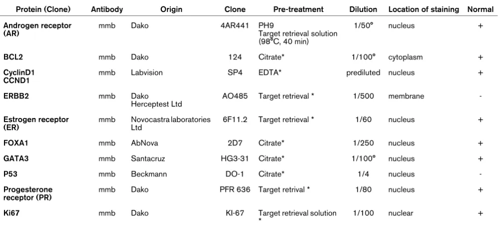
For each tumour, the mean of the score of a minimum of two core biopsies was calculated. The list of antibodies used is given in Table 1.
Statistical analysis
Survival rates were estimated by using the Kaplan-Meier method [20]. The endpoint was the MFS, which was defined as the time from the date of breast cancer diagnosis until the date of the first distant relapse. Patients without relapse were censored at the time of last follow-up. Survival analysis was computed with a stratification on treatment by chemotherapy. Relative risks of metastasis according to the baseline factors were estimated by using the Cox proportional-hazards regres-sion models [21] in univariate and multivariate analyses. In uni-variate analysis, differences in MFS were analysed by the Log-Rank test. Factors with a P value less than 0.15 in univariate analysis were included in the multivariate analysis, with a back-ward selection of variable procedure to minimise the Akaike information criterion [22]. Results are presented as mean (95% CI). Statistical analyses were performed with the R.2.7.1. Statistical language [23].
Results
Correlation between molecular subtype and oestrogen receptor immunohistochemical status
sig-nificant (Table 2). The lowest level of the Rho coefficient was observed for Ki67, P53 and ERBB2.
The frequency of the different subtypes was: 25% basal, 12% ERBB2, 46% luminal A (68.1%% Aa and 31.8%% Ab), 2% luminal B and 9% normal-like. Molecular subtype and ER sta-tus were strongly correlated. Only 8% of basal breast cancers were ER-positive for 23.5% of ERBB2, 95.5% of luminal A, 100% of luminal B and 53.8% of normal-like breast cancers (Table 3).
Univariate and multivariate analyses of survival
We studied the impact of 16 histoclinical and immunohisto-chemical factors on disease-free survival. Hormonal therapy, size of the lesion, histoprognostic grade, vascular peritumoural invasion (VPI), ER, BCL2, GATA3, Ki67 and P53 had signifi-cant impact (Table 4). Only age, CCND1, PR, FOXA1 did not have any significant value in MFS.
For ERBB2 there was a significant difference in terms of dis-ease-free survival at 60 months with 83.7% for the negative
cases and 69.1% for the 3+ cases and amplified 2+ (P =
0.017). However, when the analysis was stratified on the pres-ence or not of chemotherapy, no significant differpres-ence between the two groups was noted.
Eight factors were retained by the multivariate analysis includ-ing histopronostic grade, axillary lymph node invasion, VPI, size, then Ki67, P53, BCL2 and hormonal therapy (Table 5).
Molecular subtype and oestrogen receptor positivity
In the restricted ER-positive population studied by gene expression profiling we observed that 67 of 81 (86.4%) were luminal cases (Table 6). There was no difference in ER-positiv-ity level (with a cut off QS of 120) between luminal Aa (18 of 43 above 120, 41.8%) and luminal Ab (8 of 21 above 120, 38%) cases. However, a significant difference was observed for proliferation: luminal Ab showed a higher grade (P = 4.710; Table 6) and a higher Ki67 index (P = 0.02) than
lumi-Table 1
Immunohistochemical antibodies used to characterize the luminal cases
Protein (Clone) Antibody Origin Clone Pre-treatment Dilution Location of staining Normal Androgen receptor
(AR)
mmb Dako 4AR441 PH9
Target retrieval solution (98°C, 40 min)
1/50° nucleus +
BCL2 mmb Dako 124 Citrate* 1/100° cytoplasm +
CyclinD1 CCND1
mmb Labvision SP4 EDTA* prediluted nucleus +
ERBB2 mmb Dako
Herceptest Ltd
AO485 Target retrieval * 1/500 membrane
-Estrogen receptor (ER)
mmb Novocastra laboratories Ltd
6F11.2 Target retrieval * 1/60 nucleus +
FOXA1 mmb AbNova 2D7 Citrate* 1/250 nucleus +
GATA3 mmb Santacruz HG3-31 Citrate* 1/100° nucleus +
P53 mmb Beckmann DO-1 Citrate* 1/4 nucleus
-Progesterone receptor (PR)
mmb Dako PFR 636 Target retrival * 1/80 nucleus +
Ki67 mmb Dako KI-67 Target retrieval solution
*
1/100 nuclear +
[image:4.612.57.559.117.347.2]+ = positive expression, - = negative expression. The asterisk symbols mean that it is a buffer solution.
Table 2
Correlation between expression in microarray and quick score of the ten markers analysed
Spearman correlation test AR BCL2 CCND1 ERBB2 FOXA1 GATA3 KI67 P53 ER PR
Rho * 0.49 0.56 0.43 0.28 0.58 0.64 0.32 0.30 0.73 0.69
[image:4.612.56.564.666.715.2]nal Aa. The three luminal B were ER-positive (the percentage of ER-positive cells was below 5% for two cases) but grade 3. Two were positive for P53.
One-third of the luminal A were Ab (31.8%) and two-thirds (68.1%) were luminal Aa cases. The four ERBB2-subtype cases were ER-positive but the level of ER expression was lower than the median value of QS 120; all the cases were grade 3 and PR-negative. The seven normal-like cases were grade 1 (n = 5) or 2 (n = 2) and four showed a low level of ER protein expression.
Markers and survival
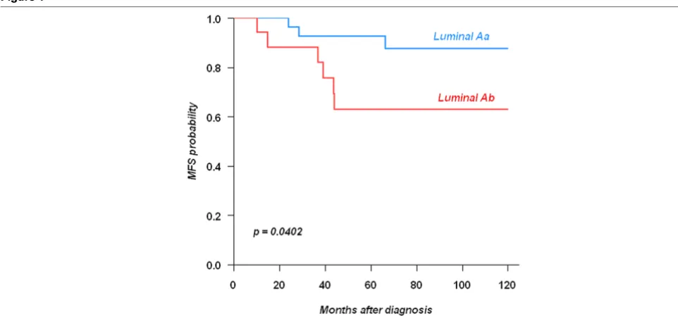
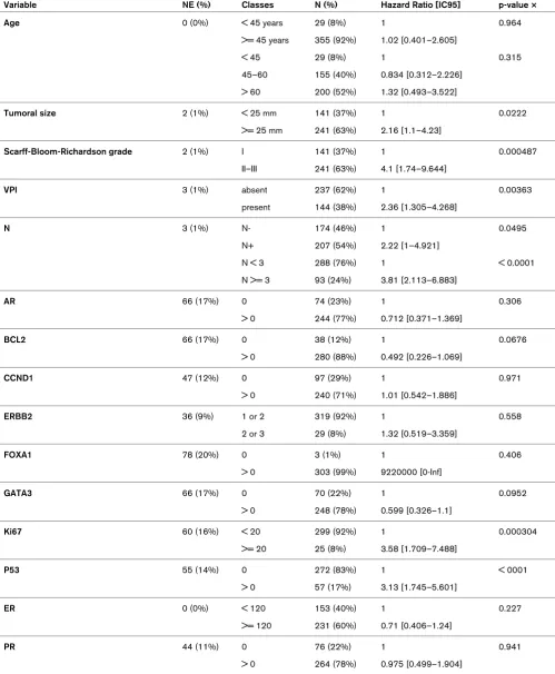
We then restricted the study to the ER-positive cases treated by hormonal therapy (n = 384). Subtype status was available for only a small series of these cases (n = 43). MFS was dif-ferent between luminal Aa and Ab cases (P = 0.042; Figure 1). Of the 14 factors studied in univariate analysis (Table 7) only 6 showed a different distribution: size, grade, VPI, lymph node invasion, GATA3 and Ki67. Oestrogen-related proteins such as FOXA1 and AR had no significant impact whatever their quantitative value. The ER and PR level of expression had no significant MFS value in univariate analysis. The multivariate analysis in terms of MFS retained four factors: VPI, Ki67, P53 and GATA3 (Table 8).
Discussion
The aim of this study was to study the expression of proteins corresponding to genes identified by gene expression profiling to be associated with luminal cases and to determine their impact on the response of patients to hormonal therapy. Due to experimental conditions (e.g. quality of the antibodies), the analysis was limited to 10 proteins. We were able to identify a score combining four factors able to predict the evolution of the luminal cases treated with adjuvant hormonal tamoxifen therapy.
Molecular subtypes and prognosis
Grossly, our 135 subtyped cases showed a similar distribution of subtypes as found in previous studies [1,2,24]. Our series contained 25% of basal cases, which is within a published range of 17 to 37%. The number of ERBB2 subtype (12%) was slightly higher than in most series. In contrast, the 45%
frequency of luminal A was high and the proportion of luminal B was low.
In a previous study [5] we focused on the kinome of luminal A breast cancers. The breast cancer kinome differs between basal and luminal A cases. Within luminal A cases, it allowed the identification of luminal Aa and Ab. Here, we have con-firmed the difference in outcome between luminal Aa and Ab by using immunohistochemistry on 43 luminal A cases treated by tamoxifen. The difference between luminal Aa and Ab was due to proliferative factors translated by a higher grade and Ki67 index in luminal Ab than in luminal Aa cases. The fact that a difference could be seen already with a small series sug-gests the importance of proliferation to distinguish outcome in ER-positive cases whatever their percentage of ER-positive cells.
Prognosis and hormonal therapy in ER-positive cases
Four factors, VPI, GATA3, P53 and Ki67, were retained by the multivariate analysis.
Two parameters were added in the 9th St Gallen meeting compared with the 8th edition: ERBB2 status and VPI. The volume of data published in the past few years provides com-pelling evidence for the importance of VPI [25] but the specific impact on luminal cases had never been described. A meta-analysis of microarray data revealed the importance of GATA3 [26]. Its expression in 10-year follow-up [27] demonstrates that its protective effect is more pronounced in patients who received tamoxifen. We showed the prognostic impact of P53 in two previous studies of luminal cases [4,5]. Ki67 higher than 20% is one of the parameters able to distinguish luminal A from luminal B [28] but its specific prognostic impact in lumi-nal cases had not been described.
[image:5.612.55.557.115.212.2]An important question is whether the combination of VPI, GATA3, P53 and Ki67 predicts pure prognosis or responsive-ness to endocrine therapy or both. Few studies using profiling of ER-positive breast cancers treated by tamoxifen have estab-lished a signature able to predict the prognosis. The oncotype DX RS [29] is a commercially available assay (Genomic Health, Redwood City, CA) that predicts recurrence in ER-positive cases. It is a PCR-based assay on paraffin-embedded
Table 3
Correlation between molecular subtype and immohistochemical ER status
Subtype N = 135
Basal N = 36 (27%)
ERBB2 N = 17 (12.5%)
LuminalAa N = 45 (33%)
LuminalAb N = 21 (15.5%)
LuminalB N = 3 (2%)
Normal-like N = 13 (10%)
ER-positive N = 81 3 (8%) 4 (23.5%) 43 (95.5%) 21 (100%) 3 (100%) 7 (53.8%) ER-negative
N = 54
33 (91.6%) 13 (76.4%) 2 (4.5%) 0 0 6 (46.2%)
Table 4
Univariate analysis of 832 consecutive cases of breast carcinomas and 16 factors including classical histopronostic and immunohistochemical parameters
Variable NE (%) Classes N (%) Hazard Ratio [IC95] p-value
Hormonal therapy 0 (0%) no 361 (43%) 1 0.000209
yes 471 (57%) 0.558 [0.408–0.763]
Age 1 (0%) < 45 years 104 (13%) 1 0.673
>= 45 years 727 (87%) 1.1 [0.711–1.696]
< 45 104 (13%) 1 0.145
Tumor Size 7 (1%) < 25 mm 381 (46%) 1 < 0.0001
>= 25 mm 444 (54%) 2.47 [1.719–3.562]
Scarff-Bloom-Richardson grade 22 (3%) I 266 (33%) 1 < 0.0001
II–III 544 (67%) 3.69 [2.244–6.083]
VPI 4 (0%) Absent 536 (65%) 1 < 0.0001
present 292 (35%) 2.1 [1.519–2.897]
Lymph node invasion 16 (2%) N- 442 (54%) 1 < 0.0001
N+ 374 (46%) 2.61 [1.693–4.013]
AR 175 (21%) 0 220 (33%) 1 0.0122
> 0 437 (67%) 0.65 [0.463–0.913]
< 80 410 (62%) 1 0.00422
>= 80 247 (38%) 0.568 [0.383–0.841]
BCL2 201 (24%) 0 162 (26%) 1 0.000944
> 0 469 (74%) 0.548 [0.381–0.787]
CCND1 142 (17%) 0 284 (41%) 1 0.562
> 0 406 (59%) 0.905 [0.647–1.268]
ERBB2 123 (15%) 0 or 1 621 (88%) 1 0.209
2 or 3 88 (12%) 1.33 [0.852–2.071]
FOXA1 187 (22%) 0 40 (6%) 1 0.324
> 0 605 (94%) 1.43 [0.698–2.952]
GATA3 188 (23%) 0 247 (38%) 1 0.00024
> 0 397 (62%) 0.534 [0.38–0.75]
Ki67 171 (21%) < 20 560 (85%) 1 0.000748
>= 20 101 (15%) 1.96 [1.316–2.923]
P53 140 (17%) 0 525 (76%) 1 0.00095
> 0 167 (24%) 1.76 [1.253–2.47]
ER 79 (9%) 0 169 (22%) 1 0.000276
> 0 584 (78%) 0.54 [0.386–0.756]
PR 120 (14%) 0 263 (37%) 1 0.0875
tissue using 16 cancer-related genes and 5 controls. It was validated on 668 node-negative cases in the National Surgical Adjuvant Breast and Bowel Project (NSABP) trial receiving tamoxifen only. The histopronostic grade and recurrence score (RS) were significant. This RS was subsequently validated for chemotherapy and tamoxifen in 645 patients from the NSABP-14 [30]. Only four of the 16 genes are common with the fac-tors we tested here (ER, PR, KI67 and BCL2). A more recent series of 255 ER-positive cases established a signature vali-dated on an independent set of 362 cases coming from differ-ent institutions and treated by tamoxifen alone [31]. A total of 181 genes belonging to 13 clusters strongly prognostic (HR = 3.26, P = 0.0002). These 13 cluster genes were the most important factor in multivariate analysis.
Immunohistochemistry has been involved in the search for a multiparametric score in ER-positive cases on a series of 257 ER-positive cases treated by tamoxifen; a multimarker model was established from nine markers and five of them were retained in a mathematic model: ER, PR, P53, ERBB2 and MYC. This model was more prognostic than the Nottingham prognostic index [4].
A previous study has looked at oestrogen-regulated genes in the MCF7 breast cancer cell line treated by 17β–oestradiol [32]. These genes were then used to develop an outcome
pre-dictor on a training set of 65 luminal breast cancers and then validated on three independent published data sets. Interest-ingly, two groups of low risk (expressing XBP1, FOXA1 and PR) and high risk (expressing MYBL2 and CCNB2) were dis-tinguished.
The study of a series of 140 cases used 23 antibodies and identified a prognostic score for ER-positive breast cancer without any notion of hormonal therapy [33]. Five factors were retained by Cox analysis (P53, NDRG1, CEACAM5, SLC7A5 and HTF9c) but regression tree analysis retained six factors (P53, PR, Ki67, NAT1, SLC7A5 and HTF9c). The best HR was obtain by the Cox model (HR = 2.21, P = 0.0008).
P53 and Ki67 are the two factors common with our series. This again underlines the impact of proliferation in luminal cases. However, our analysis, with four factors, could be an easier manner to study ER-positive cases.
[image:7.612.60.554.114.416.2]The fact that in our series the patients all received adjuvant tamoxifen stratified on chemotherapy suggests also that these factors could be more than prognostic in cases receiving hor-monal therapy.
Table 5
Significant parameters retained by Cox multivariate analysis in the estrogen receptor positive cases
N = 378 Coefficient HR IC95 p-value
Hormonal therapy no 1
yes -0.897 0.408 [0.257–0.647] 0.00014
Size < 25 mm 1
>= 25 mm 0.9 2.46 [1.449–4.175] 0.00085
Scarff-Bloom-Richardson grade I 1
II–III 0.521 1.68 [0.805–3.522] 0.17
VPI no 1
yes 0.657 1.93 [1.213–3.068] 0.0055
Lymph node N- 1
N+ 0.68 1.97 [1.208–3.222] 0.0066
BCL2 < 160 1
>= 160 -0.613 0.542 [0.331–0.886] 0.015
Ki67 < 20 1
>= 20 0.641 1.9 [1.167–3.086] 0.0098
P53 0 1
Table 6
Correlation between the different subtypes and histopronostic and immunohistochemistry factors in ER-positive cases
Basal ERBB2 Luminal Aa Luminal Ab Luminal B Normal-like p-value ER-positive
N = 84
3 4 43 (%)x 21(%)x 3 7 3 cases non evaluable
Grade I 1 0 14(32.5%) 0 0 5 4.710-6
Grade II 0 0 24(55.8%) 7(30%) 0 2
Grade III 2 4 5(11.6%) 14(66.6%) 3 0
VPI positive 0 1 20/42 (47.6%) 10/21
(47.6%)
3 1
Positive lymph nodes 1 2 26/43
(60.4%)
15/21 (71.4%)
2 2
AR positive 1 2 31/36(86%) 15/21(71.4%) 2 3
BCL2 positive 2 2 36/39 (92.3%) 14/15
(93%)
2 6/6
(100%) CCND1
Positive
2 0 26/41 (63,4%) 13/19
(68%)
2 2/5
ERBB2 (2+/3+) 0 3 (3+) 3/43 (6.9%) 0 1(2+) 0
FOX A1 positive 1 3 38/39 (97.4%) 19/19
(100%
3 4/5
GATA3 Positive
0 1 29/36 (80.5%) 12/18
(66.6%)
3 4/5
Ki67 > 20% 1 0 2/40(5%) 5/18(27.7%) 0 0 0.02
P53 positive 2 2 8/42 (19%) 4/20 (20%) 1 0/6
PR positive
0 0 37/43 (86%) 16/20 (80%) 2 5/6
Numbers in bold mean the percentage of positive available cases.
Figure 1
Influence of the molecular subtype on metastasis-free survival in ER-positive cases receiving hormonal therapy
[image:8.612.56.537.466.690.2]Table 7
Univariate analysis of ER-positive cases with adjuvant hormonal
Variable NE (%) Classes N (%) Hazard Ratio [IC95] p-value ×
Age 0 (0%) < 45 years 29 (8%) 1 0.964
>= 45 years 355 (92%) 1.02 [0.401–2.605]
< 45 29 (8%) 1 0.315
45–60 155 (40%) 0.834 [0.312–2.226] > 60 200 (52%) 1.32 [0.493–3.522]
Tumoral size 2 (1%) < 25 mm 141 (37%) 1 0.0222
>= 25 mm 241 (63%) 2.16 [1.1–4.23]
Scarff-Bloom-Richardson grade 2 (1%) I 141 (37%) 1 0.000487
II–III 241 (63%) 4.1 [1.74–9.644]
VPI 3 (1%) absent 237 (62%) 1 0.00363
present 144 (38%) 2.36 [1.305–4.268]
N 3 (1%) N- 174 (46%) 1 0.0495
N+ 207 (54%) 2.22 [1–4.921]
N < 3 288 (76%) 1 < 0.0001
N >= 3 93 (24%) 3.81 [2.113–6.883]
AR 66 (17%) 0 74 (23%) 1 0.306
> 0 244 (77%) 0.712 [0.371–1.369]
BCL2 66 (17%) 0 38 (12%) 1 0.0676
> 0 280 (88%) 0.492 [0.226–1.069]
CCND1 47 (12%) 0 97 (29%) 1 0.971
> 0 240 (71%) 1.01 [0.542–1.886]
ERBB2 36 (9%) 1 or 2 319 (92%) 1 0.558
2 or 3 29 (8%) 1.32 [0.519–3.359]
FOXA1 78 (20%) 0 3 (1%) 1 0.406
> 0 303 (99%) 9220000 [0-Inf]
GATA3 66 (17%) 0 70 (22%) 1 0.0952
> 0 248 (78%) 0.599 [0.326–1.1]
Ki67 60 (16%) < 20 299 (92%) 1 0.000304
>= 20 25 (8%) 3.58 [1.709–7.488]
P53 55 (14%) 0 272 (83%) 1 < 0001
> 0 57 (17%) 3.13 [1.745–5.601]
ER 0 (0%) < 120 153 (40%) 1 0.227
>= 120 231 (60%) 0.71 [0.406–1.24]
PR 44 (11%) 0 76 (22%) 1 0.941
Conclusions
Our study of immunohistochemistry factors in luminal breast cancers demonstrates the interest to combine prognostic markers to improve the therapeutic choice.
Competing interests
The authors declare that they have no competing interests.
Authors' contributions
JJ and DB designed the study and wrote the manuscript. JJ and ECJ read all the tissue microarrays in a double-blind man-ner. FM served as data manager. BE did the statistical analy-ses. JME, GH and LX contributed to samples and data collection. FB provided DNA microarray data.
Acknowledgements
We thank P Viens for encouragement. This work was supported by Inserm, Institut Paoli-Calmettes, and grants from Ligue Nationale Contre le Cancer (Label 2007–2009), Institut National du Cancer (PL2006, ACI2007).
References
1. Sorlie T, Tibshirani R, Parker J, Hastie T, Marron JS, Nobel A, Deng S, Johnsen H, Pesich R, Geisler S, Demeter J, Perou CM, Lonning PE, Brown PO, Borresen-Dale AL, Botstein D: Repeated obser-vation of breast tumor subtypes in independent gene expres-sion data sets. Proc Natl Acad Sci USA 2003, 100:8418-8423. 2. Bertucci F, Houlgatte R, Granjeaud S, Nasser V, Loriod B, Beau-doing E, Hingamp P, Jacquemier J, Viens P, Birnbaum D, Nguyen C: Prognosis of breast cancer and gene expression profiling using DNA arrays. Ann N Y Acad Sci 2002, 975:217-231. 3. Charafe-Jauffret E, Ginestier C, Monville F, Fekairi S, Jacquemier J,
Birnbaum D, Bertucci F: How to best classify breast cancer: conventional and novel classifications (review). Int J Oncol 2005, 27:1307-1313.
4. Bremer TM, Jacquemier J, Charafe-Jauffret E, Viens P, Birnbaum D, Linke SP: Prognostic marker profile to assess risk in stage I– III hormone receptor-positive breast cancer patients. Int J Cancer 2009, 124:896-904.
5. Finetti P, Cervera N, Charafe-Jauffret E, Chabannon C, Charpin C, Chaffanet M, Jacquemier J, Viens P, Birnbaum D, Bertucci F: Six-teen-kinase gene expression identifies luminal breast cancers with poor prognosis. Cancer Res 2008, 68:767-776.
6. Manie E, Vincent-Salomon A, Lehmann-Che J, Pierron G, Turpin E, Warcoin M, Gruel N, Lebigot I, Sastre-Garau X, Lidereau R, Remenieras A, Feunteun J, Delattre O, de The H, Stoppa-Lyonnet D, Stern MH: High frequency of TP53 mutation in BRCA1 and sporadic basal-like carcinomas but not in BRCA1 luminal breast tumors. Cancer Res 2009, 69:663-671.
7. Wilson BJ, Giguere V: Meta-analysis of human cancer microar-rays reveals GATA3 is integral to the estrogen receptor alpha pathway. Mol Cancer 2008, 7:49.
8. Wolf I, Bose S, Williamson EA, Miller CW, Karlan BY, Koeffler HP: FOXA1: Growth inhibitor and a favorable prognostic factor in human breast cancer. Int J Cancer 2007, 120:1013-1022. 9. Thorat MA, Marchio C, Morimiya A, Savage K, Nakshatri H,
Reis-Filho JS, Badve S: Forkhead box A1 expression in breast can-cer is associated with luminal subtype and good prognosis. J Clin Pathol 2008, 61:327-332.
10. Badve S, Turbin D, Thorat MA, Morimiya A, Nielsen TO, Perou CM, Dunn S, Huntsman DG, Nakshatri H: FOXA1 expression in breast cancer – correlation with luminal subtype A and sur-vival. Clin Cancer Res 2007, 13:4415-4421.
11. Nakshatri H, Badve S: FOXA1 as a therapeutic target for breast cancer. Expert Opin Ther Targets 2007, 11:507-514.
12. Moinfar F, Okcu M, Tsybrovskyy O, Regitnig P, Lax SF, Weybora W, Ratschek M, Tavassoli FA, Denk H: Androgen receptors fre-quently are expressed in breast carcinomas: potential rele-vance to new therapeutic strategies. Cancer 2003, 98:703-711.
13. Yang XR, Pfeiffer RM, Garcia-Closas M, Rimm DL, Lissowska J, Brinton LA, Peplonska B, Hewitt SM, Cartun RW, Mandich D, Sas-ano H, Evans DB, Sutter TR, Sherman ME: Hormonal markers in breast cancer: coexpression, relationship with pathologic characteristics, and risk factor associations in a population-based study. Cancer Res 2007, 67:10608-10617.
14. Agrawal AK, Jelen M, Grzebieniak Z, Zukrowski P, Rudnicki J, Nien-artowicz E: Androgen receptors as a prognostic and predictive factor in breast cancer. Folia Histochem Cytobiol 2008, 46:269-276.
15. Callagy GM, Webber MJ, Pharoah PD, Caldas C: Meta-analysis confirms BCL2 is an independent prognostic marker in breast cancer. BMC Cancer 2008, 8:153.
16. Theillet C, Adelaide J, Louason G, Bonnet-Dorion F, Jacquemier J, Adnane J, Longy M, Katsaros D, Sismondi P, Gaudray P: FGFRI and PLAT genes and DNA amplification at 8p12 in breast and ovarian cancers. Genes Chromosomes Cancer 1993, 7:219-226.
17. Bioconductor [http://www.bioconductor.org]
18. Ginestier C, Charafe-Jauffret E, Bertucci F, Eisinger F, Geneix J, Bechlian D, Conte N, Adelaide J, Toiron Y, Nguyen C, Viens P, Mozziconacci MJ, Houlgatte R, Birnbaum D, Jacquemier J: Distinct and complementary information provided by use of tissue and DNA microarrays in the study of breast tumor markers. Am J Pathol 2002, 161:1223-1233.
19. Jacquemier J, Ginestier C, Rougemont J, Bardou VJ, Charafe-Jauf-fret E, Geneix J, Adelaide J, Koki A, Houvenaeghel G, Hassoun J, Maraninchi D, Viens P, Birnbaum D, Bertucci F: Protein expres-sion profiling identifies subclasses of breast cancer and pre-dicts prognosis. Cancer Res 2005, 65:767-779.
20. Kaplan EMP: Nonparametric estimation for incomplete estima-tion. J am stat Assoc 1958:457-462.
21. Cox DR: Regression Models and Life Tables. Journal of the royal statistical Society 1972, B34:187-220.
22. Kamikubo KMHMK: microcomputer-based non linear regres-sion analysis of ligand-binding data: application of Akaike's information criteria. jpn j pharmacol 2008, 40:342-346. 23. The R-Project for Statistical Programming
[http://www.r-project.org]
24. Nielsen TO, Hsu FD, Jensen K, Cheang M, Karaca G, Hu Z, Hern-andez-Boussard T, Livasy C, Cowan D, Dressler L, Akslen LA, Ragaz J, Gown AM, Gilks CB, van de RM, Perou CM: Immunohis-tochemical and clinical characterization of the basal-like sub-type of invasive breast carcinoma. Clin Cancer Res 2004, 10:5367-5374.
[image:10.612.58.297.125.287.2]25. Mohammed RA, Ellis IO, Lee AH, Martin SG: Vascular invasion in breast cancer; an overview of recent prognostic developments and molecular pathophysiological mechanisms. Histopathol-ogy 2008.
Table 8
Multivariate analysis of ER-positive cases with adjuvant hormonal therapy
N = 240 Coefficient HR IC95 p-value
VPI No 1
yes 0.903 2.47 [1.148–5.297] 0.021
GATA3 0 1
> 0 -0.655 0.519 [0.251–1.076] 0.078
Ki67 < 20 1
>= 20 1.06 2.9 [1.272–6.604] 0.011
P53 0 1
26. Wilson BJ, Giguere V: Meta-analysis of human cancer microar-rays reveals GATA3 is integral to the estrogen receptor alpha pathway. Mol Cancer 2008, 7:49.
27. Ciocca V, Daskalakis C, Ciocca RM, Ruiz-Orrico A, Palazzo JP: The significance of GATA3 expression in breast cancer: a 10-year follow-up study. Hum Pathol 2008.
28. Spitale A, Mazzola P, Soldini D, Mazzucchelli L, Bordoni A: Breast cancer classification according to immunohistochemical markers: clinicopathologic features and short-term survival analysis in a population-based study from the South of Swit-zerland. Ann Oncol 2008.
29. Paik S, Shak S, Tang G, Kim C, Baker J, Cronin M, Baehner FL, Walker MG, Watson D, Park T, Hiller W, Fisher ER, Wickerham DL, Bryant J, Wolmark N: A multigene assay to predict recur-rence of tamoxifen-treated, node-negative breast cancer. N Engl J Med 2004, 351:2817-2826.
30. Paik S, Tang G, Shak S, Kim C, Baker J, Kim W, Cronin M, Baehner FL, Watson D, Bryant J, Costantino JP, Geyer CE Jr, Wickerham DL, Wolmark N: Gene expression and benefit of chemotherapy in women with node-negative, estrogen receptor-positive breast cancer. J Clin Oncol 2006, 24:3726-3734.
31. Loi S, Haibe-Kains B, Desmedt C, Wirapati P, Lallemand F, Tutt AM, Gillet C, Ellis P, Ryder K, Reid JF, Daidone MG, Pierotti MA, Berns EM, Jansen MP, Foekens JA, Delorenzi M, Bontempi G, Pic-cart MJ, Sotiriou C: Predicting prognosis using molecular pro-filing in estrogen receptor-positive breast cancer treated with tamoxifen. BMC Genomics 2008, 9:239.
32. Oh DS, Troester MA, Usary J, Hu Z, He X, Fan C, Wu J, Carey LA, Perou CM: Estrogen-regulated genes predict survival in hor-mone receptor-positive breast cancers. J Clin Oncol 2006, 24:1656-1664.