Open Access
Vol 10 No 2Research article
NVP-AUY922: a small molecule HSP90 inhibitor with potent
antitumor activity in preclinical breast cancer models
Michael Rugaard Jensen
1, Joseph Schoepfer
1, Thomas Radimerski
1, Andrew Massey
2,
Chantale T Guy
3, Josef Brueggen
1, Cornelia Quadt
4, Alan Buckler
3, Robert Cozens
1,
Martin J Drysdale
2, Carlos Garcia-Echeverria
1and Patrick Chène
11Novartis Institutes for BioMedical Research, Oncology Research, Klybeckstrasse 141, CH-4057 Basel, Switzerland 2Vernalis Ltd, Granta Park, Great Abington, Cambridge, CB1 6GB, UK
3Novartis Institutes for BioMedical Research, 500 Technology Square, Cambridge, MA 02139, USA 4Novartis Pharma AG, Forum 1, Novartis Campus, CH-4056 Basel, Switzerland
Corresponding author: Michael Rugaard Jensen, michael_rugaard.jensen@novartis.com
Received: 11 Mar 2008 Revisions requested: 16 Apr 2008 Revisions received: 21 Apr 2008 Accepted: 22 Apr 2008 Published: 22 Apr 2008
Breast Cancer Research 2008, 10:R33 (doi:10.1186/bcr1996)
This article is online at: http://breast-cancer-research.com/content/10/2/R33 © 2008 Jensen et al.; licensee BioMed Central Ltd.
This is an open access article distributed under the terms of the Creative Commons Attribution License (http://creativecommons.org/licenses/by/2.0), which permits unrestricted use, distribution, and reproduction in any medium, provided the original work is properly cited.
Abstract
Introduction Heat shock protein 90 (HSP90) is a key component of a multichaperone complex involved in the post-translational folding of a large number of client proteins, many of which play essential roles in tumorigenesis. HSP90 has emerged in recent years as a promising new target for anticancer therapies.
Methods The concentrations of the HSP90 inhibitor NVP-AUY922 required to reduce cell numbers by 50% (GI50 values) were established in a panel of breast cancer cell lines and patient-derived human breast tumors. To investigate the properties of the compound in vivo, the pharmacokinetic profile, antitumor effect, and dose regimen were established in a BT-474 breast cancer xenograft model. The effect on HSP90-p23 complexes, client protein degradation, and heat shock response was investigated in cell culture and breast cancer xenografts by immunohistochemistry, Western blot analysis, and immunoprecipitation.
Results We show that the novel small molecule HSP90 inhibitor NVP-AUY922 potently inhibits the proliferation of human breast
cancer cell lines with GI50 values in the range of 3 to 126 nM. NVP-AUY922 induced proliferative inhibition concurrent with HSP70 upregulation and client protein depletion – hallmarks of HSP90 inhibition. Intravenous acute administration of NVP-AUY922 to athymic mice (30 mg/kg) bearing subcutaneous BT-474 breast tumors resulted in drug levels in excess of 1,000 times the cellular GI50 value for about 2 days. Significant growth inhibition and good tolerability were observed when the compound was administered once per week. Therapeutic effects were concordant with changes in pharmacodynamic markers, including HSP90-p23 dissociation, decreases in ERBB2 and P-AKT, and increased HSP70 protein levels.
Conclusion NVP-AUY922 is a potent small molecule HSP90 inhibitor showing significant activity against breast cancer cells in cellular and in vivo settings. On the basis of its mechanism of action, preclinical activity profile, tolerability, and pharmaceutical properties, the compound recently has entered clinical phase I breast cancer trials.
Introduction
Targeted therapy against an oncogenic molecule or pathway has produced promising results for various hematological malignancies and solid tumors, such as imatinib against
chronic myelogenous leukemia [1], gefitinib against lung can-cer [2], bevacizumab and cetuximab against colon cancan-cer [3], and tamoxifen and trastuzumab against breast cancer [4]. However, considering the complexity of breast cancer with its multiple genetic abnormalities and resistance development against current therapies, targeting a single pathway by
inhibiting the activity of one component is unlikely to be effec-tive in the long term. Thus, identification of molecular targets that modulate multiple components of one or several signaling pathways in a nongenotoxic manner would be desired for anti-cancer drug discovery. For this reason, heat shock protein 90 (HSP90) has attracted considerable interest in recent years as a potential therapeutic target for the identification and devel-opment of a new generation of anticancer drugs to treat breast cancer and other malignancies [5].
HSP90 is a ubiquitously expressed molecular chaperone play-ing an important role in the post-translational conformational maturation and activation of a large number of client proteins that have been implicated in oncogenesis [6]. HSP90 is func-tional as a dimer and operates in a highly regulated ATP-fueled cycle together with a group of cochaperones (see [7] for a cur-rent overview). Inhibition of the ATPase activity at the N-termi-nus of HSP90 is being exploited by all inhibitors that have entered the clinic so far. Currently, the most advanced HSP90 inhibitors in clinical trials are of the benzoquinone ansamycin class, which have shown promising activity in human tumor xenograft models [6,8] and are currently undergoing phase II/ III clinical trials in solid tumors and hematological malignan-cies. The most studied compound of this class, tanespimycin (17-AAG), has relatively poor physiochemical properties, mak-ing formulation for clinical delivery a challenge [9]. This issue has been addressed, in part, through the identification of the water-soluble analog alvespimycin (17-DMAG) [8], but the development of HSP90 inhibitors with more favorable pharma-ceutical properties is being intensely pursued.
Breast cancer is a prime target indication for HSP90 inhibitors due to the relatively good understanding of the role of this chaperone in the turnover and folding of steroid hormone receptors [10-13]. The estrogen receptor (ER) antagonist tamoxifen is used as the standard of care in patients with ER-positive breast cancer [14]. However, there is medical need for alternative treatment strategies since most tumors eventu-ally develop tamoxifen resistance even if they remain ER-posi-tive [15]. In addition to ER, a number of other HSP90 client proteins have been shown to be involved in breast cancer pro-gression such as those that are important for signaling through the phosphahtidylinositol-3-kinase (PI3K/p110α)/protein kinase B (PKB/AKT) pathway, including epidermal growth fac-tor recepfac-tor (EGFR) 1 and 2 (ERBB2) and AKT [16,17]. In fact, one of the most well-defined client proteins in breast can-cer is the receptor tyrosine kinase ERBB2/Her2/EGFR2. Overexpression of this protein in breast tumors leads to acti-vation of the PI3K/AKT pathway [18]. This pathway is onco-genic in many tumors by controlling processes such as cell growth, proliferation, and generation of survival signals [19,20]. HSP90 inhibitors affect AKT activity indirectly through depletion of upstream signaling molecules (for exam-ple, ERBB family members) and directly by preventing HSP90-dependent conformational stability of AKT [17,21,22].
In this report, we demonstrate that the novel small molecule compound NVP-AUY922 potently inhibits HSP90 in vitro, has good pharmaceutical and pharmacological properties, and exhibits potent antitumor activity at tolerated doses in an ER-and ERBB2-positive human breast cancer model. The data provide a preclinical rationale to support phase I clinical trials with NVP-AUY922 in patients with breast cancer.
Materials and methods
NVP-AUY922 solution and formulation
The identification and structure of NVP-AUY922 have been described in detail elsewhere [23]. For in vitro experiments, stock solutions of NVP-AUY922 were prepared in 100% dimethyl sulfoxide at 10 mM and stored at -20°C. For intrave-nous (i.v.) administration, the free base of NVP-AUY922 was formulated in 60 mM lactic acid or 2.5% ethanol, 20% 50 mM tartaric acid, 77.5% (5% glucose in water [D5W] containing 1% Tween 80) vol/vol. An optimized NVP-AUY922 salt with high solubility in aqueous solutions was formulated in D5W for i.v. administration and delivered in a volume of 10 mL/kg.
Established cell lines and patient-derived primary tumors
Established cell lines were obtained from the American Type Culture Collection (ATCC) (Manassas, VA, USA) and cultured in Dulbecco's modified Eagle's medium (DMEM)/F-12 supple-mented with 10% fetal calf serum (FCS), with the exception of BT-474 cells, which were cultured in DMEM supplemented with 10% FCS. A clonogenic assay from primary human breast tumors was performed in a 24-well format as described [24] (Oncotest GmbH, Freiburg, Germany). Briefly, serially passaged solid human xenografts growing subcutaneously in nude mice (NMRI nu/nu strain) were disaggregated, and 4 × 10⁴ to 8 × 10⁴ viable cells were added to 0.2 mL of Iscove's medium (supplemented with 20% vol/vol FCS and 1% vol/vol gentamicin) containing 0.4% agar and plated on top of the base layer (0.75% agar). After 24 hours, drug was added in an additional 0.2 mL of medium and incubated at 37°C in a humidified atmosphere containing 7.5% CO2. At the time of maximum colony formation (8 to 20 days), counts were per-formed with an automatic image analysis system (OMNICON FAS IV; Bio-Sys GmbH, Karben, Germany).
Western blot analysis and immunoprecipitation
(#9271; Cell Signaling Technology, Inc.), Akt (#9272, Cell Signaling Technology, Inc.), ER-α (sc-542; Santa Cruz Bio-technology, Inc.), PI3K (p110α) (#4254; Cell Signaling Tech-nology, Inc.), PDK1 (#3062; Cell Signaling TechTech-nology, Inc.), HSP70 (SPA-810; Stressgen Bioreagents, now part of Assay Designs, Inc., Ann Arbor, MI, USA), HSP90 (SPA-845; Assay Designs, Inc.), Hsc70 (sc-7298, Santa Cruz Biotechnology, Inc.), Rb (#9309; Cell Signaling Technology, Inc.), pMEK1/2 (#9121, Cell Signaling Technology, Inc.), pERK (#9101; Cell Signaling Technology, Inc.), Bax (#2772, Cell Signaling Tech-nology, Inc.), Bcl-2 (sc-492; Santa Cruz BiotechTech-nology, Inc.), Bad (#9292; Cell Signaling Technology, Inc.), Bad (#9292; Cell Signaling Technology, Inc.), and Bcl-XL (#2762; Cell Sig-naling Technology, Inc.).
To assess total levels of AKT, phosphorylated AKT, β-tubulin, and ERBB2 in the cell extracts, 20 to 30 μg of total protein was resolved by the appropriate-percentage SDS-PAGE. The following antibodies were used for immunoblotting: anti-AKT (cat. no. 9272, rabbit polyclonal; Cell Signaling Technology, Inc.), anti-phospho-AKT (Ser473) (cat. no. 9271, rabbit poly-clonal; Cell Signaling Technology, Inc.), anti-β-tubulin (cat. no. T 4026, mouse monoclonal, clone Tub2.1; Sigma-Aldrich, St. Louis, MO, USA), and anti-ERBB2 (cat. no. 28-0004, rabbit polyclonal; Zymed Laboratories Inc., now part of Invitrogen Corporation, Carlsbad, CA, USA). Bound antibodies on PVDF immunoblots were detected by Amersham ECL (Amersham, now part of GE Healthcare, Little Chalfont, Buckinghamshire, UK) or infrared fluorescence detection (LI-COR Biosciences, Lincoln, NE, USA).
For immunoprecipitation, 300 μg of total protein was immuno-precipitated with 5 μg of rat anti-HSP90α monoclonal anti-body (SPA-840, clone 9D2, isotype: IgG2a; Assay Designs, Inc.). Proteins were resolved by 12% SDS-PAGE and trans-ferred to PVDF membranes. The antibodies used were an anti-HSP90α antibody (cat. no. SPS-771, rabbit polyclonal; Assay Designs, Inc.) and an anti-p23 antibody (cat. no. ALX-804-023, mouse monoclonal, clone JJ3, isotype: IgG1; ALEXIS Corporation, Lausen, Switzerland).
Immunohistochemistry
Formalin-fixed paraffin-embedded tissue sections were stained using the Ventana Discovery System (Ventana Medi-cal Systems, Inc., Tucson, AZ, USA). Deparaffinization of tis-sue sections and heat-induced epitope retrieval using Standard Cell Conditioning Solution 1 (Ventana Medical Sys-tems, Inc.) were performed directly on the System. A rabbit anti-human ERBB2 (HER2) monoclonal antibody (Lab Vision Corporation, Fremont, CA, USA) and a mouse anti-human HSP70 (Assay Designs, Inc.) were prepared in Dako diluent (Dako North America, Inc., Carpinteria, CA, USA) and used at concentrations of 2 ug/mL for 32 minutes and 10 ug/mL for 60 minutes, respectively. For HSP70 staining, slides were incu-bated with a biotin-labeled anti-mouse IgG1 (Research
Diag-nostics, Inc., now known as Fitzgerald Industries International, Concord, MA, USA) at a concentration of 1.25 ug/mL diluted in M.O.M. (Mouse-on-Mouse) (Vector Laboratories, Peterbor-ough, UK). Detection using a 3,3'-diaminobenzadine reaction was performed on the section by using the Ventana OmniMAP DAB for the ERBB2 antibody and the Ventana DAB Map rea-gent for the HSP70 antibody (Ventana Medical Systems, Inc.). Each tissue section was subsequently counterstained with hematoxylin. To ensure antibody specificity, consecutive tissue sections were incubated with normal isotype-matched immu-noglobulins (rabbit IgG; Jackson ImmunoResearch Laborato-ries, Inc., West Grove, PA, USA, and mouse IgG1; Lab Vision Corporation) used at concentrations equivalent to Her2 and HSP70 antibodies. Stained tissue sections were quantified using the Aperio Digital Pathology System (Aperio Technolo-gies, Inc., Vista, CA, USA).
Breast cancer xenograft model and efficacy studies The ER-positive ERBB2-overexpressing cell line BT-474, which initially was derived from a human breast ductal carci-noma established from a solid invasive ductal carcicarci-noma of the breast of a 60-year-old woman, was purchased from the ATCC (HTB-20). The cells were grown in DMEM high glucose (4.5 g/L) supplemented with 10% FCS, 200 mM l-glutamine, and 1% sodium pyruvate (BioConcept, Allschwil, Switzer-land). Two or three days prior to cell inoculation, each mouse was subcutaneously implanted on the upper dorsal side with a 17β-estradiol pellet (25 μg/day, 90-day release; Innovative Research of America, Sarasota, FL, USA) using a trocar nee-dle. BT-474 cells (5 × 10⁶) were injected in 200 μL of Matrigel/Hanks' balanced salt solution (1:1 vol) (BD Matrigel™ Basement Membrane Matrix; BD Biosciences, San Jose, CA, USA) subcutaneously in the right flank. Invasive procedures were performed under Forene anesthesia. All experiments were performed using female Harlan HsdNpa: Athymic Nude-nu mice that were obtained from Novartis internal breeding stocks (Laboratory Animal Services, Novartis Pharma AG, Basel, Switzerland). The animals were kept under optimized hygienic conditions with 12-hour dark/12-hour light condi-tions. The animals were fed food and water ad libitum. All ani-mal experiments were performed in strict adherence to the Swiss law for animal protection. The experimental protocols were approved by the Swiss Cantonal Veterinary Office of Basel-Stadt.
Tumor volume measurements
Pharmacokinetic analysis
Female athymic BT-474 tumor-bearing mice with tumors of approximately 250 mm3 received an i.v. dose of 30 mg/kg of NVP-AUY922. At various time points, mice (n = 4) were sacri-ficed and blood and tissues (tumor, liver, lung, heart, and mus-cles) were dissected. Concentrations of NVP-AUY922 in plasma and tissues were determined by high-pressure liquid chromatography/tandem mass spectrometry (HPLC/MS-MS) operated in electrospray ionization-positive mode. Frozen tis-sues were minced, then homogenized in an equal volume of ice-cold phosphate-buffered saline (Sigma P4417; Sigma-Aldrich) using a Polytron homogenizer (TP18-10; IKA, Staufen, Germany) and keeping the material cold during the homogenization. After the addition of 50 μL of internal stand-ard (1 μg/mL) to analytical aliquots (25 to 250 μL) of plasma or tissue homogenate, the proteins were precipitated by the addition of an equal volume of acetonitrile and processed fur-ther for chromatographic separation. After three repetitions of protein precipitation by the addition of an equal volume of ace-tonitrile followed always by evaporation to dryness, the sam-ples were redissolved in 100 μL of acetonitrile/water (1/9 vol/ vol) containing 0.2% vol/vol formic acid. An aliquot (5 μL) of this solution was separated on a RESECT™ Ultra Cyano reverse-phase HPLC column (column size 50 × 1 mm, particle size 3 μm, preceded by a guard column: Phenomenex™ AJO-4304 Phenylpropyl, size 4 × 2 mm (Phenomenex, Torrance, CA, USA)) with a mobile phase consisting of a mixture of 0.2% formic acid in water (solvent A) and 0.2% formic acid in ace-tonitrile (solvent B). The column eluent was introduced directly into the ion source of the triple-quadrupole mass spectrometer Quattro Ultima™ (Micromass Limited, now part of Waters Cor-poration, Milford, MA, USA) controlled by Masslynx™ 4.0 soft-ware. Positive electrospray ionization multiple-reaction monitoring was used for the MS/MS detection of the analyte. Precursors to product ion transitions of m/z 466.35 → m/z 308.20 for NVP-AUY922-NX and m/z 480.40 → m/z 308.15 for IS VER814 were used. The limits of quantification were set to 4 ng/mL and 10 ng/g for plasma and tissues, respectively (coefficient of variation and overall bias less than 30%). Regression analysis and further calculations were performed using QuanLynx™ 4.0 (Waters Corporation, Milford, MA, USA) and Excel™ 2002 (Microsoft Corporation, Redmond, WA, USA). Concentrations of unknown samples were calculated from the peak area ratio of the product ion of the analytes to the product ion of its internal standard (ordinate) against the nominal concentration (abscissa). Assay linearity was indi-cated by an overall regression coefficient of 0.9975.
Statistical analysis
When applicable, results are presented as mean ± standard error of the mean. Tumor and body weight data were analyzed by analysis of variance (ANOVA) with the post hoc Dunnett test for comparison of treatment versus control groups. The post hoc Tukey test was used for intragroup comparison. Sta-tistical analysis was performed using GraphPad Prism 5
(GraphPad Software, Inc., San Diego, CA, USA). As a meas-ure of efficacy, the %T/C value is calculated at the end of the experiment according to (Δtumor volumetreated/Δtumor volume-control) × 100, where Δtumor volumes represent the mean tumor volume on the evaluation day minus the mean tumor vol-ume at the start of the experiment.
Results and Discussion
NVP-AUY922 is a potent inhibitor of breast cancer cell proliferation in vitro
[27]. NVP-AUY922 was highly effective at inhibiting the growth of five out of the six human breast cancer explants tested, with average GI50 and GI70 (the concentration that inhibits cell growth by 70%) values of 191 and 379 nM, respectively (Table 2). One of the tumor explants was highly resistant to NVP-AUY922 treatment, exhibiting a GI50 value of greater than 10 μM (the highest concentration evaluated).
NVP-AUY922 induces HSP90-p23 dissociation and client protein depletion in a concentration- and
time-dependent manner
The classic method of following the cellular activity of HSP90 inhibitors is through the proteasome-dependent degradation of HSP90 client proteins such as ERBB2 and AKT and the
concomitant loss of signaling through the affected pathways as determined by, for example, decreased AKT phosphoryla-tion. This method, however, does not directly demonstrate that the catalytic activity of HSP90 has been blocked. An alterna-tive method of studying the cellular effects of HSP90 inhibi-tors, which more closely monitors the catalytic cycle of HSP90, is through the evaluation of disruption of the HSP90-p23 complex [28]. HSP90 recruits various cochaperones at different points in its catalytic cycle, and the association of HSP90 with p23 is essential for HSP90 activity and client pro-tein stability. HSP90-p23 interaction requires ATP binding but not ATP hydrolysis in biochemical assays, and p23 specifically recognizes HSP90-ATP complexes but not HSP90 alone [28]. Thus, treatment with the ATP competitive inhibitor 17-Figure 1
Protein expression analysis in a panel of human breast cancer cell lines and effect of NVP-AUY922 on the HSP90-p23 complex in BT-474 cells
[image:5.612.58.544.91.457.2]AAG results in the dissociation of the HSP90-p23 complex, a feature that recently has been exploited in human cell lines and xenograft using a split HSP90-p23 Renilla luciferase protein fragment-assisted complementation assay [29]. Destabiliza-tion of the HSP90-p23 interacDestabiliza-tion in tumor cells and the sub-sequent measurement using immunoprecipitation therefore can be used to monitor the effect of HSP90 inhibitors on the HSP90 catalytic cycle (Figure 1b). In BT-474 cells, NVP-AUY922 and 17-AAG caused a concentration-dependent decrease in the amount of HSP90α co-immunoprecipitating with p23. NVP-AUY922 appears to be more potent than 17-AAG at inhibiting the HSP90-p23 interaction. This observa-tion is in accordance with the increased cellular growth inhibi-tion potency obtained with NVP-AUY922 in comparison with 17-AAG (Table 1). The inhibition of the HSP90-p23 associa-tion occurs rapidly following addiassocia-tion of both compounds. Specifically, NVP-AUY922 causes HSP90-p23 complex dis-sociation to happen within 5 minutes and complete effect to happen within 15 minutes. For 17-AAG, the time to onset of complex destabilization is slightly delayed compared with NVP-AUY922 but full inhibition still occurs within 15 minutes (Figure 1b). To confirm that complex destabilization was affecting client protein status, we correlated complex dissoci-ation by immunoprecipitdissoci-ation with client protein degraddissoci-ation. Treatment of BT-474 cells with 50 nM NVP-AUY922 caused a clear dissociation between p23 and HSP90α, resulting in the loss of HSP90α from p23 immunoprecipitates (Figure 1b). This clearly coincided with a decrease in total ERBB2 levels and subsequent inhibition of downstream signaling as meas-ured by a decrease in the levels of phospho-AKT. Under these experimental conditions, significant reduction of protein AKT levels requires a higher NVP-AUY922 concentration of around 1 μM.
Biodistribution measurements
Since NVP-AUY922 exhibited a strong antiproliferative effect against established and primary breast cancer cells (Tables 1 and 2), we evaluated the properties of the compound in vivo. To this end, a breast cancer xenograft model based on the BT-474 cell line was established. The antitumor effect of 17-AAG has been thoroughly evaluated in this model, which is known to be highly sensitive to HSP90 inhibitors when grown as sub-cutaneous (s.c.) xenograft tumors [21,30]. This cell line is expressing high levels of ERBB2 and ER-α (Figure 1a) and depends on exogenous estrogen supplement for growth as s.c. xenografts in athymic nude mice. The biodistribution and pharmacokinetic profile of NVP-AUY922 was determined by HPLC/MS-MS (Figure 2a). The pharmacokinetic parameters derived from these data are summarized in Table 3. We found that NVP-AUY922 is rapidly distributed from the bloodstream into tissues and has a short half-life in plasma. Plasma clear-ance was 8.9 L/hour and a large volume of distribution of 18.9 L was determined. The pharmacokinetic profile of NVP-AUY922 is characterized by a biphasic decline in plasma. Plasma concentrations were below levels of quantitation 48 hours after compound administration. The apparent terminal elimination half-life for NVP-AUY922 in plasma was 10 hours based on the last three time points. The dose-normalized plasma exposure following i.v. dosing at 20 mg/kg resulted in a similar value as with 30 mg/kg (0.28 versus 0.24 hours*
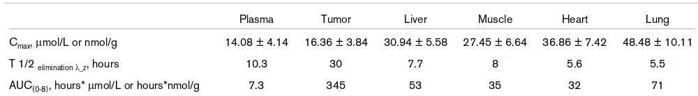
μmol/L), indicating dose linearity within this dose range (data not shown). The tissue exposures over the course of 48 hours were 5*fold (muscle and heart), 7*fold (liver), and 10*fold (lung) greater than in plasma. The terminal elimination half-life from the analyzed non-tumor tissues was between 5.5 and 8 hours. Importantly, the tumor exposure was approximately 47*fold higher than that in plasma, maintaining a concentration of 2.22 nmol/g 48 hours after compound administration. This drug level corresponded to approximately 13% of the Cmax (highest concentration) (16.3 nmol/g) and resulted in a termi-nal half-life of 25 to 30 hours. Interestingly, retention in tumor xenografts was also observed for 17*AAG [31], indicating that this may be a target-related phenomenon.
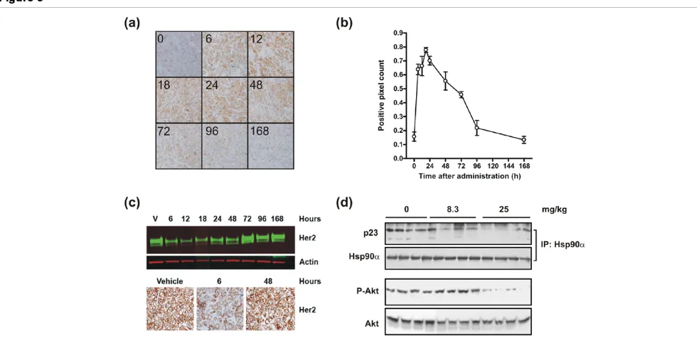
[image:6.612.59.293.125.273.2]NVP-AUY922 affects pharmacodynamic markers in vivo The biodistribution and pharmacokinetic profile of NVP-AUY922 in the BT-474 xenograft model encouraged us to assess whether NVP-AUY922 is capable of directly interfering with the catalytic cycle of HSP90 with concomitant depletion of HSP90 client proteins in vivo (Figure 2b). To asses whether systemic administration of NVP-AUY922 could affect the association between p23 and HSP90 observed in vitro, HSP90α was immunoprecipitated and co-immunoprecipitat-ing p23 was detected by Western blottco-immunoprecipitat-ing. A significant effect of NVP-AUY922 on HSP90-p23 complex dissociation was observed at the 2- and 6-hour time points. From 16 and 24 hours after compound administration, HSP90-p23 complexes reassembled in the BT-474 xenografts. To asses whether the effect of AKT phosphorylation on Ser473 observed on cell Table 1
Antiproliferative effect of NVP-AUY922 against a panel of human breast cancer cell lines
Cell line ErbB2/ER status NVP-AUY922 17-AAG
nM
BT-474 ErbB2+/ER+ 3.1 ± 1.4 17.4 ± 5.2
BT20 ErbB2-/ER- 4.0 ± 1.4 18.9 ± 4.8
MDA-MB-157 ErbB2-/ER- 126 ± 37 29.5 ± 20.8
MDA-MB-231 ErbB2-/ER- 7.0 ± 1.7 2,057 ± 571
MDA-MB-468 ErbB2-/ER- 6.3 ± 2.6 1,657 ± 390
SkBr3 ErbB2+/ER- 3.3 ± 0.9 11.9 ± 8.0
MCF-7 ErbB2-/ER- 8.8 ± 1.8 69.0 ± 18.3
lines grown in culture could be observed in the xenograft model, Western blot analysis was performed. We observed that the inhibition of HSP90 by a single dose of NVP-AUY922 was paralleled by reductions in phospho-AKT levels. Thus, NVP-AUY922 inhibits the catalytic cycle of HSP90 and reduces AKT signaling in xenograft tumor tissues, which is similar to the effects observed in vitro. Another hallmark of HSP90 inhibition is the induction of a heat shock response, which was evaluated by determining the HSP70 protein levels by immunohistochemistry (Figure 3). In this experiment, a sin-gle 50 mg/kg dose of NVP-AUY922 was administered to BT-474 xenograft-bearing mice and tumor sections were pre-pared at selected time points over the following week (Figure 3a). The staining intensity was quantified using automated imaging analysis (Figure 3b) and already demonstrated strongly increased HSP70 staining 6 hours after compound administration. The staining intensity peaked at the 24-hour time point and gradually returned to baseline over one week (Figure 3b). These data demonstrated a delayed heat shock response compared with the early response observed on the HSP90-p23 complex and AKT phosphorylation. Next, we determined the expression levels of the client protein ERBB2 in the same experiment by immunohistochemistry and West-ern blot analysis (Figure 3c). ERBB2 levels were reduced from 6 to 18 hours after administration of NVP-AUY922 but returned to baseline between 24 and 48 hours after the admin-istration. Overall, the data suggest that a cascade of timely events was initiated in which inhibition of HSP90 ATPase activity leads to immediate HSP90-p23 dissociation followed by inhibition of downstream signaling as detected by decreased phosphorylation of AKT and degradation of client proteins (for example, ERBB2) concomitant with release of HSF-1, causing transcriptional induction of HSP70.
Lowest dose that elicits pharmacodynamic response in tumors
[image:7.612.314.557.89.369.2] [image:7.612.58.294.124.280.2]The lowest dose of NVP-AUY922 which could elicit a detect-able response on HSP90-p23 complexes as well as AKT phosphorylation was determined. The effect of NVP-AUY922 on selected pharmacodynamic (PD) markers was assessed by administering the drug intravenously to s.c. BT-474 tumor-bearing mice. A single dose of 8.3 or 25 mg/kg was adminis-tered and tumors were dissected 6 hours later since HSP90-p23 dissociation and AKT phosphorylation are easily detecta-ble at this time point (Figure 2b). A dose of 25 mg/kg NVP-AUY922 caused significant disruption of HSP90-p23 com-plexes, whereas the 8.3 mg/kg dose caused only a subtle disruption (Figure 3d). Only the highest dose of 25 mg/kg Table 2
Antiproliferative effect of NVP-AUY922 against primary breast cancer
Cell line IC50 IC70
nM
MAXF 1162 304 505
MAXF 1322 29 48
MAXF 1384 209 477
MAXF 401 78 295
MAXF 574 >10,000 >10,000
MAXF 583 333 569
The concentrations of NVP-AUY922 that inhibit colony formation by 50% (IC50) or 70% (IC70) were determined (Oncotest GmbH,
Freiburg, Germany) [24].
Figure 2
Pharmacokinetic/pharmacodynamic analysis of NVP-AUY922 in BT-474 tumor-bearing nude mice
Pharmacokinetic/pharmacodynamic analysis of NVP-AUY922 in BT-474 tumor-bearing nude mice. A pharmacokinetic profile of NVP-AUY922 in BT-474 tumor xenografts, plasma, and organs (liver, heart, lung, and muscle) after administration of a single dose is shown. Female athymic mice bearing subcutaneous xenotransplants of the human ductal breast carcinoma BT-474 of approximately 250 mm3
NVP-AUY922 elicited a reduction in phospho-AKT levels. Thus, even though 8 mg/kg NVP-AUY922 appeared to cause a slight reduction in HSP90-p23 complexes, the remaining complexes are likely sufficient to maintain downstream signal-ing as evidenced by AKT phosphorylation.
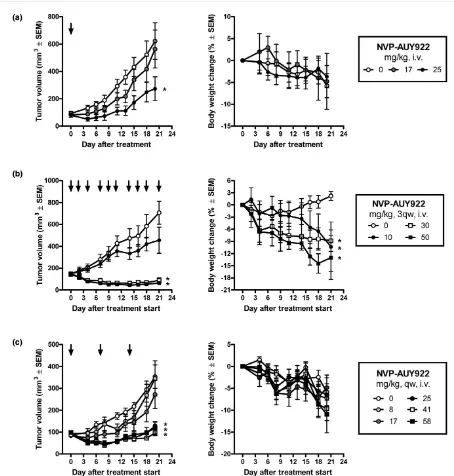
NVP-AUY922 exhibits potent antitumor activity at well-tolerated dose levels against an ERBB2-overexpressing ER-positive breast cancer xenograft
During the drug discovery selection process for the optimal development candidate, NVP-AUY922 was administered intraperitonealy once per day to HCT16 colon tumor xenograft-bearing mice at a dose of 50 mg/kg [23]. Apart from demonstrating the antitumor potential of the compound, this study demonstrated that NVP-AUY922 did not cause signifi-cant body weight changes when administered intraperitonealy at dose levels of up to 50 mg/kg daily [23]. We wanted to elaborate on these initial findings by investigating whether the effect observed on the PD markers correlated with an antitu-mor effect at well-tolerated doses and regimens in the BT-474 tumor model. During the establishment of the BT-474 model, we noticed that untreated tumor- and estrogen pellet-bearing mice generally either lost or gained minimum body weight dur-ing the experiment (data not shown and Figure 4, right panels). The body weight loss is likely to make the animals more sensi-tive to the potential additional toxicity caused by the treatment. As such, this xenograft model is ideally suited for use to opti-mize the dosing regimen that gives a good efficacy/tolerability balance. To more closely simulate the administration route to be used in clinical trials, the administration schedule was opti-mized using i.v. administration of NVP-AUY922 (Figure 4). In the first experiment, a single dose of NVP-AUY922 was admin-istered at around the lowest dose levels that elicited a reliable PD marker response (Figure 3d). Either 17 or 25 mg/kg was administered and tumor growth was monitored for 21 days (Figure 4a). A single administration at these dose levels did not affect body weights compared with vehicle. However, at the 25 mg/kg dose level, significant tumor growth was not observed for about 1 week after the administration. Also, at a dose level of 17 mg/kg, an indication of retarded tumor growth was observed (Figure 4a). Interestingly, at the end of the experiment, the animals in the group treated with 25 mg/kg had a tumor size corresponding to the tumor size in control ani-mals approximately 10 days earlier and were statistically
signif-icantly smaller in this group compared with the control group at the end of the experiment (P < 0.05, one-way ANOVA post hoc Dunnett versus control). These results combined with PD data showing a response lasting more than 24 hours clearly demonstrated a likelihood of achieving good antitumor effi-cacy when administering NVP-AUY922 in intermittent dose regimens. To evaluate this hypothesis, we administered NVP-AUY922 three times per week (Monday, Wednesday, and Fri-day) at dose levels of 10, 30, and 50 mg/kg (Figure 4b). Indeed, we achieved tumor regression at the two highest dose levels whereas treatment at 10 mg/kg, which did not elicit a reproducible effect on PD markers (Figure 3d), did not result in a significant effect on tumor growth. Although these data were highly encouraging, this treatment regimen resulted in a significant body weight loss at all dose levels (Figure 4b, right panel). To further improve the administration regimen, we eval-uated whether extending the recovery period in between dos-ing would improve tolerability without affectdos-ing antitumor effect. Since tumor growth was blocked for about 1 week after a single administration of NVP-AUY922 (Figure 4a), a once-a-week dosing regimen was investigated. Groups of eight BT-474 tumor-bearing animals were treated at dose levels of 8.3, 17, 25, 41, or 58 mg/kg. In this setting, NVP-AUY922 was highly efficacious when a dose level of 25 to 58 mg/kg was administered once per week (Figure 4c and Table 4). Dose levels of 8.3 to 17 mg/kg did not result in a significant effect on tumor growth compared with vehicle-treated control ani-mals, correlating with the data demonstrating that to observe a consistent PD response a single dose of 25 mg/kg is needed (Figure 3d). Overall, we demonstrate that NVP-AUY922 has good pharmacokinetic properties and is effica-cious and well tolerated when administered as a single agent once per week.
Conclusion
HSP90 has become an increasingly attractive target for anti-cancer therapy [6]. HSP90 inhibitors cause pleiotropic effects through degradation of a large number of client proteins as well as induction of a heat shock response [32]. The simulta-neous effect on multiple oncogenic pathways is predicted to be beneficial in a broad spectrum of cancer types that are driven by HSP90 client proteins. In this report, we focused on provided preclinical data to support the use of NVP-AUY922 for the treatment of breast cancer, an indication that is cur-Table 3
NVP-AUY922 biodistribution: pharmacokinetic parameters
Plasma Tumor Liver Muscle Heart Lung
Cmax, μmol/L or nmol/g 14.08 ± 4.14 16.36 ± 3.84 30.94 ± 5.58 27.45 ± 6.64 36.86 ± 7.42 48.48 ± 10.11
T 1/2 elimination λ_z, hours 10.3 30 7.7 8 5.6 5.5
AUC(0-8), hours* μmol/L or hours*nmol/g 7.3 345 53 35 32 71
The concentration of NVP-AUY922 was determined by high-pressure liquid chromatography/tandem mass spectrometry after a single intravenous administration of 30 mg/kg to BT-474 tumor-bearing mice. The highest concentration (Cmax), terminal half-life (T 1/2 elimination λ_z), and exposure as
[image:8.612.53.553.112.181.2]rently being pursued in clinical trials. To this end, we demon-strate that NVP-AUY922 exhibited significant antiproliferative properties against most established and primary breast cancer cells. A few breast cancer cell lines were found to be less sen-sitive, which may be due to drug transporters or metabolic activity. This is currently being investigated further.
The expression of a number of client proteins, signaling mole-cules, heat shock response proteins, and proteins involved in apoptotic pathways was determined. However, no clear corre-lation between a specific pathway and antiproliferative effect could be found. The majority of the work presented in this report was performed in the BT-474 breast cancer cell line, which is driven by ERBB2 and depends on estrogen for growth. However, although ERBB2 is an excellent client pro-tein, ERBB2-negative tumors may well be driven by suscepti-ble client proteins and respond to HSP90 inhibitors. Based on the antiproliferative profile in breast cancer cell lines, it is diffi-cult to link NVP-AUY922 antitumor activity with a specific genetic marker such as ERBB2 expression or ER status. This makes patient stratification exceedingly difficult, as discussed elsewhere [33].
NVP-AUY922 binds very potently to HSP90 in a competitive biochemical assay [23,26]. The high biochemical potency cor-related with the ability of NVP-AUY922 to directly interfere with the HSP90-p23 complex both in BT-474 cells grown in culture and as xenografts. However, for complete dissociation
of the HSP90-p23 complexes within 3 hours, a concentration of approximately 15 times the BT-474 GI50 value (determined after continuous exposure for 2 days) is needed. These data suggest that complete inhibition of HSP90 is not needed to achieve antiproliferative effect by continuous exposure. How-ever, short-term exposure at high drug concentrations may cause potent HSP90 inhibition in cells that are highly depend-ent on HSP90, such as tumors [34]. The short-term exposure causes the initiation of downstream events (for example, client protein degradation) from which affected tumor cells recover slowly. This hypothesis supports intermittent dosing of HSP90 inhibitors. When BT-474 tumor-bearing animals were adminis-tered NVP-AUY922 weekly at doses that cause tumor con-centrations to reach 1,000 times the GI50 value for approximately 2 days, significant tumor growth was not observed for about 1 week. In addition, the weekly schedule was well tolerated. Therefore, an NVP-AUY922 intermittent dosing regimen gives optimal tolerability without significantly affecting efficacy. This has also been found to be the case for other HSP90 inhibitors such as 17-AAG, which is being administered in an intermittent dose regimen [35]. Overall, our data offer further support for NVP-AUY922 to enter clinical trials.
Competing interests
[image:9.612.57.547.90.328.2]MRJ, JS, TR, CTG, JB, CQ, AB, RC, CG-E, and PC are employees and stockholders of Novartis Pharma AG. AM and MJD are employees and stockholders of Vernalis, Plc. Figure 3
Analysis of the kinetics of HSP70 induction and ERBB2 degradation following a singe dose of NVP-AUY922
Authors' contributions
MRJ participated in the design and coordination of the studies, conducted in vivo pharmacology experiments, and is the author responsible for the manuscript. JS participated in study design and coordination. TR developed and conducted the HSP90-p23 binding assay. AM conducted cellular prolifera-tion assays and PD analysis. CTG conducted
[image:10.612.65.519.94.570.2]immunohisto-chemistry and Western blot analysis. JB conducted the pharmacokinetic studies. CQ gave clinical advice. AB coordi-nated immunohistochemistry studies. RC coordicoordi-nated in vivo studies. MJD and CG-E coordinated studies. PC was respon-sible for overall study design and coordination. All authors par-ticipated in discussions and read and approved the final manuscript.
Figure 4
Antitumor effect and tolerability of NVP-AUY922 in a BT-474 human breast cancer xenograft model
Antitumor effect and tolerability of NVP-AUY922 in a BT-474 human breast cancer xenograft model. BT-474 cells were inoculated subcutaneously in female nude mice carrying an estrogen-release pellet. When the tumors reached 100 to 200 mm3, drug treatment was initiated. Each group
Acknowledgements
The authors wish to thank Kerstin Pollehn, Joelle Rudloff, Dario Sterker, Sonja Tobler, Axelle Welsch, and Kristie Wetzel for their excellent tech-nical assistance and dedication to the project.
References
1. Deininger M, Buchdunger E, Druker BJ: The development of imatinib as a therapeutic agent for chronic myeloid leukemia.
Blood 2005, 105:2640-2653.
2. Herbst RS, Fukuoka M, Baselga J: Gefitinib – a novel targeted approach to treating cancer. Nat Rev Cancer 2004, 4:956-965. 3. Adams GP, Weiner LM: Monoclonal antibody therapy of cancer.
Nat Biotechnol 2005, 23:1147-1157.
4. Demonty G, Bernard-Marty C, Puglisi F, Mancini I, Piccart M: Progress and new standards of care in the management of HER-2 positive breast cancer. Eur J Cancer 2007, 43:497-509. 5. Workman P, Burrows F, Neckers L, Rosen N: Drugging the can-cer chaperone HSP90: combinatorial therapeutic exploitation of oncogene addiction and tumor stress. Ann N Y Acad Sci
2007, 1113:202-216.
6. Drysdale MJ, Brough PA, Massey A, Jensen MR, Schoepfer J: Tar-geting Hsp90 for the treatment of cancer. Curr Opin Drug Dis-cov Devel 2006, 9:483-495.
7. Pearl LH, Prodromou C, Workman P: The Hsp90 molecular chaperone: an open and shut case for treatment. Biochem J
2008, 410:439-453.
8. Hollingshead M, Alley M, Burger AM, Borgel S, Pacula-Cox C, Fie-big HH, Sausville EA: In vivo antitumor efficacy of 17-DMAG (17-dimethylaminoethylamino-17-demethoxygeldanamycin hydrochloride), a water-soluble geldanamycin derivative. Can-cer Chemother Pharmacol 2005, 56:115-125.
9. Solit DB, Ivy SP, Kopil C, Sikorski R, Morris MJ, Slovin SF, Kelly WK, DeLaCruz A, Curley T, Heller G, Larson S, Schwartz L, Egorin MJ, Rosen N, Scher HI: Phase I trial of 17-allylamino-17-demethoxygeldanamycin in patients with advanced cancer.
Clin Cancer Res 2007, 13:1775-1782.
10. DeFranco DB, Csermely P: Steroid receptor and molecular chaperone encounters in the nucleus. Sci STKE 2000, 2000:PE1.
11. Pratt WB, Galigniana MD, Morishima Y, Murphy PJ: Role of molecular chaperones in steroid receptor action. Essays Biochem 2004, 40:41-58.
12. Beliakoff J, Whitesell L: Hsp90: an emerging target for breast cancer therapy. Anticancer Drugs 2004, 15:651-662.
13. Beliakoff J, Bagatell R, Paine-Murrieta G, Taylor CW, Lykkesfeldt AE, Whitesell L: Hormone-refractory breast cancer remains
sensitive to the antitumor activity of heat shock protein 90 inhibitors. Clin Cancer Res 2003, 9:4961-4971.
14. Osborne CK: Tamoxifen in the treatment of breast cancer. N Engl J Med 1998, 339:1609-1618.
15. Bachleitner-Hofmann T, Pichler-Gebhard B, Rudas M, Gnant M, Taucher S, Kandioler D, Janschek E, Dubsky P, Roka S, Sporn E, Jakesz R: Pattern of hormone receptor status of secondary contralateral breast cancers in patients receiving adjuvant tamoxifen. Clin Cancer Res 2002, 8:3427-3432.
16. Shimamura T, Lowell AM, Engelman JA, Shapiro GI: Epidermal growth factor receptors harboring kinase domain mutations associate with the heat shock protein 90 chaperone and are destabilized following exposure to geldanamycins. Cancer Res 2005, 65:6401-6408.
17. Basso AD, Solit DB, Chiosis G, Giri B, Tsichlis P, Rosen N: Akt forms an intracellular complex with heat shock protein 90 (Hsp90) and Cdc37 and is destabilized by inhibitors of Hsp90 function. J Biol Chem 2002, 277:39858-39866.
18. Xu W, Marcu M, Yuan X, Mimnaugh E, Patterson C, Neckers L: Chaperone-dependent E3 ubiquitin ligase CHIP mediates a degradative pathway for c-ErbB2/Neu. Proc Natl Acad Sci USA 2002, 99:12847-12852.
19. Sain N, Krishnan B, Ormerod MG, De Rienzo A, Liu WM, Kaye SB, Workman P, Jackman AL: Potentiation of paclitaxel activity by the HSP90 inhibitor 17-allylamino-17-demethoxygeldanamy-cin in human ovarian car17-allylamino-17-demethoxygeldanamy-cinoma cell lines with high levels of activated AKT. Mol Cancer Ther 2006, 5:1197-1208.
20. Maira SM, Voliva C, Garcia-Echeverria C: Class IA phosphatidyli-nositol 3-kinase: from their biologic implication in human can-cers to drug discovery. Expert Opin Ther Targets 2008, 12:223-238.
21. Solit DB, Basso AD, Olshen AB, Scher HI, Rosen N: Inhibition of heat shock protein 90 function down-regulates Akt kinase and sensitizes tumors to Taxol. Cancer Res 2003, 63:2139-2144. 22. Munster PN, Marchion DC, Basso AD, Rosen N: Degradation of
HER2 by ansamycins induces growth arrest and apoptosis in cells with HER2 overexpression via a HER3, phosphatidyli-nositol 3'-kinase-AKT-dependent pathway. Cancer Res 2002, 62:3132-3137.
23. Brough PA, Aherne W, Barril X, Borgognoni J, Boxall K, Cansfield JE, Cheung KM, Collins I, Davies NG, Drysdale MJ, Dymock B, Eccles SA, Finch H, Fink A, Hayes A, Howes R, Hubbard RE, James K, Jordan AM, Lockie A, Martins V, Massey A, Matthews TP, McDonald E, Northfield CJ, Pearl LH, Prodromou C, Ray S, Ray-naud FI, Roughley SD, et al.: 4,5-diarylisoxazole Hsp90 chaper-one inhibitors: potential therapeutic agents for the treatment of cancer. J Med Chem 2008, 51:196-218.
24. Smith V, Sausville EA, Camalier RF, Fiebig HH, Burger AM: Com-parison of 17-dimethylaminoethylamino-17-demethoxy-Table 4
Antitumor effect and tolerability of NVP-AUY922 against BT-474 tumor-bearing nude mice when administered once per week
Tumor response Host response
NVP-AUY922 dose, mg/kg
T/C, percentage Tumor volume change, cubic millimeters
Body weight change, grams
Body weight change, percentage
Survival, percentage
0 100 266 ± 51 -1.6 ± 0.9 -5.9 ± 3.2 100
8.3 95 252 ± 73 -2.2 ± 0.8 -7.3 ± 2.6 100
17 72 191 ± 61 -1.2 ± 0.7 -4.8 ± 2.9 88
25 12 32 ± 15a -1.7 ± 0.6 -6.5 ± 2.5 100
41 3 9 ± 13a -2.4 ± 0.6 -9.7 ± 2.7 100
58 8 22 ± 26a -3.0 ± 1.2 -10.9 ± 4.2 100
BT-474 cells were inoculated subcutaneously in female nude mice carrying an estrogen-release pellet. When the tumors reached 100 to 200 mm3, drug treatment was initiated. Each group consisted of eight animals. NVP-AUY922 was administered once per week at the indicated dose
levels. Tumor volumes and body weights were measured three times per week, and the experiment was evaluated 20 days after the first dose was administered. The percentage T/C represent the mean change in tumor volume of the treated group divided by the mean change in tumor volume of the vehicle treated control group. aStatistical significance compared with vehicle-treated controls (P < 0.05, one-way analysis of variance post
[image:11.612.61.553.117.257.2]geldanamycin (17DMAG) and 17-allylamino-17-demethoxy-geldanamycin (17AAG) in vitro: effects on Hsp90 and client proteins in melanoma models. Cancer Chemother Pharmacol
2005, 56:126-137.
25. Tomayko MM, Reynolds CP: Determination of subcutaneous tumor size in athymic (nude) mice. Cancer Chemother Pharmacol 1989, 24:148-154.
26. Schilb A, Riou V, Schoepfer J, Ottl J, Muller K, Chene P, Mayr LM, Filipuzzi I: Development and implementation of a highly minia-turized confocal 2D-FIDA-based high-throughput screening assay to search for active site modulators of the human heat shock protein 90beta. J Biomol Screen 2004, 9:569-577. 27. Fiebig HH, Schuler J, Bausch N, Hofmann M, Metz T, Korrat A:
Gene signatures developed from patient tumor explants grown in nude mice to predict tumor response to 11 cytotoxic drugs. Cancer Genomics Proteomics 2007, 4:197-209. 28. Sullivan WP, Owen BA, Toft DO: The influence of ATP and p23
on the conformation of hsp90. J Biol Chem 2002, 277:45942-45948.
29. Chan CT, Paulmurugan R, Gheysens OS, Kim J, Chiosis G, Gam-bhir SS: Molecular imaging of the efficacy of heat shock pro-tein 90 inhibitors in living subjects. Cancer Res 2008, 68:216-226.
30. Basso AD, Solit DB, Munster PN, Rosen N: Ansamycin antibiot-ics inhibit Akt activation and cyclin D expression in breast can-cer cells that overexpress HER2. Oncogene 2002, 21:1159-1166.
31. Banerji U, Walton M, Raynaud F, Grimshaw R, Kelland L, Valenti M, Judson I, Workman P: Pharmacokinetic-pharmacodynamic relationships for the heat shock protein 90 molecular chaper-one inhibitor 17-allylamino, 17-demethoxygeldanamycin in human ovarian cancer xenograft models. Clin Cancer Res
2005, 11(19 Pt 1):7023-7032.
32. Xu W, Neckers L: Targeting the molecular chaperone heat shock protein 90 provides a multifaceted effect on diverse cell signaling pathways of cancer cells. Clin Cancer Res 2007, 13:1625-1629.
33. Whitesell L, Lindquist SL: HSP90 and the chaperoning of cancer. Nat Rev Cancer 2005, 5:761-772.
34. Kamal A, Thao L, Sensintaffar J, Zhang L, Boehm MF, Fritz LC, Bur-rows FJ: A high-affinity conformation of Hsp90 confers tumour selectivity on Hsp90 inhibitors. Nature 2003, 425:407-410. 35. Weigel BJ, Blaney SM, Reid JM, Safgren SL, Bagatell R, Kersey J,
Neglia JP, Ivy SP, Ingle AM, Whitesell L, Gilbertson RJ, Krailo M, Ames M, Adamson PC: A phase I study of 17-allylaminogeldan-amycin in relapsed/refractory pediatric patients with solid tumors: a Children's Oncology Group study. Clin Cancer Res