The Effect of Intraportal Mannitol
on the Short-term in Vivo Distribution of
Radiolabelled A-LAK Cells
in Rats
"
Jianhua Zhou M.B.
Master of Medical Science (Surgery)
-~---university
of Tasmania
Declaration
I hereby declare that this thesis contains no material which
has been accepted for the award of any other degree or
diploma in any tertiary institution and that, to the best of my
knowledge and belief, the thesis contains no material
previously published or written by another person, except
when due reference is made in the text of the thesis.
Acknowledgements
This work is dedicated to my supervisor Mr. Stephen Wilkinson who has been developing Cancer Immunotherapy for colorectal cancer and whose support allowed me to gain confidence in this work in the face of technical and financial difficulties.
I would like to acknowledge with gratitude, support from The Paul Mackay Bolton Foundation, the Royal Hobart Hospital Research Trust, and from Professor Joseph J. Shepherd. Only with this support did it become possible for me to complete this project.
Abstract
The effectiveness of adoptive immunotherapy of cancer using LAI< cells and IL-2 depends on the accumulation of transferred effector cells at the tumour sites. In vitro LAK cell have been demonstrated to have broad cytotoxic activity to a wide variety of tumour cells in a non-major-histocompatibility complex-restricted manner, and independent of the presence of tumour specific antigens. LAK cells have now been used effectively in a small number of human trials. The effective delivery of these cells to the tumour site in vivo is one of the main aspects of this type of immunotherapy that requires further investigation. Conventional systemic infusion has shown a limited migration pattern of LAK cells. In this study the degree of entrapment of LAK cells in organs following local infusion has been determined. The large size and rigidity of LAK cells may be one mechanism which restricts the distribution of these cells. In addition, in this study the effect of mannitol, a hyperosmotic agent which increases the space between vascular endothelial cells, on the uptake of intraportal LAI< cells into liver has been determined.
Effector:Target ratio of 40:1 A-LAK cells lysed 70% P815 and 100% YAC-1 cells.
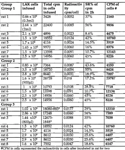
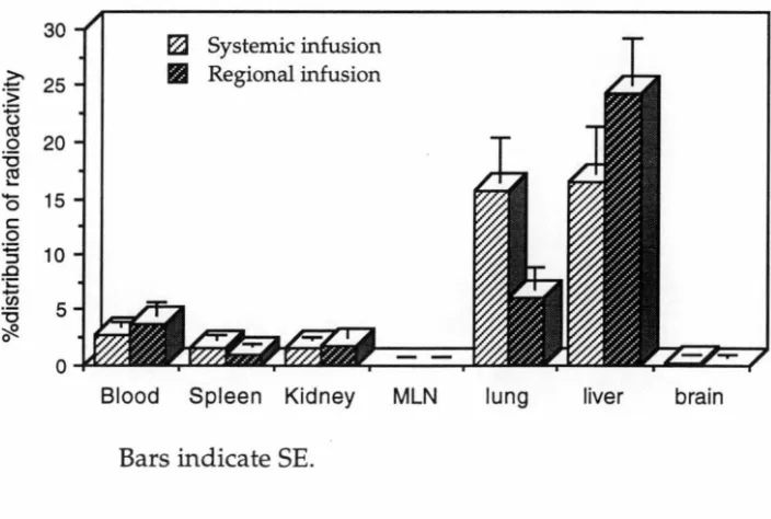
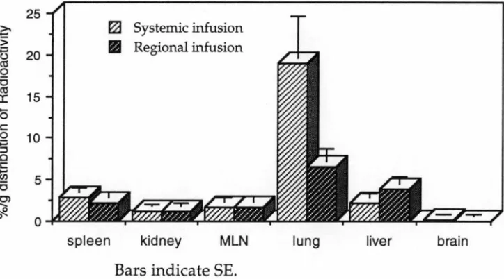
After labelling A-LAK cells with 51Cr, the effect of intraportal infussion of 30% mannitol on the distribution of intraportally infused A-LAK cells in liver was studied. The trafficking studies were carried out in three groups. In Group 1, 51Cr labelled A-LAK cells were systemically infused through the tail vein of rats as a control group. In Group 2 and Group 3, A-LAK cells were infused into syngeneic rats through the portal vein without or following prior portal infusion of 30% mannitol. Two hours after LAK cell administration the rats were sacrificed and the radioactivity in liver, lung, spleen, blood, MLN, kidney and brain were measured to determine the distribution of A-LAK cells to these organs.
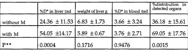
The results showed that intraportal mannitol was associated with an increased percentage of LAK cells in the liver compared with regionally infused LAK cells without mannitol (54% vs 24%; P<0.0005). The administration of intraportal mannitol was also associated with increased distribution of A-LAK cells into the brain (0.26% vs 0.08%; P<0.05) and MLN (0.05% vs 0.02%; P<0.05). There was no significant increase in uptake of A-LAK cells in lung (8.39% vs 6.01 %), spleen (1.00% vs 0.98%), or kidney (1.44% vs 1.78%) following intraportal mannitol.
CONTENTS
CHAPTER 1 LITERATURE REVIEW 1
1.1 Introduction
21.2 Development of Adoptive lmmunotherapy for
5Cancer Using LAK Cells and IL-2
1.3
In Vitro
Production and Characterisation of
12LAK Cells
1.3.1 Introduction 12
1.3.2 Morphology and cell surface characteristics of LAK 12 cells and LAK precursors
1.3.2a LAK cell morphology 12
1.3.2b Cell surface markers 13 1.3.3 Production of LAK cells in vitro 18 1.3.3a Isolation of LAK precursors from whole 18
lymphocyte population
1.3.3b Factors influencing LAK cell proliferation 21 1.3.4 Tumouricidal activity of LAK cells in vitro and in 22
vivo
1.4
In Vivo
Distribution of LAK Cells Infused
26Systemically or Regionally
1.5 The Effect of Mannitol on the Blood
34Brain Barrier (BBB)
CHAPTER 2 MATERIALS AND METHODS 37
2.1.2a Cell surface marker analysis 42 2.1.2b Morphological analysis 43
2.1.2c Cytotoxicity assay 44
2.2 Short-term in Vivo
Distribution of
46Radiolabelled A-LAK Cells in the Rat
2.2.1 Radiolabelling of A-LAK cells 46 2.2.2 Adoptive transfer of radiolabelled A-LAK cells 47
into recipient syngeneic rats
2.2.3 Determination of tissue distribution of 48 radiolabelled A- LAK cells
2.3 Administration of Mannitol
49CHAPTER 3 RESULTS 50
3.1 A-LAK cell Generation and Characterisation
51 3.1.1 Isolation of A-LAK precursors fromspleen mononuclear cells 51
3.1.2 Second-antibodies 52
3.1.3 Culture of A-LAK cells 53
3.1.4 Identification of A-LAK cells 54
3.2 LAK Cell Distribution
573.2.1 Slcr labelling of A-LAK cells 57 3.2.2 Distribution of A-LAK cells by systemic and 60
regional infusion and their comparison
3.2.3 Effect of mannitol on the distribution of A-LAK 66 cells to the liver infused via the portal vein.
CHAPTER 4 DISCUSSION 70
4.1 A-LAK Cell Generation and Characterisation
714.1.2 A-LAK cell identification 73
4.2 Distribution of s1cr-labelled A-LAK Cells in Rats
75 4.2.1 51Cr-labelling of A-LAK cells 754.2.2 A-LAK cell distribution 77
BIBLIOGRAPHY 83
ABBREVIATIONS
A-LAK adherent lymphokine-activated killer BBB blood brain barrier
BM bone marrow
BSA bovine serum albumin Ci curie
CLN clavicle lymphonodus CM complete culture medium cpm count per minute
CR complete regression CTL cytotoxic T lymphocyte CY cyclophosphamide dl deciliter
EDTA ethylenediaminetetra-acetic acid E/T effector to target ratio
FCS fetal calf serium
FITC fluorescein isothiocyanate
g gram
h hour
HEV high endothelial venules IFN interferon
lg immunoglobulin IL-2 interleukin-2 ip. intra peritoneal iv. intravenous kg kilogram L lit er
LAK lymphokine-activated killer LAL large agranular lymphocyte
LGL large granular lymphocyte (
LME lysosomotropic amine
, ___________
M mole
Mab monoclonal antibodies mg milligram
min minute ml milliliter
MLN mesenteric lymphonode MMC mitomycinC
MNC mononuclear cell
MLTC mixed lymphocyte-tumour cell culture
MW molecular weight
NH&MRC national health and medical research council
NI< natural killer NW nylon wool
NWP nylon wool passed (or non-adherent) lymphocyte NT non-tested.
PBL peripheral blood lymphocyte PBS phosphate buffered saline PMN peripheral mononuclear cell PR partial regression
P-815 murine mastocytoma rIL recombinant interleukin-2 rpm round per minute
SI small intestinal SR spontaneous release TCGF T cell growth factor TCM tissue culture medium
Th
T-helperTIL tumour infiltrate lymphocyte TNF tumour necrosis factor
Ts T-suppressor
u
unitsUV ultraviolet
YAC-1 mouse T cell lymphoma
oc
degree centigrade% per cent µg microgram µl microliter
1.1 Introduction
LAI< cells, produced by culture of lymphocytes with a high concentration of IL-2 have been demonstrated to have broad cytotoxic activity against a wide variety of tumour cells (Lotze et al 1981; Grimm
et al 1982; Rosenstein et al 1984; Lafreniere et al 1985; Mule et al 1985; Shu et al 1985; Papa et al 1986; Itoh et al 1986; Salup et al 1986; Vujanovic et al 1988b). LAI< cells lyse tumour cells m vivo after gaining access to the target cells by extravasation through the microcirculation (Harcel et al 1991; Basse et al 1991a). Clinical studies using LAI< cells with IL-2 have shown therapeutic value in cancer treatment (Rosenberg et al 1986a; 1986b; Benyunes et al 1993; Schoof et al 1993). However, systemic LAK/IL-2 therapy is associated with significant toxicity which is associated with the very large number of cells and high doses of rIL-2 infused (Rosenberg et al 1987; Kuebler et al
1993; Villani et al 1993). One solution to this problem may be the regional administration of LAK cells, possibly together with factors which enhance the distribution of LAI< cells to the target organ.
Studies have shown that regional infusion of LAI< cells could result in more effector cells accumulating at the target sites, thus decreasing the dosage of LAK cells and IL-2 required and minimising some side effects. Regional IL-2/LAK cell infusion has also been shown to be associated with anti-tumour effectiveness and lower toxicity (Lafreniere et al 1985b; Ettinghausen et al 1985; Basse et al 1991b; Kuppen et al 1992; Keilholz et al 1992; Scudeletti et al 1993; Yamamoto
1993) s~ggesting that the accumulation of effector cells in the tumour is an important factor.
LAI< cells have a limited pattern of migration after systemic administration with 47% accumulation in lungs 2h after infusion and then redistribution into liver 24h after infusion (Maghazachi et al
1988c, 1988b; Felgar et al 1990; Takai et al 1988). The limited distribution
of LAI< cells inhibits their optimal tumouricidal effect in viva. This
pattern may be the result of decreased extravasation of LAK cells because of their large size and high rigidity (Sasaki et al 1989). Mannitol
has been shown to open the Blood-Brain Barrier by a mechanism involving increase of intercellular spaces between capillary endothelial cells (Franceschini et al 1988). In this study we investigated the effect of intraportal mannitol on the distribution of LAI< cells infused into the portal vein.
During the course of these experiments there were not many reports concerning infusion of LAI< cells regionally and no report of using reagents to increase cell distribution. Recently adoptive immunotherapy has been delivered by local infusion. In the near future the utilization of LAK/IL-2 may be combined with factors which could improve their distribution, and with some reagents which could enhance LAK cell effectiveness.
The results of this study may lead to improvement in the effectiveness of adoptive immunotherapy, by determining an effective way of delivering effector cells into the tumour-bearing host. The generation of adequate numbers of highly cytotoxic cells, and the efficient delivery
. .
1.2
Development of Adoptive lmmunotherapy for Cancer
Using LAK Cells and IL-2
Adoptive immunotherapy for cancer is a therapy aimed at destroying cancer cells by transfer of activated antitumour lymphocytes into the tumour-bearing host. As early as the 1960's there were some studies which found that the intravenous delivery of lymphocytes from tumour cell-immunised mice into syngeneic tumour-bearing mice could mediate the regression of tumours. For example the treatment of fibrosarcoma in the rat with immune T-lymphocytes (Delorme et al
1964). These animal studies suggested that similar therapy could be used in human malignancy, however alternative methods for obtaining tumour-sensitised lymphocytes had to be developed. In 1970's, anti-tumour cytotoxic cells were obtained by sensitising the lymphocytes in in vitro culture with tumour cells, MLTC (Treves et al
1975; Berenson et al 1975; Gillis et al 1977; Ruscetti et al 1977; Zarling et al 1979; reviewed in Wilkinson 1993). In addition, the isolation of the lymphokine that was originally called T Cell Growth Factor (TCGF), now named interleukin-2 (IL-2), revealed that TCGF in the absence of tumour cells could stimulate lymphocytes to become cytotoxic against tumour cells (Aarden et al 1979). TCGF made it possible to maintain
cytotoxic lymphocytes in long-term culture (Morgan et al 1976; Gillis et al 197.8; Watson et al 1979).
In the early 1980's, purified IL-2 was found as a glycoprotein of MW 16,000 (Taniguch et al 1983; Louis et al 1983), and the IL-2 receptor was
isolated (Uchiyama et al 1981; Leonard et al 1982). IL-2 is synthesised
and secreted by mitogen- or antigen- activated Th lymphocytes (Smith
- .
(r-IL-2) was made possible by recombinant-DNA technology. IL-2 was found to promote the proliferation and activatio~ of antigen-stimulated T cells (Yron et al 1980; Cheever et al 1981) and to stimulate the proliferation of lymphocytes from non-immunised donors, and to provide them with the capacity to recognise and lyse a variety of tumour cells (Lotze et al 1980,1981; Rosenstein et al 1984). The activated cells which were raised from non-antigen-stimulated lymphocytes were described as Lymphokine-activated Killer (LAK) cells (Grimm et al 1982). The possibility now arose of utilizing LAK cells in adoptive immunotherapy. LAK cells could be continuously stimulated in vivo
by infusion of IL-2 systemically (Ettinghausen et al 1985a, 1985b) and could be produced by co-cultivation of peripheral blood lymphocytes with IL-2 in vitro. The generation of LAK cells both in vitro and in
vivo has the advantage that it is unnecessary to pre-sensitise the
effector cells with tumour cells. LAK cells kill tumour cells in a non-MHC-restricted manner (Hersey et al 1987).
In 1984, Rosenberg et al at the National Cancer Institute transferred LAK cells into murine tumour models and reported the first demonstration that lymphokine-activated killer cells, activated in
vitro by IL-2, can inhibit the growth of established melanoma
pulmonary metastases (Mazumder et al 1984). Systemic administration of xenogeneic IL-2 together with in vitro-sensitised and IL-2-expanded lymphocytes in animals resulted in regression of metastases (Donohue
Administration of high dose IL-2 alone resulted in significant toxicity.
In humans, initial systemic administration of recombina~t IL-2 at high doses over short periods of time showed the rapid half-life of IL-2 and it's toxicity in vivo. There was no detectible LAK cell activity in peripheral blood after short term administration -of IL-2. However there was an increase in the number of lymphocytes and their in vitro responsiveness to IL-2 (Lotze et al 1985; Atkins et al 1986). The requirement of high dose of IL-2 (over 500U/ml in vitro) to produce LAK cells made it impossible to generate them in vivo because of the toxicity. Many efforts have been made to modify the t!eatment protocol of IL-2 administration to optimise immune effects and decrease toxicity. In murine models it was demonstrated that prolonged administration of IL-2 at tolerable doses could activate sufficient in
vivo NK/LAK activity compared with a few days administration of
very high dose IL-2 (Ettinghausen et al 1986; Nishimura et al 1987; Talmadge et al 1987). It was also shown in humans that prolonged administration of low doses of rIL-2 resulted in selective expansion of NK cells in vivo with minimal toxicity (Caligiuri et al 1991). However large numbers of LAK cells could be generated in vitro. Provision of IL-2 together with administration of large quantities of LAK cells grown in vitro was more effective and less toxic (Mule et al 1984, 1985, 1986b). The antitumour effect of IL-2 appears to result from the action of LAK cells in vivo (Rosenberg et al 1985a; Ettinghausen et al 1985a, 1985b; Mule et al 1985; Hank et al 1988; Da Pozzo et al 1992).
Lafreniere et al 1985b; Shu et al 1985). Evidence of efficacy of this treatment was also obtained in established metastatic tu~ors in liver, lung, kidney and brain (Lafreniere et al 1985a; Papa et al 1986; Salup et al 1986; Takai et al 1988). A direct relationship existed between the number of LAI< cells transferred, the amount of IL-2 given, and the therapeutic effect achieved in murine models (Mule et al 1985).
Based on the knowlege from animal experiments, immunotherapy using LAK cells in conjunction with IL-2 was administered for the treatment of selected patients with advanced metastatic tumour. Rosenberg reported the result of their initial clinical trial which resulted in some responses in patients with colorectal cancer, malignant melanoma, renal cell cancer, and lung adenocarcinoma (Rosenberg et al 1985a, 1986a, 1986b). In 1987, Rosenberg et al reported their progress in treatment of patients with advanced cancer. In many patients the administration of these cells and IL-2 was limited by the toxicity of IL-2. This side effect resulted in a generalised capillary permeability leak syndrome. Further efforts were made to decrease the toxicity and complexity of this therapy.
effective in reducing established lung or liver metastases in rat models (Schwarz et al 1989). Rosenberg et al 1986c showed that LAK cells derived from lymphocytes exposed to tumour cells (such as tumour infiltrating lymphocytes) had greater tumoricidal effect than peripheral blood lymphocyte-derived LAK cells. Local adoptive immunotherapy by direct injection of LAK cells and IL-2 into brain tumours was found effective and had low toxicity (Yoshida et al 1988; Ibayashi et al 1993). In humans the effect of systemically infused LAK cells is far less in brain tumours (Monod et al 1992). In a later murine model with intracerebral metastases it was shown that there was no response to systemic infusion of LAK cells and IL-2 (McCutcheon et al 1990). The fact that histological examination of brain tissue did not reveal lymphocyte infiltration indicated that the effector cells were not delivered into tumour sites in brain by systemic infusion. The fact that there was no discernible correlation between in vitro target cell lysis by
LAK cells and theraputic efficacy in viva (Papa et al 1986; Nakano et al 1991) suggests that the effector cells may not reach the target organ in sufficient number in viva. Lack of response is not the result of loss of effectiveness in viva because it has been shown that there was corresponding in viva and in vitro susceptibility of murine tumours to lysis by LAK cells (Papa et al 1986). The tumour cells which appeared to be sensitive to LAK cells in vitro do not appear to be resistant to LAK cells in viva (Mule et al 1986). It is most likely the result of insufficient LAK cell infiltration into tumour sites. The extravasation of LAK cells into the tumour may be one of the important steps for achieving tumour damage.
results in partial or complete regression (PR and CR) of several types of tumours: prostatic carcinoma and associated pulmonary metastases (Tjota et al 1991); advanced renal cell carcinoma (Weiss et al 1992; Engelstein et al 1992); primary or metastatic liver carcinoma (Miya et al
1992; Foon et al 1992); non-Hodgkin's lymphoma (Weber et al 1992). Local and regional infusion of LAK cells and IL-2 appears to result in more responses because local infusion may be associated with greater accumulation of effector cells at the tumour site. Intrahepatic LAK cells inhibited the growth of metastatic tumours in the liver in some patients with cancers of the gastrointestinal tract (Tsugita et al 1990), and resulted in CR and PR in advanced liver metastatic or primary cancer (Han et al 1991; Keilholz et al 1992; Yamamoto et al 1993). Regional arterial administration of LAK cells also resulted in metastatic regression (Hayakawa et al 1991, 1992). Local adoptive immunotherapy using A-LAK cells and IL-2 in a nude mouse model of human squamous carcinoma of the head and neck resulted in complete tumour growth inhibition (Sacchi et al 1991). In a report on human malignant pleural mesothelioma treated with the local A-LAK immunotherapy, there was a reduction of the malignant pleural effusion (Yanagawa et al 1991).
The additional use of different lymphokines or cytokines may result in augmentation of effectiveness of LAK cells and IL-2. The combination with interferon-alpha (IFN-alpha) but not tumour necrosis factor-alpha (TNF-factor-alpha) showed augmentation of the effect of LAK cells in vitro (Wanebo et al 1991) and in vivo (Puri et al 1991). Kato et al 1991 reported that appropriately timed pretreatment of tumour-bearing mice with TNF-alpha augmented the anti-tumor efficacy of LAK cells.
human leukemia cells (Teichmann et al 1992). IL-1 and IL-3 may also increase the effect of IL-2 (Marumo et al 1992; Okuno e~ al 1992);. IL-7 was found to be able to induce significant LAK activity after bone marrow transplantation (Pavletic et al 1993).
The combination of chemotherapy with LAK cells and IL-2 adoptive immunotherapy may also result in additive effects (Yamaue et al 1991; Wakizaka et al 1992; Gazit et al 1992). Splenic or hepatic arterial injection of IL-2 with chemotherapeutic agents such as cyclophosphamide (CY), 5 fluorouracil (SFU) and mitomycin C (MMC) resulted in more LAK/NK activity in PBL in comparison to injection of IL-2 alone (Okuno et al 1991). Low-dosage CY seems increasing the LAK activity with IL-2 in patients with advanced malignancies of varying types (Abrams et al 1993). Lithium can be used as a new immunomodulator for cancer immunotherapy (Wu et al 1992). By conjugation of LAK cells with anti-tumour monoclonal antibody, LAK cell killing activity against tumour was specifically enhanced (Shiraiwa
1.3
In Vitro Production and Characterisation of LAK Cells
1.3.1 Introduction
LAI< cells are generated by culturing lymphocytes with IL-2 for several days. Since the description of the LAI< cell phenomenon there has been extensive investigation of LAI< cells including their characterisation and functions. This section summarises work on the morphology, the cell surface characteristics and the tumouricidal activity of LAI< cells and LAI< cell precursors.
1.3.2 Morphology and cell surface characteristics of LAK cells
and LAK precursors
1.3.2a LAK cell morphology
In response to culture in high concentrations of rlL-2 (500-lOOOU /ml), LGL/NK cells differentiate into highly active cytotoxic c~lls which·have been termed LAK cells. The LAK cells have similar morphology to LGL/NK. Morphological analysis of human A-LAI< cells indicate that these cells have the morphology of LGL with abundant cytoplasm and prominent cytoplasmic granules (Melder et al 1988), vacuolate cytoplasm and an undulating surface (Vujanovic et al 1988c). Sasaki et al 1989 have demonstrated that LAK cells have a greater mean diameter than nonactivated LGLs
1.3.2b Cell surface markers
Extensive studies analysing the surface phenotype of LAK progenitors and effector cells have been obtained in three species: human, rat and mouse.
In humans, most of the LAK cell progenitors from peripheral blood lymphocytes have the same characteristics as LGL/NK cells, and the human effector LAK cells also express the same phenotype as activated NK cells. Phillips et al 1986 and Ortaldo et al 1986 indicated that LAK progenitors in human peripheral blood are LGL/NK cells expressing the surface markers NKH1/Leul9 and CD16. CD16 antigen (Leull) has been found on almost all activated NK cells (Lanier et al 1983). NKHl antigen (Leu19) has been detected on virtually all NK cells and also on a small subpopulation of CD3+ T cells (Hercend et al 1985; Schmidt et al
(OKT3 or Leul) was found to be greatly augmented (Grimm et al
1983a). Killer cell precursors were also found in Leulla+ (CD16) and Leu7+ NK subsets, but not in Leu4+(CD3) or Leu3a+(CD4) T lymphocytes (Itoh et al 1986). The majority of LAI< activity is mediated
by NK cells that express the NKH1/Leul9, but not by T cells that express CD3. Talmadge et al 1986 found that the subpopulation of
lymphocytes most responsive to high doses of rIL-2 (> lOOU
I
ml) comprised LGLs, the morphologic homologue of natural killer cells. Lotzova et al 1987 further confirmed that human IL-2-stimulatedactive NK cells displayed LGL morphology and had CD16 and NKH1/Leu19 (CD56) cell surface phenotype. For human A-LAI< cells, phenotypic analysis indicates that a majority of these cells express the CD3-Leul9+ phenotype and a substantial proportion of Leul9+ cells expressed CD16 antigen (Melder et al 1988). Thus the progenitors of
LAK activity in human peripheral blood are mainly LGL/NK with CD3-, CD16+, NKHl+ (CD56) phenotype.
Grimm et al 1983a found that human LAI< progenitor cells were
present in lymphoid organs containing few active NK cells. They were null cells, distinct from NK cells or classical thymus-derived T-cells. Thymus derived LAI< cells are low to medium density lymphocytes, CD3 negative, and they possess the NK-associated marker NKH1/Leul9 (CD56), however they lack most NK-associated markers CD16 (HNKl/Leull and B73.l) (Ramsdell et al 1987).
In rats, Vujanovic et al 1988a showed that the major cell population
peripheral blood LGL express surface markers of rat NK cells such as OXs+, Laminin+, asialo-GMl +,but are OX19-, Rl-3B3-, W3/2S-, la-, Sig-. They are fully capable of generating high levels of LAI< activity by 3 to 5 days in culture with rIL-2. Analysis of the LAI< effector phenotype by
cell sorting demonstrates that the majority of cells with LAI< activity are OXs+, Laminin+, asialo-GMl+, OX6+ but not those of mature Tor B cells (i.e., OX19, Rl-3B3, W3/25, la, Slg.). LGLs have been shown to be highly associated with NK activity (Reynolds et al 1981a). LGLs express OXs, asialoGMl, L-C, and W3/13 antigens. Very few LGL express the W3/25, Thyl.l, la, or Slg antigens (Reynolds et al 1981b). Cantrell et al
1982 found that LGLs were OXs+ and W3/13+, but W3/2S-. Rat LGL are not typical T cells, B cells, monocytes, or PMN. They share some cell surface markers (eg OX1s) with T cells (Reynolds et al 1981b). Rat cytotoxic T lymphocytes were OXs+, W3/2S-, la-, Thyl- (Gilman et al
1982). Woda et al 1984 demonstrated that rat NK cells do not express the T cell-specific membrane protein OX19. The functional NK population was OX19- OXs+.
Vujanovic et al 1988 described a new method for the purification and rapid expansion of LGL by isolating the plastic adherent IL-2 stimulated LGL and then continuing culture of these cells for several days. These LAI< cells are called adherent LAK (A-LAK) cells which are found to have exceedingly high levels of broad antitumour cytotoxicity. They express surface markers of rat NK cells such as OXs(CDs)+, asialoGMl +, laminin+, but are OX19(CDs)-, Rl-3B3 (CDst, W3/25(CD3t, OX39(CD25t
, la-, and Ig- (Vujanovic et al 1988c). Further investigation of the A-LAK phenotype has also shown that 90-100% of purified A-A-LAK cells express OXs, asialo-GMl, laminin, and the structure identified by
rat LGL/NK cells. In contrast, neither OX19(CDs), W3/25(CD4), OX41 (macrophage specific), nor lg was expressed on these cells. All A-LAK cells were LGL's (Schwarz et al 1989; Chambers et al 1989). It seems that OXs and 3.2.3 are the most specific for rat LAK cells (for CDs and CD16) at the present time.
In the mouse, evidence suggests that NK cells are the progenitors of cells with LAK activity. The progenitor cells were asialo-GMl +, Lyt2-, L3T4-, Ja-, lg-, and Thy-1-, a phenotypic pattern characteristic of mouse NK cells, but not Tor B cells (Yang et al 1986; Salup et al 1987). Hackett et al 1986 found in normal adult mice that splenic NK activity was
found in the NK-1.1 + fraction and also in asialo GMl + cells. Purified NK-1.1 + cells showed a homogeneous population, each cell containing one to four cytoplasmic granules. Data on the effector phenotype of mouse LAK cells also indicate that they express surface markers similar to those of activated NK cells (asialo-GMl+, Thy-1+, 1a-, FcR+) (Yang et al 1986). Precursors of LAK cells are Thyl + (Rosenstein et al 1984).
Both Thyl- and Thyl + murine LAK precursors can give rise to LAK cells which are cytotoxic to tumour cells (Ballas et al 1987). Rosenstein et al 1984 showed that LAK lytic ~ctivity was due to Thyl +, Lyn-2+ cells. Ballas et al 1987 and Ting et al 1986 delineate at least two different
In humans and in rats, several investigators have suggested that LGL/NK cells are derived from noncytotoxic agranular precursor cells which are called large agranular lymphocytes (LAL). Human thoracic-duct lymphocytes are devoid of NK activity, but when incubated with IL-2, develop into LAK cells (Andriole et al 1985). Lotzova et al 1987
found that impairment of NK cells tumour-binding and lytic activity in leukemia patients could be corrected by culture of peripheral blood effector cells with IL-2. Thus non-cytotoxic pre-LAK cells exist. That NK activity could be generated from nongranular precursors was first demonstrated by Shau et al in 1985. These workers found that after
selectively depleting LGL from human PBL with a lysosomotropic amine (LME) and subsequently culturing the LME-resistant cells with IL-2, LAK cells were obtained from the culture. The precursors are probably LAL because these cells have the same low buoyant density as LGL/NK cells, and thus can be selected together with LGLs from Percoll density gradient separation columns. However they are resistant to LeuOMe or LME which depletes LGL/NK cells. These cells are distinct from mature NK cells which express HNKl, OX16 (B73.l, Leu-llb) and OKMl markers. Gray et al 1985 also found that depletion of NK activity
from human PBL does not impair the development of LAK activity. That development of LAK activity does not require mature NK cells is showii. by the generation of activated LAK cells from fresh thymocytes and lymph node cells which lack natural cytotoxic activity. Thymocyte LAK precursors are of low to medium density, with their phenotypic identification devoid of OX16 and NKHl, T cell surface marker OXl negative, and predominantly OX3-negative (Gray et al 1985; Ramsdell et al 1987). These phenomena suggest that the precursors are LAL. LAL
and LAK cells appear to represent sequential developmental and activation stages. In response to rIL-2, enriched populati.ons of splenic LAL can first differentiate into LGL with NK activity and then these LGL/NK cells can be further differentiated and/ or activated by high doses of rIL-2, to become the cells with LAK activity (Maghazachi et al
1988a). These LAL express asialo-GMl but only some of them express OXS (30%) which are expressed on virtually all rat LGL/NK cells (Vujanovic et al 1988c; Woda et al 1984; Reynolds et al 1981b;
Vujanovic et al 1988a) and no surface laminin. They lack the T cell
surface marker OX19. Kumagai et al 1982 also found LAL were
asialo-GMl + but had no NK activity.. During the transformation of LAL into LGL/NK, these cells acquire LGL/NK cell surface markers as well as NK activity. Andriole et al in 1985 also found that there was no
correlation between the presence of NK cells and the capacity to generate LAK cells after in vitro incubation of splenocytes with IL-2 in
some immunodeficient mouse strains. It has also been shown that active NK cells can be induced from precursors from bone marrow (BM). BM is devoid of, or has a low frequency of cytotoxic NK cells (Hackett et al 1985, 1986). Thymocytes and bone marrow cells need a
longer time to generate LAK activity in culture with IL-2 (Vujanovic et al 1988b). LAL may comprise a major source of LAK progenitors in
lymphoid populations having few LGL or mature active NK cells.
1.3.3 Production of LAK cells
in vitro
1.3.3a Isolation of LAK precursors from whole lymphocyte populations
been generated from lymph node, thymus, thoracic duct cells, bone marrow, spleen and PBL (Grimm et al 1983b). Since .the LAL and
LGL/NK cells are the major precursors of LAK cells, information about their presence in different organs provides a guide for the selection of tissue for LAK cell generation in vitro. In rat the LGL precursor frequency in different organs has been shown to be in the order of peripheral blood> spleen> peritoneal exudate> lymph node> thymus or bone marrow (Reynolds et al 1981a). The frequency of LGL and NK
activity in different strains appears different. The low NK activity in older animals or in strains with low NK cells is not always associated with low number of LGL (Grimm et al 1983). Grimm et al 1983 found
in the W /Fu rat that the frequency of LGL among the spleen cells is 2-6%, and among PBL is 4-14%. Timonen et al 1981 also reported the
frequency of LGL among human blood leukocytes varied from 2% to 6%.
There are several different techniques available to isolate LGL from mixed leukocyte populations. Nylon wool (NW) was described as a material which could induce B cells and macrophages to adhere to it. LGL/NK cells are of low density because of their relatively high cytoplasmic: nuclear ratio, and can ~e separated from higher density lymphocytes on Percoll discontinuous density gradients. Different types of gradients have been used. In a 7-step gradient, the highest frequency of LGL (82%) with enriched NK activity was found in fraction 2. In a 4-step gradient, the fraction 2/3 contained 73% LGL (Reynolds et al 198la}. Human LGL could be enriched to a purity of
>90% by depleting high affinity rosette-forming cells from the cells collected from low density Percoll fractions (Timonen et al 1981).
depleting high affinity sheep erythrocyte rosette-forming cells from the LGL-enriched Percoll fractions, resulting in >90% purity. Other techniques are available to obtain the LAK precursors. One technique is to deplete the T cells from NWP lymphocytes with monoclonal antibody (Mab). The Mab Rl-3B3 can react with T cell membrane antigens but not with that of LGL. Use of this Mab with complement can remove pan T cells. Another technique makes use of LGL-selective antibodies and cell sorting (Ortaldo et al 1981). The disadvantages of
this technique are the small recovery of LGL and the risk of changing cell membrane structure and function.
In 1988 Vujanovic et al first described a new procedure for the
purification and rapid expansion of LGL. The antitumour effector cells were generated by isolating IL-2 stimulated plastic adherent LGL then continuously culturing these for several days. They found that one of the first responses of a small subpopulation of cells (LGL) to IL-2 is their adherence to the plastic surfaces of a culture flask. After 24-48h, about 4.5% of input cells could adhere to the plastic surface and 97% of these adherent cells are LGLs. Forty eight hours culture seems to be the optimal time for selecting the adherent cells for expansion. These LGLs express surface markers of rat NK cells. When plastic-adherent LGL/NK cells were cultured over 3-4 days in IL-2 in conditioned medium, these cells expanded 30-100 fold and they comprised highly purified LAK cells with a very high level of broad antitumour cytotoxicity. Schwarz et al 1989 showed that purified populations of
LAI< cells prepared in a conventional manner) are obtained. This relatively new technique has been utilised in human A-LAI< cell production (Melder et al 1988).
1.3.3b Factors influencing LAK cell proliferation
It has been described that LAK cells could be generated by coculture of LAI< precursors with IL-2 in vitro, and that IL-2 alone directly stimulates LAK precursors to become cytolytic effector cells (Grimm et al 1983b). Recombinant human interleukin-2 in the presence or
absence of additional stimuli was found to be able to induce and maintain the proliferation of human PBL. However when low concentrations (<lOU/ml) of interleukin-2 was used, proliferation was observed only when additional signals (antigen, mitogen) were provided (Talmadge et al 1986). Higher concentration (>lOOU/ml) of
IL-2 stimulated the proliferation of LAK cells in the absence of exogenous lectin, antigen or allogeneic serum. Rosenstein et al 1984 also reported
that IL-2 is the key stimulus for the generation of LAI< cells. The lytic capacity of LAK cells is due only to IL-2 and not Con A, fetal calf serum or other lymphokines. However Vujanovic et al 1988c suggested that
factors in addition to IL-2 present in conditioned medium may be required for optimal expansion of A-LAK cells. LGL synthesise DNA and proliferate very rapidly in response to rIL-2 in culture (Talmadge et al 1986). Up to 85% of adherent LGL were synthesising DNA in the
culture (Vujanovic et al 1988c). The reaction of LGL to IL-2 seems
dependent on the expression of the TAC receptor on the cell membrane (Talmadge et al 1986). The cells from discontinuous Percoll
There is evidence that the presence of some other cell populations in
vitro may influence the LGL proliferation. Macrophages have been
shown to have suppressive effects on NK activity in vitro. Vujanovic
et al 1988c found that both suppressor T cells and monocytes in bulk
culture inhibit LAI< cell generation. Triozzi et al 1991 found that human monocytes markedly inhibit LAI< cell expansion in PBL culture with IL-2. The addition of monocytes to IL-2 stimulated lymphocytes decreased LAK cell activity. This inhibition was enhanced in the presence of rIFN gamma (Weng et al 1991).
1.3.4 Tumouricidal activity of LAK cells
in vitro
andin vivo
Lymphokine-activated Killer (LAK) cells are immune effector cells which can non-specifically lyse neoplastic cells including NK-resistant tumour cells. In vitro, LAK cells have been shown to have broad cytotoxic activity against a wide variety of tumour cells. Rosenstein et al 1984 found that murine LAK cells produced by culture of lymphocytes with IL-2 manifested significant lysis of several fresh NK-resistant syngenic cell lines (MCA-102, MCA-106, EL-4 lymphoma) and the NK-sensitive YAC target. Lafreniere et al 1985 and Papa et al 1986
demonstrated that LAK cells can mediate anti-tumour effectiveness despite the lack of specific antigen recognition by the immune system to the MCA-102 sarcoma which is a non-immunogenic tumour. LAK cells can effectively lyse autologous fresh tumour cells in addition to allogeneic fresh tumours and all cultured tumours tested, including those NK-resistant targets, in a 4h chromium-release assay (Grimm et
al 1982; Lotze et al 1981). Vujanovic et al 1988b found that rat LAI< cells
from rat, mice or human. LAK cells could easily lyse fresh syngenic ascitic or solid tumour cells. LAK cells were unable to lyse fresh normal targets. LAI< cells had antitumour activity in standard adoptive transfer (Winn-type) assays to an NK-resistant syngenic adenocarcinoma-MADB106 (Vujanovic et al 1988b). Human MNC
incubated in the presence of high concentrations of rIL2 develop broad cytotoxic reactivity against autologous and allogeneic fresh tumour cell targets and tumour cell lines (Grimm et al 1982; Itoh et al 1986). The
broad spectrum of tumour lysis distinguishes LAK cells from that of conventional CTL (cytotoxic T lymphocytes) which require the recognition of specific antigens and MHC molecules on tumour cells.
Cytotoxicity of LAK cells has been demonstrated in vivo. LAK cells and
IL-2 are effective for a variety of histological tumour types (sarcoma, adenocarcinoma, and melanoma) and for both immunogenic and nonimmunogenic tumours (Papa et al 1986; Lafreniere et al 1985a,
1985b; Shu et al 1985). Evidence of tumouricidal activity has also been
obtained in different sites aganst established metastatic tumours such as the hepatic metastases (Lafreniere et al 1985), pulmonary metastases
(Papa et al 1986), renal (Salup et al 1986), and brain metastases (Takai et al 1988). A direct relationship has been shown between the number of
LAK cells transferred, the amount of IL-2 given and the therapeutic effect achieved in murine models (Mule et al 1985).
The mechanisms responsible for the tumouricidal effect of LAK and A-LAK cells remains obscure, but clearly require cell : cell contact and this is dependent on gaining access to the target cells. In vivo this includes
cells were able to migrate out of tumour vessels and establish direct contact with tumour cells within 16h of injection. These authors· also obtained direct evidence that A-LAK cells migrated to and heavily infiltrated metastases of murine tumours in different organs. Ultrastructural study of LAK cell lysis has shown that LAK cells make close contact with the target cell followed by the delivery of as yet unknown products into the tumour cells (Chen et al 1991). Harcel et al
1991 found that the frequency of LAK cells in the developing liver metastases was 3-6 times higher than that in the surrounding normal liver tissue, which indicated that LAK cells lyse tumour cells by direct attack. In liver metastasis-bearing animals, very few A-LAK cells were seen in normal and metastatic tissue following systemic injection. Lotzova et al 1987 found that following systemic injection, very few
LAK cells arrive at the sites of tumour and relatively small fraction of total injected cells localised in tumour. Basse et al 1991b indicated that
the effectiveness of LAK cells in viva depands on the route of
administration. Substantial infiltration of lung metastases was seen after systemic intravenous injection, whilst significant infiltration of liver metastases was seen only after intraportal injection of the A-LAK cells, indicating impaired trafficking of systemically injected A-LAK cells through the lung capillaries. Sasaki et al 1989 indicated that
increased rigidity of LAK cells coupled with their large cell size may explain the poor localisation of LAK cells into tumour targets in viva,
because of trapping of the cells in the lungs. Felgar et al 1990 pointed
out that LAK cells do not actively migrate towards tumour sites. A-LAK cells appeared to enhance suboptimal but ongoing host antitumour effector mechanisms by release of cytokines, functioning as a form of "helper cell". Sasaki et al 1991 suggested that A-LAK cells
supply. Jain et al 1989 indicated that cytokines produced by A-LAK cells,
such as interferon, tumour necrosis factor-alpha (Futami. et al 1991),
1.4 In Vivo Distribution of IAK Cells Infused Systemically or
Regionally
Only a few studies of LAK cell distribution following systemic or regional infusion have been reported. Studies of cell distribution in the rat have shown that purified LAK cells are primarily trapped in the lungs 2h after systemic infusion. By 24h, the cells had left the lungs and were largely located in the liver and spleen. Forty seven per cent of 51Cr-labelled purified A-LAK cells injected systemically into syngeneic F344 rats accumulated in the lungs 2h after injection, then redistributed to the liver and the spleen by 24h (Maghazachi et al
followed by 33.9%24h after injection), which suggested a rapid release of [3H]uridine from cells (Ettinghausen et al 1985; Rolstad et al 1986).
Rolstad et al 1986 demonstrated that both 51Cr and lllin-oxine
radiolabells resulted in a similar distribution pattern for LGL. Marincola et al 1988 investigated the distribution of human PBL and
LAK cell in v iv o in cancer-bearing nude mice by labelling cells with
lllindium-Oxine. After administration of these cells into human pancreatic cancer-bearing nude mice, they found that LAK cells were taken up predominantly by liver and spleen, but PBL located mainly to lung, kidney, skin and pancreatic tumours.
Maghazachi et al 1990 investigated the different distribution of LAK
cells labeled with four radioisotopes: 51Cr; lllin-oxine, 125I-dUrd and lllin-Cl. They found that 30min and 2h after systemic iv. injection, cells labelled with lllin-oxine had an equivalent distribution into the lung and liver, those labelled with 51Cr or 125I-dUrd showed a higher accumulation in the lungs, whereas cells labelled with 111In-Cl entered into the liver and blood. Twenty four hours after injection, LAK cells labelled with 111In-Cl, lllin-oxine or 51Cr redistributed to the liver and spleen in variable concentrations. Cells labelled with 125I-dUrd were not detected in any organ tested, probably because of the elution of 12sr-dUrd from the labelled cells after iv. administration. But the regional administration of 125I-dUrd labelled cells through portal vein did not show the elution of 125I-dUrd from cells (Basse 1992b, 1992a).
The pattern of in vivo distribution of rat LAK cells is similar to that of
LGL/NK cells but different from that of activated T cells (Maghazachi
et al 1988c). Maghazachi et al 1988b indicated that a LAK-like migration
pattern of distribution from LGL/LAK cells. Reynolds et al 1984 also
showed that the distribution of LGL/NK differed markedly from that of the other leukocytes. By 2h-4h following transfusion, there were significantly more LGL (13.5%) than T cells (6.4%) remaining in the lungs and the difference persisted through 48h (5.4% vs 0.8%). Zoller et al 1982 suggested that rat LGL have a lower capacity than T or B cells to
traverse the blood-lymph barrier. The migration pattern of cultured pre-immunised CTL is markedly different from immunised cells that had not been cultured with IL-2. With an increased CTL frequency after culture in MLC with IL-2, the cell migration was progressively decreased in spleen, lymph nodes and femurs, while increased levels of radioactivity were detected in the lung and liver (Lefevre et al 1987).
Lotze et al 1980 investigated the migration patterns of human
MLC-generated cells and murine CTL clones cultured with IL-2. The results showed that 51Cr labelled murine activated cells were located in the lungs 4h after iv injection and redistributed to the liver and spleen over the next 24h. When compared with labelled uncultured spler:iocytes, the cells cultured with 'IL-2 showed an early increased accumulation in the lung. Human activated cells exhibited a similar trafficking pattern to cells of the mouse. It was found that only 5-6% of infused activated cells could be detected in blood 20min after injection, compared to other studies showing 50% of uncultured cells detected in the blood shortly after iv infusion. This difference suggests that the large IL-2 cultured cells are quickly removed from the circulation. It
were localised to the lungs but not to the tumour site (Hayakawa 1992). These studies generally reveal an accumulation of cells in the lungs and a paucity of CTL in lymphoid sites. There are different views as to what mechanisms are responsible for the differential entrapment of activated cells. Lotze et al 1980 suggested that cell membrane alterations
may be induced by culturing cells in IL-2 and these changes may explain the altered trafficking pattern. The cultured cells are large and blastic, and may become trapped in the capillary bed of the lung. Comparing the results from studies of in viva LGL or LAK cell
distribution, after systemic iv. injection, there is a higher entrappement of LAK cells in rat lungs (Maghazachi et al 1988c;
Reynolds et al 1984). Similarly, more activated-T cells are entrapped in
the lungs than fresh-T cells. LAK cells and activated T cells have a relatively large diameter (11.0µm, 9.7µm) compared with LGL (mean diameter 7.2µm) and fresh T cells (6.6µm). LAK cells are significantly less deformable than other cell types such as nonactivated lymphocytes. Cell deformability was independent of cell size. LAK cells contain numerous cytoplasmic granules. Both the cell membrane and the cytoplasmic factors contribute to the rigidity of LAK cells. Structural rigidity is known to influence the behaviour of cells in capillaries, by hindering their passage through small capillaries and altering their haemodynamic behaviour in postcapillary venules (Sasaki et al 1989;
Worthen et al 1989). Maghazachi et al 1988b showed that LAK cells may
The migration of lymphocytes to Peyer's patches and lymph nodes appears to be associated with the presence of receptors on lymphocytes for high endothelial venules (HEV) (LeFever et al 1984; Lotze et al 1980; Zoller et al 1982). The paucity of production of CTL lymph node lymphocytes may result from the loss of HEV receptors and other differences in cell surface carbohydrate composition (Carroll et al 1983). McCoy et al 1990 found that rat LGL/NK cells cultured with IL-2 results in altered expression of specific cell-surface carbohydrates which alter their 1 ytic function.
It is possible that the limited ability of LAK cells to extravasate could be modified. Migliori et al 1987 demonstrated that macrophage activation factor could effectively attract LAK cells from the circulation. By stimulation of macrophages at the sites of tumour growth, more LAK cells were attracted. Marincola et al 1988 found that when the nude mice were pretreated with human recombinant tumour necrosis factor {TNF), localisation of LAK cells was enhanced both in implanted tumours in the pancreas and in the surrounding normal tissue.
The administration of IL-2 may be necessary to maintain the cytotoxic activity of injected LAK cells and their proliferation in v iv o
Harcel et al 1991 found that the frequency of LAI< cells localised in
developing liver metastases was 3-6 times higher than that iri the surrounding normal liver tissue. Ames et al 1989 have shown that the
preferential localisation of TIL in tumour sites to normal tissue. Griffith et al 1989 demonstrated that transferred human autologous
TIL pretreated with IL-2 preferentially localised to metastatic tumour sites. They indicated that this localisation was based on the ability of TIL to distinguish tumour from normal tissue. LAI< cells showed increased migration to tumour site compared to splenocytes (Midis et al 1992). Morita et al 1987 found that the localisation of IL-2-activated
PBL to hypernephroma was increased when these cells were given as an intra-operative, intra-arterial infusion just prior to nephrectomy. Hayakawa et al 1991, 1992 found that by transarterial regional infusion,
LAK cells showed short-term but appreciable accumulation of LAK cells in the tumour site.
There are several reports regarding the regional infusion of LAI< cells in tumour immunotherapy and in experimental systems. Keilholz et al 1992 showed that the regional administration of LAI< cells was
essential for successful treatment. Tumour regression was observed only in anatomic areas of the liver into which the LAI< cells were locally perfused. Basse et al 199lb studied whether a more direct
5 times as many as after systemic iv. injection. These findings indicate that the route of injection is an important factor . in achieving accumulation of LAK cells within liver metastases. Conventional systemic iv. injection could be viewed as regional infusion for the lung. It is found that adoptively transferred cells reach the metastatic lesions in different organs in a time-dependent manner. 0-lh after injection, almost no infiltration of the metastases by A-LAK cells is seen. Many A-LAK cells are located at the nomal lung tissue at the begining, then disappear from nomal lung tissue at 16h whereas they are found in 90%-95% of pulmonary metastases. Kuppen et al 1992
found that 2h after local infussion (through hepatic artery for liver and jugular vein for lungs), a high concentration of LAK cells in the first capillary bed can be obtained. They suggested that local administration of LAK cells may be more effective against tumour. Lafreniere et al
1985 and Ettingha1:1sen et al 1985 have mentioned that the intraportal
or intrahepatic administration of LAK cells is significantly more effective than the systemic iv. administration of these cells in treatment of liver metastases. Nelson et al 1990 also demonstrated that
cultured human PBL distributed to intraperitoneal tumour when administered intraperitoneally, but did not do so when administered systemically intravenously. Basse et al 1992b found that a substantial
1.5 The Effect of Mannitol on the Blood Brain Barrier (BBB)
In 1990 McCutcheon et al reported their investigation of adoptive immunotherapy for intracerebral metastases in mice. They demonstrated that intracerebral tumours are completely resistant to treatment with IL-2 and LAK cells. It has been demonstrated that after intravenous administration, LAK cells can migrate to some extracerebral tumour sites. The demonstration that intracerebral tumours were resistant to treatment with IL-2 and LAK cells suggests that the trafficking pattern of LAK cells may exclude them from the cerebral circulation, or that these cells are unable to breach the BBB. Takai et al 1988 reported controversial results suggesting intravenous or intracerebral injections of LAK cells resulted in longer survival of Fischer rats that had received inoculation of T9 gliosarcoma cells into the basal ganglia. Other experiments have failed to demonstrate an increased survival time in rodents with primary brain tumours treated with activated lymphocytes and IL-2.
both tumours and adjacent tissue were increased. Groothuis et al 1990
found that there was no change in the rate of delivery of cells to brain tumours as a result of hyperosmotic BBB disruption, however they found that there was a marked enhancement of delivery to tumour-free brain. It was suggested that the reason is probably due to the type of tumour used for investigation (Rapoport et al 1990), i.e. the efficacy of
hyperosmolar mannitol depends on the permeability characteristics of the tumor type. In addition there was a direct correlation between tumour size and increased capillary permeability. Mannitol at a concentration of l.37M did not increase the K values for either tumour or adjacent tissue.
Osmotic shrinkge of cerebrovascular endothelial cells is generally considered to be the prodominant mechanism of BBB breakdown, resulting in passive diffusion across separated interendothelial cell junctions and enhanced endocytosis and vesicular transport across the endothelium (Houthoff et al 1982). Franceschini et al 1988 demonstrated that treatment with hypertonic solutions of mannitol (0.25M, O.SM, lM) caused opening of the blood brain barrier in the newt due to the opening of the tight junctions between the endothelial cells, which was demonstrated by the presence of the tracer in interjunctional regions between adjacent endothlial cells. They found in the specimens treated with hyperosmolar mannitol that increasing the osmolarity of the solution caused more extensive disruption of the BBB. The molecular mechanisms mediating BBB breakdown in rats has been investigated by Koenig et al 1989. They reported that rapid
2.1 Generation and Characterisation of A-LAK Cells
A-LAK cells were generated according to the procedure described by
Vujanovic et al in 1988. The characterisation of A-LAK cells was defined in three ways: (i). Cell surface marker analysis by
immunofluorescent staining; (ii). morphological analysis by Giemsa-stained cytocentrifuge preparations; (iii). cytotoxicity assay in a standard 4h 51Cr-release microcytotoxicity assay.
2.1.1 Generation of A-LAK cells
Rats were obtained from the animal house of University of Tasmania. They were housed and kept in the animal room in the Clinical School in accordance with NH&MRC guidelines. Young male rats 100g-120g were used for splenectomy to obtain cells from spleen for cell culture. Syngeneic rats about 120g were used for the cell trafficking study.
During the experiments, the rats were anaesthetised with inhalation of ether combined with ip. injection of 1:10 diluted pentobarbitone sodium (Boehringer Ingelheim, Australia). For the inhalation anaesthesia, the rat was placed into a glass box containing with Sml-lOml ether and kept there until it's whiskers stoped moving. Usually the rat could wake up in 1-Smin without continuous inhalation of further ether or ip. injection of pentobarbitone. The Pentobarbitone sodium(60mg/ml) was diluted with PBS-A which was sterilised by
combined anaesthesia method gave a satisfactory result which usually lasted for more than 30min.
Dose of Pentobarbitone Na used in the experiments (ip.)
minimum Volume of Maximum Volume of weight of rats body 1:10 Pentobarbitone 1:10 Pentobarbitone
Na Na
lOOg O.Sml O.Sml
llOg O.Sml O.SSml
120g O.Sml 0.60ml
130g 0.Sml 0.65ml
140g O.Sml 0.70ml
lSOg O.Sml 0.75ml
Under the anaesthesia, laparotomy was carried out aseptically using standard procedures (Van Dongen et al 1990). All the operative
instruments were autoclaved at 120°C for lSmin and dried for 20min in the autoclave machine. Rat skin was prepared with 70% ethanol and the abdomen was opened through the midline. After the spleen or mesenteric lymph nodes were removed, the abdomen was closed using 4-0 absorbable suture and the skin edges were brought together and secured with clips. Rats which underwent splenectomy were sacrificed by neck spine dislocation immediately after operation and under full anaesthesia.
centrifugation of splenocytes on Ficoll-Hypaque gradients (Flow, UK, density l.077g/ml, at 300g for 20min).
To deplete B cells and macrophages, these mononuclear cells were incubated in a nylon wool column (Julius et al 1973). The nylon wool
(Polysciences, Inc., US.) was previously soaked extensively in 0.9% saline for one week to remove toxic substances. The nylon wool column (20ml syringe containing 0.6g of sterile nylon wool) had been incubated with 15ml warm TCM in 37°C for lh. 0.5xl08 to l.Ox108 cells in 1-2ml were incubated on the columns at 37°C for lh. Nylon wool non-adherent lymphocytes were collected by gently washing the column through with 20ml-30ml warm TCM.
For A-LAI< cell generation, 10-20ml nylon wool nonadherent lymphocytes at a density of 2.0-3.Sx106 cells/ml were cultured in Complete Medium [CM: RPMI 1640 medium (Flow, UK.) supplemented with 10% heat-inactivated FCS (Flow, UK.), 2mM L-glutamine (Flow, UK.), 50µg/ml streptomycin and 100 U/ml penicillin and 1 % nonessential amino acids (Flow, UK), Hepes buffer (CSL, Australia), 2-Mercaptoethanol (7x10⁻⁴ M) and 1000 U /ml human rIL-2 (Boehringer Mannheim, W.Germany)] in 75cm2 tissue culture flasks (Corning, US.) using the method described by Vujanovic et al 1989.
medium was added back to the adherent LGL cells in the flask. The adherent cells were then cultured in the pure conditioned medium or in conditioned medium diluted with fresh CM for several more day!:! till the adherent LGL had undergone adequate expansion.
2.1.2 Characterisation of A-LAK cells
2.1.2a Cell surface marker analysis
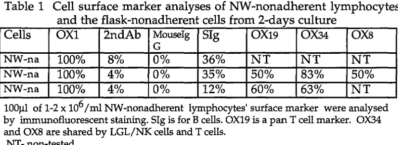
Cell surface marker analysis was carried out using immunofluorescent staining. A-LAK cell surface phenotype has been discussed in section 1.3.2. The monoclonal antibodies (Mab's) used for A-LAK cell surface marker analysis in this experiment were:
OXs: a murine IgGl for the surface marker of CDs on rat NK cells, LAK cells, T suppressor/cytotoxic lymphocytes; OX19: murine IgGl for the surface marker of CDs on rat pan-T cells; OX34: murine IgG2a for the surface marker of CD2; OX1: murine IgGl for rat leucocyte common antigen. These Mab's were obtained from Serotec, Australia. The second-step reagent was sheep-antimouse IgG F(ab')2 fragment conjugated with FITC (Silenus, Australia); Sheep anti-rat immunoglobulin antibody (Silenus, Australia) conjugated with FITC were used for rat B cells; Mouse IgGl conjugated with FITC (Becton Dickinson, US.) were used as negative controls.
Chambers et al 1989 introduced a more specific Mab for rat A-LAK cells
and NK cells named 3.2.3 antibody, an IgGlk (CD16). This Mab was not available in Australia when cell surface marker analysis was performed in this study.
For surface marker analysis, nylon wool nonadherent lymphocytes or A-LAK cells from culture were washed for 3 times in cold PBS-A (phosphate buffered saline; Oxoid limited, UK) and once in staining buffer. The staining buffer comprised PBS-A, 0.1%sodium azide (BDH, UK) and 20% bovine serum albumin (CSL, Australia). Then 1.0-2.0xlos lymphocytes or A-LAK cells in 12x75-mm plastic tubes in 100µ1 cold staining buffer were mixed with 10µ1 of primary Mab's (1:10) at 4°C for 45-60min. The cells were washed three times in staining buffer and resuspended with 10µ1 of the second antibody and kept at 4°C for 30min. After three further washes the cells were resuspen~ed in 10µ1 staining buffer and put on glass slides (Knittel Glaser, Germany). They were fixed in 100% Ethanol after dried on slides at room temperature. 20µ1 PBS-Azide-Glycerol (Heidelberg, US) was added and cells were covered by a glass cover slip. Cells were counted using alternate incidental or UV light.
2.1.2b Morphological analysis
cover slips. The cells were examined using a Lietz Ortheplan microscope and photographic records were made.
2.1.2c Cytotoxicity assay
complete cell lysis. Percentage cytotoxicity was calculated using the formula:
2.2 Short-term in Vivo Distribution of Radiolabelled A-LAK
Cells in the Rat
.2.2.1 Radiolabelling of A-LAK cells
A-LAK cells harvested from culture were labelled with 51Cr. After suspending l-9xl06 cells in lml RPMI 1640 + 10%FCS, lOOµCi 51Cr was added and the cells were incubated at 37°C in a water bath for lh. The radiolabelled A-LAK cells were washed in RPMI-1640 5 times. 50µ1 of supernatant of the first, 4th and last washing were taken for gamma-counting to ensure that there was no free 51Cr remaining in the supernatant. After labelling, A-LAK cells were resuspended in serum-free PBS-A, and cell viability was checked by trypan blue exclusion before infusion.
Radiolabelled A-LAK cells were suspended in l.2ml PBS-A. lml labelled A-LAK cells were placed in a 2ml-syringe for infusion into rats for the trafficking study. Three aliquots of 25µ1 labelled cells were taken at the same concentration as the injected cells and counted on gamma-counter. The radioactivity (cpm) in the total volume of infused cells was obtained by correcting the cpm obtained from 25µ1 labelled cells to the total injected cell volume. (total injected cpm in lml= 40 x cpm in 25µ1). The remaining labelled cells were used to check the spontaneous release of 51Cr from the cells. These remaining cells were resuspended