IN THE PATHOPHYSIOLOGY AND MANAGEMENT
OF HEART FAILURE
BY
MICHAEL ANDREW FITZPATRICK
B MED Sc, MB, BS,
SUBMITTED IN FULFILLMENT OF THE REQUIREMENTS FOR
THE DEGREE OF
DOCTOR OF MEDICINE,
UNIVERSITY OF TASMANIA.
THIS THESIS IS DEDICATED
TO MY WIFE,
CONTENTS:
Page
Contents
Work Done Personally
Acknowledgements vi
Abstract viii
Contribution to Scientific and Medical Milieu xi
Chapter 1: INTRODUCTION
1.1 Heart Failure - The problem 1
1.2 Historical perspectives 4
1.3 The kidney in heart failure 8
1.4 The role of the renin-angiotensin-aldosterone system
in heart failure 10
1.5 The role of the sympathetic nervous system in heart failure 17
1.6 The role of ADH in heart failure 20
1.7 Possible role of peripheral vasoconstriction in the natural
history of heart failure 22
1.8 Objectives of this thesis 23
Chapter 2: PATIENT SELECTION
2.1 Introduction 24
2.2 Definition of Heart Failure 25
.2.3 Aetiology of Heart Failure 26
2.4 Inclusion criteria 27
2.5 Exclusion criteria 27
Chapter
3:
ASSESSMENT OF SEVERITY OF HEART FAILURE AND THE RESPONSE TO THERAPY3.1 Introduction 29
3.2 Clinical assessment 31
3.3
Haemodynamic assessment of cardiac function 323.4 Radionuclide angiography 36
3.5
Echocardiographic assessment of cardiac function 433.6 Exercise testing 44
3.7
Evaluation of respiratory gas exchange during exercise 46Chapter 4: MEASUREMENT OF HAEMODYNAMIC PARAMETERS
4.1 Introduction 50
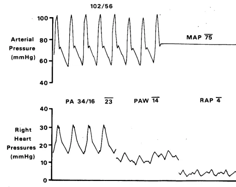
4.2 Catheter insertion 1
4.3 Pressure measurement 53
4•4 Measurement of cardiac output 58
4.5 Indices derived from flow and pressure measurements 60
4.6 Forearm plethysmography 61
Chapter 5: HORMONE ASSAYS AND METABOLIC BALANCE
5.1 Introduction
66
5.2 Plasma angiotensin II
68
5.3
Plasma renin activity 705.4 Plasma aldosterone 71
5.5 Urine aldosterone excretion 72
5.6 Plasma and urine cortisol
73
5.7
Plasma catecholamines 745.8 Plasma ADH 75
6.1 Introduction 79
6.2 Vasodilator therapy for heart failure 80
6.3 Methods 84
6.4 Results 88
6.4 Discussion 105
6.5 Conclusion 110
Chapter 7: HAEMODYNAMIC, HORMONAL AND ELECTROLYTE EFFECTS OF PRENALTEROL INFUSION IN HEART FAILURE
7.1 •Introduction 111
7.2 Inotropic therapy for heart failure 112
7.3 Methods 115
7.4 Results 120
7.5 Discussion 131
7.6 Conclusion 137
Chapter 8: STABILITY OF HAEMODYNAMIC, HORMONAL AND ELECTROLYTE DATA AND THEIR INTER-RELATIONSHIPS
8.1 Introduction 138
8.2 Patients and Methods 139
8.3 Results 143
8.4 Discussion 159
8.5 Conclusion 166
Chapter 9: BETA-BLOCKADE IN DILATED CARDIOMYOPATHY: IS IT BENEFICIAL?
9.1 Introduction 167
9.2 Patients and methods 169
9.3
Results 1739.4 Discussion 176
Chapter 10: WITHDRAWAL OF LONG-TERM CAPTOPRIL THERAPY FOR HEART FAILURE: A DOUBLE-BLIND, CONTROLLED TRIAL
10.1 Introduction 183
10.2 Patients and Methods 184
10.3 Results 190
10.4 Discussion 197
10.5 Conclusion 200
Chapter 11: .CONCLUSIONS 201
REFERENCES 0 206
WORK DONE PERSONALLY:
I was involved in the design of the protocol for each investigation and I selected the patients involved in these studies, explained in detail the nature of each study, and obtained informed, written consent. All catheters were inserted by me, and I was available at all times to attend the patients during their hospital stay in the event of problems arising. I routinely checked on their physical well-being at least daily while they were in hospital. After discharge, I saw all patients for follow-up care at a special "Heart Failure" clinic.
All exercise tests were supervised by me, and I performed all radionuclide angiographic studies using the "Nuclear Stethoscope". I was present on every occasion when blood sampling and haemodynamic measurements were made, except when arrangements for cover were made with Dr. Nicholls or . Dr. Ikram (which occurred infrequently). A cardiology technician was present at most measurement times and calculated results which were checked by me on a weekly basis. I tabled the primary data and performed statistical analyses. All figures were initially drawn by myself then copied and photographed by the Dept. of Medical Illustrations. The investigations were performed over the last three years, while I was employed as Registrar, and later as Senior Registrar in Cardiology at the Princess Margaret Hospital, Christchurch.
This thesis contains no material which has been accepted for the award of any other degree or graduate diploma in any university. It contains no material previously published or written by another person, except when due reference has been made in the text of this thesis.
ACKNOWLEDGEMENTS:
Firstly, my sincere thanks to my wife, Carolyn, without whose support this thesis could not have been completed. I am most grateful to her for her ability to master a word-processor enabling her to type the manuscript.
During the course of work, I benefited greatly from the advice, guidance, and encouragement given freely by my supervisors, Dr. Gary Nicholls and Dr. Hamid Ikram. I am grateful to them for their patience in teaching me the basic methodology that allowed me to carry out the following investigations. I must also acknowledge the help of the physicians and general practioners who allowed me to investigate their patients.
I wish to thank the nursing staff of the PMH Theatre Recovery Ward who cared very diligently for our patients while they underwent
investigation. Without this care, none of the first three studies to be reported would have been possible. I also wish to thank the dieticians for carefully preparing meals for metabolic balance studies, and the Special Test Sisters who collected and stored most blood samples. The staff of the Pathology and Endocrinology laboratories patiently taught me the basic techniques for assaying hormones and electrolytes.
I am very grateful to Sarah Jones and Lise Brabant (Cardiology research technicians), for their assistance with haemodynamic measurements and exercise testing, while echocardiograms documented in Chapter 9 were performed by Christine Wilson. The technicians of the Respiratory Department helped to perform spirometry and on-line breath-by-breath gas analyses during exercise. Their help is most gratefully acknowledged.
I am indebted to Dr. Elisabeth Wells for valuable statistical advice and helping me to use the BMDP Statistics Package; and to Dr. John Turner, of the Nuclear Medicine Department, Christchurch Hospital for teaching me safety with radioactive isotopes, performing radionuclide angiograms by gamma-camera and for helping me to validate
the "Nuclear Stethoscope". The help of the Department of Medical Illustrations, The Princess Margaret Hospital is gratefully acknowledged for their work in preparing the figures from drawings and graphs that I had prepared.
The studies were supported by several generous grants from the National Heart Foundation of New Zealand and the Medical Research Council of New Zealand.
ABSTRACT: CHAPTER 1:
Congestive heart failure is a common and lethal disorder. Improved understanding of the pathophysiology has led in recent years to more rational therapeutic regimes. The historical development of modern concepts of the pathophysiology and management of heart failure are outlined, concentrating mainly on the role of neurohumoral systems - the renin-angiotensin-aldosterone system and sympathetic nervous system. Deficiencies in current knowledge are discussed, from which I outline the objectives of the studies presented in this thesis.
CHAPTER 2:
Only those patients with documented left ventricular dysfunction were included in the studies. In this chapter, I outline methods of patient selection: referral; definition of heart failure and aetiology; and finally, inclusion and exclusion criteria. Ethical guidelines followed implicitly in the studies are then outlined.
CHAPTER 3:
Current methods of assessment of the severity of heart failure and underlying left ventricular dysfunction are reviewed. Emphasis is placed on those methods used in this thesis: NYHA Functional Classification; invasive haemodynamics; radionuclide angiography; echocardiography; and exercise testing.
CHAPTER 4:
CHAPTER 5:
Correlation of hormone and haemodynamic measurements in heart failure under control conditions and following therapeutic intervention forms the basis of three studies reported in this thesis. In this chapter, I briefly outline assay methods used in our laboratory. Metabolic balance was instituted to facilitate the interpretation of hormone levels in the studies. Methods for performing these metabolic studies are dealt with in this section.
CHAPTER 6:
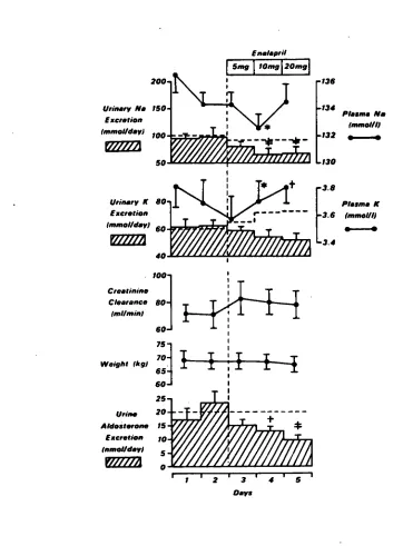
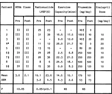
In this study, I document the acute haemodynamic, hormone, and electrolyte response to enalapril in heart failure, and correlate these changes with the short-term clinical response: Enalapril appears to be a long-acting angiotensin converting enzyme inhibitor that effectively reduces elevated angiotensin II levels found in heart failure. The greatest haemodynamic improvement occurred in those patients with the highest baseline angiotensin II levels. Over a period of - 4 to 8 weeks, exercise capacity improved in those patients who were most severely afflicted, on higher frusemide doses, with the greatest activation of the renin-angiotensin-aldosterone system.
CHAPTER 7:
CHAPTER 8:
I retrospectively analysed the control data from the preceeding two studies and an earlier study performed in our unit (Maslowski et al, 1981a). Cardiac catheterisation appears to significantly influence cardiac and hormone parameters for a period of up to twelve to eighteen hours. Thereagter, these parameters are relatively stable and more truly represent "baseline" levels, from which haemodynamic-hormone relationships and the effects of therapeutic intervention can be more accurately assessed. Activation of the renin-angiotensin-aldosterone system depends largely on the severity of underlying myocardial dysfunction and frusemide dosage, while the sympathetic nervous system appears to play a lesser role in determining cardiac function at rest.
CHAPTER 9:
This study was the first double-blind, placebo controlled trial to investigate the effects of beta-blockade in dilated cardiomyopathy. I refute the claims of Swedish workers for therapeutic benefit in this condition, claims which are contrary to our current notions concerning the role of the sympathetic nervous system in heart failure, and the administration of beta-blocking agents in this syndrome.
CHAPTER 10:
In this double-blind, controlled study I document haemodynamic deterioration associated with diminished exercise capacity following withdrawal of long-term captopril therapy for heart failure. This confirms the sustained effectiveness of angiotensin-converting enzyme inhibitors in the long-term management of heart failure.
CHAPTER 11:
x.
CONTRIBUTION TO THE SCIENTIFIC AND MEDICAL MILIEU:
The studies embodied in this thesis contribute significantly to knowledge concerning the mac.: of neurohumoral systems in the pathophysiology and management of heart failure. The following publications (excluding abstracts) have or will appear in the medical literature:
1. Double-Blind Trial of Chronic Oral Beta-Blockade in Congestive
Cardiomyopathy.
Lancet
ii:490-3, 1981.
2. Beta-Blockade for DilatedCardiomyopathy: the Evidence Against
Therapeutic Benefit.
.Eur Ht J 4(A):179-80, 1983..
3. Haemodynamic, Hormonal and Electrolyte Effects of Prenalterol
Infusion in Heart Failure.
Circulation
67:613-9,
1983.4
•
Haemodynamic, Hormonal and Electrolyte Effects of Enalapril inHeart Failure. Br Ht J (in press).
5. Acute Haemodynamic, Hormonal, and Electrolyte Effects and
Short-term Clinical Response to Enalapril in Heart Failure.
J Hypertension (in
press).
6. Stability of Haemodynamic and Hormonal Parameters, and Their
Inter-relationships in
Heart
Failure.To be submitted to
J
Clin Sol.7. Withdrawal of Long-term Captcpril Therapy for Heart Failure:
So far, data from the studies in this thesis have been presented at the following international meetings:
1. 54 th Annual Scientific Session of the American Heart Association, Dallas, Nov, 1981 (Ch 9).
2. 30 th Annual Scientific Meeting of the Cardiac Society of Australia and New Zealand, Canberra, May, 1982 (Ch 7).
3. Symposium on "The Failing Myocardium - What we have learned since Withering", Salzburg, June, 1982 (Ch 9).
4. 9 th World Congress of Cardiology, Moscow, June, 1982 (Ch 7).
5. 32nd Annual Scientific Session of the American College of Cardiology, New Orleans, March, 1983 (Ch 6).
" Heart failure, the consequence of many forms of heart disease, is one of the most common and serious disorders that afflicts individuals of all ages"
Braunwald (1981)
1.1 HEART FAILURE - THE PROBLEM:
Congestive heart failure is a very common clinical problem which leads to incapacitating symptoms and often progresses to the point of refractoriness to therapy. From the time of diagnosis, mortality over the following five years is about 50% (McKee et al,
1971). In more severe cases, characterised by marked haemodynamic abnormalities, the mortality rate may be even higher (Fuster et al,
1981). While initial therapy produces symptomatic relief in many patients early in the course, progressive worsening of symptoms with time is the usual outcome, although sudden death may intervene.
In the last three decades, cardiology has benefited from extensive research efforts. With application of this knowledge, we have witnessed more than a 25% decline in age-corrected mortality from ischaemic heart disease and a
37%
decrease in stroke mortality (Fromer 1982). Furthermore, there has been dramatic progress in our ability to manage most major types of cardiovascular disease (coronary artery disease, arrhythmias, valve and congenital heart diseases and hypertension).INTRODUCTION: 2.
Despite the prevalence of the syndrome, there has been a remarkable lack of information regarding:
1. Prognosis and mode of death;
2. Mechanisms of progression of the syndrome; 3. Timing of therapeutic intervention; and
4• Assessment of severity and therapeutic benefit with regard to symptoms, and life-expectancy.
Part of the problem lies in the heterogeneous nature of the syndrome, which may arise from any form of heart disease, and from imprecision in making a definitive diagnosis and evaluating its severity, which are likely to be a strong factors in determining prognosis. In the few studies where adequate definitive diagnosis has been made (Bruschke et al, 1973; Fuster et al, 1981), the prognosis in the presence of heart failure is poor and in many cases is worse than that for many forms of malignancy (McKee et al, 1971).
Traditional therapy involves exercise curtailment, salt . restriction and administration of digitalis and diuretics, however
many patients fail to respond or respond poorly. New therapeutic modalities (vasodilators, blockade of the renin-angiotensin-aldosterone system and non-glycoside inotropes) show promise, making management of heart failure one of the most rapidly expanding areas of cardiac therapeutics (Braunwald, 1982).
In recent years, increasing attention has been devoted to the neurohumoral vasoconstrictor, and dilator systems which contribute to fluid retention, increase heart rate and alter regional blood flow. These systems include:
1. The Renin-Angiotensin-Aldosterone system; 2. The Sympathetic Nervous System;
3. Antidiuretic hormone; 4. Kinins;
& 5.
Prostaglandins;The causes and consequences of these disturbed mechanisms are not well understood (Braunwald, 1982). Reversal of the increased ventricular afterload observed in heart failure is beneficial (Franciosa, 1981), however "tolerance" to some vasodilators has been attributed to activation of those neurohumoral constrictor systems not initially blocked (Colucci et al, 1980b). Furthermore, tolerance to inotropic therapy may occur for similar reasons.
INTRODUCTION:
4.
1.2 HISTORICAL PERSPECTIVES: (Jarcho 1980 & Braunwald 1981)
The principal clinical manifestations of heart failure, dyspnoea and oedema, were recognised in antiquity, although heart failure as an entity could not be described until the fundamental function of the circulation had been identified. The Greeks and Romans attributed dyspnoea and oedema to an obstruction of the upper airways, and to an abnormality of the urinary system respectively.
William Harvey (1578-1657), despite his monumental contribution to cardiac physiology and his lifelong involvement in the practice of medicine, did not write about disorders of the organ whose function he had described. In his book "De Corde" (1669), Richard Lower pointed out that it is necessary for the two sides of the heart to have similar strengths to maintain the circulation. He appreciated that inequality of the two sides could lead to symptoms, . and he presaged much later work on heart failure by pointing out that the cardiac parenchyma may be subject "to various illnesses and inflammation", which could interfere with its "pulsations", leading to a feeble cardiac output. In this manner, the concept of what we now call "forward heart failure" was first formulated.
A current formulation of this concept would suggest that the inability of cardiac muscle to shorten against a load alters the relationship between ventricular pressure and volume, so that end-systolic volume rises. The following sequence of adaptations then occurs:
1. ventricular end-diastolic volume and pressure increases; 2. pressure rises in the venous and capillary beds;
3. transudation occurs;
4• extracellular volume icreases.
Although these adaptations at first tend to maintain normal cardiac output, many of the symptoms that are characteristic of heart failure result directly from this sequence of fluid sequestration in the interstitial spaces of the lungs, liver, subcutaneous tissues, and serous cavities.
In contrast to the "backward theory", proponents of the "forward failure theory" expounded most clearly by MacKenzie in 1913, maintain that the clinical manifestations of heart failure result directly from an inadequate discharge of blood into the arterial system. According to this formulation, the principal clinical manifestations of heart failure arise from reduced cardiac output, which results in diminished perfusion of vital organs including: the brain, leading to mental confusion; the skeletal muscles, leading to weakness; and the kidneys, leading to sodium and water retention. Although these two seemingly opposing views concerning the pathogenesis of heart failure led to lively controversy during the first half of the century, a rigid distinction between backward and forward heart failure now seems artificial, since both mechanisms appear to operate to varying extents in most patients with heart failure.
INTRODUCTION:
6.
urinary excretion of sodium was suggested by Schrodeder (1941), and later confirmed (Urquhart & Davis, 1963).
It was not until Parrish (1949) and Deming & Luetscher (1950) reported increased sodium-retaining activity in the urine from patients with heart failure, that the possibility of hormone-induced renal sodium retention received scientific support. Davis et al (1956) and Singer (1957) confirmed that this active material was aldosterone when they demonstrated increased levels of this substance in urine and adrenal vein blood respectively. Davis et al . (1962) subsequently suggested that increased circulating angiotensin II levels were responsible for hypersecretion of aldosterone in experimental heart failure.
The manifestations of heart failure are now thought to be largely due to a disturbance of feed-back control of fluid homeostasis in response to a change in renal perfusion (forward failure), in association with redistribution of blood flow by . neurohumoral reflexes (Hamer, 1982 - p.1). Congestive heart failure ' appears when a cardiac output necessary for tissue needs cannot be produced by the diseased heart using compensatory mechanisms of the sympathetic nervous system, which include:
1. tachycardia;
2. ventricular hypertrophy (Strobeck & Sonnenblick, 1981); & 3. moving to a higher ventricular function curve (Sarnoff &
Beglund, 1954) so that compensation is attempted through the Starling response.
The last factor is aided by renal sodium and water retention. The retained fluid is distributed by the venous system to provide suitable filling pressures in each ventricle to maintain an adequate cardiac output (Guyton, 1963), however, this fluid cannot be retained in the intravascular compartment. Sequestration in the interstitial spaces results in the appearance of oedema.
congestive heart failure is seen less frequently. Patients are more often troubled by symptoms related to diminished cardiac output, namely fatigue and reduced exercise capacity. As a result, greater research emphasis is now being directed toward agents that can improve cardiac output by direct inotropic action or blockade of neurohumoral vasoconstrictor systems that adversely effect the regional distribution of blood flow in heart failure.
INTRODUCTION:
8.
1.3 THE KIDNEY IN HEART FAILURE:
Salt and water retention by the kidney is a major compensatory adjustment brought about in an attempt to restore the effectiveness of the circulation (Braunwald et al, 1965). Although the quantitative role of each mechanism has not been clarified, there are at least five mechanisms which have been proposed in heart failure:
1. Renal Haemodynamic changes:
Renal haemodynamic changes are currently thought to play the major role in sodium and water retention in heart failure (Hume et al, 1978). A decline in renal blood flow and plasma flow is commonly observed (Merrill, 1949 & Mokotoff et al, 1948), but this is associated with little or no change in glomerular filtration rate (Heller & Johnson, 1950). As a result of this, a rise in filtration fraction is consistently found in heart failure (Vander et al, 1958). The mechanisms by which diminished sodium excretion results from these haemodynamic alterations are detailed elsewhere (Hume et al,
1978), and as they are not pertinent to this thesis, they will not be discussed further.
2. Aldosterone:
Aldosterone contributes to sodium retention by enhancing distal sodium reabsorption in exchange for potassium. Its role in heart failure will be discussed further in the next section.
3. "Third Factor":
A disturbance of such a mechanism could contribute to the sodium retention seen in heart failure.
4. Intra-Renal Redistribution of Blood Flow:
Alterations in the distribution of blood flow within the kidney (Kilcoyne et al, 1971) occur as part of the response to the general reduction in renal blood flow; the tendency to medullary, rather than cortical blood flow may favour nephrons with more potent sodium-retaining properties. A local intrarenal effect of renin may play a part here (Levens et al, 1981).
There are two populations of nephrons in the kidney (Britton, 1981):
1. a cortical group with a juxtaglomerular apparatus which•
maintains perfusion by autoregulation of arterial pressure; and
2. a juxtamedullary group subjected to a passive increase in flow as arterial pressure rises.
A redistribution of blood flow to the juxtamedullary group in response to a falling cardiac output could account for the salt retention of heart failure (Hamer, 1982 - p.7).
5. Other Humoral Agents:
Anti-diuretic hormone contributes to water retention in heart failure by effecting distal renal tubular function (see section 1.6). Other vasoactive substances such as prostaglandins, kallikreins and kinins have also been implicated as important factors in sodium balance (McGiff & Itslovitz,
INTRODUCTION:
10.
1.4 THE ROLE OF THE RENIN-ANGIOTENSIN-ALDOSTERONE SYSTEM IN HEART FAILURE :
1. Physiology:
Aldosterone, the most potent mineralocorticoid secreted by the adrenal cortex, plays a major role in sodium and potassium homeostasis. Enhanced distal tubular sodium-potassium exchange by aldosterone promotes sodium retention and potassium depletion. Four regulators of aldosterone release are well defined:
1. the renin-angiotensin system; 2. plasma potassium;
3. ACTH;
4. plasma sodium.
Of these, the first two are generally considered to be the most important (Hollenberg & Williams, 1981). Other regulators have been suggested, including dopamine, which is thought to have a tonic inhibitory effect on aldosterone secretion (Campbell et al, 1981). Normally, more than 75% of the circulating aldosterone is inactivated during a single passage through the liver. However, in the presence of heart failure, this percentage may be reduced (Tait et al, 1965; Camargo et al, 1965). In an otherwise normal subject, and in primary aldosteronism, excess levels of aldosterone per se do not usually lead to oedema formation because of the so-called "escape phenomenon". To prevent this from occurring proximal sodium reabsorption decreases, but the precise "escape" mechanism remains to be explained (Urqbart & Davis, 1963; Johnson et al, 1968). In heart failure, however, proximal tubular sodium reabsorption is enhanced, and the "escape" from aldosterone effect does not occur, thus this hormone contributes to the increase in extracellular volume.
the alpha-2 globulin called angiotensinogen. Circulating renin has a plasma half-life of 10-15 minutes (Levens, Peach & Carey 1981). In the presence of angiotensin converting enzyme (found in all tissues, but predominently pulmonary endothelium), angiotensin I is converted into the biologically active octapeptide, angiotensin II. This hormone is a potent direct stimulus both to aldosterone and constriction of vascular smooth muscle, while it is destroyed rapidly by angiotensinases, thus it has a half-life in the order of minutes. Renin release is controlled by a composite of four interdependent factors (Oparil & Haber, 1974):
1. The juxtaglomerular cells act as miniature pressure transducers that sense changes in afferent arteriolar perfusion pressure. When the circulating blood volume is reduced, a corresponding fall in afferent arteriolar pressure and renal perfusion pressure occurs. Renin is then released by these cells to restore blood volume to normal through the effect of angiotensin II on aldosterone, and perhaps directly on the kidney (Levens et al, 1981).
2. The Macula Densa cells are thought to function as chemoreceptors, monitoring the sodium or chloride load present in the distal tubule, and feeding this information back to the juxtaglomerular cells, where appropriate modifications in renin release occur.
3. The sympathetic nervous system plays a prominent role in regulating renin secretion mediated by beta-receptors.
INTRODUCTION: 12.
2. Activity of the Renin-Angiotensin-Aldosterone System in Heart Failure:
Elevation of plasma renin activity and aldosterone concentration in subjects with heart failure has not been consistently observed (Merrill et al 1946, Brown et al 1970, Sanders and Melby 1964, Wolff et al 1959, Genest et al 1968, Chonko et al 1977). Recent experimental studies in conscious animals with cardiac failure have suggested that the renin-angiotensin-aldosterone system is activated soon after the induction of a low cardiac output (Watkins et al, 1976; Freeman et al, 1979; Morris et al, 1977; Davis 1962). During the chronic compensated state of experimental heart failure, plasma renin activity and plasma aldosterone concentration decrease toward normal as the extracellular fluid volume expands (Watkins et al, 1976; Davis, 1962). Thus, the discrepancies in the state of the renin-angiotensin-aldosterone system in the clinical literature are probably due to lack of clear definition of the clinical status of the patients studied, and failure to control other factors affecting both renin and aldosterone secretion.
Dzau et al (1981) found that during acute, severe left ventricular decompensation, before the development of extracellular fluid volume expansion and restoration of systemic blood pressure, plasma renin activity and aldosterone were markedly elevated. With stabilisation of cardiac failure and extracellular fluid expansion, plasma renin activity and aldosterone returned to apparently normal levels although they remained abnormally elevated for the degree of blood volume expansion.
the renin-angiotensin-aldosterone system, presumably as a result of increased distal tubular sodium load (Vander, 1967). As the patients approached dry body weight with continued diuretic therapy, plasma aldosterone concentrations rose to very high levels (Nicholls et al 1974; Knight et al, 1979). The parallel rise in plasma renin activity (Nicholls et al, 1974) suggested that the renin-angiotensin system (rather than other known secretagogues) controlled this pattern of aldosterone change. Sodium load to the distal tubule will be low at this stage, as there is little further diuresis, and continued intense diuretic therapy can maintain dry body weight, while augmenting renin release.
INTRODUCTION:
14.
3. Pharmacological Interruption of Renin-Angiotensin Activity:
The development of agents that block the renin-angiotensin system has provided pharmacological probes that are useful in assessing more directly angiotensin's contribution to heart failure. Blockade may be effected at three levels:
1. Beta-adrenergic antagonists suppress ream n release, but this class of drug has contributed little to our understanding of renin's role in heart failure because of confounding effects on myocardial function in heart failure (Hollenberg & Williams 1981);
2. Angiotensin converting enzyme inhibitors (eg. teprotide, captopril, and more recently, enalapril) which block the formation of angiotensin II;
3. Angiotensin II analogues (eg. saralasin) which compete directly with angiotensin II at its receptor site. These antagonists appear to be more specific in their action than converting enzyme inhibitors, however they can only be given intravenously, are expensive to synthesise, and like most receptor antagonists, they have some agonist action.
It should be noted that there is much debate concerning the specificity of the action of converting enzyme inhibitors. This enzyme is responsible, in part, for degradation of bradykinin, a powerful vasodilator, but levels do not appear to be elevated by captopril (Johnston et al 1979, Dzau et al 1980). Clarification
L. Renin-Angiotensin Blockade in Heart Failure:
Agents that interrupt the renin-angiotensin axis activity have shown promise in animal models (Watkins et al 1976, Freeman et al 1979) and clinical studies-(Dzau et al 1980, Faxon et al 1980, Maslowski et al 1981a, Turini et al 1979). Acute pharmacological blockade of the renin-angiotensin system with converting enzyme inhibitors or angiotensin analogues has produced salutary response in a large majority of patients with severe heart failure. Haemodynamic improvement resembles the response seen with nonspecific vasodilators (eg. nitroprusside - Vrobel et al, 1980). Since both classes of agents have been effective, it is very likely that a substantial portion of the vascular response in heart failure reflects reversal of the vasoconstriction induced by angiotensin (Hollenberg & Williams, 1981).
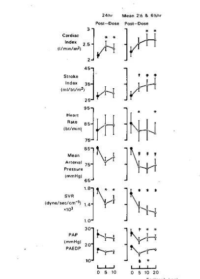
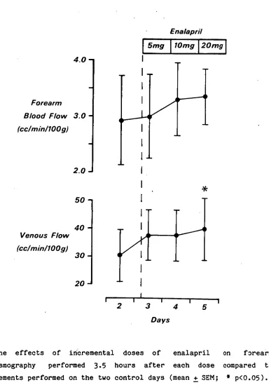
Captopril, the only currently available oral converting enzyme inhibitor, has been studied in patients resistant to conventional therapy. Although side effects are not common, potentially serious complications have been reported (Vidt, Bravo & Fouad, 1982). Enalapril, a member of a new group of converting enzyme inhibitors which lacks a mercapto function and is characterised by weak chelating properties, has recently been synthesised (Patchett et al, 1980) and used successfully in hypertension (Gavras et al, 1981). Preliminary data suggest it is as effective and longer acting than captopril in hypertension (Gavras et al, 1981), and so far serious side effects have not been observed, paving the way for its use in patients with milder degrees of cardiac failure. To date the haemodynamic, hormonal and electrolyte response to enalapril in heart failure have not been documented. In chapter 6, I report the results of such a study in nine patients with heart failure stable on digoxin and diuretic therapy.
INTRODUCTION: 16.
1.5 THE ROLE OF THE SYMPATHETIC NERVOUS SYSTEM :
In view of the well established importance of the sympathetic nervous system in normal regulation of the circulation, considerable attention has been directed to the activity of this system in heart failure. In 1962, Chidsey et al noted that either no change or very small increases in plasma norepinephrine occurred in normal subjects with exercise, while much greater rises occurred in patients with heart failure, presumably reflecting greater activity of the sympathetic nervous system during exercise. Measurements of 24 hour urinary norepinephrine excretion revealed marked elevations in patients with heart failure, suggesting greater activity at rest as well (Chidsey, Braunwald & Morrow; 1965).
Plasma catecholamines provide a view of "global" sympathetic activity (Goldstein, 1981), but they do not provide information on regional tone. A further limitation is that plasma norepinephrine reflects both "spillover" rate from sympathetic nerve endings and clearance rate from plasma (Esler et al, 1981). Hence, some caution must be exercised in equating plasma norepinephrine with sympathetic tone.
INTRODUCTION: 18.
The investigation described in Chapter 9 provides the first double-blind controlled study of the effect of beta-blockade in dilated cardiomyopathy. The results from this study do not support the Swedish viewpoint, and in that chapter I look critically at the Swedish data.
Depletion of myocardial norepinephrine stores (Chidsey et al 1965,1966) provides further evidence for abnormal adrenergic activity in heart failure. As a result, the heart fails to respond to sympathetic input with increasing heart failure. Goldstein et al (1975) demonstrated an impaired chronotropic response to atropine and baroreceptor-mediated reflexes which appears to be related to severity of heart disease. They observed a normal response to isoprenaline indicating the abnormality in sympathetic response results from presynaptic norepinephrine depletion, rather than a reduction in responsiveness of beta-receptors. In pre-terminal patients undergoing cardiac transplantation, however, depletion of beta-receptors and diminished responsiveness to clatecholamines in vitro has recently been reported (Bristow et al, 1982).
This peripheral vasoconstriction is mediated by alpha-adrenergic receptors (Kramer et al, 1968). In patients with heart failure, blockade of these receptors with agents such as prazosin reduces ventricular afterload, and acute haemodynamic improvement associated with symptomatic improvement has been reported (Miller et al, 1977; Mehla et al, 1978). At least two centres have presented objective haemodynamic data to suggest that tolerance to prazosin occurs and develops rapidly (Packer et al, 1978; Arnold et al, 1978). Furthermore, Colucci et al (1980b) noted an increase in plasma renin activity, with many patients requiring an increase in diuretic dosage. Thus activation of the renin-angiotensin system may be involved in the development of tolerance to long-term prazosin therapy.
INTRODUCTION:
20.
1.6 ROLE OF ANTIDIURETIC HORMONE IN HEART FAILURE :
1. Physiology:
Antidiuretic hormone (ADH or arginine -vasopressin) is synthesised in the supraoptic and paraventricular nuclei of the hypothalamus. Packaged in neurosecretory granules by the endoplasmic reticulum, ADH is then transported along the axons to their bulbs in the posterior pituitary where it may be released into the circulation by exocytosis (Schrier & Leaf,
1981). The role of ADH in water homeostasis has received most attention but it does have vasoconstrictor properties which may be of physiological and pathophysiological importance (Johnston et al, 1981).
Release of ADH is controlled by osmotic and non-osmotic pathways (Schrier, Berl & Anderson, 1979):
1. "Osmoreceptor cells" which apparently lie outside the blood-brain barrier sense changes in extracellular osmolality induced by fluid deprivation or ingestion and increase or decrease ADH release respectively. ADH enhances distal tubular water reabsorption, thus water deprivation decreases free water clearance, while water excess increases its clearance.
2. The major non-osmotic stimuli for ADH release include depletion of the extra-cellular fluid and hypotension, however pain, fright, nausea, and hypoxia may also stimulate release (Robertson et al,
1977;
Schrier et al, 1979). Major parasympathetic afferent pathways appear to arise from low-pressure atrial receptors (vagal), which perceive early changes in the volume of the extra-cellular fluid, and from high-pressure baroreceptors of the carotid sinus2. ADH Activity in Heart Failure:
Hyponatraemia occurs frequently in patients with heart failure who have a moderate water intake (Bartter, 1964). At present it is not clear whether persistent release of ADH which diminishes water clearance, or intrarenal factors account for this hyponatraemia. The vasoconstrictor role of ADH in heart failure has received scant attention. A specific inhibitor of the vasoactive action of ADH is now available (Seto et al, 1980), but it has not yet been used to investigate the role of ADH induced direct vasoconstriction in heart failure.
Early studies using bioassay to measure plasma ADH in patients with heart failure have not been definitive (Sztalowicz et al, 1981). Using a radioimmunoassay for ADH, these authors recently demonstrated higher ADH levels in heart failure patients when hyponatraemia and hypo-osmolarity were more severe. This supports the role of ADH in impaired water excretion in heart failure and implies that nonosmotic pathways, rather than osmotic pathways, provide the main stimulus for ADH release under these circumstances, confirming findings from animal studies (Anderson et al 1975,1976; Handelman et al,
1979). What factor(s) dictate this "inappropriate" elevation in ADH levels is not clear, but possibilities include impaired inhibition of ADH release in response to atrial stretch or volume receptors (Greenberg et al, 1973) or parasympathetic stimulation from high pressure baroreceptors when "effective" blood volume is reduced (Schrier & Humphries, 1971).
INTRODUCTION: 22.
1.7 POSSIBLE ROLE OF PERIPHERAL VASOCONSTRICTION IN THE NATURAL HISTORY OF HEART FAILURE :
The syndrome of heart failure is a naturally progressive disorder that often appears to worsen without any evidence for an active process within the myocardium (Cohn et al, 1981). Studies of cardiac mechanics have suggested that progression of heart failure may represent an inappropriate increase in wall tension associated with an enlarging chamber without concomitant increase in wall thickness (Strauer, 1979). Peripheral vasoconstriction may well be an important mechanism in the genesis of this cardiac dilatation (Cohn et al, 1981). Constriction of small arteries and reduced distensibility of large arteries increases impedance to left ventricular ejection and increases left ventricular end-systolic volume.
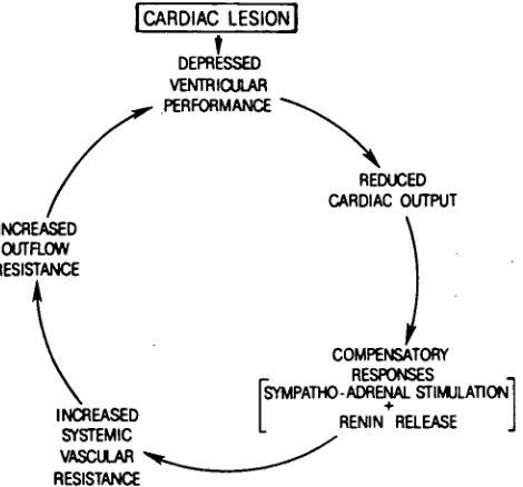
Chronic heart failure can be looked upon as a vicious cycle initiated by a cardiac lesion that impairs cardiac performance and results in reduced cardiac output (see fig 6.2). This low output may initially be compensated by activation of neurohumoral systems, which eventually elevate systemic vascular resistance, in turn increasing resistance to ventricular outflow. For the failing ventricle, this further depresses ventricular performance, thereby completing a positive feedback loop.
1.8 OBJECTIVES OF THIS THESIS :
To summarise, heart failure is associated with a poor prognosis, and it is one of the most common causes of death in our society. Despite advances in other fields of cardiology, research has produced little impact on the morbidity and mortality of this serious syndrome. Evidence has been produced to show that neurohumoral systems are integrally involved in the pathophysiology and possibly with the progression of heart failure. Better therapy is likely to follow improved knowledge of the role of these systems in heart failure.
In the studies incorporated in this thesis, I explore some facets of this large, and fascinating field of research. The acute haemodynamic, hormone and electrolye effects of blockade of the renin-angiotensin-aldosterone system and beta-1 adrenergic stimulation are discussed in Chapters 6 & 7 respectively. The current role of vasodilator and inotropic therapy in heart failure will be outlined at the beginning of each chapter. From these Studies, control data is utilised to document stability of haemodynamic, hormone and electrolyte measurements and their inter-relationships in heart failure (Chapter 8). In Chapter 9, I investigate the long-term effects of blocking the beta receptor of the sympathetic nervous system in dilated cardiomyopathy. Finally, in Chapter 10, I discuss the effect of withdrawal of blockade of the renin-angiotensin-aldosterone system. These studies contribute significantly to our growing fund of knowledge concerning the role of neurohumoral systems and applications of this knowledge in medical therapy are considered.
CHAPTER 2
PATIENT SELECTION:
2.1 INTRODUCTION :
Over the past decade the Cardiology and Endocrinology Departments of The Princess Margaret Hospital, Christchurch have had an ongoing interest in the investigation of the pathophysiology and management of heart failure. Consequently, a good rapport has been established between these departments and general practitioners and physicians in the region so that most patients developing heart failure are referred for diagnosis and management of their condition. The Cardiology Department serves the province of Canterbury, thus it services a population of approximately 400,000 people.
2.2 DEFINITION OF HEART FAILURE:
"Heart failure may be defined as the pathophysiological state in which an abnormality of cardiac function is responsible for failure of the heart to pump blood at a rate commensurate with the requirements of the metabolising tissues."
Braunwald (1982)
PATIENT SELECTION: 26.
2.3 AETIOLOGY OF HEART FAILURE:
Abnormal cardiac function resulting in circulatory failure may be due to:
1. Pressure overload: eg. aortic or pulmonary stenosis or hypertension.
2. Volume overload: eg. valvular incompetence or shunts.
3. Impaired myocardial function: eg. myocardial ischaemia/infarction, scarring, infiltrative disorders, toxic insults, primary and secondary cardiomyopathy.
4. Extrinsic compression of the heart: eg. pericardial constriction or effusion.
2.4 INCLUSION CRITERIA WERE AS FOLLOWS:
1. Presence of symptoms directly attributable to left ventricular failure;
2. Absence of .a surgically correctable lesion eg. coronary artery disease or valvular lesion;
3. Clinical, radiographic, and/or haemodynamic evidence of left ventricular failure;
4. Absence of exacerbating factors eg. infection, anaemia, thyrotoxicosis, pregnancy, arrhythmias other than atrial fibrillation, recent myocardial infarction ( < six months), infection or malignant hypertension.
2.5 EXCLUSION CRITERIA INCLUDED:
1. Absence of symptoms attributable to myocardial failure;
2. Unstable clinical condition;
3. Presence of exacerbating factors;
4. Unstable angina or myocardial infarction within the previous six months;
5. Diabetes mellitus was an exclusion criteria in some studies because hormone responses, especially catecholamines, may be abnormal;
6. Severe concomitant illness;
PATIENT SELECTION: 28.
2.6 ETHICAL CONSIDERATIONS:
The guidelines of the Helsinki Convention for human experimentation were followed implicitly (18 th World Medical Assembly, Helsinki, 1964 and subsequently - revised by the 29 th World Medical Assembly, Tokyo, 1975). For each investigation included in this thesis, the protocol was perused by the physicians involved, then submitted to and approved by the Ethical Committee of the North Canterbury Hospital Board. Patients were fully informed of the techniques used in each study, possible dangers and were free to withdraw from the investigation at any time. All patients gave written consent.
AND
RESPONSE TO THERAPY:3.1 INTRODUCTION:
Over the last decade we have seen a rapid increase in the number of methods available for cardiac diagnosis. Aetiology may now be determined with greater accuracy, and prognosis may be forecast with greater certainty. The quantitative assessment of the severity of heart failure and underlying myocardial dysfunction remains a problem in clinical cardiology as no single method provides a "gold standard" to compare patients or their response to therapy. This is a challenging and important task for clinicians, thus a plethora of methods have been devised to suit different purposes:
1. Clinical Assessment: Symptoms and physical findings.
2. Invasive Methods:
1. Haemodynamics - Pressure, flow relationships;
2. Left ventricular force-velocity-length relationships; 3. Quantitative Angiocardiography.
3.
Non-invasive Methods:1. Radionuclide Angiography; 2. Echocardiography;
3. Systolic Time intervals; 4. Apex and phonocardiography;
ASSESSMENT OF THE SEVERITY OF HEART FAILURE:
30.
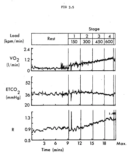
4. Exercise Testing: Exercise may be accurately assessed on a treadmill or bicycle ergometer. Useful adjuncts to this assessment include invasive haemodynamics measured during exercise and quantification of oxygen uptake, carbon dioxide production and anaerobic threshold.
Quantitative angiocardiography and left ventricular force-velocity-length relationships require left ventricular catheterisation. Due to the risk of embolisation studies can only be performed over a short period of time, and were not suitable for the present studies. Systolic time intervals and phonocardiography are too imprecise for adequate assessment (Braunwald, 1980).
3.2 CLINICAL ASSESSMENT:
As muscular exercise places the greatest metabolic stress on the heart, it is hardly surprising that the severity of heart failure may be assessed clinically by paying attention to exertional symptoms particularly dyspnoea and fatigue. The New York Heart Association (NYHA) Classification (1973) has been the most widely accepted, and has been used throughout this thesis. Patients were classified as follows:
1. Class I: No limitation - ordinary physical activity does not cause undue fatigue or dyspnoea.
2. Class II: Slight limitation of physical activity - such patients are comfortable at rest. Ordinary physical activity results in fatigue or dyspnoea.
3. Class III: Marked limitation of physical activity - although patients are comfortable at rest, less than ordinary activity will lead to symptoms.
4• Class IV: Inability to carry on any physical activity without discomfort - symptoms of congestive failure are present even at rest. With any physical activity, increased discomfort is experienced.
ASSESSMENT OF THE SEVERITY OF HEART FAILURE: 32.
3.3 HAEMODYNAMIC ASSESSMENT OF LEFT VENTRICULAR FUNCTION:
Direct measurement of haemodynamic parameters, cardiac output and vascular pressures, has become the time honoured method for assessing the severity of cardiac function and the effects of therapeutic intervention (Braunwald, 1980). To aid the interpretation of the haemodynamic studies reported in this thesis, it is useful to review circulatory dynamics at rest and during exercise in normal and pathological circumstances according to the Frank-Starling mechanism:
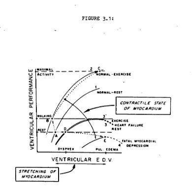
1. Normal Myocardial Function:
The normal relationship between ventricular end-diastolic volume and performance is shown in Fig 3.1, curve 1. Assumption of the upright posture reduces venous return, thus cardiac output is lower than in the recumbent position. During exercise, venous return is augmented by increased ventilation, the pumping action of exercising muscles and venoconstriction. Increased sympathetic activity simultaneously augments the contractile state of the myocardium and stroke volume, with either no change or a decline in end-diastolic pressure and volume (resulting in a shift from point A on curve 1 to point B on curve 2 in fig 3.1). Vasodilatation occurs in the exercising muscles, thus cardiac output is greatly elevated during exercise at an arterial pressure only slightly higher than that in the resting state. During intense exercise, cardiac output may be further augmented by utilisation of the Frank-Starling mechanism (B - C, fig 3.1).
2. Impaired Myocardial Function:
NORMAL-REST
CONTRACTILE STATE OF MYOCARDIUM
EXERCISE 3 • HEART FAILURE
REST •
E FATAL MYOCARDIAL 4' DEPRESSION PUL EDEMA
WALK IND
REST • - • •
DYS•NE A
2 cr 0
Li
a.
V
E
N
T
RICU
L
AR
[image:47.562.107.497.79.466.2]ASSESSMENT OF THE SEVERITY OF HEART FAILURE: 33.
FIGURE 3.1:
w MAXIMA L 2
(...) ACTIVITY • NORMAL • EXERCISE
VENT RICULAR E DV
STRETCH/NO OF MTOCARDIUM
ASSESSMENT OF THE SEVERITY OF HEART FAILURE: 34.
Ventricular performance curves or contractility cannot be elevated to the same extent during exercise (compare curves 3 & 3', fig 3.1) because cardiac epinephrine stores are depleted and the inotropic response to impulses from the cardiac sympathetic nerves is diminished (see Chapter 1). Factors that tend to augment ventricular filling during exercise in the normal subject, push the failing heart along its flattened length to active tension curve. Although ventricular performance may be augmented somewhat, this occurs only as a consequence of an inordinate elevation of ventricular end-diastolic volume and pressure, and therefore of pulmonary capillary wedge pressure. This intensifies dyspnoea and plays an important role in limiting the level of exercise that the patient can perform.
Left ventricular failure is thus characterised haemodynamically by elevated left ventricular filling pressure and reduced cardiac output (especially during exercise), which account for the pulmonary congestion and peripheral underperfusion. Heart rate is increased in an effort to maintain cardiac output, while systemic arterial pressure is usually supported at normal levels, despite reduced cardiac output, consequently systemic vascular resistance is usually elevated. Quantitative measurement of these parameters allows accurate assessment of the degree of myocardial dysfunction (Braunwald, 1980). Furthermore, invasive measurement of haemodynamic changes provides a useful tool for assessing the efficacy of therapeutic agents (Liander, 1982). Therapeutically induced reduction in left ventricular filling pressure and increase in cardiac output, is beneficial according to Frank-Starling function curves, and has been associated with acute symptomatic benefit and long-term efficacy (Braunwald, 1980).
As with all methods of assessing cardiac function, there are limitations which need to be appreciated:
2. Left ventricular filling pressure is used as an approximation for left ventricular end-diastolic volume which determines stretch of myocardial fibres. This is a valid extrapolation in the presence of normal compliance, however in disease states compliance is often abnormal (Liander, 1982);
3. In disease and health, cardiac output is closely controlled by auto-regulation (Guyton, 1981), thus resting cardiac output is only depressed when ventricular dysfunction is severe;
4. Some studies have demonstrated improved exercise capacity during long-term therapeutic interventions that were not associated with significant haemodynamic changes at rest (Franciosa et al 1978, Rubin et al 1979).
ASSESSMENT OF THE SEVERITY OF HEART FAILURE: 36.
3.4 RADIONUCLIDE ANGIOGRAPHY:
1. General:
Since Blumgart and Yens (1927) first used a radioactive tracer to evaluate the velocity of blood flow in man, substantial improvements have been made in imaging devices, isotopes and computer techniques. Consequently, these techniques have been applied to radionuclide angiographic assessment of cardiac function, and particularly to left ventricular function, for which ejection fraction (LVEF) has become the most widely used parameter.
Studies were initially performed with a gamma-camera located at the Nuclear Medicine Department of The Christchurch Hospital four miles from the Cardiology Department situated at The Princess Margaret Hospital. This necessitated the transfer of patients for cardiac scans. To obviate this; a "Nuclear Stethoscope" (Bios Instruments) was purchased by the department and isotopes were delivered to the Department in a lead cannister prior to use.
2. Blood Labelling:
The need to perform counts for several minutes requires that the isotope remains within the circulation. In the present studies, in-vivo labelling of red blood cells (Pavel, Zimmer and Patterson, 1977) was achieved by intravenous administration of
7
mg of stannous pyrophosphate thirty minutes before intravenous injection of 15-20 mCi of technetium-99m sodium pertechnetate. This provided sufficient "tag" for studies to be performed for several hours after administration of the isotope if required.3. Principle of calculations:
background activity arising from the lung fields, left . atrium, and right heart. In all studies, a small zone just lateral to the left ventricle was used.
Ejection fraction was calculated by the equation:
LVEF = End-diastolic - End-systolic counts X 100
End-diastolic - Background counts
• Gamma-camera studies:
Resting studies were performed with the patient supine and images obtained in the modified (caudal tilt) left anterior oblique position using a picker Dyna camera equipped with a high-resolution parallel-hole collimator. Scintillation data was accumulated in histogram mode (64 x 64 ward mode images) in a PDP 11-34 computer system using the R Wave of the ECG as the synchronising impulse. The cardiac cycle was divided into twelve equal frames, and three hundred cycles were counted.
Calculation of LVEF was performed using a commercially available modified Fortran Decus HRTIMG program (Decus No. 11-363, August 1978). This program uses an automatic stepping, automatic , edge finder routine after selection of an end-diastolic region of
interest. Background was calculated from a simple level subtraction and was chosen from the end-systolic frame as a two pixel postero-lateral 90 0 crescent immediately adjacent to the end-systolic left ventricular border. A background corrected time activity curve was displayed and LVEF calculated from the formula given above.
Validation studies from the Nuclear Medicine Department have shown:
ASSESSMENT OF THE SEVERITY OF HEART FAILURE: 38.
2. Studies in normals (n=10) showed mean resting EF=62% (range 50-73%).
3. Mean serial variability of absolute ejection fraction in repeated studies on two different days: 6 + 4.3% (9 normal patients) and 3 + 2.4% (10 patients with coronary artery disease).
5. Nuclear Stethoscope:
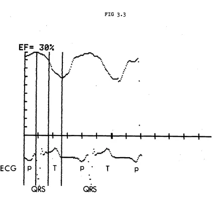
The "Nuclear Stethoscope" consists of a single scintillation ' probe with a 2 by 1.5 inch sodium iodide crystal and a slightly conical collimator with an outer opening of 3 cm diameter. The probe is carried by an adjustable arm allowing angulation in two different planes. Radioactivity is sampled and displayed with a temporal resolution of 10 msec (ventricular function mode) or 50 msec (beat-to-beat mode) per datum point. A dedicated microcomputer calculates left ventricular ejection fraction and other parameters. Together with the time-activity curve these data are displayed on a screen and could be reproduced by a Tecktronix Recorder.
With the probe positioned over the chest in a 35 0 left anterior oblique position with 5 0 caudal tilt, the precordium was scanned in parallel movements from right to left and in the cranio-caudal direction to approximate the left ventricular position. Small adjustments of the initial probe angulations were made if required according to individual chamber localisations. Correct positioning was guided by a search for maximal extension of a broad horizontal bar (fig 3.2B). The length of this bar is proportional to the amplitude of the time-activity curve and inversely proportional to the mean count rate.
of left ventricular ejection fraction. The probe was subsequently moved back to the left ventricular position . for recording of the gated time-activity curve (for two R-R intervals - Ventricular Function Curve). Left ventricular ejection fraction was calculated by moving two vertical cursors to positions corresponding to the end-diastolic and end-systolic count rate of the displayed mean time-activity (fig 3.3).
ASSESSMENT OF THE SEVERITY OF HEART FAILURE: 40.
FIG 3.2 A.
B.
EF=
33%
Determination of ejection fraction by "Nuclear Stethoscope":
A. Determination of background counts (note minimum excursion of horizontal bar)
FIG
3.3
EF= 30%
ECG
QkS
QkS
[image:55.559.90.514.88.503.2]ASSESSMENT OF THE SEVERITY OF HEART FAILURE: 42.
FIG 3.4
EJECTION FRACTION (
%)
NUCLEAR STETHOSCOPE
70-
60-
50
40-
30
20-
10
•
•
y =
0.9X+ 6.7
r =
0.847
P< 0.001
10 20 30 40 50 60 70
EJECTION FRACTION (%) GATED BLOOD POOL
3.5
M-Mode Echocardiographic Assessment of Left Ventricular Function:This technique provides an alternative non-invasive method for measuring left ventricular dimensions during systole and diastole. By making several assumptions about ventricular shape, volume, and ejection fraction may be determined. It should be noted that for M-mode echocardiography assumptions are false when regional wall motion abnormalities are present thus volume and ejection fraction measurement is inaccurate.
Left ventricular chamber dimension can most accurately be assessed by echocardiography at a level between the papillary muscles and the free edge of the mitral valve. To standardise the procedure, the transducer is placed on the chest wall in that intercostal space which permits recording of the mitral valve leaflet when the transducer is perpendicular to the chest wall. By tilting the transducer inferiorly and slightly laterally, characteristic left ventricular echoes are seen, allowing highly reproducible quantitation of ventricular dimensions
(Popp, 1979). Assuming a uniform geometric model for the ventricle, theoretical ventricular volume may be calculated from the single known left ventricular dimension. Techniques and equipment used in assessing patients with dilated cardiomyopathy are described in Chapter
9.
ASSESSMENT OF THE SEVERITY OF HEART FAILURE: 44.
3.6 Exercise Testing:
Although exercise testing has commonly been used in the diagnostic evaluation and follow-up of patients with angina pectoris, exercise has only recently been employed to assess the degree of functional limitation in patients with heart failure. Franciosa et al (1979) have found that the exercise test is better able to categorise patients with heart failure than physical examination or resting haemodynamic characteristics. Although Patterson et al (1972) demonstrated good overall correlation between NYHA Functional Classification and treadmill exercise tolerance, significant differences occurred in 25% of patients highlighting the subjective nature of historical classification. Furthermore, recent studies in patients with heart failure have shown that physical findings, chest films, echocardiograms and resting haemodynamics are inconsistently altered by therapeutic interventions which do increase exercise capacity (Franciosa et al, 1978; Awan et al, 1977; Aronow et al, 1977). Thus, exercise capacity has become another important parameter in assessing the severity of heart failure, as well as the response to treatment._