REVIEW
STLV-1 as a model for studying HTLV-1
infection
Brice Jégado
1, Fatah Kashanchi
2, Hélène Dutartre
1and Renaud Mahieux
1*Abstract
Few years after HTLV-1 identification and isolation in humans, STLV-1, its simian counterpart, was discovered. It then
became clear that STLV-1 is present almost in all simian species. Subsequent molecular epidemiology studies
dem-onstrated that, apart from HTLV-1 subtype A, all human subtypes have a simian homolog. As HTLV-1, STLV-1 is the
etiological agent of ATL, while no case of TSP/HAM has been described. Given its similarities with HTLV-1, STLV-1
repre-sents a unique tool used for performing clinical studies, vaccine studies as well as basic science.
Keywords:
HTLV-1, STLV-1, ATL, Prevalence, Interspecies transmission, Animal model, Therapy
© The Author(s) 2019. This article is licensed under a Creative Commons Attribution 4.0 International License, which permits use, sharing, adaptation, distribution and reproduction in any medium or format, as long as you give appropriate credit to the original author(s) and the source, provide a link to the Creative Commons licence, and indicate if changes were made. The images or other third party material in this article are included in the article’s Creative Commons licence, unless indicated otherwise in a credit line to the material. If material is not included in the article’s Creative Commons licence and your intended use is not permitted by statutory regulation or exceeds the permitted use, you will need to obtain permission directly from the copyright holder. To view a copy of this licence, visit http://creat iveco mmons .org/licen ses/by/4.0/. The Creative Commons Public Domain Dedication waiver (http://creat iveco mmons .org/publi cdoma in/ zero/1.0/) applies to the data made available in this article, unless otherwise stated in a credit line to the data.
Background
The first human oncogenic retrovirus was discovered in the
USA, in a T cell line obtained from blood cells of a patient
suffering from a disease then called “cutaneous T-cell
lym-phoma” [
1
,
2
]. Few years earlier, Adult T-cell Leukemia/
Lymphoma or ATLL (i.e. an aggressive malignancy of
CD4
+
T-cells) had been described in Japan [
3
,
4
]. In 1982,
Japanese researchers also reported the presence of a
ret-rovirus among ATLL patients. They named it Adult T cell
leukemia virus (ATLV). Further work demonstrated that
HTLV-1 specific antibodies were present among
Japa-nese ATLL patients, thus allowing identification of the first
HTLV-1 endemic area [
5
]. Later, it was decided to name this
virus HTLV-1 for Human T-cell Leukemia Virus type 1.
Few years later, Tropical Spastic Paraparesis/HTLV-1
associated myelopathy (TSP/HAM), a severe
neuromy-elopathy, was also identified as another disease caused
by HTLV-1 [
6
]. Thus, ATLL and TSP/HAM are the main
pathologies present among HTLV-1 infected
individu-als. It was recently estimated that 5 to 10 million people
are infected by HTLV-1 worldwide, although HTLV-1
prevalence is likely to be underestimated. Two to 4%
of HTLV-1 carriers will develop either ATLL or TSP/
HAM, while most of them will remain asymptomatic
[
7
]. HTLV-1 is endemic in areas such as Japan, central
Africa, the Caribbean region and South America [
8
].
Because HTLV-1 mostly replicates through clonal
expan-sion of infected cells even in asymptomatic carriers [
9
],
its retroviral genome displays a remarkable genetic
stabil-ity. HTLV-1 molecular epidemiology studies have been
carried out throughout the world. The very low genetic
variability allowed identification of different HTLV-1
subtypes. All but one of these subtypes, i.e.
Cosmopoli-tan subtype A that is present all over the world, are
spe-cific to a given African or Asian region [
8
]. ATL cases
were described in HTLV-1 carriers infected by HTLV-1
subtype A but also subtype B and subtype C [
10
,
11
],
thus suggesting that ATL occurrence is not linked to the
most frequent HTLV-1 subtype. Of note, HTLV-1
sub-type B and subsub-type C lack p12 and/or p30 auxiliary
pro-tein. Whether the lower ATL frequency in type B and C
infected individuals is linked to the absence of these
pro-teins remains to be determined.
In 1982, lymphocytes from a Japanese monkey
(
Macaca fuscata
) were co-cultured with chronically and
productively infected T-cells from the MT-2 cells, an
HTLV-1-transformed cell line. This allowed the authors
to obtain a simian cell line persistently infected by
Open Access
*Correspondence: renaud.mahieux@ens-lyon.fr
1 International Center for Research in Infectiology, Retroviral Oncogenesis
Laboratory, INSERM U1111 - Université Claude Bernard Lyon 1, CNRS, UMR5308, Ecole Normale Supérieure de Lyon, Université Lyon, Fondation pour la Recherche Médicale, Labex Ecofect, Lyon, France
HTLV-1, thus suggesting that Japanese monkeys might
be susceptible to HTLV-1 natural infection [
12
]. Later,
seroepidemiological studies were performed in Japan
and demonstrated that many Japanese monkeys were
infected by HTLV-1-like viruses [
13
]. Sera from New
World Monkeys (NWM), Old World Monkeys (OWM)
and Apes were then tested and revealed the presence
of antibodies reacting against HTLV-1 antigens. Such
antibodies were detected in OWM and Apes, but not
in NWM, suggesting endemicity of HTLV-1-related
viruses in African and Asian monkeys, but not in
Ameri-can animals [
14
]. Sequence analyses characterized these
viruses as Simian T-cell Leukemia Viruses (STLVs) [
15
,
16
]. To date, it is well established that Old World
Non-Human Primates (NHPs) and Apes are naturally infected
with a great variety of STLV-1 viruses and that HTLV-1
appeared in Humans following STLV-1 cross-species
transmission approximately 27,300 years ago (95% CI
19,100–35,500) in Africa, even if interspecies
transmis-sion episodes still occur [
17
–
19
]. Given the high degree
of similarity between HTLV-1 and STLV-1 sequences, it
was suggested to cluster these viruses in the single PTLV
(Primate T lymphotropic virus) family [
20
–
22
]. Because
STLV-1 induces ATLL in naturally infected NHPs [
23
,
24
], and even if some auxiliary proteins are lacking [
25
],
it represents a suitable tool that contributes to our
under-standing of HTLV-1 pathogenesis. This review will
com-pare HTLV-1 and STLV-1 retroviruses from different
aspects and will focus on the use of STLV-1 as a model of
HTLV-1 infection.
STLV‑1 epidemiology
Around 132 non-human primate species represent Old
World Monkeys (OWM). They are divided in two
sub-families,
Cercopithecinae
and
Colobinae,
distributed in
African and Asian continents [
26
].
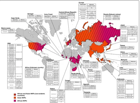
To determine which simian species carry STLV-1,
seroepidemiological studies were performed using kits
that had been previously developed for the detection
of anti-HTLV-1 human antibodies, as well as by PCR
(Fig.
1
). Sera from Japanese monkeys were tested, and
25% scored seropositive. As in humans, STLV-1
inci-dence increased with age and was higher in females than
males. Other species were tested later. A high
seropreva-lence was observed in African Green monkeys (AGM).
Two studies then reported STLV-1 infection in captive
Old World NHPs and Apes [
27
,
28
]. Ishikawa et al. [
29
]
performed an STLV-1 survey using 567 NHPs’ blood
samples covering 30 species caught in the wild or kept
in zoos, institutes or private owners from Kenya, Gabon,
Ghana, Cameroon, Ethiopia and Indonesia. STLV-1 was
detected in African Green monkeys and Sykes’ monkeys,
in Olive baboons, Patas monkeys, Mandrills and Gorillas.
STLV-1 was also found in different species of macaques
from Indonesia, with a seroprevalence ranging from 11
to 25%. Other studies reported natural STLV-1
infec-tions in AGM, Vervet monkeys and among baboon
spe-cies (
Papio anubis
,
Papio hamadryas
,
Papio papio
and
Papio cynocephalus
) originating from South Africa and
Ethiopia [
30
–
33
]. As in Japan, the infection status
posi-tively correlates with age, and disease incidence is higher
in females than males. Other seroepidemiological
stud-ies were also performed [
34
–
44
] (Fig.
1
). Thirty-one Old
World NHP species were reported as naturally infected
with STLV-1 [
33
,
45
–
50
].
STLV-1 sequence analyses were then performed in
order to determine relationship between STLV-1 and
HTLV-1 and whether HTLV-1 originated from a
non-human primate virus.
STLV‑1 phylogeny
Since the first publication of a complete HTLV-1
provi-ral genome [
51
], phylogenetic studies enabled to
iden-tify several HTLV-1 subtypes: Cosmopolitan subtype A,
which is found all over the world; subtypes B, D, E, F, G,
which are restricted to Central Africa; and
Australo-Mel-anesian subtype C which is the most divergent HTLV-1
subtype [
8
]. Based on molecular clock and phylogenetic
analyses, origin of HTLV-1 subtypes A, B, D, E was
inferred in a time frame of 27,300
±
8200 years, whereas
subtype F arose more than 10,000 years ago.
In 1984, Watanabe et al. [
52
] demonstrated
similari-ties between restriction maps obtained using HTLV-1
from Robert Gallo’s laboratory or using Japanese simian
Adult T-cell Leukemia Virus (ATLV). These results
sug-gested that HTLV-1 and simian ATLV shared a
com-mon ancestor. Other studies reported that HTLV-1 and
STLV-1 from Japanese monkeys, Red-faced monkeys,
Pig-tailed monkeys, AGM, Chimpanzees and baboons
(
Papio cynocephalus
) had the same genomic organization
i.e.
LTR
-
gag
-
pol
-
env
-
pX
-
LTR
[
15
,
20
]. Sequence analyses
comparing Pig-tailed (Asian NHP) and AGM (African
NHP) STLV-1 sequences to HTLV-1 revealed 90% and
95% identity respectively. These results suggested that
(1) STLV-1 could be separated into two subgroups: Asian
and African and that (2) HTLV-1 originated from the
African STLV-1 subgroup [
16
].
the
Env
region clusters with STLV-1 isolated from two
baboon species,
Papio ursinus
and
Papio cynocephalus
[
59
]. No data has been so far reported about a simian
counterpart of HTLV-1G and HTLV-1A. Altogether,
the diversity of STLV-1 strains found in different NHPs
species and related to a given HTLV-1 subtype from the
same geographical areas is strongly supporting the
con-cept of multiple cross-species transmissions between
NHPs but also from NHPs to humans.
Most divergent STLV-1 strains were described in
Asian
Macaca tonkeana
(living in Indonesia) and
Macaca arctoides
(living in India, Thailand and China)
[
60
–
62
].
Macaca tonkeana
virus is related to the most
divergent HTLV-1 subtype C that is present in
Melane-sia and Australia. Molecular clock data inferred STLV-1
introduction around 156,000 to 269,000 years ago on
the Asian continent [
59
]. These results suggest that
macaque infection with STLV-1 might have led to the
emergence of HTLV-1 in Asian human population.
Finally, Calvignac et al. [
63
] demonstrated that STLV-1
sequences could be amplified from bones samples
origi-nating from an early 20th century
Chlorocebus
pygeryth-rus
sample. Therefore, it should now be possible to use
this technique to determine STLV-1 virus evolution over
time using available Egyptian or Asian NHP mummies.
STLV‑1 interspecies transmission
Prevalence of HTLV-1 may reach 1 to 40% in adults
depending on age, sex and geographic location [
8
]. It is
well known that HTLV-1 can be transmitted under
dif-ferent routes: sexual, mother-to-child and contact with
infected blood. However, STLV-1 transmission occurs
mostly through aggressive contacts instead of mother
to infant or sexual transmissions [
64
–
68
], even if sexual
transmission of STLV-1 is more important in NHPs such
as vervet [
40
].
[image:3.595.58.542.88.443.2]STLV‑1 associated‑disease in naturally infected
animals
As it is the case for HTLV-1-infected individuals, most
STLV-1-infected monkeys remain lifelong
asympto-matic hosts [
69
]. For some unexplained reasons, TSP/
HAM cases have never been observed in infected
NHPs, even when those animals were living in animal
facilities for a long period. Phylogenetic studies
per-formed using samples from an African human TSP/
HAM patient showed that the viral sequence was highly
related to an STLV-1 sequence obtained from
asympto-matic West-African sooty mangabey [
70
]. Other strains
obtained from HTLV-1 African TSP/HAM patients also
clustered with STLV-1 strains obtained from
asympto-matic animals [
71
,
72
]. It is well established that there is
no specific mutation in HTLV-1 genome that would be
associated with a given disease. Altogether, these data
suggest that the lack of TSP/HAM described cases in
NHPs might only be linked to the mode of viral
trans-mission rather than the age of infection.
On the contrary, a number of ATLL-like diseases
sharing clinical and pathological features with human
ATLL were reported in NHPs [
24
,
69
,
73
–
79
]. The first
report was made in STLV-1 infected macaques which
developed malignant lymphoma [
80
]. Subsequent
stud-ies reported similar symptoms in captive
Papio anubis
,
Gorillas and AGM [
75
–
78
,
81
,
82
]. In a recent study,
Tax-positive cells were detected in lymphoid and
non-lymphoid organs, mesenteric and axillary lymph nodes
and lung, but not in the blood from an infected
Papio
anubis
suffering from ATL [
24
]. In that case, skin lesion
biopsies also showed a massive dermal, hypodermic
and muscular cell infiltrates of positive CD3
+
CD25
+
T
cells, as described in human ATL.
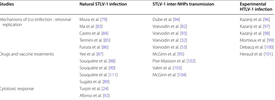
Using STLV‑1 infected animals
After natural STLV‑1 infection
Given the high degree of sequence similarities between
STLV-1 and HTLV-1 genomes and the fact that both
viruses cause ATL, STLV-1 infected NHPs (Japanese
macaques,
Mandrillus sphinx
and
Papio anubis
) have
been used for performing molecular studies [
79
,
83
–
89
]
(Table
1
). As HTLV-1, STLV-1 infection is mostly
occur-ring in CD4
+
T-cells, although STLV-1 Tax expression
was also detected in bone marrow hematopoietic stem
cells in vivo, and viral DNA was retrieved in all myeloid
and lymphoid cells derived from these infected
progeni-tors [
86
].
[image:4.595.305.539.112.182.2]STLV-1 natural infection leads to Tax and SBZ
(sim-ian equivalent of HBZ) expression. Sim(sim-ian SBZ and Tax
amino-acid sequences are highly similar to human HBZ
and Tax (see Tables
2
and
3
). These viral proteins also
Table 1 STLV‑1 naturally or experimentally infected non‑human primates (NHPs) described in published biological
studies
STLV-1 infection mechanisms, experimental treatments and immune response were analyzed in several NHP species
Studies Natural STLV‑1 infection STLV‑1 inter‑NHPs transmission Experimental
HTLV‑1 infection
Mechanisms of (co-)infection : retroviral
replication Miura et al. [Ma et al. [83]79] Voevodin et al. [Dube et al. [94]82] Kazanji et al. [Kazanji et al. [9796]] Castro et al. [84] Voevodin et al. [93] Kazanji et al. [98] Termini et al. [85] Voevodin et al. [32] Mortreux et al. [99] Furuta et al. [86] Voevodin et al. [53] Debacq et al. [100] Drugs and vaccine treatments Yee et al. [87] McGinn et al. [95] Heraud et al. [101]
Souquière et al. [88] Pise-Masison et al. [102] Souquière et al. [90] Valeri et al. [103] Souquière et al. [111] McGinn et al. [104] Sugata et al. [89]
Cytotoxic response Turpin et al. [24] Afonso et al. [92]
Table 2 Amino acid sequence comparison of HTLV‑1 HBZ
vs. STLV‑1 SBZ
ATK belongs to HTLV-1 A cosmopolitan subtype, EL to HTLV-1 B subtype, STLV-1
Papio anubis was obtained from an African NHP, while STLV-1 Mf5 was obtained from an Asian NHP (Macaca fuscata)
HTLV‑1a ATK HTLV‑1b EL
HTLV-1a ATK – 74.27%
HTLV-1b EL 74.27% –
STLV-1 Papio anubis 83.01% 71.36%
[image:4.595.58.540.540.715.2]display activating properties on viral LTR and NF-κB
signaling pathways. As an example, a high STLV-1
pro-viral load (PVL) is linked to IL-2, IL-6, IL-10, IFNγ and
TNF-α elevated expression in asymptomatic
STLV-1-infected
Mandrillus sphinx
[
90
]. Given
well-estab-lished results pubwell-estab-lished in the HTLV-1 situation, this
is likely due to STLV-1 Tax expression, although this
hypothesis has not been formally demonstrated. IL-2
and IFNγ results were also obtained in asymptomatic
STLV-1-positive
Macaca mulatta
[
87
], while anti IFNγ
and TNF-α responses against Tax expressing cells were
also observed in STLV-1 infected baboons [
85
]. STLV-1
infection also promotes CTL response against STLV-1
Tax protein [
84
,
85
].
Interestingly, TCF1 and LEF1, two T-cell specific
pro-teins, prevent Tax effect on viral LTR. Their expression
is high in thymocytes and thus counteract STLV-1
rep-lication in thymus. On the opposite, their expression
and thus their effect is down-regulated in peripheral
blood T-cells (both in human and simian cells), thanks
to a Tax effect on STAT5a. This might explain why Tax
is more potent in these cells, and why HTLV-1 induces
ATL in the periphery [
83
].
Depending upon STLV-1 strain, SBZ protein
sequence is highly similar or contain insertions and
deletion compared to HBZ (see Table
2
). Nevertheless,
in both cases, animals can develop ATL [
24
,
79
]. This
might be due to conservation of the N-terminal region
as well as of C-terminus basic leucin zipper domain
between human and simian viral proteins.
As its human counterpart, STLV-1 replication occurs
through clonal expansion of infected cells, both in
asymptomatic and ATL animals [
24
,
79
]. Antiviral
ther-apy based on the use of azidothymidine (AZT)
com-bined with interferon-α (IFN-α) improves the survival
rate of ATL patients suffering from acute and chronic/
smoldering forms. A confirmation clinical trial using
these compounds was reported in an STLV-1 infected
Papio anubis
suffering from ATL. The animal was
treated with a combination of AZT and interferon-α.
However, and contrary to human ATL, no clinical
improvement was observed. It would now be
interest-ing to determine post-mortem whether, this absence of
remission was linked to
p53
mutation already present
when treatment started as shown in human ATL cases
who were not responding to AZT [
91
].
Given the fact that treating ATL patients is difficult,
and because an elevated PVL is a characteristic of ATL,
a study tested whether PVL decreases when valproate
and AZT were delivered to asymptomatic
STLV-1-in-fected animals [
92
]. This was indeed the case and it was
associated to an increased anti-Tax CTL response, thus
confirming the importance of immune response for
con-trolling viral infection [
92
]. In another study, STLV-1
infected asymptomatic Japanese monkey were
inocu-lated with mogamulizumab (anti-CCR4), a component
that is also used for human relapsed ATL cases. This led
to a strong reduction of STLV-1 proviral load [
79
,
89
].
Altogether, these results support the fact that STLV-1
infected animals represent a useful tool for testing drugs.
Finally, a recent study was performed in two
asympto-matic STLV-1-infected animals. This showed that
immu-nization using recombinant vaccinia viruses expressing
either Tax-22 (which cannot activate the NF-kB pathway)
or an HBZ LL/AA mutant (which is partially impaired for
blocking Tax ability to induce transcription) was linked
to a temporary decrease of STLV-1 PVL [
89
].
After STLV‑1 interspecies transmission
A limited number of reports described STLV-1
inter-sim-ian species transmission [
32
,
53
,
93
,
94
] (Table
1
). In one
report and following an unknown mode of transmission,
it was shown that baboons accidentally infected with a
rhesus macaque STLV-1 virus, developed
leukemia/lym-phoma at a high frequency [
93
]. This is the only reported
case suggesting that inter-simian species transmission
might impact viral pathogenesis. Experimental infection
of pig-tailed macaques with sooty mangabey STLV-1 was
also tested. Animals maintained low antibody titers and
displayed a high mortality rate without any identified
cause [
95
]. Finally, another work reported tantalus and
patas animals artificially infected with STLV-1 from other
species. All animals became infected, as shown by PCR
results, even if one stayed seronegative due to mutations
in the genome [
94
]. Why were these
pol
mutant viruses
still able to infect animals remains unexplained.
After artificial HTLV‑1 infection
[image:5.595.56.292.111.182.2]Finally, given the high degree of similarity between
HTLV-1 and STLV-1 genomes and the abundance of
molecular tools available in the HTLV-1 field, some
labo-ratories decided to use the HTLV-1 molecular clone or
HTLV-1 infected cells to perform studies in non-human
primates (Table
1
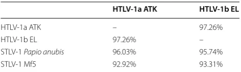
). Artificial infection after inoculation
Table 3 Amino acid sequence comparison of HTLV‑1 Tax
vs. STLV‑1 Tax
ATK belongs to HTLV-1 A cosmopolitan subtype, EL to HTLV-1 B subtype, STLV-1
Papio anubis was obtained from an African NHP, while STLV-1 Mf5 was obtained from an Asian NHP (Macaca fuscata)
HTLV‑1a ATK HTLV‑1b EL
HTLV-1a ATK – 97.26%
HTLV-1b EL 97.26% –
STLV-1 Papio anubis 96.03% 95.74%
of HTLV-1 to primates provides an inestimable tool to
study primo-infection and viral dissemination, in vivo, a
process that is inaccessible in humans. HTLV-1 infection
of
Saimiri sciureus
, i.e. non-human primates that are not
naturally infected with STLV-1 [
96
], demonstrated that
lymphoid organs represent the major viral reservoir [
97
].
As in HTLV-1 infected humans and STLV-1
naturally-infected animals, IL-2, IL-10, IFNγ levels also increased
after HTLV-1 infection [
98
]. In
Saimiri sciureus
, the
virus also replicates through clonal expansion after
hav-ing used reverse transcription (RT) at the initial stages
[
99
] and it causes ATL [
100
]. As in baboons treated with
AZT/IFN [
24
], arsenic combined to IFN-α was not able
to lead to HTLV-1 proviral load reduction, even if the
number of circulating ATL flower cells decreased for
some unexplained reason [
101
].
Studies were also performed in pig-tailed and rhesus
macaques inoculated with autologous cells previously
transfected with the HTLV-1 ACH molecular clone
[
102
–
104
]. Following infection with wild-type
HTLV-1, pig-tailed macaques developed a series of extremely
aggressive diseases that were different from ATL. These
results therefore suggest that this animal model
can-not be used for studying events that are resulting from
HTLV-1 infection.
Consequences of rhesus macaque infection with the
same molecular clone were different since animals
remained asymptomatic. HTLV-1 p12 and p8 proteins
have been shown previously to increase NFAT activity,
IL-2 production and STAT-5 activity, while p30 controls
viral expression at the post-transcriptional level in vitro
(for a review, see [
105
,
106
]). Thus, this simian model was
useful to investigate the role of p12, p13, and p30
auxil-iary proteins in vivo [
102
,
103
]. This allowed
research-ers to show that p12 and p30 are required for allowing
HTLV-1 presence and replication in dendritic cells [
103
],
while p12 and p8 are necessary for allowing a viral
resist-ance to CTL responses. These studies provided the first
in vivo evidence on the mechanisms that HTLV-1 uses
to establish chronic infection and on the crucial role of
myeloid cells in that process.
Interestingly, the authors also demonstrated that the
results obtained in rhesus macaques were different from
those obtained in rabbits infected with the same viral
clones, thus reinforcing the fact that NHPs are the more
relevant system for studying HTLV-1 pathogenesis.
PTLV retroviral coinfection in NHPs and in humans
In addition to STLV-1, other retroviruses, i.e. Simian
Immunodeficiency Virus (SIV) and Simian Foamy Virus
(SFV) infect NHPs. Cases of natural coinfection have
been reported both in humans and in NHPs: HTLV-1/
HIV-1, HTLV-1/HFV, STLV-1/SFV or STLV-1/SIV-1
[
67
,
107
–
115
]. HIV-1/HTLV-1 coinfection leads to
sig-nificant increase of HTLV-1 PVL as well as on a possible
delay in HIV-1 pathogenesis in humans [
107
,
108
,
116
].
Anti-HIV-1 therapy promotes an increase in HTLV-1
PVL in HIV-1/HTLV-1 coinfected carriers. These
results strongly suggest that both retroviruses compete
for CD4
+
T-cell infection. However, it is worth
not-ing that opposite results were obtained in other studies
[
117
–
121
].
Natural STLV-1/SIV-1 co-infection induces the
devel-opment of a neoplastic disease in sooty mangabey [
122
]
and of a lymphoproliferative disease in AGM [
123
].
Souquière et al. described pathological manifestations,
i.e. infective dermatitis and scabies, in two STLV-1/SIV-1
co-infected mandrills [
111
], while no clinical sign has
been reported previously in STLV-1 naturally infected
mandrills [
90
]. Thus, these symptoms could be due to
co-infection. Ongoing experiments should allow us to
determine whether STLV-1 clonal expansion impacts SIV
replication in vivo.
Finally, blood SFV proviral load from STLV-1/SFV
nat-urally co-infected
Papio anubis,
was recently shown to be
much higher compared to SFV mono-infected animals
[
124
]. These results either suggest that cells might be
co-infected with both retroviruses, with STLV-1 promoting
clonal expansion, or that soluble STLV-1 Tax
transactiva-tor enters SFV-infected cells where it promotes viral
rep-lication. Ongoing experiments should allow us to answer
this question.
Altogether, these data demonstrate that STLV-1 is a
useful tool to understand mechanisms of HTLV-1
trans-mission and ATL pathogenesis. PTLV-1 mono-infected
as well as SIV co-infected animals could also be used to
develop possible new anti-HTLV-1 clinical approaches
and to modify anti-HIV treatment.
Acknowledgements
BJ is supported by Labex Ecofect, RM is supported by Ecole Normale Supé-rieure de Lyon. RM is part of the French Laboratory of Excellence project ECOFECT (ANR-11-LABX-0048). The authors acknowledge the support Fonda-tion pour la recherche médicale (équipe Labellisée). The authors thank Dr C. Journo for her helpful comments.
Authors’ contributions
BJ, HD, FK and RM wrote the manuscript. All authors read and approved the final manuscript.
Funding
RM is part of the French Laboratory of Excellence project ECOFECT (ANR-11-LABX-0048). The authors acknowledge the support Fondation pour la recherche médicale (équipe Labellisée DEQ20180339200). BJ is supported by Labex Ecofect, RM is supported by Ecole Normale Supérieure de Lyon. HD is funded by INSERM.
Availability of data and materials
Not applicable.
Ethics approval and consent to participate
Consent for publication
Not applicable.
Competing interests
Not applicable.
Author details
1 International Center for Research in Infectiology, Retroviral
Oncogen-esis Laboratory, INSERM U1111 - Université Claude Bernard Lyon 1, CNRS, UMR5308, Ecole Normale Supérieure de Lyon, Université Lyon, Fondation pour la Recherche Médicale, Labex Ecofect, Lyon, France. 2 Laboratory of Molecular
Virology, George Mason University, Manassas, VA, USA.
Received: 24 July 2019 Accepted: 7 December 2019
References
1. Poiesz BJ, Ruscetti FW, Gazdar AF, Bunn PA, Minna JD, Gallo RC. Detec-tion and isolaDetec-tion of type C retrovirus particles from fresh and cultured lymphocytes of a patient with cutaneous T-cell lymphoma. Proc Natl Acad Sci USA. 1980;77:7415–9.
2. Poiesz BJ, Ruscetti FW, Reitz MS, Kalyanaraman VS, Gallo RC. Isolation of a new type C retrovirus (HTLV) in primary uncultured cells of a patient with Sézary T-cell leukaemia. Nature. 1981;294:268–71.
3. Takatsuki K, Uchiyama T, Sagawa K. Hattori T [Lymphoma and immu-noglobulin abnormalities, with special reference to M proteinemia]. Nippon Rinsho. 1977;35:3757–67.
4. Uchiyama T, Yodoi J, Sagawa K, Takatsuki K, Uchino H. Adult T-cell leukemia: clinical and hematologic features of 16 cases. Blood. 1977;50:481–92.
5. Gallo RC. History of the discoveries of the first human retroviruses: HTLV-1 and HTLV-2. Oncogene. 2005;24:5926–30.
6. Gessain A, Francis H, Sonan T, Giordano C, Akani F, Piquemal M, et al. HTLV-I and tropical spastic paraparesis in Africa. Lancet. 1986;2:698. 7. Futsch N, Mahieux R, Dutartre H. HTLV-1, the other pathogenic yet neglected human retrovirus: from transmission to therapeutic treat-ment. Viruses. 2017;10:1.
8. Gessain A, Cassar O. Epidemiological aspects and world distribution of HTLV-1 infection. Front Microbiol. 2012;3:388.
9. Wattel E, Vartanian JP, Pannetier C, Wain-Hobson S. Clonal expansion of human T-cell leukemia virus type I-infected cells in asymptomatic and symptomatic carriers without malignancy. J Virol. 1995;69:2863–8. 10. Mahieux R, Ibrahim F, Mauclere P, Herve V, Michel P, Tekaia F, et al. Molecular epidemiology of 58 new African human T-cell leukemia virus type 1 (HTLV-1) strains: identification of a new and distinct HTLV-1 molecular subtype in Central Africa and in pygmies. J Virol. 1997;71:17. 11. Einsiedel L, Cassar O, Bardy P, Kearney D, Gessain A. Variant human T-cell
lymphotropic virus type 1c and adult T-cell leukemia, Australia. Emerg Infect Dis. 2013;19:1639–41.
12. Miyoshi I, Taguchi H, Fujishita M, Yoshimoto S, Kubonishi I, Ohtsuki Y, et al. Transformation of monkey lymphocytes with adult T-cell leukae-mia virus. Lancet. 1982;1:1016.
13. Miyoshi I, Yoshimoto S, Fujishita M, Taguchi H, Kubonishi I, Niiya K, et al. Natural adult T-cell leukemia virus infection in Japanese monkeys. Lancet. 1982;2:658.
14. Hayami M, Komuro A, Nozawa K, Shotake T, Ishikawa K, Yamamoto K, et al. Prevalence of antibody to adult T-cell leukemia virus-associated antigens (ATLA) in Japanese monkeys and other non-human primates. Int J Cancer. 1984;33:179–83.
15. Komuro A, Watanabe T, Miyoshi I, Hayami M, Tsujimoto H, Seiki M, et al. Detection and characterization of simian retroviruses homologous to human T-cell leukemia virus type I. Virology. 1984;138:373–8. 16. Watanabe T, Seiki M, Hirayama Y, Yoshida M. Human T-cell leukemia
virus type I is a member of the African subtype of simian viruses (STLV). Virology. 1986;148:385–8.
17. Mossoun A, Calvignac-Spencer S, Anoh AE, Pauly MS, Driscoll DA, Michel AO, et al. Bushmeat hunting and zoonotic transmission of sim-ian T-lymphotropic virus 1 in tropical West and Central Africa. J Virol. 2017;91:e02479-16.
18. Kazanji M, Mouinga-Ondémé A, Lekana-Douki-Etenna S, Caron M, Makuwa M, Mahieux R, et al. Origin of HTLV-1 in hunters of nonhuman primates in Central Africa. J Infect Dis. 2015;211:361–5.
19. Filippone C, Betsem E, Tortevoye P, Cassar O, Bassot S, Froment A, et al. A severe bite from a nonhuman primate is a major risk factor for HTLV-1 infection in hunters from Central Africa. Clin Infect Dis. 2015;60:1667–76.
20. Guo HG, Wong-Stall F, Gallo RC. Novel viral sequences related to human T-cell leukemia virus in T cells of a seropositive baboon. Science. 1984;223:1195–7.
21. Watanabe T, Seiki M, Tsujimoto H, Miyoshi I, Hayami M, Yoshida M. Sequence homology of the simian retrovirus genome with human T-cell leukemia virus type I. Virology. 1985;144:59–65.
22. Sherman MP, Saksena NK, Dube DK, Yanagihara R, Poiesz BJ. Evolution-ary insights on the origin of human T-cell lymphoma/leukemia virus type I (HTLV-I) derived from sequence analysis of a new HTLV-I variant from Papua New Guinea. J Virol. 1992;66:2556–63.
23. Locatelli S, Peeters M. Cross-species transmission of simian retroviruses: how and why they could lead to the emergence of new diseases in the human population. AIDS. 2012;26:659–73.
24. Turpin J, Alais S, Marçais A, Bruneau J, Melamed A, Gadot N, et al. Whole body clonality analysis in an aggressive STLV-1 associated leukemia (ATLL) reveals an unexpected clonal complexity. Cancer Lett. 2017;389:78–85.
25. Afonso PV, Fagrouch Z, Deijs M, Niphuis H, Bogers W, Gessain A, et al. Absence of accessory genes in a divergent simian T-lymphotropic virus type 1 isolated from a bonnet macaque (Macaca radiata). PLoS Negl Trop Dis. 2019;13:e0007521.
26. Lawrence JM, Cords M. Old World Monkeys. Nat Educ Knowl. 2012;3(7):13.
27. Lowenstine LJ, Pedersen NC, Higgins J, Pallis KC, Uyeda A, Marx P, et al. Seroepidemiologic survey of captive Old-World primates for antibodies to human and simian retroviruses, and isolation of a lentivirus from sooty mangabeys (Cercocebus atys). Int J Cancer. 1986;38:563–74. 28. Hunsmann G, Schneider J, Schmitt J, Yamamoto N. Detection of serum
antibodies to adult T-cell leukemia virus in non-human primates and in people from Africa. Int J Cancer. 1983;32:329–32.
29. Ishikawa K, Fukasawa M, Tsujimoto H, Else JG, Isahakia M, Ubhi NK, et al. Serological survey and virus isolation of simian T-cell leukemia/T-lymphotropic virus type I (STLV-I) in non-human primates in their native countries. Int J Cancer. 1987;40:233–9.
30. Becker WB, Becker ML, Homma T, Brede HD, Kurth R. Serum antibodies to human T-cell leukaemia virus type I in different ethnic groups and in non-human primates in South Africa. S Afr Med J. 1985;67:445–9. 31. Ishida T, Yamamoto K, Shotake T, Nozawa K, Hayami M, Hinuma Y. A
field study of infection with human T-cell leukemia virus among African primates. Microbiol Immunol. 1986;30:315–21.
32. Voevodin A, Samilchuk E, Allan J, Rogers J, Broussard S. Simian T-lymphotropic virus type 1 (STLV-1) infection in wild yellow baboons (Papio hamadryas cynocephalus) from Mikumi National Park, Tanzania. Virology. 1997;228:350–9.
33. Moné J, Whitehead E, Leland M, Hubbard G, Allan JS. Simian T-cell leukemia virus type I infection in captive baboons. AIDS Res Hum Retroviruses. 1992;8:1653–61.
34. Hayami M, Ishikawa K, Komuro A, Kawamoto Y, Nozawa K, Yamamoto K, et al. ATLV antibody in cynomolgus monkeys in the wild. Lancet. 1983;2:620.
35. Yamamoto N, Hinuma Y, zur Hausen H, Schneider J, Hunsmann G. African green monkeys are infected with adult T-cell leukaemia virus or closely related agent. Lancet. 1983;1:240–1.
36. Yamamoto N, Okada M, Hinuma Y, Hirsch FW, Chosa T, Schneider J, et al. Human adult T-cell leukaemia virus is distinct from a similar isolate of Japanese monkeys. J Gen Virol. 1984;65(Pt 12):2259–64.
37. Yamamoto N, Kobayashi N, Takeuchi K, Koyanagi Y, Hatanaka M, Hinuma Y, et al. Characterization of African green monkey B-cell lines releasing an adult T-cell leukemia-virus-related agent. Int J Cancer. 1984;34:77–82. 38. Botha MC, Jones M, de Klerk WA, Yamamoto N. Spread and
39. Coursaget P, Barres JL, Yvonnet B, Chiron JP, Cornet M, Ferrara L, et al. Antibodies to human T-cell leukemia virus (HTLV-1) in non human primates from Senegal. Biomed Pharmacother. 1985;39:198–9. 40. Dracopoli NC, Turner TR, Else JG, Jolly CJ, Anthony R, Gallo RC, et al.
STLV-I antibodies in feral populations of East African vervet monkeys (Cercopithecus aethiops). Int J Cancer. 1986;38:523–9.
41. Daniel MD, Letvin NL, Sehgal PK, Schmidt DK, Silva DP, Solomon KR, et al. Prevalence of antibodies to 3 retroviruses in a captive colony of macaque monkeys. Int J Cancer. 1988;41:601–8.
42. Fultz PN, Gordon TP, Anderson DC, McClure HM. Prevalence of natural infection with simian immunodeficiency virus and simian T-cell leu-kemia virus type I in a breeding colony of sooty mangabey monkeys. AIDS. 1990;4:619–25.
43. Estaquier J, Peeters M, Bedjabaga L, Honoré C, Bussi P, Dixson A, et al. Prevalence and transmission of simian immunodeficiency virus and simian T-cell leukemia virus in a semi-free-range breeding colony of mandrills in Gabon. AIDS. 1991;5:1385–6.
44. Courgnaud V, Van Dooren S, Liegeois F, Pourrut X, Abela B, Loul S, et al. Simian T-cell leukemia virus (STLV) infection in wild primate popula-tions in Cameroon: evidence for dual STLV type 1 and type 3 infection in agile mangabeys (Cercocebus agilis). J Virol. 2004;78:4700–9. 45. Koralnik IJ, Boeri E, Saxinger WC, Monico AL, Fullen J, Gessain A, et al.
Phylogenetic associations of human and simian T-cell leukemia/lym-photropic virus type I strains: evidence for interspecies transmission. J Virol. 1994;68:2693–707.
46. Mahieux R, Pecon-Slattery J, Chen GM, Gessain A. Evolutionary infer-ences of novel simian T lymphotropic virus type 1 from Wild-Caught Chacma (Papio ursinus) and olive baboons (Papio anubis). Virology. 1998;251:71–84.
47. Saksena NK, Herve V, Durand JP, Leguenno B, Diop OM, Digouette JP, et al. Seroepidemiologic, molecular, and phylogenetic analyses of simian T-cell leukemia viruses (STLV-I) from various naturally infected monkey species from central and western Africa. Virology. 1994;198:297–310.
48. Schatzl H, Yakovleva L, Lapin B, Rose D, Inzhiia L, Gaedigk-Nitschko K, et al. Detection and characterization of T-cell leukemia virus-like proviral sequences in PBL and tissues of baboons by PCR. Leukemia. 1992;6(Suppl 3):158S–60S.
49. Sintasath DM, Wolfe ND, LeBreton M, Jia H, Garcia AD, Diffo JLD, et al. Simian T-lymphotropic virus diversity among nonhuman primates, Cameroon. Emerg Infect Dis. 2009;15:175–84.
50. Van Dooren S, Verschoor EJ, Fagrouch Z, Vandamme A-M. Phylogeny of primate T lymphotropic virus type 1 (PTLV-1) including various new Asian and African non-human primate strains. Infect Genet Evol. 2007;7:374–81.
51. Seiki M, Hattori S, Hirayama Y, Yoshida M. Human adult T-cell leukemia virus: complete nucleotide sequence of the provirus genome inte-grated in leukemia cell DNA. Proc Natl Acad Sci USA. 1983;80:3618–22. 52. Watanabe T, Seiki M, Yoshida M. HTLV type I (U. S. isolate) and ATLV
(Japanese isolate) are the same species of human retrovirus. Virology. 1984;133:238–41.
53. Voevodin AF, Johnson BK, Samilchuk EI, Stone GA, Druilhet R, Greer WJ, et al. Phylogenetic analysis of simian T-lymphotropic virus type I (STLV-I) in common chimpanzees (Pan troglodytes): evidence for interspecies transmission of the virus between chimpanzees and humans in Central Africa. Virology. 1997;238:212–20.
54. Meertens L, Rigoulet J, Mauclère P, Van Beveren M, Chen GM, Diop O, et al. Molecular and phylogenetic analyses of 16 novel simian T cell leukemia virus type 1 from Africa: close relationship of STLV-1 from Allenopithecus nigroviridis to HTLV-1 subtype B strains. Virology. 2001;287:275–85.
55. Ayouba A, Michem A, Peeters M, Vercammen F. Full-genome characteri-zation of simian T-cell leukemia virus type 1 subtype b from a wild-born captive gorilla gorilla gorilla with T-cell lymphoma. Genome Announc. 2017;5:e01117-17.
56. Mahieux R, Chappey C, Georges-Courbot MC, Dubreuil G, Mauclere P, Georges A, et al. Simian T-cell lymphotropic virus type 1 from Mandrillus sphinx as a simian counterpart of human T-cell lymphotropic virus type 1 subtype D. J Virol. 1998;72:10316–22.
57. Makuwa M, Souquière S, Clifford SL, Telfer PT, Sallé B, Bourry O, et al. Two distinct STLV-1 subtypes infecting Mandrillus sphinx follow the
geographic distribution of their hosts. AIDS Res Hum Retroviruses. 2004;20:1137–43.
58. Liégeois F, Lafay B, Switzer WM, Locatelli S, Mpoudi-Ngolé E, Loul S, et al. Identification and molecular characterization of new STLV-1 and STLV-3 strains in wild-caught nonhuman primates in Cameroon. Virology. 2008;371:405–17.
59. Van Dooren S, Salemi M, Vandamme AM. Dating the origin of the Afri-can human T-cell lymphotropic virus type-i (HTLV-I) subtypes. Mol Biol Evol. 2001;18:661–71.
60. Mahieux R, Pecon-Slattery J, Gessain A. Molecular characterization and phylogenetic analyses of a new, highly divergent simian T-cell lymphotropic virus type 1 (STLV-1marc1) in Macaca arctoides. J Virol. 1997;71:6253–8.
61. Ibrahim F, de Thé G, Gessain A. Isolation and characterization of a new simian T-cell leukemia virus type 1 from naturally infected celebes macaques (Macaca tonkeana): complete nucleotide sequence and phylogenetic relationship with the Australo-Melanesian human T-cell leukemia virus type 1. J Virol. 1995;69:6980–93.
62. Van Dooren S, Meertens L, Lemey P, Gessain A, Vandamme A-M. Full-genome analysis of a highly divergent simian T-cell lymphotropic virus type 1 strain in Macaca arctoides. J Gen Virol. 2005;86:1953–9. 63. Calvignac S, Terme J-M, Hensley SM, Jalinot P, Greenwood AD, Hänni C.
Ancient DNA identification of early 20th century simian T-cell leukemia virus type 1. Mol Biol Evol. 2008;25:1093–8.
64. Niphuis H, Verschoor EJ, Bontjer I, Peeters M, Heeney JL. Reduced trans-mission and prevalence of simian T-cell lymphotropic virus in a closed breeding colony of chimpanzees (Pan troglodytes verus). J Gen Virol. 2003;84:615–20.
65. Leendertz FH, Junglen S, Boesch C, Formenty P, Couacy-Hymann E, Courgnaud V, et al. High variety of different simian T-cell leukemia virus type 1 strains in chimpanzees (Pan troglodytes verus) of the Taï National Park, Côte d’Ivoire. J Virol. 2004;78:4352–6.
66. d’Offay JM, Eberle R, Sucol Y, Schoelkopf L, White MA, Valentine BD, et al. Transmission dynamics of simian T-lymphotropic virus type 1 (STLV1) in a baboon breeding colony: predominance of female-to-female trans-mission. Comp Med. 2007;57:105–14.
67. Nerrienet E, Amouretti X, Müller-Trutwin MC, Poaty-Mavoungou V, Bed-jebaga I, Nguyen HT, et al. Phylogenetic analysis of SIV and STLV type I in mandrills (Mandrillus sphinx): indications that intracolony transmis-sions are predominantly the result of male-to-male aggressive contacts. AIDS Res Hum Retroviruses. 1998;14:785–96.
68. Roussel M, Pontier D, Ngoubangoye B, Kazanji M, Verrier D, Fouchet D. Modes of transmission of simian T-lymphotropic virus type 1 in semi-captive mandrills (Mandrillus sphinx). Vet Microbiol. 2015;179:155–61. 69. Allan JS, Leland M, Broussard S, Mone J, Hubbard G. Simian T-cell
lymphotropic viruses (STLVs) and lymphomas in african nonhuman primates. Cancer Invest. 2001;19:383–95.
70. Enose-Akahata Y, Caruso B, Haner B, Charlip E, Nair G, Massoud R, et al. Development of neurologic diseases in a patient with primate T lym-photropic virus type 1 (PTLV-1). Retrovirology. 2016;13:56.
71. Salemi M, Van Dooren S, Audenaert E, Delaporte E, Goubau P, Desmyter J, et al. Two new human T-lymphotropic virus type I phylogenetic subtypes in seroindeterminates, a Mbuti pygmy and a Gabonese, have closest relatives among African STLV-I strains. Virology. 1998;246:277–87. 72. Touzé E, Gessain A, Lyon-Caen O, Gout O. Tropical spastic paraparesis/
HTLV-I-associated myelopathy in Europe and in Africa: clinical and epidemiologic aspects. J Acquir Immune Defic Syndr Hum Retrovirol. 1996;13(Suppl 1):S38–45.
73. Noda Y, Ishikawa K, Sasagawa A, Honjo S, Mori S, Tsujimoto H, et al. Hematologic abnormalities similar to the preleukemic state of adult T-cell leukemia in African green monkeys naturally infected with simian T-cell leukemia virus. Jpn J Cancer Res. 1986;77:1227–34.
74. Sakakibara I, Sugimoto Y, Sasagawa A, Honjo S, Tsujimoto H, Nakamura H, et al. Spontaneous malignant lymphoma in an African green monkey naturally infected with simian T-lymphotropic virus (STLV). J Med Prima-tol. 1986;15:311–8.
76. McCarthy TJ, Kennedy JL, Blakeslee JR, Bennett BT. Spontaneous malignant lymphoma and leukemia in a simian T-lymphotropic virus type I (STLV-I) antibody positive olive baboon. Lab Anim Sci. 1990;40:79–81.
77. Hubbard GB, Moné JP, Allan JS, Davis KJ, Leland MM, Banks PM, et al. Spontaneously generated non-Hodgkin’s lymphoma in twenty-seven simian T-cell leukemia virus type 1 antibody-positive baboons (Papio species). Lab Anim Sci. 1993;43:301–9.
78. d’Offay JM, Eberle R, Wolf RF, Kosanke SD, Doocy KR, Ayalew S, et al. Simian T-lymphotropic Virus-associated lymphoma in 2 naturally infected baboons: T-cell clonal expansion and immune response dur-ing tumor development. Comp Med. 2013;63:288–94.
79. Miura M, Yasunaga J, Tanabe J, Sugata K, Zhao T, Ma G, et al. Charac-terization of simian T-cell leukemia virus type 1 in naturally infected Japanese macaques as a model of HTLV-1 infection. Retrovirology. 2013;10:118.
80. Homma T, Kanki PJ, King NW, Hunt RD, O’Connell MJ, Letvin NL, et al. Lymphoma in macaques: association with virus of human T lympho-trophic family. Science. 1984;225:716–8.
81. Srivastava BI, Wong-Staal F, Getchell JP. Human T-cell leukemia virus I provirus and antibodies in a captive gorilla with non-Hodgkin’s lymphoma. Cancer Res. 1986;46:4756–8.
82. Voevodin AF, Lapin BA, Yakovleva LA, Ponomaryeva TI, Oganyan TE, Razmadze EN. Antibodies reacting with human T-lymphotropic retrovirus (HTLV-I) or related antigens in lymphomatous and healthy hamadryas baboons. Int J Cancer. 1985;36:579–84.
83. Ma G, Yasunaga J, Akari H, Matsuoka M. TCF1 and LEF1 act as T-cell intrinsic HTLV-1 antagonists by targeting Tax. Proc Natl Acad Sci USA. 2015;112:2216–21.
84. Castro I, Giret TM, Magnani DM, Maxwell HS, Umland O, Perry JK, et al. Cellular immune responses against simian T-lymphotropic virus type 1 target tax in infected baboons. J Virol. 2016;90:5280–91.
85. Termini JM, Magnani DM, Maxwell HS, Lauer W, Castro I, Pecotte J, et al. Simian T lymphotropic virus 1 infection of Papio anu-bis: tax sequence heterogeneity and T cell recognition. J Virol. 2017;91:e00950-17.
86. Furuta R, Yasunaga J-I, Miura M, Sugata K, Saito A, Akari H, et al. Human T-cell leukemia virus type 1 infects multiple lineage hematopoietic cells in vivo. PLoS Pathog. 2017;13:e1006722.
87. Yee JL, Montiel NA, Ardeshir A, Ardeshr A, Lerche NW. Constitutive release of IFNγ and IL2 from peripheral blood mononuclear cells of rhe-sus macaques (Macaca mulatta) infected with simian T-lymphotropic virus type 1. Comp Med. 2013;63:508–14.
88. Souquière S, Mouinga-Ondemé A, Makuwa M, Hermine O, Kazanji M. Dynamic interaction between STLV-1 proviral load and T-cell response during chronic infection and after immunosuppression in non-human primates. PLoS ONE. 2009;4:e6050.
89. Sugata K, Yasunaga J-I, Miura M, Akari H, Utsunomiya A, Nosaka K, et al. Enhancement of anti-STLV-1/HTLV-1 immune responses through multimodal effects of anti-CCR4 antibody. Sci Rep. 2016;6:27150. 90. Souquière S, Mouinga-Ondeme A, Makuwa M, Beggio P, Radaelli A, De
Giuli Morghen C, et al. T-cell tropism of simian T-cell leukaemia virus type 1 and cytokine profiles in relation to proviral load and immuno-logical changes during chronic infection of naturally infected mandrills (Mandrillus sphinx). J Med Primatol. 2009;38:279–89.
91. Datta A, Bellon M, Sinha-Datta U, Bazarbachi A, Lepelletier Y, Canioni D, et al. Persistent inhibition of telomerase reprograms adult T-cell leuke-mia to p53-dependent senescence. Blood. 2006;108:1021–9. 92. Afonso PV, Mekaouche M, Mortreux F, Toulza F, Moriceau A, Wattel
E, et al. Highly active antiretroviral treatment against STLV-1 infec-tion combining reverse transcriptase and HDAC inhibitors. Blood. 2010;116:3802–8.
93. Voevodin A, Samilchuk E, Schätzl H, Boeri E, Franchini G. Interspecies transmission of macaque simian T-cell leukemia/lymphoma virus type 1 in baboons resulted in an outbreak of malignant lymphoma. J Virol. 1996;70:1633–9.
94. Dube S, Saksena N, Spicer T, Healey J, Benz P, Dube DK, et al. Delayed seroconversion to STLV-1 infection is associated with mutations in the pol and rex genes. Virol J. 2013;10:282.
95. McGinn TM, Tao B, Cartner S, Schoeb T, Davis I, Ratner L, et al. Asso-ciation of primate T-cell lymphotropic virus infection of pig-tailed macaques with high mortality. Virology. 2002;304:364–78. 96. Kazanji M, Moreau JP, Mahieux R, Bonnemains B, Bomford R, Gessain
A, et al. HTLV-I infection in squirrel monkeys (Saïmiri sciureus) using autologous, homologous, or heterologous HTLV-I-transformed cell lines. Virology. 1997;231:258–66.
97. Kazanji M, Ureta-Vidal A, Ozden S, Tangy F, de Thoisy B, Fiette L, et al. Lymphoid organs as a major reservoir for human T-cell leukemia virus type 1 in experimentally infected squirrel monkeys (Saimiri sciureus): provirus expression, persistence, and humoral and cellular immune responses. J Virol. 2000;74:4860–7.
98. Kazanji M, Heraud J-M, Merien F, Pique C, de Thé G, Gessain A, et al. Chimeric peptide vaccine composed of B- and T-cell epitopes of human T-cell leukemia virus type 1 induces humoral and cellular immune responses and reduces the proviral load in immunized squirrel monkeys (Saimiri sciureus). J Gen Virol. 2006;87:1331–7.
99. Mortreux F, Kazanji M, Gabet AS, de Thoisy B, Wattel E. Two-step nature of human T-cell leukemia virus type 1 replication in experimentally infected squirrel monkeys (Saimiri sciureus). J Virol. 2001;75:1083–9. 100. Debacq C, Héraud J-M, Asquith B, Bangham C, Merien F, Moules V, et al.
Reduced cell turnover in lymphocytic monkeys infected by human T-lymphotropic virus type 1. Oncogene. 2005;24:7514–23. 101. Heraud JM, Mortreux F, Merien F, Contamin H, Mahieux R, Pouliquen
JF, et al. The efficacy of combined therapy of arsenic trioxide and alpha interferon in human T-cell leukemia virus type-1-infected squirrel monkeys (Saimiri sciureus). Antiviral Res. 2006;70:132–9.
102. Pise-Masison CA, de Castro-Amarante MF, Enose-Akahata Y, Buchmann RC, Fenizia C, Washington Parks R, et al. Co-dependence of HTLV-1 p12 and p8 functions in virus persistence. PLoS Pathog. 2014;10:e1004454. 103. Valeri VW, Hryniewicz A, Andresen V, Jones K, Fenizia C, Bialuk I, et al.
Requirement of the human T-cell leukemia virus p12 and p30 products for infectivity of human dendritic cells and macaques but not rabbits. Blood. 2010;116:3809–17.
104. McGinn TM, Wei Q, Stallworth J, Fultz PN. Immune responses to HTLV-I(ACH) during acute infection of pig-tailed macaques. AIDS Res Hum Retroviruses. 2004;20:443–56.
105. Bai XT, Nicot C. Overview on HTLV-1 p12, p8, p30, p13: accomplices in persistent infection and viral pathogenesis. Front Microbiol. 2012;3:400. 106. Anupam R, Doueiri R, Green PL. The need to accessorize: molecular
roles of HTLV-1 p30 and HTLV-2 p28 accessory proteins in the viral life cycle. Front Microbiol. 2013;4:275.
107. Beilke MA, Theall KP, O’Brien M, Clayton JL, Benjamin SM, Winsor EL, et al. Clinical outcomes and disease progression among patients coin-fected with HIV and human T lymphotropic virus types 1 and 2. Clin Infect Dis. 2004;39:256–63.
108. Oo Z, Barrios CS, Castillo L, Beilke MA. High levels of CC-chemokine expression and downregulated levels of CCR5 during HIV-1/HTLV-1 and HIV-1/HTLV-2 coinfections: CC-chemokines and CCR5 levels during HIV/ HTLV coinfections. J Med Virol. 2015;87:790–7.
109. Switzer WM, Garcia AD, Yang C, Wright A, Kalish ML, Folks TM, et al. Coinfection with HIV-1 and simian foamy virus in West Central Africans. J Infect Dis. 2008;197:1389–93.
110. Switzer WM, Tang S, Zheng H, Shankar A, Sprinkle PS, Sullivan V, et al. Dual simian foamy virus/human immunodeficiency virus type 1 infec-tions in persons from Côte d’Ivoire. PLoS ONE. 2016;11:e0157709. 111. Souquière S, Makuwa M, Sallé B, Lepelletier Y, Mortreux F, Hermine O,
et al. Immunological alterations and associated diseases in mandrills (Mandrillus sphinx) naturally co-infected with SIV and STLV. Virology. 2014;454–455:184–96.
112. Traina-Dorge VL, Martin LN, Lorino R, Winsor EL, Beilke MA. Human T cell leukemia virus type 1 up-regulation after simian immunode-ficiency virus-1 coinfection in the nonhuman primate. J Infect Dis. 2007;195:562–71.
113. Harrison LH, Quinn TC, Schechter M. Human T cell lymphotropic virus type I does not increase human immunodeficiency virus viral load in vivo. J Infect Dis. 1997;175:438–40.
•fast, convenient online submission •
thorough peer review by experienced researchers in your field • rapid publication on acceptance
• support for research data, including large and complex data types •
gold Open Access which fosters wider collaboration and increased citations maximum visibility for your research: over 100M website views per year •
At BMC, research is always in progress.
Learn more biomedcentral.com/submissions
Ready to submit your research? Choose BMC and benefit from:
115. Durand JP, Tuppin P, Maison P, Galat G, Galat-Luong A, Jeannel D, et al. Increased risk for a second retroviral infection (SIV or STLV type I) for wild African green monkeys already infected by one retrovirus in Sen-egal (West Africa). AIDS Res Hum Retroviruses. 1995;11:985–8. 116. Beilke MA, Dorge VLT, Sirois M, Bhuiyan A, Murphy EL, Walls JM, et al.
Relationship between human T lymphotropic virus (HTLV) type 1/2 viral burden and clinical and treatment parameters among patients with HIV type 1 and HTLV-1/2 coinfection. Clin Infect Dis. 2007;44:1229–34. 117. Beilke MA, Japa S, Moeller-Hadi C, Martin-Schild S. Tropical spastic
paraparesis/human T leukemia virus type 1-associated myelopathy in HIV type 1-coinfected patients. Clin Infect Dis. 2005;41:e57–63. 118. Casseb J, Posada-Vergara MP, Montanheiro P, Fukumori LMI, Olah I, Smid
J, et al. T CD4+ cells count among patients co-infected with human immunodeficiency virus type 1 (HIV-1) and human T-cell leukemia virus type 1 (HTLV-1): high prevalence of tropical spastic paraparesis/HTLV-1-associated myelopathy (TSP/HAM). Revista do Instituto de Medicina Tropical de São Paulo. 2007;49:231–3.
119. Casseb J, de Oliveira ACP, Vergara MPP, Montanheiro P, Bonasser F, Meil-man Ferreira C, et al. Presence of tropical spastic paraparesis/huMeil-man T-cell lymphotropic virus type 1-associated myelopathy (TSP/HAM)-like among HIV-1-infected patients. J Med Virol. 2008;80:392–8.
120. Casoli C, Pilotti E, Bertazzoni U. Molecular and cellular interactions of HIV-1/HTLV coinfection and impact on AIDS progression. AIDS Rev. 2007;9:140–9.
121. Rahimi H, Rezaee SA, Valizade N, Vakili R, Rafatpanah H. Assessment of HTLV-I proviral load, HIV viral load and CD4 T cell count in infected subjects; with an emphasis on viral replication in co-infection. Iran J Basic Med Sci. 2014;17:49–54.
122. Fultz PN, Su L, May P, West JT. Isolation of sooty mangabey simian T-cell leukemia virus type I [STLV-I(sm)] and characterization of a mangabey T-cell line coinfected with STLV-I(sm) and simian immunodeficiency virus SIVsmmPBj14. Virology. 1997;235:271–85.
123. Traina-Dorge V, Blanchard J, Martin L, Murphey-Corb M. Immunode-ficiency and lymphoproliferative disease in an African green monkey dually infected with SIV and STLV-I. AIDS Res Hum Retroviruses. 1992;8:97–100.
124. Alais S, Pasquier A, Jegado B, Journo C, Rua R, Gessain A, et al. STLV-1 co-infection is correlated with an increased SFV proviral load in the peripheral blood of SFV/STLV-1 naturally infected non-human primates. PLoS Negl Trop Dis. 2018;12:e0006812.