R E S E A R C H
Open Access
Molecular detection of tick-borne pathogens in
captive wild felids, Zimbabwe
Patrick Kelly
1, Lisa Marabini
2, Keith Dutlow
2, Jilei Zhang
3, Amanda Loftis
4and Chengming Wang
3*Abstract
Background:The populations of wild felids in Africa, of especially lions (Panthera leo) and cheetahs (Acinonyx jubatus), are declining and the species are classified as vulnerable to extinction by the International Union for Conservation of Nature. As infections with tick-borne pathogens (TBP) can become more of a problem in wild felids, there are relatively few studies on TBP in wild felids in Africa and on how these infections might influence
population numbers.
Methods:To gain further knowledge on TBP in captive wild felids in Southern Africa, we collected whole blood from captive lions, Southern African wildcats, cheetahs and servals in Zimbabwe for PCRs against the 18S rRNA gene of the piroplasmids (Babesia, Theileria, Cytauxzoon) andHepatozoonspp., and the 16S rRNA gene ofEhrlichia
andAnaplasmaspp.
Results:Overall, 78% of the lions (67/86) and all the Southern African wildcats (6/6), cheetahs (4/4) and servals (2/2) had evidence of infection with at least one organism. The organisms most commonly detected in the lions wereB. leo(59%; 51/86),B. vogeli(12%; 10/86) andH. felis(11%; 9/86) while all the Southern African wildcats and servals were positive forB. vogeliand all the cheetahs were positive forB. leo. Mixed infections were found in 22% (15/67) of the PCR positive lions, most commonlyB. leoandH. felis(27%; 4/15), and in 1 (50%) of the servals (B. vogeliand A. phagocytophilum). Two lions were infected with three TBP, mainlyB. leo, H. canisandT. parva, andB. leo,A. phagocytophilumandT. sinensis. Mixed infections withB. vogeliandA. phagocytophilumwere seen in a serval and a Southern African wildcat. Other TBP were detected at a low prevalence (≤2%) in lions, mainlyH. canis,T. sinensis, T. parva,C. manul, E. canis, and E. canis-like andB. odocoilei-like organisms.
Conclusions:Infections with tick-borne agents are common in captive wild felids in Zimbabwe.
Keywords:Babesia,Ehrlichia,Anaplasma, Wild felids, Zimbabwe
Background
The populations of wild felids in Africa, especially lions (Panthera leo) and cheetahs (Acinonyx jubatus), are de-clining and the species are classified as vulnerable to ex-tinction by the International Union for Conservation of Nature (http://www.iucnredlist.org, accessed 21st July 2014). Similarly, although figures are not available, the Southern African wildcat (Felis silvestris cafra) is thought to be endangered due to hybridization with domestic cats and contracting their infectious diseases [1]. Human ex-pansion has resulted in loss of natural habitat for the
endangered wild felids which are increasingly being incor-porated into wildlife reserves or parks where they play an important role in ecotourism, a major source of revenue for many African countries. In these smaller territories, ec-toparasites can become more of a problem and this may increase the possibility of infections with tick-borne agents [2].
There are relatively few studies on TBP in wild felids in Africa and thus little information on how these infec-tions might influence population numbers. The earlier studies in which parasites were identified microscopic-ally [3,4] are of limited value, as many TBP are difficult to detect in blood films and it is not generally possible to determine the species of pathogens by morphological criteria. More recent studies using molecular methods
* Correspondence:[email protected]
3Jiangsu Co-innovation Center for Prevention and Control of Important
Animal Infectious Diseases and Zoonoses, Yangzhou University College of Animal Science and Technology, Yangzhou, Jiangsu 225009, China Full list of author information is available at the end of the article
have provided greater details on the agents that infect lions and cheetahs but only five have been carried out to date de-scribing the situation in 436 lions and 142 cheetah in seven countries: 81 lions from South Africa and Swaziland and 137 cheetah from South Africa and Namibia [5], 21 lions in Botswana [6], 301 lions in Tanzania [7], 9 lions and 5 cheetah in Kenya [8] and 24 lions in Zambia [9]. To add to this current knowledge, we studied TBP in lions, Southern African wildcats, cheetahs and serval (Leptailurus serval) from Zimbabwe and describe our findings below.
Methods
Whole blood samples
Whole blood samples (n = 98) were collected into EDTA from apparently healthy captive lions in the towns of Gweru (n = 67) and Masvingo (n = 6) and Dollar Block farm (n = 8). They were also collected from captive Southern African wildcats (n = 6), cheetahs (n = 4) and servals (n = 2) in the city of Harare. Five further lions were sampled in the town of Gweru, within days of be-ing translocated from the town of Hwange (n = 3) and the village of Fig tree (n = 2) where they had been kept in captivity. All animals in the study were over 6 months of age and all had been in captivity for at least 6 months. They were apparently healthy and were sampled while being anesthetized for microchipping and routine health monitoring or procedures. The study was reviewed and approved by the Institutional Animal Care and Use Committee of the Ross University School of Veterinary Medicine (RUSVM), St Kitts.
DNA extraction and quality control
After collection, blood samples were frozen in Zimbabwe at -20°C before being sent on ice to RUSVM where DNA was extracted from aliquots (200 μl) using the QIAamp DNA Blood Mini Kit (Qiagen, Valencia, CA, USA) accord-ing to the manufacturer’s instructions. The DNA was eluted in 200μl elution buffer and shipped to Yangzhou University College of Veterinary Medicine of China at room temperature where it was frozen at -80°C until PCRs were performed.
PCRs
The PCRs were performed on a Roche LightCycler 480-II platform. The HMBS gene was used as an endogenous control [10] and a conventional PCR was used to detect a 848-bp fragment of the 18S rRNA gene with previously described primers (BTF1 - forward: 5′ -GGCTCATTA-CAACAGTTATAG-3′; BTR2 - reverse: 5′ -GGACTAC-GACGGTATCTGATCG-3′) [11]. These primers are reported to detect most species ofBabesiaandTheileria. To ensure the primers detected the feline Babesia spp., we used them in the BLASTN program to search the se-quence data available on GenBank.
A conventional PCR was also performed to detect a 478 base pair fragment of the 16S rRNA gene ofEhrlichiaspp. and Anaplasma spp. using primers (ECC - forward: 5′ -AGAACGAACGCTGGCGGCAAGCC-3′; ECB - reverse: 5′-CGTATTACCGCGGCTGCTGGCA-3′) described pre-viously [12].
Positive and negative controls were as described in the relevant references. For all PCRs, positive products were verified by gel electrophoresis and nucleotide sequencing using forward and antisense primers (GenScript, Nanjing, China).
Results
The HMBS-based FRET-PCR showed average HMBS copy numbers in the 97 samples to be 3.5 ± 1.2 × 10⁶ copies/ml whole blood (range 4.0 × 10⁴to 2.5 × 10⁷), in-dicating the DNA in each sample was of sufficient qual-ity for further PCR studies. The BLASTN program showed the BTF1 and BTR2 primers had 100% identity with the 18S rRNA gene of three genera (Babesia spp.,
Theileria spp., and Hepatozoon spp.) and only a single base pair mismatch in the BTF1 with Cytauxzoon spp. The primers recognized all the feline Babesia spp. with 100% identity, except forB. feliswhere there was a single mismatch in the BTF1.
Following generally accepted rules of taxonomy [13], or-ganisms we identified with sequences that were at least 97% identical to a recognized TBP on GenBank were regarded to be that species while those with lesser identity were regarded to be species-like. Sequences with under 100% identity and at least 200 base pairs in length were submitted to GenBank and the following accession num-bers were obtained: KJ598879 - 94% identity withE. canis, KJ598888 - 97.6% identity withE. canis, KJ598889 - 99.4% identity withA. phagocytophilum; KJ598890 - 99.5% iden-tity withA. phagocytophilum; KJ598891 – 99.3% identity with A. phagocytophilum, KJ598880 - 97.5% identity with
B. vogeli; KJ598881 - 94.8% identity with B. odocoilei; KJ598882 - 94.6% identity with B. odocoilei; KJ598886 -99.3% identity withH. felis; KJ598887 - 99.2% identity with
H. felis; KM211712 - 99.5% identity withT. parva.
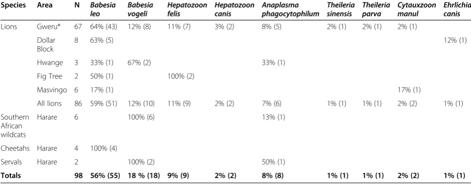
Overall, very high percentages of lions (78%; 67/86), Southern African wildcats (100%; 6/6), cheetahs (100%; 4/4) and servals (100%; 2/2) had evidence of infection with at least one of the TBP (Table 1). Each of the nine recognized TBP we found, as well asB. odocoilei-like andE. canis-like organisms, were present in the lions while only B. vogeli
(3), orC. manul(1). Mixed infections were seen in 22% of the PCR positive lions (15/67) [B. leoand H. canis(1); B. leoandE. canis(1);B. leoandE. canis-like (1);B. leo and
H. felis (4); B. leoand A. phagocytophilum (3); B. leo, H. canisand T. parva(1); B. leo, A. phagocytophilumandT. sinensis(1);B. leo,A. phagocytophilum andH. felis(1);B. vogeli and H. felis (1); or B. odocoilei-like andC. manul
(1)]. A mixed infection withB. vogeliandA. phagocytophi-lumwas seen in one of the servals and one of the Southern African wildcats, but there were no mixed infections in the cheetahs (Table 1).
Discussion
The wild felid species we studied in Zimbabwe were commonly infected with Babesia with 73% (63/86) of the lions and all the Southern African wildcats (6), chee-tahs (4) and servals (2) being PCR positive. All animals were apparently healthy at the time of blood collection, indicating a high prevalence of chronic subclinical infec-tions. The most common TBP we identified was B. leo
which we identified in 59% of the lions and all the chee-tahs (4) we studied. Babesia leo is a smallBabesia spp. which was first described in 2001 [14] and has been re-ported to be very common in lions (28%) in Southern Africa [5] but far less common in Tanzania (<1%) [7]. It has only been previously reported at low prevalence (0-3%) in cheetahs in Southern Africa. Phylogenetic analysis shows it forms a group with B. felis which infects do-mestic cats, B. microti which infects rodents and humans, andB. rodhainiwhich infects rodents. The vec-tor of B. leo is unknown, but infected lions have been found to be commonly infested with Amblyomma heb-raeum, Haemaphysalis leachi and Rhipicephalus simus
[14]. While we did not study the ectoparasites on the an-imals we tested, local experience is that captive lions are
more commonly infested with ticks while cheetahs have light tick burdens but more severe flea infestations. Tick and flea control products used by owners of wild felids in Zimbabwe include Deltatick Pour On® (Chemplex, Harare, Zimbabwe), a topical 5% deltamethrin product for use in cattle, and Frontline Spray® (Merial, South Africa), a fipro-nil based product reported to control fleas and ticks on dogs. To the best of our knowledge these products have not been evaluated for efficacy or safety in wild felids.
Although around 1% of stray domestic cats in Thailand have been found to be infected withB. vogeli[15] ours is the first report of the organism in Southern African wild-cats (100%; 6/6), lions (12%; 10/86) and servals (100%; 2/2).
Babesia vogelicommonly infects dogs in tropical and sub-tropical areas with prevalences of 4-60% [16]. The organ-ism is transmitted by R. sanguineus and infections are mostly asymptomatic with dogs becoming subclinical car-riers [17]. Rhipicephalus sanguineus was not found on Southern African domestic or wild felids in a recent sur-vey which was regarded as consistent with the near-strict host preference of the tick for domestic dogs [2]. Further studies are needed to determine the transmission mecha-nisms in wild felids.
We found no evidence of B. felisin the wild felids we studied which was also the case in a study of 2 lions and 5 cheetahs from Kenya [8]. Nevertheless, infections have been described in lions and cheetahs, both free-living and captive, from Southern Africa (4-28%) [5] as well as in lions from Zambia (number not given) [9] and Tanzania (number not given) [7]. One infected serval has also been reported [18]. A possible reason we failed to detect the or-ganism is because the BTF1 primer we used in our PCR had a mismatch with B. felis which might have reduced the sensitivity of the reaction and required relatively high copy numbers of B. felis to give positive reactions. Also,
Table 1 Survey sites, animals tested and results of PCR analyses for tick borne pathogens
Species Area N Babesia
leo Babesiavogeli Hepatozoonfelis Hepatozooncanis phagocytophilumAnaplasma Theileriasinensis Theileriaparva Cytauxzoonmanul Ehrlichiacanis
Lions Gweru* 67 64% (43) 12% (8) 11% (7) 3% (2) 8% (5) 2% (1) 2% (1) 2% (1)
Dollar Block
8 63% (5) 12% (1)
[image:3.595.56.538.102.291.2]Hwange 3 33% (1) 67% (2) 33% (1)
Fig Tree 2 50% (1) 100% (2)
Masvingo 6 17% (1) 17% (1)
All lions 86 59% (51) 12% (10) 11% (9) 2% (2) 7% (6) 1% (1) 1% (1) 2% (2) 1% (1)
Southern African wildcats
Harare 6 100% (6) 13% (1)
Cheetahs Harare 4 100% (4)
Servals Harare 2 100% (2) 50% (1)
Totals 98 56% (55) 18 % (18) 9% (9) 2% (2) 8% (8) 1% (1) 1% (1) 2% (2) 1% (1)
our detection system using a standard PCR and sequen-cing is not sensitive for mixed infections which are re-ported to be common using methods which are more accurate in their detection [5]. Investigations with more specificB. felisprimers are planned to resolve this issue.
We also failed to identify the other Babesia spp. that have been described in wild felids in Africa. In Tanzania and Kenya, lions have been found to be infected with an organism most similar toB. gibsoni[7] andB. canis(98% identity) [8], respectively, both of which have previously only been described in dogs.Babesia lengauis a recently described species that was found in a high percentage (29%) of cheetahs in South Africa [19]. A closely related species has been found in spotted hyenas (Crocuta cro-cuta) and a lion in Zambia [9]. The organism appears to result in asymptomatic infections in cheetahs, but in do-mestic cats [20] and sheep [21],B. lengauand a closely re-lated organism, have been reported to cause cerebral and hemolytic babesiosis, and hemolytic disease, respectively.
Undescribed Babesia spp. have been reported to be common in African wild felids [5] and we found an B. odocoilei-like organism in two lions. This large Babesia
has, to date, only been described in North America in a variety of wild ruminants [22]. Recently seropositive rab-bits have been reported [23] and the amplicons of PCRs for Babesia on two Florida panthers (Puma concolor coryi) had similar sequences [24]. Also, Ixodes ovatus
from dogs in Japan have been found to contain B. odo-coilei-like organisms (97.7% identical) [25]. It is of note that a large Babesia has been described in a domestic cat from Zimbabwe [26] and further studies are indi-cated to more clearly describe these novelBabesiaspp.
AlthoughCytauxzoonspp. have been described by mo-lecular studies in wild felids from US [27], Spain [28] and Brazil [29], ours is the first report of the organism in wild felids in Africa. Sequencing of the organism showed it was most similar to C. manul (99% identity) which is found in Pallas cats (Otocolobus manul) in Mongolia [30]. While a cytauxzoonosis-like disease has been described in a domestic cat in Zimbabwe [31], ex-perimental infection studies usingC. manul in domestic cats resulted in no clinical signs although there was a low parasitemia [32].
Our finding ofHepatozoonspp. in both lions and chee-tahs was not unexpected as they have been seen frequently in blood smears from African wild felids [3,4]. There is lit-tle data, however, on their molecular biology with organ-isms being identified by molecular means in 38% of Zambian lions [9], an unspecified number in lions from Tanzania [7], and 55% of Asiatic lions [33]. The species identified in the Asian and Tanzanian lions was H. felis
whileH. canis-like organism were detected in the Zambian lions. There is considerable diversity in Hepatozoon spp. in wild canids in the US [34] and further studies are
indicated to determine if this is also the case in African wild felids. Although generally regarded as being non-pathogenic in wild felids, there is a report ofHepatozoon
infection perhaps contributing to the death of spotted hy-enas (Crocuta crocuta) in Tanzania. The organism in-volved had a 18S RNA gene sequence identical to that of a
Hepatozoon sp. described previously in domestic cats in Spain which is distinct fromH. canis[35].
AlthoughTheileria-like parasites have been seen in blood films of cheetahs from Tanzania [4] and South Africa [36] there has been only a single report of the molecular identi-fication of organisms in African wild felids. With reverse line blots,Theileriaspp. were found in cheetahs and a lion from Kenya with sequencing indicating the parasites were closely related to those infecting sheep and giraffes [8]. We had positive PCR reactions for two lions with sequencing showing the organisms were most similar toT. parva, the agent of Corridor disease and East Coast Fever in domestic ruminants, and T. sinensisthat infects cattle and yaks in China [37]. Neither of these species has been described in felids and further genomic studies are required to more precisely characterize the organisms and determine their pathogenicity.
Six of the lions, one of the servals, and one of the Southern African wildcats we studied were infected with an organism most similar toA. phagocytophilum, a tick-borne rickettsia infecting a wide range of species includ-ing domestic and wild ruminants, horses, dogs, humans and domestic cats. It has been found in wild felids out-side of Africa, including up to 10% of lions in Italy and California [38,39], but none of 21 free ranging lions in Botswana were seropositive [6]. Although widely distrib-uted in the Northern Hemisphere where it’sIxodes spp. vectors are common, in Sub-Saharan Africa A. phagocy-tophilum has only been described in three sheep in Senegal [40] and a closely related organism was reported in a dog in South Africa [41]. A. phagocytophilum can cause severe disease in animals and people and in do-mestic cats has been reported to cause fever, lethargy and inappetence with non-specific signs including tach-ypnea, muscle and joint pain, neurological signs and lymphadenomegaly that respond to tetracycline therapy [42-46].
in Zambia [9] were negative, there is evidence ofE. canis
infections in domestic cats in Southern Africa [51]. Our finding of two lions infected withE. canisorE. canis-like organisms is consistent with the available data showing infections are uncommon in felids generally. This may be related to the fact that the vector of E. canis, the brown dog tickR. sanguineus, very seldom feeds on spe-cies other than the domestic dog [2].
Conclusions
In summary, our study has added to the knowledge of the tick-borne infections of wild felids in Southern Africa. Although further molecular studies are needed to more precisely determine the range of species involved, the available evidence is that infections with tick-borne agents are common, perhaps more particularly in captive animals where ticks can become more prevalent. Al-though most of the infected wild felids we tested ap-peared healthy and most likely had chronic infections, studies are needed to determine if there are signs associ-ated with acute infections and if the tick-borne organ-isms can potentiate other conditions. Studies are also needed on optimal ways of controlling ticks in captive wild felids to control infections.
Competing interests
The authors declare that they have no competing interests.
Authors’contributions
PK, CW and AL designed the study and wrote the manuscript. LM, KD, and JZ collected the samples, performed the experiments and reviewed the manuscript. All authors participated in the revision of the manuscript, read, and approved the submitted version.
Acknowledgments
This project was supported by the Ross University School of Veterinary Medicine, the Animal and Wildlife Area Research and Rehabilitation (AWARE) Trust of Zimbabwe, the National Natural Science Foundation of China (NO: 31272575, 31472225), and the Priority Academic Program Development of Jiangsu Higher Education Institutions.
Author details
1Ross University School of Veterinary Medicine, Basseterre, St. Kitts and Nevis. 2AWARE Trust, Harare, Zimbabwe.3Jiangsu Co-innovation Center for
Prevention and Control of Important Animal Infectious Diseases and Zoonoses, Yangzhou University College of Animal Science and Technology, Yangzhou, Jiangsu 225009, China.4Department of Microbiology &
Immunology, Midwestern University, Glendale, AZ, USA.
Received: 27 August 2014 Accepted: 1 November 2014
References
1. Driscoll CA, Menotti-Raymond M, Roca AL, Hupe K, Johnson WE, Geffen E, Harley EH, Delibes M, Pontier D, Kitchener AC, Yamaguchi N, O’brien SJ, Macdonald DW:The Near Eastern origin of cat domestication.Science 2007,317:519–523.
2. Horak IG, Heyne H, Donkin EF:Parasites of domestic and wild animals in South Africa. XLVIII. Ticks (Acari: Ixodidae) infesting domestic cats and wild felids in Southern Africa.Onderstepoort J Vet Res2010,77:E1–E7. 3. McCully RM, Basson PA, Bigalke RD, De Vos V, Young E:Observations on
naturally acquired hepatozoonosis of wild carnivores and dogs in the Republic of South Africa.Onderstepoort J Vet Res1975,42:117–133.
4. Averbeck GA, Bjork KE, Packer C, Herbst L:Prevalence of hematozoans in lions (Panthera leo) and cheetah (Acinonyx jubatus) in Serengeti National Park and Ngorongoro Crater, Tanzania.J Wildl Dis1990,26:392–394. 5. Bosman AM, Venter EH, Penzhorn BL:Occurrence ofBabesia felisand
Babesia leoin various wild felid species and domestic cats in Southern Africa, based on reverse line blot analysis.Vet Parasitol2007,144:33–38. 6. Ramsauer S, Bay G, Meli M, Hofmann-Lehmann R, Lutz H:Seroprevalence
of selected infectious agents in a free-ranging, low-density lion population in the Central Kalahari Game Reserves in Botswana.
Clin Vaccine Immunol2007,14:808–810.
7. Munson L, Terio KA, Kock R, Mlengeya T, Roelke ME, Dubovi E, Summers B, Sinclair AR, Packer C:Climate extremes promote fatal co-infections during canine distemper epidemics in African lions.PLoS One2008,3:e2545. 8. Githaka N, Konnai S, Kariuki E, Kanduma E, Murata S, Ohashi K:Molecular
detection and characterization of potentially newBabesiaandTheileria species/variants in wild felids from Kenya.Acta Trop2012,124:71–78. 9. Williams BM, Berentsen A, Shock BC, Teixiera M, Dunbar MR, Becker MS, Yabsley MJ:Prevalence and diversity ofBabesia,Hepatozoon,Ehrlichia, andBartonellain wild and domestic carnivores from Zambia, Africa.
Parasitol Res2014,113:911–918.
10. Wei L, Kelly P, Zhang J, Yang Y, Zheng X, Tao J, Zhang Z, Wang C:Use of a universal hydroxymethylbilane synthase (HMBS)-based PCR as an endogenous internal control and to enable typing of mammalian DNAs.
Appl Microbiol Biotechnol2014,98:5579–5587.
11. Jefferies R, Ryan UM, Irwin PJ:PCR-RFLP for the detection and differentiation of the canine piroplasm species and its use with filter paper-based technologies.Vet Parasitol2007,144:20–27.
12. Dawson JE, Stallknecht DE, Howerth EW, Warner C, Biggie K, Davidson WR, Lockhart JM, Nettles VF, Olson JG, Childs JE:Susceptibility of white-tailed deer (Odocoileus virginianus) to infection withEhrlichia chaffeensis, the etiologic agent of human ehrlichiosis.J Clin Microbiol1994,32:2725–2728. 13. Petti CA:Detection and identification of microorganisms by gene
amplification and sequencing.Clin Infect Dis2007,44:1108–1114. 14. Penzhorn BL, Kjemtrup AM, López-Rebollar LM, Conrad PA:Babesia leon.
sp. from lions in the Kruger National Park, South Africa, and its relation to other small piroplasms.J Parasitol2001,87:681–685.
15. Simking P, Wongnakphet S, Stich RW, Jittapalapong S:Detection ofBabesia vogeliin stray cats of metropolitan Bangkok, Thailand.Vet Parasitol2010,
173:70–75.
16. Taboada J, Lobetti R:Babesiosis.InInfectious Diseases of the Dog and Cat. Edited by Greene CE. Missouri: Saunders Elsevier; 2006:722–736. 17. Kelly PJ, Xu C, Lucas H, Loftis A, Abete J, Zeoli F, Stevens A, Jaegersen K,
Ackerson K, Gessner A, Kaltenboeck B, Wang C:Ehrlichiosis, babesiosis, anaplasmosis and hepatozoonosis in dogs from St Kitts, West Indies.
PLoS One2013,8:e53450.
18. Penzhorn BL:Babesiosis of wild carnivores and ungulates.Vet Parasitol 2006,138:11–21.
19. Bosman AM, Oosthuizen MC, Peirce MA, Venter EH, Penzhorn BL:Babesia lengau sp. nov., a novelBabesiaspecies in cheetah (Acinonyx jubatus, Schreber, 1775) populations in South Africa.J Clin Microbiol2010,48:2703–2708.
20. Bosman AM, Oosthuizen MC, Venter EH, Steyl JC, Gous TA, Penzhorn BL: Babesia lengauassociated with cerebral and haemolytic babesiosis in two domestic cats.Parasit Vectors2013,6:128.
21. Giadinis ND, Chochlakis D, Kritsepi-Konstantinou M, Makridaki E, Tselentis Y, Kostopoulou D, Karatzias H, Psaroulaki A:Hemolytic disease in sheep attributed to aBabesia lengau-like organism.Vet Rec2012,170:155. 22. Pattullo KM, Wobeser G, Lockerbie BP, Burgess HJ:Babesia odocoilei
infection in a Saskatchewan elk (Cervus elaphus canadensis) herd.
J Vet Diagn Invest2013,25:535–540.
23. Fritzen C, Mosites E, Applegate RD, Telford SR 3rd, Huang J, Yabsley MJ, Carpenter LR, Dunn JR, Moncayo AC:Environmental investigation following the first human case of babesiosis in Tennessee.J Parasitol 2014,100:106–109.
24. Yabsley MJ, Murphy SM, Cunningham MW:Molecular detection and characterization ofCytauxzoon felisand aBabesiaspecies in cougars from Florida.J Wildl Dis2006,42:366–374.
25. Inokuma H, Beppu T, Okuda M, Shimada Y, Sakata Y:Epidemiological survey ofAnaplasma platysandEhrlichia canisusing ticks collected from dogs in Japan.Vet Parasitol2003,115:343–348.
27. Shock BC, Moncayo A, Cohen S, Mitchell EA, Williamson PC, Lopez G, Garrison LE, Yabsley MJ:Diversity of piroplasms detected in blood-fed and questing ticks from several states in United States.Ticks Tick Borne Dis2014,5:373–380.
28. Meli ML, Cattori V, Martínez F, López G, Vargas A, Simón MA, Zorrilla I, Muñoz A, Palomares F, López-Bao JV, Pastor J, Tandon R, Willi B, Hofmann-Lehmann R, Lutz H:Feline leukemia virus and other pathogens as important threats to the survival of the critically endangered Iberian lynx (Lynx pardinus).PLoS One2009,4:e4744.
29. André MR, Adania CH, Machado RZ, Allegretti SM, Felippe PA, Silva KF, Nakaghi AC, Dagnone AS:Molecular detection ofCytauxzoonspp. in asymptomatic Brazilian wild captive felids.J Wildl Dis2009,45:234–237. 30. Reichard MV, Van Den Bussche RA, Meinkoth JH, Hoover JP, Kocan AA:A
new species ofCytauxzoonfrom Pallas’cats caught in Mongolia and comments on the systematics and taxonomy of piroplasmids.J Parasitol 2005,91:420–426.
31. Foggin CA:A cytauxzoonosis-like disease in a cat in Zimbabwe.
Zimbabwe Vet J1982,13:28–30.
32. Joyner PH, Reichard MV, Meinkoth JH, Milne VE, Confer AW, Kocan AA, Hoover JP:Experimental infection of domestic cats (Felis domesticus) with Cytauxzoon manulfrom Pallas’cats (Otocolobus manul).Vet Parasitol2007,
146:302–306.
33. Pawar RM, Poornachandar A, Srinivas P, Rao KR, Lakshmikantan U, Shivaji S:
Molecular characterization ofHepatozoonspp. infection in endangered Indian wild felids and canids.Vet Parasitol2012,186:475–479. 34. Starkey LA, Panciera RJ, Paras K, Allen KE, Reiskind MH, Reichard MV,
Johnson EM, Little SE:Genetic diversity ofHepatozoonspp. in coyotes from the south-central United States.J Parasitol2013,99:375–378. 35. East ML, Wibbelt G, Lieckfeldt D, Ludwig A, Goller K, Wilhelm K, Schares G,
Thierer D, Hofer H:AHepatozoonspecies genetically distinct fromH. canisinfecting spotted hyenas in the Serengeti ecosystem, Tanzania.
J Wildl Dis2008,44:45–52.
36. Zinkl JG, McDonald SE, Kier AB, Cippa SJ, Small PJ:Cytauxzoon-like organisms in erythrocytes of two cheetahs.J Am Vet Med Assoc1981,
179:1261–1262.
37. Liu J, Guan G, Liu Z, Liu A, Ma M, Bai Q, Yin H, Luo J:Additional data for a newTheileriasp. from China based on the sequences of ribosomal RNA internal transcribed spacers.Exp Parasitol2013,133:217–221.
38. Torina A, Naranjo V, Pennisi MG, Patania T, Vitale F, Laricchiuta P, Alongi A, Scimeca S, Kocan KM, de la Fuente J:Serologic and molecular
characterization of tickborne pathogens in lions (Panthera leo) from the Fasano Safari Park, Italy.J Zoo Wildl Med2007,38:591–593.
39. Girard YA, Swift P, Chomel BB, Kasten RW, Fleer K, Foley JE, Torres SG, Johnson CK:Zoonotic vector-borne bacterial pathogens in California mountain lions (Puma concolor), 1987-2010.Vector Borne Zoonotic Dis 2012,12:913–921.
40. Djiba ML, Mediannikov O, Mbengue M, Thiongane Y, Molez JF, Seck MT, Fenollar F, Raoult D, Ndiaye M:Survey ofAnaplasmataceaebacteria in sheep from Senegal.Trop Anim Health Prod2013,45:1557–1561. 41. Inokuma H, Oyamada M, Kelly PJ, Jacobson LA, Fournier PE, Itamoto K,
Okuda M, Brouqui P:Molecular detection of a newAnaplasmaspecies closely related toAnaplasma phagocytophilumin canine blood from South Africa.J Clin Microbiol2005,43:2934–2937.
42. Adaszek L, Górna M, Skrzypczak M, Buczek K, Balicki I, Winiarczyk S:Three clinical cases ofAnaplasma phagocytophiluminfection in cats in Poland.
J Feline Med Surg2013,15:333–337.
43. Heikkilä HM, Bondarenko A, Mihalkov A, Pfister K, Spillmann T:Anaplasma phagocytophiluminfection in a domestic cat in Finland: case report.
Acta Vet Scand2010,52:62.
44. Palomar AM, García-Álvarez L, Santibáñez S, Portillo A, Oteo JA:Detection of tick-borne‘Candidatus Neoehrlichia mikurensis’andAnaplasma phagocytophilumin Spain in 2013.Parasit Vectors2014,7:57. 45. Breitschwerdt EB, Hegarty BC, Qurollo BA, Saito TB, Maggi RG, Blanton LS,
Bouyer DH:Intravascular persistence ofAnaplasma platys,Ehrlichia chaffeensis, andEhrlichia ewingiiDNA in the blood of a dog and two family members.Parasit Vectors2014,1(7):298.
46. Jahfari S, Coipan EC, Fonville M, van Leeuwen AD, Hengeveld P, Heylen D, Heyman P, van Maanen C, Butler CM, Földvári G, Szekeres S, van Duijvendijk G, Tack W, Rijks JM, van der Giessen J, Takken W, van Wieren SE, Takumi K, Sprong H:Circulation of four Anaplasma phagocytophilum ecotypes in Europe.Parasit Vectors2014,7:365.
47. Braga Mdo S, André MR, Freschi CR, Teixeira MC, Machado RZ:Molecular and serological detection ofEhrlichiaspp. in cats on São Luís Island, Maranhão, Brazil.Rev Bras Parasitol Vet2012,21:37–41.
48. André MR, Dumler JS, Scorpio DG, Teixeira RH, Allegretti SM, Machado RZ:
Molecular detection of tick-borne bacterial agents in Brazilian and exotic captive carnivores.Ticks Tick Borne Dis2012,3:247–253.
49. Filoni C, Catão-Dias JL, Cattori V, Willi B, Meli ML, Corrêa SH, Marques MC, Adania CH, Silva JC, Marvulo MF, Ferreira Neto JS, Durigon EL, de Carvalho VM, Coutinho SD, Lutz H, Hofmann-Lehmann R:Surveillance using serological and molecular methods for the detection of infectious agents in captive Brazilian neotropic and exotic felids.J Vet Diagn Invest2012,
24:166–173.
50. Tateno M, Nishio T, Sakuma M, Nakanishi N, Izawa M, Asari Y, Okamura M, Maruyama S, Miyama TS, Setoguchi A, Endo Y:Molecular epidemiologic survey ofBartonella,Ehrlichia, andAnaplasmainfections in Japanese Iriomote and Tsushima leopard cats.J Wildl Dis2013,49:646–652. 51. Matthewman LA, Kelly PJ, Wray K, Bryson NR, Rycroft AN, Raoult D, Mahan
SM:Antibodies in cat sera from southern Africa react with antigens of Ehrlichia canis.Vet Rec1996,138:364–365.
doi:10.1186/s13071-014-0514-6
Cite this article as:Kellyet al.:Molecular detection of tick-borne pathogens in captive wild felids, Zimbabwe.Parasites & Vectors
20147:514.
Submit your next manuscript to BioMed Central and take full advantage of:
• Convenient online submission
• Thorough peer review
• No space constraints or color figure charges
• Immediate publication on acceptance
• Inclusion in PubMed, CAS, Scopus and Google Scholar
• Research which is freely available for redistribution