R E S E A R C H
Open Access
Development of a novel IGRA assay to test
T cell responsiveness to HBV antigens in
whole blood of chronic Hepatitis B patients
Werner Dammermann
1*, Frank Bentzien
2, Eva-Maria Stiel
1, Claudia Kühne
1, Sebastian Ullrich
3,
Julian Schulze zur Wiesch
1,4,5and Stefan Lüth
1,4Abstract
Background:Interferon gamma release assays (IGRA) have been developed to support easy and fast diagnosis of diseases like tuberculosis, and CMV in transplant patients. IGRAs focus on cellular immunity especially memory T cells and thus also allow rapid screening prior to complex flow cytometric testing. Here, we describe a novel, sensitive whole blood based cytokine release assay capable of assessing T cell responsiveness to HBV antigens in Hepatitis B patients and assessing hepatitis B vaccination status in healthy individuals.
Methods:Seventy two chronic Hepatitis B patients (CHB), 8 acute hepatitis B patients (AHB) and 80 healthy controls (HC) were tested by ELISA for IFNγ- and IL2-secretion in whole blood after challenge with synthetic peptide libraries of hepatitis B core antigen (HBcAg) or hepatitis B surface antigen (HBsAg).
Results:The developed IGRA test reliably differentiated between Hepatitis B patients, vaccinees and unvaccinated healthy controls. Treatment naïve and treated CHB patients showed a weaker IFNγresponse to HBcAg (16 ± 5 and 35 ± 28 pg/ml, respectively) compared to the AHB group (82 ± 39 pg/ml), whereas HC remained unresponsive (6 ± 1 pg/ml). IL2 levels after HBcAg challenge were also higher in the AHB group compared to naive and treated CHB as well as HC (47 ± 21 vs. 12 ± 3, 15 ± 10 and 12 ± 9 pg/ml, respectively). HBsAg stimulation led to increased IFNγ and IL2 levels in the AHB group (33 ± 12 and 22 ± 12 pg/ml) and even higher levels in HC due to a high hepatitis B vaccination rate (41 ± 10 and 167 ± 58 pg/ml). Naive and treated CHB patients developed no or only weaker IFNγor IL2 responses to HBsAg (5 ± 2 and 12 ± 7 pg/ml, for naive CHB, 12 ± 10 and 18 ± 15 pg/ml, for treated CHB). For HC, IL2 release after HBsAg stimulation depicted hepatitis B vaccination status with a diagnostic sensitivity and specificity of 85 % and 90 %.
Conclusion:Our novel whole blood based cytokine release assay constitutes an easy and robust tool for screening HBV specific cellular immunity as alternative to flow cytometry or ELISPOT assays.
Keywords:Chronic hepatitis B, HBV, Interferon gamma release assay, Cytokine release assay
Introduction
More than 240 million individuals are infected with chronic hepatitis B worldwide and are at risk to develop severe liver disease, liver cirrhosis or hepatocellular car-cinoma [1]. A prophylactic hepatitis B vaccine has been available for over 3 decades and ensures long-term pro-tection from infection [2]. However, development of a
therapeutic vaccine for chronic hepatitis B patients has not met with success so far [3]. It is thought that one reason for lacking efficacy of such a therapeutic vaccine is that HBV-specific T lymphocytes show functional de-fects and exhaustion and lack proliferation in chronic hepatitis B [4, 5].
Effective prophylactic and therapeutic vaccines depend on strong humoral and cellular immune responses to in-fectious antigens as part of the vaccine formulation. Whereas in vaccine trials the humoral immune response is analyzed by standard serological assays,e.g. ELISA or * Correspondence:w.dammermann@uke.de
1
Department of Medicine, University Medical Center Hamburg-Eppendorf, Martinistrasse 52, Hamburg 20246, Germany
Full list of author information is available at the end of the article
western blot [6,7], cell-mediated immunity (CMI) is usu-ally assessed by established and technicusu-ally demanding analytical methods like flow cytometry, intracellular cytokine staining (ICS), 3H-thymidine proliferation as-says or ELISPOT [5]. Therefore, whole blood based cyto-kine release assays or interferon gamma release assays (IGRA), respectively, may constitute a robust, easy and cost effective alternative as screening tools in studies of HBV Immunology and HBV vaccination studies.
Here, we describe the establishment of a whole blood based cytokine release assay capable of assessing T cell responsiveness to HBV peptide pools in chronic hepa-titis B patients and hepahepa-titis B vaccination status in healthy individuals.
Methods Patient selection
Hepatitis B patients treated at the hepatitis outpatient department of the University Medical Center Hamburg-Eppendorf were enrolled in the study for which all pa-tients gave written consent and which was approved by the Ethics Committee of the Hamburg Chamber of Phy-sicians (PV3941). Patients were stratified according to their clinical course into 3 groups (Table 1) with either NUC treatment naïve chronic Hepatitis B (CHB, NUC treatment naive, n = 40), NUC treated chronic Hepatitis B (CHB, NUC treated, n = 32) or acute hepatitis B (AHB, n = 8). NUC are nucleoside/nucleotide analogues (NUC, tenofovir and/or entecavir) used for HBV specific antiviral treatment. Healthy donors of the blood transfu-sion service at the University Medical Center
Hamburg-Eppendorf were anonymously enrolled in the study as healthy controls (HC, n = 80). Additional clinical data for all patients and HC was provided (Table 1).
Reagents
[image:2.595.59.539.488.709.2]The recall peptide pool CEFT was purchased from JPT, Germany (#PM-CEFT) and solved in sterile dimethyl sulfoxide (DMSO; #RO/A9941/000100, Th. Geyer, Germany) 25 mg/ml followed by storage at −20 °C. CEFT peptide pool consisted of antigenic peptides from human Cytomegalovirus (HHV-5; CMV), Epstein-Barr virus (HHV-4; EBV), Influenza A andClostridium tetani. This positive control pool contained 27 peptides selected from defined HLA class I and II-restricted T-cell epi-topes. Considering the high vaccination frequency against Influenza and C.tetani and the high prevalence of CMV and EBV in the general population in Germany recall antigen responses were expected for all patient samples.Staphylococcus aureusenterotoxin B superanti-gen (SEB) was obtained from Sigma Aldrich GmbH, Germany (#S4881) and stored 1 mg/ml in sterile, endotoxin-free H2O and −20 °C. Synthetic HBV peptide libraries of HBcAg and HBsAg were purchased from JPT, Germany (#PM-HBV-CP and #PM-HBV-lEP, re-spectively) and solved in sterile dimethyl sulfoxide (DMSO; #RO/A9941/000100, Th. Geyer, Germany) 50 mg/ml followed by storage at −20 °C. Synthetic HBcAg represented a mix of 44 peptides (15 amino acid length each, 11 aa overlap, peptide scan 15/11) compris-ing the whole amino acid sequence of HBcAg, the 21.5 kDa capsid protein of HBV (genotype A2 subtype
Table 1Characteristics of all subjects included in the study
Characteristic
HCb AHBb CHBb, NUC treatment naive CHBb, NUC treated
n 80 8 40 32
Male 69 (86.3 %) 4 (50 %) 20 (50 %) 23 (71.9 %)
Female 11 (13.8 %) 4 (50 %) 20 (50 %) 9 (28.1 %)
Age (yr)a 40.6 ± 12.0 45.9 ± 15 43.4 ± 14.9 47.3 ± 11.4
Male 40.2 ± 12.1 49.7 ± 11.8 41.2 ± 11.6 50.1 ± 11.2
Female 43.7 ± 11.5 42.2 ± 18.7 45.7 ± 17.6 40.0 ± 8.6
ALT (U/l)a n.d.c 1046.0 ± 1035.0 37.7 ± 60.2 42.9 ± 39.3
AST (U/l)a n.d.c 610.6 ± 702.5 26.6 ± 27.9 29.5 ± 27.7
HBV-DNA (IU/ml)a n.d.c 2.88*106± 7.7*106 4.2*107± 1.8*108 1.6*107± 8.8*107
HBsAg (IU/ml)a n.d.c 9153.4 ± 10,378.9 12,588.6 ± 20,958.8 4783.1 ± 6326.3
HBeAg Negative n.d.c 4 (50 %) 34 (85 %) 24 (77.4 %)
Positive n.d.c 4 (50 %) 6 (15 %) 7 (22.6 %)
Anti-HBs Negative 41 (51.2 %) 8 (100 %) 36 (90 %) 32 (100 %)
Positive 33 (41.3 %) 0 (0 %) 4 (10 %) 0 (0 %)
a
The data are shown as means ± standard deviations
b
HC = healthy controls, AHB = acute hepatitis B, CHB = chronic hepatitis B
c
adw2,UniProt: P0C693). Synthetic HBsAg represented a mix of 98 peptides (15 amino acid length each, 11 aa overlap, peptide scan 15/11) comprising the whole amino acid sequence of HBsAg, the 43.7 kDa surface protein of HBV (genotype A2 subtype adw2, UniProt: P17101).
Cytokine release assay in whole bloodex vivo
Venous blood was collected from hepatitis B patients and HC into sterile 7.5 ml Lithium heparin Monovettes (Sarstedt, Germany). 1 ml of whole blood was then dis-pensed into sterile, pyrogen free 2 ml tubes with screw caps (Sarstedt, Germany) within 4 h of collection pre-loaded with sterile 0.9 % (w/v) NaCl solution (negative control), SEB (1st positive control), recall CEFT peptide pools (2ndpositive control) or HBV peptide pools. In all tubes, glucose (2 mg/mI final concentration; pre-diluted in sterile 0.9 % (w/v) NaCl solution; #HN06.1, Carl Roth, Germany) was used to further enhance cytokine secre-tion, which had been tested before using various anti-gens (Additional file 1: Online resource 1).
Thus, whole blood was stimulated with a total volume of 120 μl/ tube of NaCl & glucose (negative control), 120μl/ tube SEB & glucose (1μg/ml final concentration after dilution out of stock, 1st positive control), 120 μl/ tube recall antigen CEFT & glucose (10μg/ml final con-centration after dilution out of stock, 2nd positive con-trol) or 120μl/ tube HBV peptide pools (50μg/ml final concentration after dilution out of stock). All HBV pep-tide pools were titrated for maximum cytokine responses in advance. SEB served as a 1st positive control due to its superantigenic properties by cross-linking MHC mol-ecules with T-cell receptors, which proved the general viability of all samples. CEFT was introduced as a 2nd positive control to prove the functionality of antigen presenting cells in all samples.
The tubes were closed and incubated at 37 °C for 24 h. Thereafter, plasma supernatants were aspirated, pooled, stabilized with 0.045 % (w/v) NaN3 and stored at −20 °C until assayed for cytokines within the next 7 days. A 5 % (v/v) CO2atmosphere was proven to be unnecessary (data not shown) and yielded results com-parable to stimulation in the presence of CO2.
IFNγand IL2 ELISAs
Detection of total / overall amounts of IFNγ and IL2 in human plasma was conducted using ELISA MAX De-luxe sets from Biolegend, Germany: IFNγ(#430106) and IL2 (#431806). The manufacturer’s Avidin-horseradish peroxidase conjugate was replaced by PolyHRP80 strep-tavidine conjugate (#SP80C, SDT Reagents, Germany) in order to achieve a tenfold lower limit of detection. Lower limit of detection (Background + 3x S.D.) was generally at 2– 5 pg/ml for IFNγ and IL2, respectively.
Initial dilution of control and test samples was per-formed described as follows: negative control (NaCl) 1/5, 1st positive control (SEB) 1/2500 for IFNγ and 1/500 for IL2, 2ndpositive control (CEFT) 1/50 for IFNγ and 1/25 for IL2, test samples (HBcAg and HBsAg) 1/5. If a test sample’s absorbance value fell outside the max-imum standard curve range, these samples were sub-sequently retested with a tenfold higher dilution, e.g. 1/5→1/50, 1/500→1/5000, 1/2500→1/25000. A seven-point standard curve from 1 – 64 pg/ml IFNγ or IL2 was used for quantitation. Standards, controls and test samples were measured in duplicate. The samples were analyzed using Magellan software (version 6.5) equipped on a Tecan M200 plate reader.
Data analysis Software
The ELISA data was analyzed using SigmaPlot software (Systat Software Inc., version number 12.2) and Graph-Pad Prism software (Graphpad Software Inc., version number 6.04).
Statistical analysis
Descriptive statistics, ANOVA including Tamhane T2 post hoc tests and prior normality tests, unpaired t-test and receiver operating characteristic (ROC) analysis were performed using SPSS Statistics software (IBM, version number 22), SigmaPlot software (Systat software Inc., version number 12.2) and GraphPad Prism software (Graphpad Software Inc., version number 6.04). Negative control values were deducted from the peptide induced responses prior to statistical analysis. Intra-assay and inter-assay variability could not be assessed due to lim-ited amounts of whole blood per patient.
Results
IFNγresponses to HBcAg in whole blood of CHB patients are reduced compared to AHB patients
Two major HBV proteins, HBcAg and HBsAg, were tested in an HBV specific cytokine release assay with HC and 3 different hepatitis B patient groups: acute, NUC treatment naïve and NUC treated chronic hepatitis B. Stimulation of whole blood with sterile 0.9 % (w/v) NaCl solution (negative control), SEB (1st positive control) and CEFT (2nd positive control) confirmed the viability of all collected samples (Fig. 1. and Table 2).
33 ± 12 pg/ml; p = 0.99), but weaker responses in naïve as well as treated CHB patients (5 ± 2 and 12 ± 10 pg/ml; naïve CHB vs. HC p = 0.003 and naïve CHB vs. AHB p = 0.46; treated CHB vs. HC p = 0.28 and treated CHB vs. AHB p = 0.9).
IL2 responses were generally comparable to IFNγ re-sponses regarding total cytokine amounts (Fig. 2b). HBcAg induced IL2 synthesis in AHB (47 ± 21 pg/ml), but not naïve and treated CHB patients as well as HC (12 ± 3, 15 ± 10 and 12 ± 9 pg/ml; p = 0.78, p = 0.89 and p = 0.82). HBsAg elicited significantly higher IL2
responses in HC (167 ± 58 pg/ml) compared to AHB, naïve and treated CHB patients (22 ± 12, 12 ± 7 and 18 ± 15 pg/m; p = 0.05, p = 0.03 and p = 0.04).
Taken together, cytokine release assays using HBcAg stimulation in whole blood are able to depict the (hypo-) responsiveness of HBV-specific T cells in CHB patients.
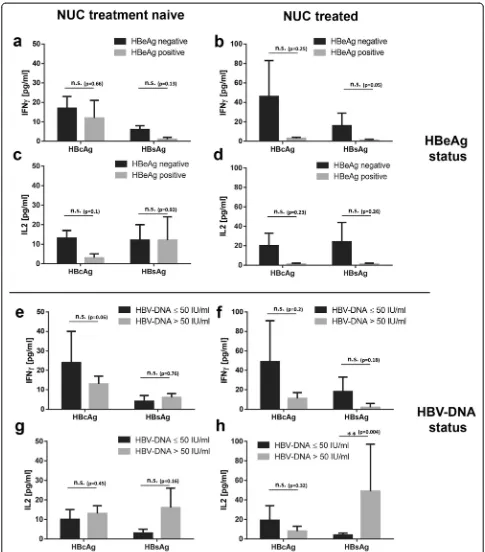
HBeAg + CHB patients show weaker cytokine responses to HBcAg and HBsAg than HBeAg- CHB patients
All NUC treatment naïve and NUC treated 72 CHB pa-tients were stratified according to HBeAg status as well as HBV-DNA status and subsequently tested for IFNγ -and IL2-secretion in whole blood after challenge with synthetic HBcAg- and HBsAg-specific peptides by ELISA. Stimulation with HBcAg-specific peptides led to higher IFNγconcentrations in plasma of treated, but not naive HBeAg- CHB patients compared to HBeAg + pa-tients (Fig. 3a - b; 17 ± 6 vs. 12 ± 9 pg/ml for naïve CHB; 46 ± 37 vs. 3 ± 1 pg/ml for treated CHB, respectively). Stimulation with HBsAg-specific peptides was also more prominent in treated, but not naive HBeAg- CHB pa-tients compared to HBeAg + papa-tients (6 ± 2 vs. 1 ± 1 pg/ ml for naïve CHB and 16 ± 13 vs. 1 ± 1 pg/ml for treated CHB, respectively). IL2 responses yielded a comparable pattern with stronger cytokine production in plasma of HBeAg- CHB patients compared to HBeAg + patients: treated HBeAg- CHB patients gave higher IL2 levels than HBeAg + CHB patients (Fig. 3d; 20 ± 13 vs. 1 ± 1 pg/ml for HBcAg and 24 ± 20 vs. 1 ± 1 pg/ml for HBsAg, respectively), whereas effects of HBcAg and HBsAg stimulation on IL2 levels in plasma of naïve CHB patients were negligible (Fig. 3c).
Regarding HBV-DNA status we looked for effects of viral load on the cytokine responses directed against HBcAg and HBsAg, but unlike HBeAg status viral load did not seem to influence cytokine production in a simi-lar fashion: low HBV-DNA titers≤50 IU/ml did not al-ways coincide with higher cytokine levels after HBcAg or HBsAg stimulation in treatment naïve and NUC treated CHB patients (Fig. 3e–h).
In conclusion, HBeAg + treatment naïve and NUC treated CHB patients showed the weakest cytokine Fig. 1HC and hepatitis B patients show comparable and distinct
[image:4.595.57.290.87.310.2]IFNγand IL2 responses in whole blood towards control stimulations with SEB and CEFT. n = 80 HC, n = 8 AHB patients, n = 40 NUC treatment naive CHB patients and n = 32 NUC treated patients, for each tested control antigen. Every HC or patient was tested against buffer control, SEB and CEFT. (a) IFNγ, (b) IL2. Negative control values were deducted from the antigen induced responses. Lower limit of detection (Background + 3x S.D.) was at 2–5 pg/ml for IFNγ and IL2. All values are given as mean concentration pg/ml ± S.E.M. ANOVA and Tamhane T2 post hoc tests, following symbol pinpoints significant differences: *. One symbol equals 0.05, two symbols 0.01, three symbols 0.001
Table 2Positive and negative controls
Assay NaCl (negative control)a SEB (1. positive control)a CEFT (2. positive control)a
IFNγ[pg/ml] IL2 [pg/ml] IFNγ[pg/ml] IL2 [pg/ml] IFNγ[pg/ml] IL2 [pg/ml] N
Group
HCb 8 ± 2 2 ± 1 66,537 ± 8485 22,762 ± 1399 1681 ± 361 125 ± 36 80
AHBb 15 ± 5 3 ± 1 45,083 ± 14,487 10,737 ± 2148 458 ± 278 22 ± 11 8
CHBb, NUC treatment naive 41 ± 22 1 ± 1 65,734 ± 8837 18,439 ± 1244 1490 ± 669 220 ± 173 40
CHBb, NUC treated 17 ± 6 2 ± 1 107,200 ± 30,799 20,838 ± 1357 3186 ± 1383 318 ± 293 32
a
The data are shown as means ± standard error of mean (S.E.M.). SEB and CEFT values are given after deduction of background values (NaCl, negative control)
b
[image:4.595.57.539.619.716.2]responses to HBV antigen-specific peptide challenge of all hepatitis B patient groups.
Treatment naïve CHB patients show comparable cytokine responses to HBcAg and HBsAg compared to NUC treated CHB patients in whole blood
72 CHB patients were stratified according to treatment status with nucleoside/nucleotide analogues (NUC, teno-fovir and/or entecavir) and subsequently tested for IFNγ- and IL2-secretion in whole blood after challenge with synthetic HBcAg and HBsAg peptides by ELISA.
HBcAg and HBsAg stimulation did not result in sig-nificant differences regarding IFNγ concentrations in plasma of treatment naïve patients compared to NUC treated patients (Fig. 4a; 16 ± 5 vs. 35 ± 28 pg/ml for HBcAg and 5 ± 2 vs. 12 ± 10 pg/ml for HBsAg, respect-ively). Likewise, IL2 responses to HBcAg and HBsAg in whole blood yielded no significant differences in view to treatment status (Fig. 4b; 12 ± 3 vs. 15 ± 1 pg/ml for HBcAg and 12 ± 10 vs. 18 ± 15 pg/ml for HBsAg, respectively).
NUC treatment had no significant positive effect on cytokine release in whole blood after stimulation with HBcAg and HBsAg-specifc peptidesex vivo.
IL2 responses to HBsAg in HC correlate with hepatitis B vaccination status
HBsAg constitutes the single main antigen in hepatitis B vaccine formulations. Thus, 74 HC with known hepatitis B vaccination status were also analyzed regarding their HBsAg-dependent IFNγand IL2 release. 33/74 HC pos-sessed a positive anti-HBs antibody titer and 41/74 were anti-HBs negative. Whereas IFNγ responses were negli-gible, IL2 values reached marked heights if the tested in-dividual had been successfully vaccinated (Fig. 5b), but these IL2 levels did not correlate with the corresponding anti-HBs antibody titer (Fig. 6). To determine the cut-off value of our newly developed assay, we performed a ROC analysis of the IL2 readings (Fig. 5a). This HBV specific cytokine release assay with HBsAg-specific pep-tides using IL2 release as the primary readout (cut-off = 11 pg/ml IL2) reached 85 % diagnostic sensitivity and 90 % diagnostic specificity. The corresponding AUC value was at 0.92. Using the cut-off defined by the ROC analysis, as shown in Fig. 4, 28 (of 33) vaccinated HC showed positive scores, while 4 (of 41) not vaccinated HC slightly exceeded the cut-off value (Fig. 5b).
Discussion
Cytokine release assays using whole blood depict (hypo-) responsiveness of HBV-specific T cells in CHB patients
[image:5.595.57.291.88.316.2]The group of Boniet al.delivered key data regarding the HBV specific cellular immune status under treatment
Table 3 HBcAg and HBsAg stimulations
Assay HBcAga HBsAga
IFNγ[pg/ml] IL2 [pg/ml] IFNγ[pg/ml] IL2 [pg/ml] N
Group
HCb 6 ± 1 12 ± 9 41 ± 10 167 ± 58 80
AHBb 82 ± 39 47 ± 21 33 ± 12 22 ± 12 8
CHBb, NUC treatment naive 16 ± 5 12 ± 3 5 ± 2 12 ± 7 40
CHBb, NUC treated 35 ± 28 15 ± 10 12 ± 10 18 ± 15 32
a
The data are shown as means ± standard error of mean (S.E.M.). HBcAg and HBsAg values are given after deduction of background values (NaCl, negative control)
b
HC = healthy controls, AHB = acute hepatitis B, CHB = chronic hepatitis B
[image:5.595.58.539.619.716.2]with first (lamivudine) or second generation drugs (teno-fovir and entecavir) in consecutive and longitudinal clin-ical studies on HBV specific cellular immunity in CHB patients [4, 5]. In most cases a restoration of anti-viral T cell responses could be observed afterin vitroexpansion, but not directlyex vivo[4, 5].
Our current results mirror these findings, since no sig-nificant recovery of the cytokine responses to HBcAg and HBsAg could be observed in NUC treated CHB patients in whole blood ex vivo. Interestingly, we detected the strongest IFNγ and IL2 responses to HBcAg stimulation in a small pilot group of resolved hepatitis B patients (109 ± 27 and 309 ± 143 pg/ml, n = 3) that we measured for exploratory reasons compared to all other groups, whereas the IL2 response to HBsAg was negligible in con-trast to HC (35 ± 4 vs 165 ± 58 pg/ml). It will be intriguing to further analyze this group of patients in follow-up stud-iesi.e.to understand in how far the size of the immune re-sponse in our assays can predict patients that are at risk of HBV reactivation under immune suppression.
This suppressed T cell state is ongoing even after initi-ating successful treatment probably due to initially high
viral or antigen load and might partially explain why clinical trials with therapeutic hepatitis B vaccines and anti-viral drugs have not been effective so far, whereas treatment safety was generally achieved [8–11]. In these studies, HBV-specific cell mediated immunity and espe-cially the T cell (hypo)-responsiveness of CHB patients was analyzed by established techniques,e.g.flow cytome-try, intracellular cytokine staining (ICS), 3H-thymidine proliferation assays or ELISPOT.
We have demonstrated the suitability of whole blood based cytokine release assays to analyze T cell (hypo-) re-sponsiveness in CHB patients and were even able to differ-entiate the suppressed T cell state further into HBeAg + CHB patients with stronger suppression than HBeAg-CHB patients. Thus, we propose our protocol as an add-itional easy-to-use, cost efficient and robust tool for future therapeutic hepatitis B vaccination studies.
IL2 responses after HBsAg-specific peptide stimulation allow assessment of Hepatitis B vaccination status in HC
Another important aspect of hepatitis B vaccination studies is the emergence of non- and low-responders to Fig. 4NUC treated CHB patients show comparable cytokine responses to HBcAg and HBsAg compared to treatment naïve CHB patients in whole blood. Lower limit of detection (Background + 3x S.D.) was at 2–5 pg/ml for IFNγand IL2. n = 72 CHB patients, whereas n = 40 NUC treatment naive and n = 32 NUC treated. Every patient was tested against HBV antigens HBcAg and HBsAg. (a) IFNγ, (b) IL2. Negative control values were deducted from the antigen induced responses. All values are given as mean concentration pg/ml ± S.E.M. Unpairedt-test, following symbol pinpoints significant differences: *. One symbol equals 0.05, two symbols 0.01, three symbols 0.001, n.s. not significant
[image:7.595.59.539.89.203.2] [image:7.595.57.540.561.683.2]vaccination and of previously successfully vaccinated subjects who lost their protective anti-HBs titers over time [12, 13]. In these cases the question arises whether hepatitis B immunity is completely absent with no humoral and cellular immunologic memory or only par-tial, namely with hepatitis B specific cellular immunity present. The group of Bauer and Jilg proved the pres-ence of significant numbers of HBsAg-specific memory T and B cells in a group of 15 healthy individuals who were successfully vaccinated but had lost anti-HBs titers [13]. A different study used a prototypic third generation HBV vaccine, Sci-B-Vac™, containing small S, PreS1 and PreS2 antigens to trigger cellular and humoral immunity in healthy individuals who failed immunization with conventional vaccines [12]. 15 non-responders and 6 low-responders were included. After three vaccinations, 20/ 21 subjects developed protective anti-HBs titers ≥10 IU/l, whereas 8/15 non-responders and 5/6 low-responders showed HBsAg-specific T cell immunity using proliferation assays and IFNγrelease assays.
In these studies 3H-thymidine proliferation assays or IFNγ ELISPOT assays were used to analyze HBV-specific cell-mediated immunity. Both assay types repre-sent well-established techniques using lymphocytes isolated from peripheral blood (PBMCs). However, the direct use of whole blood ex vivo and the cytokine IL2 as a novel T cell readout marker might be an attractive alternative in case of future hepatitis B vaccination stud-ies. Our results clearly depicted the hepatitis B vaccin-ation status in healthy individuals and thus should be considered equal to the aforementioned techniques. Interestingly, a strong IL2 response was not mirrored by an equally high antibody titer thus adding valuable infor-mation on the immune status. Integrating both humoral
and cellular data potentially could give us critical in-sights regarding overall humoral and cellular immunity towards HBV.
We propose our assay as fast and cost-effective tool for initial screenings of specimen to identify specific can-didates which could then subsequently be tested by more technically demanding analytical methods like flow cytometry or ELISPOT.
Conclusion
We have established a protocol which is capable of ana-lyzing the responsiveness of HBV-specific T cells in pa-tients with chronic hepatitis B infection using whole blood directly for testing without further sample prepar-ation. In addition, we are able to assess the hepatitis B vaccination status of healthy blood donors on the cellu-lar immunity level.
This novel IGRA constitutes an additional easy-to-use, cost-efficient and robust tool for screening HBV specific cellular immunity alone or in addition to other more technically demanding follow-up analytical methods like flow cytometry or ELISPOT.
Additional file
Additional file 1:Online Resource 1 Glucose supplementation to whole blood from HC enhances cytokine secretion during antigen responses.Stimulations were performed with (a) & (d) VZV antigen peptide pools gE and IE63 (b) & (e) mumps virus lysate or (c) & (f) glucose only. Lower limit of detection (background + 3x S.D.) was at 2–5 pg/ml for IFNγand IL2. Only varicella zoster virus (VZV) and mumps virus seropositive HC are shown. All values are given as single concentration pg/ml. Blue lines indicate median concentrations. Reproducibility was ensured by applying every antigen in two independent experiments with n = 3+ HC per experiment.
Competing interests
The authors declare that they have no competing interest.
Authors’contributions
Study design: WD, SU, JSzW and SL; patient selection: WD, SL, JSzW and FB; Sample analysis and interpretation: WD, EMS and CK; Statistics: WD and EMS; Manuscript writing: WD, JSzW, SU and SL. All authors have read and approved the final manuscript.
Acknowledgments
We thank the donors who participate in this study. We also thank Katharina Heinzel (1. Department of Medicine, University Medical Center Hamburg-Eppendorf, Germany) for excellent technical assistance. SL is supported by the Deutsche Forschungsgemeinschaft (DFG), grant # LU 1362/2-1 581069. JSzW is supported by the Deutsche Forschungsgemeinschaft (SFB 841, A6) and the German center for Infection Research (DZIF).
Author details
1
Department of Medicine, University Medical Center Hamburg-Eppendorf, Martinistrasse 52, Hamburg 20246, Germany.2Department of Transfusion Medicine, University Medical Center Hamburg-Eppendorf, Martinistrasse 52, Hamburg 20246, Germany.3Department of Anatomy and Experimental Morphology, University Medical Center Hamburg-Eppendorf, Hamburg, Germany.4German Center for Infection Research (DZIF), partner site Hamburg, Hamburg, Germany.5Heinrich Pette Institute–Leibniz Institute for Experimental Virology, Hamburg, Germany.
[image:8.595.56.290.88.247.2]Received: 21 November 2014 Accepted: 4 May 2015
References
1. Ott JJ, Stevens GA, Groeger J, Wiersma ST. Global epidemiology of hepatitis B virus infection: new estimates of age-specific HBsAg seroprevalence and endemicity. Vaccine. 2012;30(12):2212–9.
2. Huang LM, Lu CY, Chen DS. Hepatitis B virus infection, its sequelae, and prevention by vaccination. Curr Opin Immunol. 2011;23(2):237–43. 3. Liu J, Kosinska A, Lu M, Roggendorf M. New therapeutic vaccination
strategies for the treatment of chronic hepatitis B. Virol Sin. 2014;29(1):10–6. 4. Boni C, Penna A, Bertoletti A, Lamonaca V, Rapti I, Missale G, et al. Transient restoration of anti-viral T cell responses induced by lamivudine therapy in chronic hepatitis B. J Hepatol. 2003;39(4):595–605.
5. Boni C, Laccabue D, Lampertico P, Giuberti T, Vigano M, Schivazappa S, et al. Restored function of HBV-Specific T Cells after Long-term effective therapy with Nucleos(t)ide Analogues. Gastroenterology. 2012;143(4):963–73. 6. Cooper C, Mackie D. Hepatitis B surface antigen-1018 ISS
adjuvant-containing vaccine: a review of HEPLISAV safety and efficacy. Expert Rev Vaccines. 2011;10(4):417–27.
7. Halperin SA, Ward BJ, Dionne M, Langley JM, McNeil SA, Smith B, et al. Immunogenicity of an investigational hepatitis B vaccine (hepatitis B surface antigen co-administered with an immunostimulatory phosphorothioate oligodeoxyribonucleotide) in nonresponders to licensed hepatitis B vaccine. Hum Vaccin Immunother. 2013;9:9(7).
8. Vandepapeliere P, Lau GK, Leroux-Roels G, Horsmans Y, Gane E, Tawandee T, et al. Therapeutic vaccination of chronic hepatitis B patients with virus suppression by antiviral therapy: a randomized, controlled study of co-administration of HBsAg/AS02 candidate vaccine and lamivudine. Vaccine. 2007;25(51):8585–97.
9. Xu DZ, Zhao K, Guo LM, Li LJ, Xie Q, Ren H, et al. A randomized controlled phase IIb trial of antigen-antibody immunogenic complex therapeutic vaccine in chronic hepatitis B patients. PLoS One. 2008;3(7), e2565. 10. Hoa PT, Huy NT, Thu lT, Nga CN, Nakao K, Eguchi K. Randomized controlled
study investigating viral suppression and serological response following pre-S1/pre-S2/S vaccine therapy combined with lamivudine treatment in HBeAg-positive patients with chronic hepatitis B. Antimicrob Agents Chemother. 2009;53(12):5134–40.
11. Gaggar A, Coeshott C, Apelian D, Rodell T, Armstrong BR, Shen G, et al. Safety, tolerability and immunogenicity of GS-4774, a hepatitis B virus-specific therapeutic vaccine, in healthy subjects: a randomized study. Vaccine. 2014;32(39):4925–31.
12. Krawczyk A, Ludwig C, Jochum C, Fiedler M, Heinemann FM, Shouval D, et al. Induction of a robust T- and B-cell immune response in non- and low-responders to conventional vaccination against hepatitis B by using a third generation PreS/S vaccine. Vaccine. 2014;32(39):5077–82.
13. Bauer T, Jilg W. Hepatitis B surface antigen-specific T and B cell memory in individuals who had lost protective antibodies after hepatitis B vaccination. Vaccine. 2006;24(5):572–7.
Submit your next manuscript to BioMed Central and take full advantage of:
• Convenient online submission
• Thorough peer review
• No space constraints or color figure charges
• Immediate publication on acceptance
• Inclusion in PubMed, CAS, Scopus and Google Scholar
• Research which is freely available for redistribution