R E S E A C H
Open Access
Latent class models for
Echinococcus
multilocularis
diagnosis in foxes in
Switzerland in the absence of a gold
standard
Belen Otero-Abad
1, Maria Teresa Armua-Fernandez
2,3, Peter Deplazes
2, Paul R. Torgerson
1and Sonja Hartnack
1*Abstract
Background:In Europe the principal definitive host forEchinococcus multilocularis, causing alveolar echinococcosis in humans, is the red fox (Vulpes vulpes). Obtaining reliable estimates of the prevalence ofE. multilocularisand relevant risk factors for infection in foxes can be difficult if diagnostic tests with unknown test accuracies are used. Latent-class analysis can be used to obtain estimates of diagnostic test sensitivities and specificities in the absence of a perfect gold standard. Samples from 300 foxes in Switzerland were assessed by four different diagnostic tests including necropsy followed by sedimentation and counting technique (SCT), an egg-PCR, a monoclonal and a polyclonal copro-antigen ELISA. Information on sex, age and presence of other cestode species was assessed as potential covariates in the Bayesian latent class models. Different Bayesian latent-class models were run, considering dichotomized test results and, additionally, continuous readings resulting in empirical ROC curves.
Results:The model without covariates estimated a true parasite prevalence of 59.5% (95% CI: 43.1–66.4%). SCT, assuming a specificity of 100%, performed best among the four tests with a sensitivity of 88.5% (95% CI: 82.7–93.4%). The egg-PCR showed a specificity of 93.4% (95% CI: 87.3–99.1%), although its sensitivity of 54.8% was found moderately low (95% CI: 48.5–61.0%). Relatively higher sensitivity (63.2%, 95% CI: 55.3–70.8%) and specificity (70.0%, 95% CI: 60.1–79.4%) were estimated for the monoclonal ELISA compared to the polyclonal ELISA with a sensitivity and specificity of 56.0% (95% CI: 48.0–63.9%) and 65.9% (95% CI: 55.8–75.6%), respectively. In the Bayesian models, adult foxes were found to be less likely infected than juveniles. Foxes with a concomitant cestode infection had double the odds of anE. multilocularis
infection. ROC curves following a Bayesian approach enabled the empirical determination of the best cut-off point. While varying the cut-offs of both ELISAs, sensitivity and specificity of the egg-PCR and SCT remained constant in the Bayesian latent class models.
Conclusions:Adoption of a Bayesian latent class approach helps to overcome the absence of a perfectly accurate diagnostic test and gives a more reliable indication of the test performance and the impact of covariates on the prevalence adjusted for diagnostic uncertainty.
Keywords:Echinococcus multilocularis, Foxes, Diagnostic test, Diagnostic sensitivity, Diagnostic specificity
* Correspondence:sonja.hartnack@access.uzh.ch
1Section of Veterinary Epidemiology, Vetsuisse Faculty, University of Zurich, Zurich, Switzerland
Full list of author information is available at the end of the article
Background
Echinococcus multilocularis is a zoonotic tapeworm found in the northern hemisphere and mainly transmit-ted between foxes and small mammals [1]. Humans are accidental hosts that can become infected through the oral intake of parasite eggs. In the absence of treatment, potentially fatal alveolar echinococcosis (AE) develops [2]. There is evidence of a geographical expansion of the knownE. multilocularisendemic area in Central Europe towards the north, west and east of the continent [1]. Expert consensus foresees a delayed increase in the oc-currence of AE cases in Europe within the next decades due to its long incubation period [3]. As a consequence, information on the parasite distribution in the red fox (Vulpes vulpes), the principal definitive host in Europe ofE. multilocularis, is paramount to estimate the poten-tial risk of human infection and assist in prevention ef-forts [4, 5]. Three of the diagnostic techniques frequently used forE. multilocularisdetection in the de-finitive host include the visual identification of adult worms in the small intestine at necropsy through the sedimentation and counting technique (SCT), the para-site coproantigen detection and the amplification of DNA from parasitic eggs present in the fox faeces [6]. The performance of these tests, for a given population, are commonly measured based on their diagnostic sensi-tivity and specificity. The necropsy followed by SCT is considered the reference test with a very high specificity (around 99%), as the morphological features ofE. multi-locularis allow an unequivocal diagnosis in most cases [7]. However, some limitations concerning SCT’s sensi-tivity must be taken into consideration [8, 9], as high worm burdens are required. Despite some available modifications in its performance [10, 11], this technique remains laboratory intensive, time-consuming and ex-pensive, and entails the implementation of strict safety precautions to minimize the risk of infection of the personnel involved. Also, this procedure requires the collection of dead red foxes limiting its practicality for population studies. The detection of parasite antigens in the fox faeces through the binding of antigen-antibody in an enzyme-linked immunosorbent assay (ELISA) re-mains an alternative method for the diagnosis of parasite infection in foxes. The coproantigen test has the advan-tage of detecting also pre-patent infections [12–14]. Polyclonal- and monoclonal-antibody-based ELISAs have been developed for the detection of E. multilocu-laris [12, 13, 15, 16]. High sensitivities (80–95%) and specificities (≈ 0–99%) have been originally reported for the coproantigen test [12, 13] although sensitivities are strongly dependent on fox worm burdens [13, 17–19]. Being a relatively safe, rapid and inexpensive test, it qualifies as a potential technique for mass screening in the fox population from endemic areas where false
positives are acceptable. The parasite distribution is known to be skewed with a small number of foxes har-bouring a high number of worms [20]. It is believed that foxes with moderate to high worm burdens might con-tribute to most of the environmental contamination and hence, to human exposure [21]. Thus, it is paramount that the diagnostic test could adequately identify them. Consequently, the present study included a scenario where foxes were harbouring worm loads of 100 or more parasites to evaluate the potential performance of one of the coproantigen test for population studies. A third diagnosis option is the detection of E. multilocularis genetic material excreted with the faeces of the definitive host through the amplification by the polymerase chain reaction (PCR). Since the first publication of this tech-nique for E. multilocularis diagnosis [22] different ap-proaches have been developed to improve its performance on faeces [23–31]. This method is highly specific, but low worm burdens and the presence of in-hibitory components may compromise its sensitivity [29, 32]. However, these limitations might be overcome by the development of newly magnetic capture-PCR and the implementation of real-time PCR procedures assign-ing this diagnostic procedure with a sensitivity compar-able to SCT’s [9]. Nevertheless, it remains a labour intensive and expensive technique, so its application in population studies is commonly restricted as a confirma-tory test for coproantigen positive samples [13, 14, 26]. Despite several available E. multilocularis diagnosis op-tions in foxes none of them can be regarded as a perfect gold standard test, with 100% specificity and 100% sensi-tivity. Therefore, prevalence studies in foxes rely on im-perfect diagnostic methods and these limitations in tests’ accuracies should be taken into account when reporting and interpreting their results [6].
information can be incorporated and potentially condi-tional dependencies assessed. The evaluation of the accuracy of the diagnostic methods forE. multilocularis detection by latent class analysis has become increas-ingly common [9, 31, 35].
Here, we applied Bayesian latent class models using the results of four diagnostic tests for E. multilocularis in foxes, the necropsy and SCT, the monoclonal ELISA, the polyclonal ELISA and the egg-PCR, to a single refer-ence fox population in Switzerland aiming to address the following research questions: (i) what is the true parasite prevalence?; (ii) what are the performance char-acteristics of the diagnostic tests?; (iii) have any of the three covariates assessed (fox age, sex and presence of co-infection with other cestodes) an effect on the true infection status?; (iv) do any differences exist between the selection of the cut-off point for the ELISA by adopt-ing Bayesian latent class models compared with the em-ployment of the classic method of considering the necropsy and SCT as the gold standard test?; (v) has the selection of the ELISA cut-off point any effect on the es-timation of performance of the other tests?; and (vi) what is the impact on the performance of the monoclonal-ELISA if we change the threshold for the necropsy and SCT results to be considered a sample positive only with 100 or moreE. multilocularis?
Methods Fox samples
A total of 300 red foxes (Vulpes vulpes) were examined at the Parasitology Institute, University of Zurich, forE. multilocularis as part of the European Research Programme on Emerging and Major Infectious Diseases of Livestock (EU-Project EMIDA). The animals were shot and collected by hunters at different locations in the midlands of Switzerland during the official hunting seasons between 2012 and 2014. Thus, it is representa-tive of this area and not of the alpine regions, which tend to have a lower prevalence of infection. According to the Swiss Animal Welfare act, article 3, this research project is not considered as an animal experiment. Due to the risk associated with the handling of infectious ma-terials, a fraction of the small intestines retrieved from the fox carcasses was frozen at -80 °C for 5 days before proceeding with their parasitological examination [36]. However, 163 of them were only kept at 4 °C as there was a need to collect viableE. multiloculariseggs for ex-perimental infection of rodents in the context of the EMIRO project, a research project in the framework of the EMIDA ERA-NET [37].
Diagnostic tests
Four diagnostic procedures were performed for each fox. The original data file with the diagnostic test
results and information of covariates can be found in the Additional file 1: Table S1.
Necropsy and sedimentation counting technique (SCT) The small intestines were removed during the necropsy of the fox carcasses to be later used for the identification of adult stages ofE. multilocularisby SCT. This proced-ure was carried out as previously described in [20]. The suggested sensitivity of this procedure is 98% [38]. Re-sults were recorded for fox classification as positive (1) or negative (0) for E. multilocularis presence. During necropsy, information related to the sex of the fox, pres-ence of other cestode species and fox age was recorded for each animal. This information was registered by assigning numerical values of 0 and 1 as follows: female = 0 and male = 1; young = 0 and adult = 1; and absence of cestodes = 0 and presence of cestodes = 1. The propor-tion of foxes by age, sex and presence of cestodes coin-fection are displayed in Table 1. The age determination of the fox was roughly estimated based on the displaying level of tooth wear [39]. Animals with front upper inci-sors showing a sharp and visible fleur-de-lys pattern were regarded as young foxes (< 1 year-old) while ani-mals displaying a high degree of attrition were classified as adults (> 1 year old). Also, fresh faecal samples were collected from the rectum of each fox and kept at -80 °C for at least 1 week before being processed.
Coproantigen enzyme-linked immunosorbent assay (ELISA)
[image:3.595.307.539.612.724.2]Part of the faecal samples was analysed using two coproantigen tests, specific for E. multilocularis diagno-sis. Both ELISAs have been produced by the Institute of Parasitology of Zurich: the polyclonal antibodies based ELISA (pAb- ELISA) using rabbit and chicken egg anti-bodies was performed as described [13] and the recently modified monoclonal antibody-based ELISA (mAb-ELISA) using a rat monoclonal antibody directed against E. multilocularis integument antigen and rabbit anti-bodies as described [40]. The ELISA results were expressed in corrected A405nm reading values obtained
Table 1Observed proportions of collected foxes by age, sex and presence of cestodes co-infection
Sex Total
Female Male
Age Young 0.21 0.19 0.40
Adult 0.25 0.35 0.60
Total 0.46 0.54 1a
Cestodes Yes 0.25 0.36 0.61
No 0.21 0.18 0.39
Total 0.46 0.54 1a
a
from the subtraction of the specific reaction minus the unspecific reaction [40]. The original overall reported sensitivity of the pAb-ELISA, calculated as the mean A405nm reading value plus three times the standard de-viation of faecal samples or intestinal contents of Echino-coccus-free dogs and foxes, was 84%, strongly dependent on worm burdens [13]. The ELISA results were classified as positive (1) or negative (0) considering the necropsy and subsequent SCT as the perfect gold standard test. The receiver operating characteristic (ROC) curve was built by comparing the ELISA’s numerical continuous reading values to the dichotomous necropsy and SCT results by using thepROCR package [41].
Copro-DNA detection by multiplex polymerase chain reaction (egg-PCR)
The remainder of the faecal material was used for the isolation and microscopy identification of taeniid eggs as described in [24], followed by egg-DNA extraction and egg-DNA detection by a multiplex PCR following indications of [27].
The originally proposed sensitivity for this procedure, estimated by comparison with the results derived from the microscopic examination of the deep intestinal mu-cosal scrapings after necropsy, was 89% dependent on worm burdens and the maturity of the worms [25]. The combination of egg isolation and egg-DNA detection by PCR gave the information to classify the samples as posi-tive (1) or negaposi-tive (0) forE. multilocularisinfection.
Bayesian latent class models
The test results on E. multilocularis infection in foxes were analysed using latent class models within the Bayesian framework described in detail in [42]. This ap-proach aims to identify appropriate models, which jointly estimate the diagnostic test accuracies, condi-tional dependencies and disease prevalence and simul-taneously to identify those covariates which are related to the true prevalence (and not solely to the apparent prevalence) in the absence of a true gold standard. The probability model used is the binomial distribution to model prevalence. The description of the Bayesian latent-class model code used for the analysis of three and four diagnostic tests is available in the Additional files 2 and 3.
Latent class analysis of three tests
The first part of the latent class analysis included the re-sults of three of the diagnostic tests including necropsy and SCT, pAb-ELISA and egg-PCR. The model parame-ters encompassed the true parasite prevalence, the sensitivities and specificities of the three diagnostic tests (Se1, Se2, Se3, Sp1, Sp2 and Sp3) and their corre-sponding two-way covariance terms. With the aim to adjust for conditional dependencies, first, all potential
covariances (σSe12, σSe23,σSe13 and σSp23) were in-cluded simultaneously. Subsequently, in the absence of evident covariances (i.e. the posterior mean was equal to zero), they were set to 0. Since the specificity of the necropsy and SCT has been reported to be close to 99% [36] this parameter (Sp1) was fixed to 1.
Latent class analysis of four tests
The second part of the latent class analysis included the results of the four diagnostic tests, which included nec-ropsy and SCT, pAb-ELISA, mAb-ELISA and egg-PCR. The model parameters encompassed the true parasite prevalence, the sensitivities and specificities of the four diagnostic tests (Se1, Se2, Se3, Se4, Sp1, Sp2, Sp3 and Sp4) and their covariance terms. Once more the specifi-city of the necropsy and SCT was fixed to 1. Similarly, first, all potential nine covariance terms (σSe12, σSe23,
σSe34, σSe13, σSe14, σSe24, σSp23, σSp24 and σSp34) were included simultaneously, and set to 0 subsequently, when the posterior means were equal to zero.
Model priors
Model fitting and comparison
Latent class models were fitted using Markov chain Monte Carlo (MCMC) simulation by employing the free statistical software JAGS version 3.1.0 [43]. For each model, three chains of the Gibbs sampler were run inde-pendently for 200,000 iterations after an initial burn-in of 50,000 iterations. The behaviour of the MCMC chains was monitored through the plotting of the posterior values to identify potential converging problems. The output files from the Gibbs sampler were analyzed through the package coda [44] calculating the multivari-ate potential scale factor within the open source software R [45]. The model comparison of goodness-of-fit to the data was based on three criteria. The first criterion in-cluded the histograms resulting from the marginal pos-terior distribution for each covariance term. If the histograms showed the higher frequencies around 0, and the posterior mean was zero, then it was assumed that this term was negligible and thus, its addition did not improve the model. The second criterion was based on the impact experimented by the parameters esti-mates and their credibility intervals following the addition of a covariance term. The parameter point es-timates were reported as the mean of their marginal posterior distributions. If the parameter estimates did not vary greatly, it indicated the redundancy of add-ing the extra term to the model. The third criterion was based on the deviance information criterion (DIC), which takes into account the deviance of the posterior mean of the parameters and the effective number of parameters used in the model. The smaller the value of the DIC, the better the model fits the data without overfitting.
Model with covariate pattern
The three covariates,“sex”, “age” and“presence of other cestodes”, were included in the best model one at a time to explore their potential association with the fox infec-tion status. We used a binomial regression model with a logit link function between the true unknown prevalence and the covariate term including an intercept and a slope. The improvement of the model after adding each covariate was established if there was a significant reduc-tion in the DIC (by at least two units) and depending on the impact on the parameter estimates and accuracies. The covariates were regarded as statistically significant associated with E. multilocularis infection when the credibility intervals of the slope (expressed in odds ratio) did not include 1. The three MCMC chains ran inde-pendently for 200,000 iterations after a burn-in of 50,000 iterations and the plots of the posterior values for each chain were visually checked to identify potential converging problems and multivariate potential scale factors were obtained.
The receiver operating characteristic (ROC) curve
The ROC curve describes graphically the ELISA per-formance by plotting the sensitivity on the y-axis against 1-specificity on the x-axis for many different cut-off points. The area under the ROC curve (AUC) provides an overall measure of the accuracy of the ELISA. We produced first two ROC curves, one for the pAb-ELISA and one for the mAb-ELISA with the model for three tests. Subsequently, two ROC curves for both ELISAs with the four-test model, including the cut-offs esti-mated from the previous analyses, were generated.
Bayesian empirical pAb- and mAb-ELISA ROC curves The ROC curves for the ELISA tests were produced by initially considering the results of three tests, then con-sidering the results of all four tests together. For the analyses including three of the tests, two ROC curves were produced, one curve based on the results of the necropsy and SCT, pAb-ELISA and egg-PCR and the other curve based on the results of the necropsy and SCT, mAb-ELISA and egg-PCR. To that end, a hundred potential cut-off values were obtained from the percent-ile values of the ELISAs’optical readings (Specific minus Unspecific), ranging from the 1st to the 100th. For each of these 100 cut-off points, the results of the pAb- and mAb-ELISA were classified as positive or negative. Therefore, a hundred different classifications were ob-tained for the results of both ELISAs. Next, the best-fitting model (without covariates) was run 100 times using each of these hundred classifications obtained for the results of the ELISA. Afterwards, the estimated values of the sensitivities and specificities for both ELISAs obtained from the model were used to produce the two ROC curves for 100 possible cut-off points. Next, the same procedure was carried out to produce the ROC curves for the ELISAs, but now the results of all four tests were included in the analysis. Also, this time the value used to classify the results of the ELISA were the best cut-off determined in the previous three-test models.
Bayesian empirical mAb-ELISA ROC curve after changing the threshold for the necropsy and SCT
Finally, we changed the threshold criteria for the nec-ropsy and SCT results by assigning a positive value only to the fox samples where 100 or more parasites were counted. The best-fitting model (without covariates) to the results of the four diagnostic tests was run a hun-dred times, following the same procedure as above, to produce a new mAb-ELISA ROC curve.
Results
in the final model and are presented in the Add-itional file 7: Table S4. In contrast, due to the absence of evident covariance (posterior mean equal to zero), the specificity covariance between PCR and pAb-ELISA was set to 0. The addition of sensitivity covariance terms compared to the independence model, without any co-variances included led to a decrease of approximately 2% points in the posterior means.
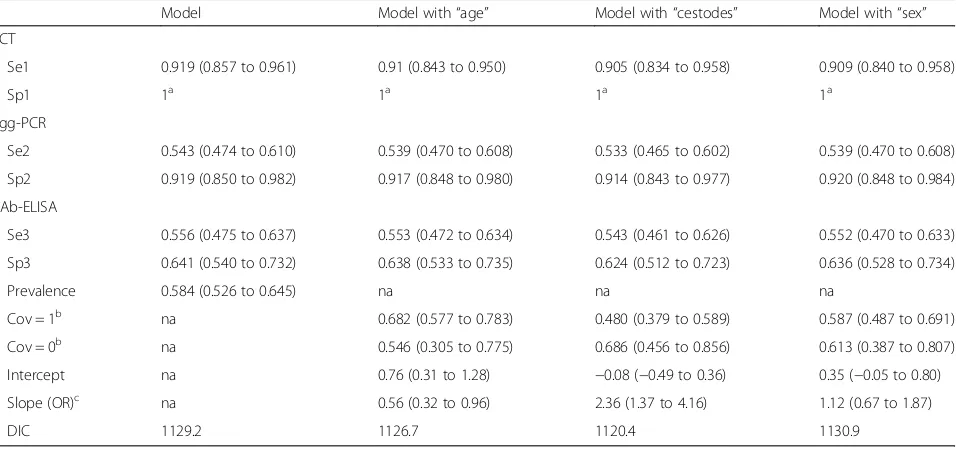
The estimated parameter values with their 95% cred-ibility intervals and DIC for the best-fitting model with and without covariates are presented in Table 2. Figures 1 and 2 show estimated true E. multilocularis prevalence in foxes with and without the significant covariates,“ ces-todes”and“age”.
Two covariates, “cestodes” and “age”, were found sig-nificantly associated withE. multilocularisoccurrence in foxes. The addition of the covariate “cestodes” brought the largest improvement in DIC and suggested that foxes with a concomitant cestode infection had double the odds of presenting E. multilocularis compared to foxes without it. The model including the covariate “age” experienced a less remarkable improvement in DIC and implied that adult foxes were less likely to be in-fected with E. multilocularis compared to younger an-imals. The covariate “sex” was found not significant, with no differences in E. multilocularis infection between males and females. The addition of covari-ates to the model had a negligible influence on the parameter estimates.
Bayesian latent class models for four diagnostic tests Similarly to the three-test models, all six sensitivity co-variances had posterior means unequal to zero and were therefore included in the final model (Additional file 7: Table S4). In contrast, there was no evidence for covari-ances between specificities (i.e. posterior mean equal to zero), and all three potential specificity covariances were set equal to 0.
The parameters estimates with their related 95% credibility intervals and DIC for the best-fitting model with and without covariates are presented in Table 3. Figures 3 and 4 show the E. multilocularis prevalence in foxes with and without the significant covariates as well as “cestodes” and “age”.
[image:6.595.61.539.466.691.2]Once more, the covariates “cestodes” and “age” were found significantly associated with E. multilocularis presence in the fox. Again, the model including the co-variate “cestodes” displayed the lowest DIC indicating that the odds of E. multilocularis infection doubled in foxes with concurrent cestode infection in comparison to foxes without it. The covariate “age” was also found significant although its addition to the model did not cause a remarkable reduction in the DIC. The model suggested lower odds of E. multilocularis infection in adults than younger foxes. The covariate “sex” was found not significant, with no differences in E. multi-locularis infection between male and female foxes. The addition of covariates to the model did not change the parameter estimates.
Table 2Parameters estimates (posterior means) with their corresponding 95% credibility intervals and the model goodness-of-fit to the data of the best model for three tests with and without covariates
Model Model with“age” Model with“cestodes” Model with“sex”
SCT
Se1 0.919 (0.857 to 0.961) 0.91 (0.843 to 0.950) 0.905 (0.834 to 0.958) 0.909 (0.840 to 0.958)
Sp1 1a 1a 1a 1a
Egg-PCR
Se2 0.543 (0.474 to 0.610) 0.539 (0.470 to 0.608) 0.533 (0.465 to 0.602) 0.539 (0.470 to 0.608)
Sp2 0.919 (0.850 to 0.982) 0.917 (0.848 to 0.980) 0.914 (0.843 to 0.977) 0.920 (0.848 to 0.984)
pAb-ELISA
Se3 0.556 (0.475 to 0.637) 0.553 (0.472 to 0.634) 0.543 (0.461 to 0.626) 0.552 (0.470 to 0.633)
Sp3 0.641 (0.540 to 0.732) 0.638 (0.533 to 0.735) 0.624 (0.512 to 0.723) 0.636 (0.528 to 0.734)
Prevalence 0.584 (0.526 to 0.645) na na na
Cov = 1b na 0.682 (0.577 to 0.783) 0.480 (0.379 to 0.589) 0.587 (0.487 to 0.691)
Cov = 0b na 0.546 (0.305 to 0.775) 0.686 (0.456 to 0.856) 0.613 (0.387 to 0.807)
Intercept na 0.76 (0.31 to 1.28) −0.08 (−0.49 to 0.36) 0.35 (−0.05 to 0.80)
Slope (OR)c na 0.56 (0.32 to 0.96) 2.36 (1.37 to 4.16) 1.12 (0.67 to 1.87)
DIC 1129.2 1126.7 1120.4 1130.9
a
Specificity of necropsy fixed to 1
b
Prevalence for respective covariate = 1 (adult, with other cestodes and male) and covariate = 0 (young, without other cestodes and female)
c
Odds ratio
The receiver operating characteristic (ROC) curve results
Bayesian empirical pAb-ELISA ROC curve from the three-test model
The best cut-off point obtained from the pAb-ELISA ROC curve using the classical method of considering necropsy and SCT as a gold standard test was 0.21, assigning the coproantigen test with 58.5% sensitivity, 65.4% specificity and an overall accuracy of 63.8%
(95% CI: 57.6–70.1%) given by the AUC. The optimal cut-off value from the Bayesian pAb-ELISA ROC curve using the three-test model was 0.29, assigning the coproantigen test with 42.2% sensitivity, 77.8%, specificity and an overall accuracy of 60.7% given by the AUC. Figure 5 shows both pAb-ELISA ROC curves derived using the classical and the Bayesian approach.
Bayesian empirical mAb-ELISA ROC curve from the three-test model
The best cut-off point obtained from the mAb-ELISA ROC curve using the classical method was 0.10, assign-ing the coproantigen test with 65.2% sensitivity, 68.4% specificity and an overall accuracy of 71.2% (95% CI: 65.4–77.0%) given by the AUC. The optimal cut-off value from the Bayesian mAb-ELISA ROC curve using the three-test model was 0.16, assigning the coproanti-gen test with 68.3% sensitivity, 75.3% specificity and an overall accuracy of 71.7% given by the AUC. Figure 6 shows both mAb-ELISA ROC curves derived using the classical and the Bayesian approach.
Bayesian empirical pAb- and mAb-ELISA ROC curves from the four-test model
When including the mAb-ELISA cut-off based on a Bayesian approach in the four-test model, AUC for the pAb-ELISA ROC curve was similar to the three-test model, e.g. 60.7%. The highest sum of the sensitivity plus specificity was 1.20 with an associated sensitivity and specificity of 69.9 and 50.6%, respectively. The corre-sponding cut-off was 0.17. The second highest sum of sen-sitivity and specificity was 1.198 with the same cut-off as in the three-test model of 0.29. For this cut-off, the sensi-tivity and specificity were 41.6 and 78.3%, respectively.
When including the pAb-ELISA cut-off based on a Bayesian approach in the four-test model, the AUC for the mAb-ELISA ROC curve was 76.2% for the same cut-off 0.16 with associated sensitivity and specificity of 70.5% and 80.0%. In Additional file 8: Figure S1 and Additional file 9: Figure S2, ROC curves for both ELISAs with the classical and the Bayesian approach are shown.
The variation of the cut-off points for the classification of both ELISA tests, pAb- and mAb-ELISAs, had virtu-ally no impact on the estimations of the other parame-ters of interest. The analysis was performed once more using the four-test model and a new classification for the necropsy and SCT results, being positive only the samples with 100 or more E. multilocularis. In this case, the optimal cut-off point determined by the Bayesian mAb-ELISA ROC was still 0.16, conferring to the coproantigen test with 70.5% sensitivity, 80.0% specificity and an overall accuracy of 76.2% given by the AUC. Figure 7 shows the corresponding mAb-ELISA ROC curve.
Fig. 2Posterior distribution ofE. multilocularisprevalence in foxes with and without the significant covariate“age”for the best-fitting model to the results of three diagnostic tests
[image:7.595.56.291.86.302.2] [image:7.595.57.290.478.693.2]Table 3Parameters estimates (posterior means) with their corresponding 95% credibility intervals and the model goodness-of-fit to the data of the best model for four tests with and without covariates
Model Model with“age” Model with“cestodes” Model with“sex”
SCT
Se1 0.885 (0.827–0.934) 0.879 (0.816 to 0.931) 0.876 (0.811 to 0.930) 0.878 (0.814 to 0.930)
Sp1 1a 1a 1a 1a
Egg-PCR
Se2 0.548 (0.485 to 0.610) 0.544 (0.482 to 0.608) 0.544 (0.482 to 0.606) 0.546 (0.483 to 60.8)
Sp2 0.934 (0.873 to 0.991) 0.936 (0.872 to 0.990) 0.940 (0.874 to 0.992) 0.940 (0.874 to 0.993)
pAb-ELISA
Se3 0.560 (0.480 to 0.639) 0.558 (0.477 to 0.637) 0.551 (0.471 to 0.631) 0.557 (0.476 to 0.638)
Sp3 0.659 (0.558 to 0.756) 0.659 (0.555 to 0.758) 0.648 (0.540 to 0.749) 0.659 (0.552 to 0.759)
mAb-ELISA
Se4 0.632 (0.553 to 0.708) 0.629 (0.550 to 0.706) 0.623 (0.544 to 0.701) 0.629 (0.549 to 0.707)
Sp4 0.700 (0.601 to 0.794) 0.701 (0.600 to 0.797) 0.693 (0.590 to 0.791) 0.701 (0.598 to 0.799)
Prevalence 0.595 (0.431 to 0.664) na na na
Cov = 1b na 0.697 (0.594 to 0.794) 0.500 (0.398 to 0.606) 0.596 (0.594 to 0.794)
Cov = 0b na 0.558 (0.312 to 0.784) 0.692 (0.464 to 0.857) 0.631 (0.312 to 0.784)
Intercept na 0.83 (0.38 to 1.34) 0.00 (−0.04 to 0.43) 0.39 (−0.01 to 0.83)
Slope (OR)c na 0.55 (0.31 to 0.94) 2.24 (1.31 to 3.90) 1.16 (0.96 to 1.96)
DIC 1507.0 1501.9 1497.2 1506.2
a
Specificity of necropsy fixed to 1
b
Prevalence for respective covariate = 1 (adult, with other cestodes and male) and covariate = 0 (young, without other cestodes and female)
c
Odds ratio
Abbreviations: Sesensitivity,Spspecificity,Egg-PCRpolymerase chain reaction,pAb-ELISApolyclonal enzyme-linked immunosorbent assay,mAb-ELISAmonoclonal enzyme-linked immunosorbent assay (cut-off for both ELISAs determined by considering necropsy and SCT as the gold-standard test),DICdeviance information criterion,nanot applicable
Fig. 3Posterior distribution ofE. multilocularisprevalence in foxes with and without the significant covariate,“cestodes”for the best-fitting model to the results of four diagnostic tests
[image:8.595.57.298.477.693.2] [image:8.595.307.538.477.695.2]Discussion
The employment of latent class models to analyse the results of the diagnostic tests for E. multilocularis allowed the determination of the performance of the test in the study population and the estimation of the true parasite prevalence in the absence of a perfect gold standard test. Furthermore, it was also possible to adjust for potential conditional dependence between tests. Also, these models could evaluate the association be-tween three covariates and parasite infection occurrence in the fox. Likewise, the application of latent class models permitted the building of ROC curves for the ELISAs following a Bayesian approach that enabled the empirical determination of the best cut-off point and the evaluation of the impact that the selection of the cut-off had in the estimation of the rest of the characteristics of the test.
In the present study, the latent class models including all potential covariances between sensitivities proved to be robust and their parameter estimates showed to be consistent with previous knowledge. The point estimates for the true E. multilocularis prevalence in foxes given by the three and four-test models (without covariates) were 58.4 and 59.5%, respectively. Similar high parasite prevalences have been previously reported in Swiss foxes [46–48]. In regard to the tests performances, the model estimates are also in line with prior information on diag-nostics accuracy of these techniques. The best-fitted models (without covariates) gave high point estimates for the necropsy and SCT sensitivities, 91.9 and 88.5%. The SCT’s sensitivity has commonly been considered relatively high, 98–100% [38] since the immersion of the
Fig. 7Bayesian monoclonal ELISA ROC when the criteria to be positive by necropsy and SCT is to present 100 or more E. multilocularis
Fig. 5Polyclonal ELISA (pAb-ELISA) ROC curves produced using the classical and the Bayesian approach
[image:9.595.57.293.87.342.2] [image:9.595.306.539.89.294.2] [image:9.595.56.291.375.702.2]intestines in saline solution and the posterior scrapping of the intestinal wall ensures the release of most of the worms [36]. Hence, if a fox has intestinal worms this method should identify them reliably. However, an ex-perimental study determined that intestinal samples should contain at least 10 tapeworms to achieve a 60% probability of obtaining positive detection [8]. Although experimental conditions differ from natural infection, this study highlights SCT’s sensitivity limitations related to worm burdens. In addition, the combination of worms’degradation during post-mortem conditions plus the intestines deep-freezing stage involved in the SCT process could also affect the SCT’s sensitivity. Moreover, a recent latent class analysis ofE. multilocularis diagnos-tic tests estimated the SCT-sensitivity to be between 76 and 88% [9]. Hence, SCT should not be regarded as a
“gold standard” test [6]. The estimated specificities of the pAb-ELISA from the three and four-test models (without covariates) ranged between 54.0–73.2% and 55.8–75.6%, respectively. The estimated specificity of the mAb-ELISA was found amid 60.1–79.4%. Coproantigen specificities can be altered by the occurrence of cross-reactions with antigens from concomitant helminths in-fections [13] or even the persistence ofE. multilocularis antigens in the faeces after the fox is no longer infected resulting in false positives results. The pAb-ELISA and the mAb-ELISA’s estimated sensitivities from the three and four-test models (without covariates) ranged be-tween 47.5–63.7% and 48–63.9% for the pAb-ELISA and 55.3–70.8% for the mAb-ELISA. Coproantigen sensitiv-ities strongly depend on the intensity ofE. multilocularis infection [13, 17–19], so foxes with low worm burdens are more likely to result in false negatives. Knowing how highly aggregated distributed is E. multilocularis in the fox population, it is likely that some foxes harboring low worm burdens will be misclassified as negatives by this type of test. Overall, the best model showed that the mAb-ELISA performed slightly better than the pAb-ELISA. Our pAb-ELISA estimates are in line with a prior latent class study that included arecoline purgation and egg-PCR in their analysis (SEdog 55%, 95% CI: 40.8– 68.9% and SPdog 70.6%, 95% CI: 65.3–76.7%) [35], but lower than the originally test characteristics reported (SEfox ~80%, SPfox 95–99%) [13]. Often the coproanti-gen test has been evaluated using the SCT as the gold standard test [13, 18] even though, as we have discussed previously, its sensitivity is not perfect. Taking this into account the coproantigen test’s actual sensitivity in the field can be realistically considered to be around 60% [6]. Furthermore, ELISA assays using polyclonal anti-bodies are prone to batch-to-batch variation and thus their performance reproducibility cannot be guaranteed. In this study however, sufficient quantities of polyclonal antibodies were produced in one batch to allow 400,000
tests, which could be the basis of minimizing this issue. In addition, the use of the polyclonal antibody test per-mitted the use of the three or four-test models and thus was important to help define the parameters of the other tests used, which do not suffer from this potential issue. Lastly, the estimates obtained from the three and four-test models for the egg-PCR specificities ranged between 85.0–98.2% and 87.3–99.1% and their sensitivities amid 47.4–61.0% and 48.5–61.0%. A field study in Kyrgyzstan also described the performance of this multiplex PCR as a highly specific but low sensitive test (SEdog 50%, 95% CI: 29–72% and SPdog 100%, 95% CI: 97–100%) [49]. High specificities are expected because the primers of this egg-PCR can identify and differentiate specifically the Echinococcus egg-DNA found in the faeces, even though there is always the possibility of false positive an-imals resulting from cross-contamination [50]. In gen-eral, the PCR’s sensitivity might be low under low worm burdens conditions or the presence of juvenile worms (characteristic during pre-patent infections) [25]. Fur-thermore, during the DNA isolation procedure PCR-inhibitory substances could be in the sample increasing the number of false negative results [25, 50].
In our models we considered conditional dependen-cies between sensitivities, but not specificities. The absence of evident covariances among specificities can at least partly be explained by the relatively high specificities and hence a low number of false positives resulting in a too small sample size to gain any infor-mation for these covariances.
age-differences is the potential existence of an acquired im-munological response after repeated infection [46, 48, 53]. However, other plausible causes such as differences in their predatory or territorial behaviour might result in juvenile animals with higher exposure to E. multilocu-laris infection compared to adults [54, 55]. A recent study modelling E. multilocularis abundance in Zurich foxes suggests that variations in infection pressure among age groups might be behind the observed differ-ences in parasite loads between juveniles and adults [56]. Nevertheless, in our study, the fox age was estimated on visual examination of teeth wear assessed by the re-searcher who was identifying the animals. Despite being a quick and easy method to distinguish between older and younger animals, it is also known to be less than 100% reliable as the teeth wear is subjected to individual characteristics such as the type of diet or the occurrence of missing teeth [39, 57]. There is less evidence that sus-tains the potential association betweenE. multilocularis infection and sex of the fox [58]. Although young male foxes are known to expand their territory during the mating season [59] and thus, might have a higher risk of infection if, during their roaming behaviour, they tres-pass clusters presenting an active parasite cycle with in-fected rodents. Nevertheless, the models did not find any significant differences in the odds of E. multilocu-laris infection between male and female foxes. This might be caused due to the small size of the study popu-lation or because of an unbalance of proportions in the data set, although the difference between numbers of collected males and females was not remarkable. Due to the small sample size, no internal validation was pos-sible. Potentially, two sources of bias might have oc-curred. First, it could be that due to the sampling of the foxes during the hunting seasons a seasonal variation in cestode infection [56] might have introduced some sort of bias. Secondly, the PCR is designed to detect patent, but not pre-patent infections. With a life duration of 90 days and a third of this time being in a pre-patent state, the PCR results will never be unbiased in detecting allE. multilocularisinfections [60].
For this analysis, uninformative as well as informative priors based on existing knowledge were used. By sensi-tivity analyses varying our prior information systematic-ally, we found that our results are robust and are driven by the data and not by the prior information. Further-more, the specificity of the necropsy and SCT was fixed at 100% [36]. Also, the assumption of a high specificity in the identification of parasites by necropsy and SCT is supported by the lack of a potential differential diagnosis as, to the authors’knowledge,E. granulosushas not been yet found in foxes in Switzerland.
Here, we wanted to assess the difference in the deter-mination of the cut-off by using two methods: the
classical approach of considering the necropsy and SCT as a perfectly accurate test and the empirical method of deriving the ROC curve using the parameter estimations of the Bayesian latent class model. On this occasion, some differences were found, as the cut-off points ob-tained from the Bayesian methods were slightly higher than those obtained from the classical approach. To some extent, the use of the classic method of treating the necropsy and SCT results as true infection status to establish the coproantigen test accuracy could underesti-mate the specificity of the ELISA, in the case of having several necropsy and SCT false negatives. Also, the building of the Bayesian ROC curves proved that the variation in the selection of the cut-off point for the ELISA did not affect the estimations of the other tests when including just one ELISA in the analysis. When in-cluding the two ELISAs the selection of the mAb-ELISA cut-off point did have an impact only on the pAb-ELISA estimations as the model structure accounted for condi-tional dependency between both coproantigen tests.
Finally, we employed the Bayesian latent class models to evaluate the test accuracy of the monoclonal ELISA to identify foxes presenting high parasite burdens of 100 or more worms. The distribution of E. multilocularis in the fox population is highly aggregated with few animals making the largest contribution to the environmental contamination with parasitic eggs, and thus representing the majority of the zoonotic risk [21]. However, it is also possible that foxes with low worm burdens at the time of sampling could have had much higher burdens a short period before due to the dynamics of infection [60]. The highly infected foxes are believed to play a critical role in E. multilocularis transmission and ultimately human in-fection. Therefore, when monitoring this zoonotic parasite in the fox population, it is paramount that surveillance programs employ diagnostic tests that can identify foxes effectively harbouring high parasite loads. The monoclonal coproantigen test proved to be a good tool for this pur-pose, showing high sensitivity and specificity to identify animals with moderate-to-high parasite burdens (≥ 100 worms). Furthermore, its good test performance along with its economic implementation and the fact that it can be performed on the faecal field samples without the need to collect dead animals, make this diagnostic test suitable for population studies in endemic areas.
Conclusions
Through the implementation of Bayesian latent class models, we could estimate the prevalence of infection and the specific performance of four diagnostic tests for E. multilocularis on the study population. As we have seen, there is a lack of a gold standard test forE. multi-locularis diagnosis in the definitive host. Furthermore, we know that the performance of these diagnostic tech-niques varies depending on the population investigated. Thus, the particular test performance on the population investigated has to be accounted for to be able to cor-rectly interpret the diagnosis results [61]. The adoption of a Bayesian latent class approach helps to overcome the absence of a perfectly accurate test and therefore gives a more reliable indication of the tests performance to ensure that meaningful conclusions can be drawn. Furthermore, the flexibility inherent in this type of models allows the incorporation of the potential de-pendence between diagnostic tests and permits the in-vestigation of the association of potential risk factors with true disease status [35, 49]. Finally, in the case of using a diagnostic test that needs the establishment of a cut-off point for the interpretation of its results, the Bayesian modelling facilitates the selection of this threshold value more reliably and comprehensively than the classical method.
Additional files
Additional file 1: Table S1.Original data file. (XLS 74 kb)
Additional file 2:Bayesian latent-class model code for three diagnostic tests. (DOC 33 kb)
Additional file 3:Bayesian latent-class model code for four diagnostic tests. (DOC 43 kb)
Additional file 4: Table S2.Description of the prior information used in the latent class models for three diagnostic tests. (DOC 35 kb)
Additional file 5: Table S3.Description of the prior information used in the latent class models for four diagnostic tests. (DOC 38 kb)
Additional file 6:The sensitivity analysis for the sensitivity of PCR. (PDF 320 kb)
Additional file 7:Table S4. Resulting covariances between sensitivities of the 3 and 4 test model. (DOCX 13 kb)
Additional file 8: Figure S1.Polyclonal ELISA ROC curves produced using the classical and the Bayesian approach (4 tests). (TIFF 501 kb)
Additional file 9: Figure S2.Monoclonal ELISA ROC curves produced using the classical and the Bayesian approach (4 tests). (TIFF 501 kb)
Abbreviations
AE:Alveolar echinococcosis; DIC: Deviance information criterion; DNA: Deoxyribonucleic acid; ELISA: Enzyme-linked immunosorbent assay; mAb: Monoclonal antibodies; MCMC: Markov chain Monte Carlo; pAb: Polyclonal antibodies; ROC: Receiver operating characteristic; SCT: Sedimentation and counting technique; Se: Sensitivity; SLT: Sedimentation counting technique; Sp: Specificity
Acknowledgements
The authors would like also to acknowledge the valuable contribution of the people of the Institute of Parasitology at the Vetsuisse Faculty of Zurich involved in the data collection and diagnosis performance of the samples.
Funding
This study has been funded by the Swiss National Science Foundation (SNSF) (grant 650 CR3313_132482/1 to PT). The funding body was not involved in the design of the study and collection, analysis, and interpretation of data and in writing the manuscript.
Availability of data and materials
The datasets supporting the conclusions of this article are included within the article and its additional files. The original data file with the diagnostic test results and information of covariates can be found in the Additional file 1.
Authors’contributions
Conceived and designed the experiments: BOA, SH, PRT and PD. Collection of data: MTAF, BOA and PD. Performed the experiments: MTAF, BOA and PD. Analyzed the data: BOA, SH and PRT. Wrote the paper: BOA, SH and PRT. All authors read and approved the final manuscript.
Ethics approval
According the Swiss animal welfare legislation (English translation http:// www.zuerchertierschutz.ch/fileadmin/user_upload/Tierschutzthemen/pdf/ Tierschutzgesetz_e.pdf) Article 3, our study is not considered as animal experimentation. Therefore, no ethical approval is needed.
Consent for publication Not applicable.
Competing interests
The authors declare that they have no competing interests.
Author details
1Section of Veterinary Epidemiology, Vetsuisse Faculty, University of Zurich, Zurich, Switzerland.2Institute of Parasitology, Vetsuisse Faculty, University of Zurich, Zurich, Switzerland.3Laboratorio de Vectores y Enfermedades transmitidas, Facultad de Veterinaria, CENUR Litoral Norte - Salto- Universidad de la República, Rivera 1350, 50000 Salto, Uruguay.
Received: 27 July 2017 Accepted: 29 November 2017
References
1. Deplazes P, Rinaldi L, Alvarez Rojas C, Torgerson P, Harandi M, Romig T, et al. Global distribution of alveolar and cystic echinococcosis. Adv Parasitol. 2017;95:315–493.
2. Torgerson PR, Keller K, Magnotta M, Ragland N. The global burden of alveolar echinococcosis. PLoS Negl Trop Dis. 2010;4:e722.
3. Gottstein B, Stojkovic M, Vuitton DA, Millon L, Marcinkute A, Deplazes P. Threat of alveolar echinococcosis to public health - a challenge for Europe. Trends Parasitol. 2015;31:407–12.
4. Romig T. Spread ofEchinococcus multilocularisin Europe? In: Craig P, Pawlowski Z, editors. Cestode zoonoses: Echinococcosis and cysticercosis. Amsterdam: IOS Press; 2002. p. 65–80.
5. Konig A, Romig T. Fox tapewormEchinococcus multilocularis, an underestimated threat: a model for estimating risk of contact. Wildlife Biol. 2010;16:258–66.
6. Conraths FJ, Deplazes P.Echinococcus multilocularis: epidemiology, surveillance and state-of-the-art diagnostics from a veterinary public health perspective. Vet Parasitol. 2015;213:149–61.
7. Eckert J, Deplazes P, Craig P, Gemmell M, Gottstein B, Heath D, et al. Echinococcosis in animals: clinical aspects, diagnosis and treatment. In: Eckert J, Gemmell M, Meslin F-X, Pawlowski Z, editors. WHOI/OIE manual on echinococcosis in humans and animals: a public health problem of global concern. World Organisation for Animal Health; 2001. p. 72–99. 8. Karamon J, Sroka J, Cencek T. Limit of detection of sedimentation and
counting technique (SCT) forEchinococcus multilocularisdiagnosis, estimated under experimental conditions. Exp Parasitol. 2010;124:244–6. 9. Wahlstrom H, Comin A, Isaksson M, Deplazes P. Detection ofEchinococcus
multilocularisby MC-PCR: evaluation of diagnostic sensitivity and specificity without gold standard. Infect Ecol Epidemiol. 2016;1:1–6.
11. Umhang G, Woronoff-Rhen N, Combes B, Boué F. Segmental sedimentation and counting technique (SSCT): an adaptable method for qualitative diagnosis ofEchinococcus multilocularisin fox intestines. Exp Parasitol. 2011; 128:57–60.
12. Sakai H, Nonaka N, Yagi K, Oku Y, Kamiya M. Coproantigen detection in a routine fox survey ofEchinococcus multilocularisinfection in Hokkaido, Japan. Parasitol Int. 1998;47:47–51.
13. Deplazes P, Alther P, Tanner I, Thompson RCA, Eckert J.Echinococcus multiloculariscoproantigen detection by enzyme-linked immunosorbent assay in fox, dog, and cat populations. J Parasitol. 1999;85:115–21. 14. Al-Sabi M, Kapel C, Deplazes P, Mathis A. Comparative copro-diagnosis of
Echinococcus multilocularisin experimentally infected foxes. Parasitol Res. 2007;101:731–6.
15. Craig PS, Gasser RB, Parada L, Cabrera P, Parietti S, Borgues C, et al. Diagnosis of canine echinococcosis: comparison of coproantigen and serum antibody tests with arecoline purgation in Uruguay. Vet Parasitol. 1995;56: 293–301.
16. Kohno H, Sakai H, Okamoto M, Ito M, Oku Y, Kamiya M. Development and characterization of murine monoclonal antibodies toEchinococcus multilocularisadult worms and its use for the coproantigen detection. Jpn J Parasitol. 1995;44:404–12.
17. Morishima Y, Tsukada H, Nonaka N, Oku Y, Kamiya M. Coproantigen survey forEchinococcus multilocularisprevalence of red foxes in Hokkaido, Japan. Parasitol Int. 1999;48:121–34.
18. Reiterová K, Miterpáková M, Turčeková L, Antolová D, Dubinský P. Field evaluation of an intravital diagnostic test ofEchinococcus multilocularis infection in red foxes. Vet Parasitol. 2005;128:65–71.
19. Yimam AE, Nonaka N, Oku Y, Kamiya M. Prevalence and intensity of Echinococcus multilocularisin red foxes (Vulpes vulpes schrencki) and raccoon dogs (Nyctereutes procyonoides albus) in Otaru city, Hokkaido, Japan. Jpn J Vet Res. 2002;49:287–96.
20. Hofer S, Gloor S, Müller U, Mathis A, Hegglin D, Deplazes P. High prevalence ofEchinococcus multilocularisin urban red foxes (Vulpes vulpes) and voles (Arvicola terrestris) in the city of Zürich, Switzerland. Parasitology. 2000;120: 135–42.
21. Deplazes P, Hegglin D, Gloor S, Romig T. Wilderness in the city: the urbanization ofEchinococcus multilocularis. Trends Parasitol. 2004;20:77–84. 22. Bretagne S, Guillou J, Morand M, Houin R. Detection ofEchinococcus multilocularis
DNA in fox faeces using DNA amplification. Parasitology. 1993;106:193–9. 23. Monnier P, Cliquet F, Aubert MF, Bretagne S. Improvement of apolymerase
chain reaction assay for the detection ofEchinococcus multilocularisDNA in faecal samples of foxes. Vet Parasitol. 1996;67:185–95.
24. Mathis A, Deplazes P, Eckert J. An improved test system for PCR-based specific detection ofEchinococcus multiloculariseggs. J Helminthol. 1996;70:219–22. 25. Dinkel A, von Nickisch-Rosenegk M, Bilger B, Merli M, Lucius R, Romig T.
Detection ofEchinococcus multilocularisin the definitive host:
coprodiagnosis by PCR as an alternative to necropsy. J Clin Microbiol. 1998; 36:1871–6.
26. Ziadinov I, Mathis A, Trachsel D, Rysmukhambetova A, Abdyjaparov T, Kuttubaev O, et al. Canine echinococcosis in Kyrgyzstan: using prevalence data adjusted for measurement error to develop transmission dynamics models. Int J Parasitol. 2008;38:1179–90.
27. Trachsel D, Deplazes P, Mathis A. Identification of taeniid eggs in the faeces from carnivores based on multiplex PCR using targets in mitochondrial DNA. Parasitology. 2007;134:911–20.
28. Klein C, Liccioli S, Massolo A. Egg intensity and freeze-thawing of fecal samples affect sensitivity ofEchinococcus multilocularisdetection by PCR. Parasitol Res. 2014;113:3867–73.
29. Knapp J, Millon L, Mouzon L, Umhang G, Raoul F, Ali ZS, et al. Real time PCR to detect the environmental faecal contamination byEchinococcus multilocularisfrom red fox stools. Vet Parasitol. 2014;201:40–7.
30. Isaksson M, Hagstöm A, Armua-Fernandez M, Wahlström H, Ågren E, Miller A, et al. A semi-automated magnetic capture probe based DNA extraction and real-time PCR method applied in the Swedish surveillance of Echinococcus multilocularisin red fox (Vulpes vulpes) faecal samples. Parasit Vectors. 2014;19:583.
31. Maas M, van Roon A, Dam-Deisz C, Opsteegh M, Massolo A, Deksne G, et al. Evaluation by latent class analysis of a magnetic capture based DNA extraction followed by real-time qPCR as a new diagnostic method for detection ofEchinococcus multilocularisin definitive hosts. Vet Parasitol. 2016;230:20–4.
32. Boufana B, Umhang G, Qiu J, Chen X, Lahmar S, Boué F, et al. Development of three PCR assays for the differentiation betweenEchinococcus shiquicus, E. granulosus(G1genotype), andE. multilocularisDNA inthe co-endemic region of Qinghai-Tibet plateau, China. Am J Trop Med Hyg. 2013;88:795–802. 33. Hui S, Walter S. Estimating the error rates of diagnostic tests. Biometrics.
1980;36:167–71.
34. Enøe C, Georgiadis MJW. Estimation of sensitivity and specificity of diagnostic tests and disease prevalence when the true state of disease is unknown. Prev Vet Med. 2000;45:61–81.
35. Hartnack S, Budke C, Craig P, Jiamin Q, Boufana B, Campos-Ponce M, et al. Latent-class methods to evaluate diagnostics tests forEchinococcus infections in dogs. PLoS Negl Trop Dis. 2013;7:e2068.
36. Eckert J, Deplazes P, Craig P, Gemmell M, Gottstein B, Heath D, et al. Chapter 3: Echinococcosis in animals: clinical aspects, diagnosis and treatment. In: Eckert J, Gemmell M, Meslin F-X, Pawlowsk Z, editors. WHO/ OIE manual on Echinococcosis in humans and animals: a public health problem of global concern. Paris, France: WHO/OIE; 2001.
37. Woolsey I, Bune N, Jensen P, Deplazes P, Kapel C.Echinococcus multilocularis infection in the field vole (Microtus agrestis): an ecological model for studies on transmission dynamics. Parasitol Res. 2015;114:1703–9.
38. Eckert J. Predictive values and quality control of techniques for the diagnosis ofEchinococcus multilocularisin definitive hosts. Acta Trop. 2003;85:157–63. 39. Harris S. Age determination in the red fox (Vulpes vulpes): an evaluation of
technique efficiency as applied to a sample of suburban foxes. J Zool. 1978; 184:91–117.
40. Deplazes P, Gottstein B, Eckert J, Jenkins D, Ewald D, Jimenez-Palacios S. Detection ofEchinococcuscoproantigens by enzyme-linked immunosorbent assay in dogs, dingoes and foxes. Parasitol Res. 1992;78:303–8.
41. Robin X, Turck N, Hainard A, Tiberti N, Lisacek F, Sanchez J, et al. pROC: an open-source package for R and S+ to analyze and compare ROC curves. BMC Bioinformatics. 2011;12:77.
42. Lewis FI, Torgerson PR. A tutorial in estimating the prevalence of disease in humans and animals in the absence of a gold standard diagnostic. Emerg Themes Epidemiol. 2012;9:9.
43. Plummer M. JAGS: a program for analysis of Bayesian graphical models using Gibbs sampling. In: Hornik K, Leisch F, Zeileis A, editors. Proceedings of the 3rd international workshop on distributed statistical computing (DSC 2003). Vienna, Austria; 2003. p. 1–10.
44. Plummer M, Best N, Cowles K, Vines K. CODA: convergence diagnosis and output analysis for MCMC. R News. 2006;6:7–11.
45. R Development Core Team. R: A language and environment for statistical computing. 2008. https://www.r-project.org/.
46. Oksanen A, Siles-Lucas M, Karamon J, Possenti A, Conraths FJ, Romig T, et al. The geographical distribution and prevalence of Echinococcus multilocularis in animals in the European Union and adjacent countries: a systematic review and meta-analysis. Parasit Vectors. 2016;9:519.
47. Fischer C, Reperant LA, Weber JM, Hegglin D, Deplazes P.Echinococcus multilocularisinfections of rural, residential and urban foxes (Vulpes vulpes) in the canton of Geneva, Switzerland. Parasite. 2005;12:339–46.
48. Brossard M, Andreutti C, Siegenthaler M. Infection of red foxes withEchinococcus multilocularisin western Switzerland. J Helminthol. 2007;81:369–76.
49. Ziadinov I, Mathis A, Trachsel D, Rysmukhambetova A, Abdyjaparov TA, Kuttubaev OT, et al. Canine echinococcosis in Kyrgyzstan: using prevalence data adjusted for measurement error to develop transmission dynamics models. Int J Parasitol. 2008;38:1179–90.
50. Mathis A, Deplazes P. Copro-DNA tests for diagnosis of animal taeniid cestodes. Parasitol Int. 2006;55(Suppl):S87–90.
51. Reperant LA, Hegglin D, Fischer C, Kohler L, Weber JM, Deplazes P. Influence of urbanization on the epidemiology of intestinal helminths of the red fox (Vulpes vulpes) in Geneva, Switzerland. Parasitol Res. 2007;101:605–11. 52. Robardet E, Giraudoux P, Caillot C, Boue F, Cliquet F, Augot D, et al.
Infection of foxes byEchinococcus multilocularisin urban and suburban areas of Nancy, France: influence of feeding habits and environment. Parasite. 2008;15:77–85.
53. Tackmann K, Löschner U, Mix H, Staubach C, Thulke HH, Conraths FJ. Spatial distribution patterns ofEchinococcus multilocularis(Leuckart 1863) (Cestoda: Cyclophyllidea: Taeniidae) among red foxes in an endemic focus in Brandenburg, Germany. Epidemiol Infect. 1998;120:101–9. 54. Vervaeke M, Davies S, Leirs H, Verhagen R. Implications of increased
55. Hegglin D, Bontadina F, Contesse P, Gloor S, Deplazes P. Plasticity of predation behaviour as a putative driving force for parasite life-cycle dynamics: the case of urban foxes andEchinococcus multilocularis tapeworm. Funct Ecol. 2007;21:552–60.
56. Otero-Abad B, Rüegg S, Hegglin D, Deplazes P, Torgerson P. Mathematical modelling ofEchinococcus multilocularisabundance in foxes in Zurich, Switzerland. Parasit Vectors. 2017;10:21.
57. Zapata S, Travaini A, Delibes M. Comparacion entre varias tecnicas de estimacion de la edad en zorros,Vulpes vulpes, de Doñana (sur de la peninsula iberica). Acta Vertebr. 1995;22:29–50.
58. Otero-Abad B, Torgerson PR. A Systematic Review of the Epidemiology of Echinococcosis in Domestic and Wild Animals. PLoS Negl Trop Dis. 2013; 7(6):e2249. https://doi.org/10.1371/journal.pntd.0002249.
59. Cavallini P. Variation in the social system of the red fox. Ethol Ecol Evo. 1996;8:323–42.
60. Kapel C, Torgerson P, Thompson R, Deplazes P. Reproductive potential of Echinococcus multilocularisin experimentally infected foxes, dogs, raccoon dogs and cats. Int J Parasitol. 2006;36:79–86.
61. Torgerson P, Deplazes P. Echinococcosis: diagnosis and diagnostic interpretation in population studies. Trends Parasitol. 2009;25:164–70.
• We accept pre-submission inquiries
• Our selector tool helps you to find the most relevant journal • We provide round the clock customer support
• Convenient online submission • Thorough peer review
• Inclusion in PubMed and all major indexing services • Maximum visibility for your research
Submit your manuscript at www.biomedcentral.com/submit