BioMedCentral
Page 1 of 9
(page number not for citation purposes)
Journal of Translational Medicine
Open Access
Research
Differential in vitro inhibitory activity against HIV-1 of alpha-(1-3)-
and alpha-(1-6)-D-mannose specific plant lectins : Implication for
microbicide development
Hela Saïdi
†1, Nadine Nasreddine
†, Mohammad-Ali Jenabian
1,
Maxime Lecerf
1, Dominique Schols
2, Corinne Krief
1, Jan Balzarini
2and
Laurent Bélec*
1Address: 1Unité INSERM U743, Equipe « Immunité et Biothérapie Muqueuse », Centre de Recherches Biomédicales des Cordeliers, Paris, France
and 2Rega Institute for Medical Research, Leuven, Belgium
Email: Hela Saïdi - [email protected]; Nadine Nasreddine - [email protected]; Mohammad-Ali Jenabian - [email protected]; Maxime Lecerf - [email protected];
Dominique Schols - [email protected]; Corinne Krief - [email protected]; Jan Balzarini - [email protected]; Laurent Bélec* - [email protected]
* Corresponding author †Equal contributors
Abstract
Background: Plant lectins such as Galanthus nivalis agglutinin (GNA) and Hippeastrum hybrid agglutinin (HHA) are natural proteins able to link mannose residues, and therefore inhibit HIV-target cell interactions. Plant lectins are candidate for microbicide development.
Objective: To evaluate the activity against HIV of the mannose-specific plant lectins HHA and GNA at the cellular membrane level of epithelial cells and monocyte-derived dendritic cells (MDDC), two potential target cells of HIV at the genital mucosal level.
Methods: The inhibitory effects of HHA and GNA were evaluated on HIV adsorption to genital epithelial HEC-1A cell line, on HIV transcytosis throughout a monolayer of polarized epithelial HEC-1A cells, on HIV adsorption to MDDC and on transfer of HIV from MDDC to autologous T lymphocytes.
Results: HHA faintly inhibited attachment to HEC-1A cells of the R5-tropic HIV-1Ba-L strain, in a dose-dependent manner, whereas GNA moderately inhibited HIV adsorption in the same context, but only at high drug doses. Only HHA, but not GNA, inhibited HIV-1JR-CSF transcytosis in a dose-dependent manner. By confocal microscopy, HHA, but not GNA, was adsorbed at the epithelial cell surface, suggesting that HHA interacts specifically with receptors mediating HIV-1 transcytosis. Both plant lectins partially inhibited HIV attachment to MDDC. HHA inhibited more efficiently the transfer of HIV from MDDC to T cell, than GNA. Both HHA and GNA lacked toxicity below 200 µg/ml irrespective the cellular system used and do not disturb the monolayer integrity of epithelial cells.
Conclusion: These observations demonstrate higher inhibitory activities of the lectin plant HHA by comparison to GNA, on HIV adsorption to HEC-1A cell line, HIV transcytosis through HEC-1A cell line monolayer, HIV adsorption to MDDC and HIV transfer from MDDC to T cells, highlighting the potential interest of HHA as effective microbicide against HIV.
Published: 12 June 2007
Journal of Translational Medicine 2007, 5:28 doi:10.1186/1479-5876-5-28
Received: 5 March 2007 Accepted: 12 June 2007
This article is available from: http://www.translational-medicine.com/content/5/1/28
© 2007 Saïdi et al; licensee BioMed Central Ltd.
Background
Heterosexual contact is the primary mode of human immunodeficiency virus type 1 (HIV-1) transmission worldwide. The majority of new HIV-1-infected people are women. Effective topical microbicides, as molecules with potent activity against HIV-1, inserted into the vagina prior to sexual intercourse, would offer an alternative to condom use that would be controlled by the receptive partner [1].
Plant lectins represent a well-defined class of antiretroviral compounds that are most advantageous than other antivi-ral drugs: lectins are natuantivi-ral and not synthetic products (proteins) and target the sugar moieties of a wide variety of glycoproteins [2,3]. Galanthus nivalis agglutinin (Snow-drop) (GNA) has specificity for terminal α(1–3)-linked mannose residues, whereas Hippeastrum hybrid agglutinin (Amaryllis) (HHA) recognizes both terminal and internal
α(1–3)- and α(1–6)-linked mannose residues [4]. These lectins occur as tetramers exhibiting a molecular mass of 50 kDa. They are potent and highly selective inhibitors of the spread of HIV by virus- to- lymphocytes, as well as infected lymphocytes- to- uninfected lymphocytes. HHA and GNA selectively inhibited a wide variety of HIV-1 and HIV-2 strains and clinical (CXCR4- and CCR5-using) iso-lates in different cell types [4]. It was also shown that these lectins inhibited infection of T lymphocytes by a variety of mutant virus strains, but also prevented syncytium forma-tion between persistently HIV-infected cells and unin-fected T lymphocytes [4]. There is strong evidence that plant lectins with anti-HIV activity predominantly target the heavily glycosylated gp120 envelope glycoprotein, as assessed by HIV-lectins pre-incubation assays [4]. In con-trast, no marked lectin-lymphocyte interactions were observed [4]. In addition, HHA and GNA efficiently abro-gated the directed HIV-1 capture in DC-SIGN-expressing B-lymphoblast Raji cells (Raji/DC-SIGN) and its subsequent transfer to CD4+ T lymphocytes [5]. HHA and GNA are also perfectly fitted to microbicide guide-lines in the way they are odorless, tasteless, colorless, not mitogenic and not anti-metabolically active at anti-viral concentrations [4].
Since early microbicide trials raised concerns about testing incompletely characterized compounds in humans [6], we propose to complete the in vitro pre-clinical evaluation of microbicide molecule candidates against HIV-1 adsorp-tion on epithelial cells, and against HIV-1 transcytosis through a tight monolayer of epithelial cells used as an in vitro model mimicking the penetration of HIV-1 through unstratified epithelia [7]. Indeed, epithelial cells represent the most important interface between host and environ-ment. Hocini et al. have previously hypothesized that sim-ple epithelial monolayers, such as the endocervix and rectal mucosae, may support rapid transcellular transport
(or " transcytosis ") from the apical to basolateral side of the cell, and thus provide viral access to the target cells of the submucosa, such as dendritic cells, CD4+ T cells and macrophages [8].
In the present study, we thus aimed at characterizing whether interactions exist between mannose-specific lectins and mannosylated HIV-interacting molecules. Indeed, HIV was described to interact with a large number of molecules onto cell surface during the adsorption phase. If some of these receptors are mannosylated close to the HIV-binding sites, the plant lectins may recognise such mannosylated residues, and may mask the HIV bind-ing site by steric hindrance, thus limitbind-ing HIV attachment onto cells. The effects of carbohydrate-binding plant lectins on T lymphocytes [4] and on DC-SIGN+ B-lym-phoblast Raji cells [5] have been previously investigated. To complete these latter studies, we herein evaluated the inhibitory effect of HHA and GNA on the attachment of HIV on other possible mucosal target cells, including epi-thelial and dendritic cells.
Methods
Cells lines, virus strains and plant lectins
the epithelial cell line HEC-1A was maintained in RPMI 1640 containing 10% FCS and antibiotics (100 µg of streptomycin per ml, 100 IU of penicillin per ml). Primary HIV-1NDK X4-tropic viruses (gift from Prof. F. Barré-Sinoussi, Institut Pasteur, Paris, France) were grown in peripheral blood lymphocytes (PBL) of healthy donors stimulated with phytohemagglutinin-P (PHA; Sigma-Aldrich, Saint Louis, MO) and interleukin-2 (IL-2; Pepro-tech Inc, Rocky Hill, NJ). R5-tropic HIV-1J-RCSF and HIV-1Ba-L were amplified in monocyte-derived macrophages (MDM) of healthy donors. Tropism of viruses was deter-mined using U87 cells (provided by Dr. E. Menue, Institut Pasteur) transfected with DNA encoding for human CD4, CCR5, or CXCR4. HIV was quantified in cell culture super-natants by means of the DuPont HIV-p24 antigen ELISA (HIV-1 core profile ELISA; DuPont de Nemours, Les Ulis, France). The mannose-specific lectins HHA and GNA were derived and purified from the bulbs of these plants, as pre-viously described [9]. Mannan was provided by Sigma.
In vitro differentiation of monocyte-derived dendritic cells (MDDC)
Journal of Translational Medicine 2007, 5:28 http://www.translational-medicine.com/content/5/1/28
Page 3 of 9
(page number not for citation purposes)
1640medium supplemented with L-glutamine (BioWhit-taker Europe, Verviers, Belgium), penicillin (100 IU/ml) and streptomycin (100 µg/ml) (Gibco BRL, Paisley, Scot-land). Cells were seeded into 24 well-plates (Costar, Cam-bridge, MA) at the concentration 1 × 10⁶ adherent cells/ ml. Cells were incubated at 37°C for 45 min. Non-adher-ent cells were removed by 4 gNon-adher-ently washes. AdherNon-adher-ent monocytes were further incubated in RPMI medium sup-plemented with 10% fetal calf serum (FCS), L-glutamine, and antibiotics in the presence of 10 ng/ml rhIL-4 and 10 ng/ml rhGM-CSF (both from R&D Systems, Oxon, UK), in order to differentiate MDDC. Half of the medium, including all supplements, was replaced every 2 days. After 6 days of culture, non-adherent cells corresponding to the DC-enriched fraction were harvested, washed, and used for subsequent experiments. Flow cytometry analysis demonstrated that the DC were 90% or more pure DC at an immature stage (CD14-, CD16-, CD1a+, CD83-, DC-SIGN+).
Isolation of autologous T lymphocytes
PBL were subsequently prepared from the monocyte-depleted cell fraction. PBL were cultured for 48 h in fresh medium supplemented with PHA (2.5 µg/ml) and IL-2 (1
µg/ml). PBL were then washed and further cultured in growth medium containing IL-2 (1 µg/ml) for 24 h.
Cytotoxicity assay
the cytotoxicity of the plant lectins against epithelial cells and MDDC was analysed using the MTT (3- [4,5-dimeth-ylthiazol-2-yl]-2,5-diphenyl tetrazolium bromide) assay (Sigma). Briefly, cells were seeded onto 96-well plates at a density of 2 × 10⁵ cells/well and incubated for 24 h at 37°C prior to drug exposure. On the day of treatment, cul-ture medium was carefully aspirated from the wells and replaced with fresh medium containing the plant lectins at concentrations ranging from 1 to 500 µg/ml. Triplicate wells were used for each treatment. The cells were incu-bated with the various compounds for 24 h at 37°C in a humidified 5% CO2 atmosphere. To each well, 20 µl of MTT (0.5 mg/ml final concentration) was added and the plates were incubated at 37°C for 4 h to allow MTT to form formazan crystals by reacting with metabolically active cells. The formazan crystals were solubilized 30 min at 37°C in a solution containing 10% sodium dodecyl sulphate in 0.01 M HCl. The absorbance of each well was measured in a microtitre reader at 490 nm. To translate the OD490 values into the number of live cells in each well, the OD490 values were compared with those of standard OD490 versus cell number curves generated for each cell type. The percentage survival was calculated using the for-mula :
% survival = live cell number (test)/live cell number (con-trol) × 100
Epithelial monolayer integrity
the effect of the plant lectins on their ability to maintain an intact epithelium was determined by measuring tran-sepithelial resistance (TER). HEC-1A cells were grown as a tight polarized monolayer on a permeable support of 0.4
µm-pore-diameter polycarbonate (Transwell, Costar, Cambridge, Mass.). Apical and basolateral media were replaced, and TER was measured daily with a Millicell-ERS (electrical resistance system) instrument (Millipore Cor-poration, Bedford, Mass.). When plateau TER was reached, plant lectins or media alone were added in dupli-cate wells, and the TER was measured at 30 min and 2, 4, 9, and 24 h. The epithelial resistance was expressed as fol-lows :
epithelial resistance = (Ω/cm2) - the resistance of tran-swells without cells.
HIV cell-free particles transcytosis
the epithelial cell line HEC-1A was grown as a tight polar-ized monolayer on a permeable support of 0.4-µ m-pore-diameter polycarbonate (Transwell), as previously described [10]. The tightness of the monolayer of HEC-1A cells was monitored by measuring resistivity above 200 Ω/ cm2. HIV-1 (5 ng of p24 antigen) was added together with increasing doses of each plant lectin or mannan (100 µg/ ml) on the apical side of the HEC-1A monolayer. Cells were incubated for 1 h at 37°C. HIV-1 transcytosis was assessed by detecting the presence of p24 antigen (HIV-1 core profile ELISA) in the basolateral chamber of the tran-swell. The inhibition of transcytosis was expressed as the percentage of p24 antigen recovered in the basolateral chamber in the presence of each plant lectin by compari-son to that recovered without the plant lectins.
HIV-1 attachment assay on epithelial cells
HEC-1A cells, seeded at confluency in 48-well plates, were incubated with increasing doses of each lectins or mannan (100 µg/ml) and HIV-1 (5 ng p24 antigen). Each sample was performed in duplicate. After 1 h of incubation, unat-tached virus was removed (4 washes) and cells were lysed (1% Triton X-100 for 45 min at 37°C). Cell lysates were harvested and centrifuged at 1,800 rpm for 5 min. The amount of HIV-p24 antigen associated to cell lysates was determined using the HIV-1 p24 core profile ELISA.
HIV-1 attachment on MDDC
min at 37°C with 1% Triton X-100. Cell lysates were har-vested and centrifuged at 1,800 rpm for 5 min. The amount of cell-associated HIV-1 was evaluated using p24 antigen capture ELISA.
MDDC-mediated infection of autologous T cells
to assess the transmission of HIV-1 from MDDC to autol-ogous T-cells, MDDC were incubated into 96-well culture plates (1 × 10⁵ cells/well) and infected with HIV-1 (0.5 ng
p24 antigen) in the presence of increasing concentrations of each plant lectin or mannan (100 Pg/ml) for 3 h at 37°C in a 5% CO2 atmosphere. Cells were washed four
times and autologous stimulated T cells were added onto HIV-exposed MDDC at a DC:T-cell ratio of 1:5. Each sam-ple was performed in triplicate. Culture supernatants were harvested every 3 days and fresh medium was added. Supernatants were inactivated with 1% Triton X-100 and frozen at -20°C. The viral production by T lymphocytes was evaluated at the sixth day of the co-culture by meas-urement of p24 antigen in supernatants using capture ELISA.
Confocal microscopy
confluent epithelial cells HEC-1A were trypsinized, washed, and 2 × 10⁵ cells were adsorbed on a
microscopy-adapted slide for 3 days. After 6 days of differentiation MDDC are washed and 2 × 10⁵ cells were adsorbed on a
microscopy-adapted slide. Cells were incubated with or without HHA-TRITC or GNA-TRITC (200 Pg/ml; Eylabs) at 4°C for 30 min. Cells were washed with PBS 0.01% azide 0.5% BSA and then fixed with 1% paraformalde-hyde. The coverslides were mounted in Mowiol (Sigma-Aldrich). Fluorescence analysis was performed with a Zeiss LSM510 confocal microscope.
Statistical analysis
unpaired and non-parametric Mann-Whitney U-test was performed for all tests to determine the statistical signifi-cance of the data. P < 0.05 was considered the level of sta-tistical significance.
Results
HHA and GNA activities in the epithelial cell line HEC-1A (i) Toxicity
Any topical microbicide approved for human use first need to be evaluated for epithelial toxicity, because of the direct contact between the product and the vaginal mucosal surface. Thus, non-toxic concentrations of the plant lectins were determined by culturing the epithelial cell line, HEC-1A, for 24 h in the presence of different lec-tin concentrations (1 Pg/ml to 200 Pg/ml) or medium (Fig. 1A). Plant lectins concentrations that gave culture viabilities of more than 80% compared to control cultures were considered to be non-toxic and were further evalu-ated. Overall, the products exhibited no measurable
toxic-ity towards HEC-1A epithelial cells within a tested concentration range of 1 to 200 Pg/ml.
(ii) Epithelial monolayer integrity
The ability of mucosal epithelial cells to maintain an intact polarized monolayer in the presence of the plant lectins is a possible predictor of the safety of products on vaginal tissues. Thus, HHA and GNA were added to con-fluent HEC-1A cell-monolayers, as determined by TER = 200 :/cm2. The TER was evaluated at 24 h after addition
(A). Non-toxic concentration of the study plant lectins in epi-thelial cell line (HEC-1A) and primary immune cells (MDDC and MDM)
Figure 1
(A). Non-toxic concentration of the study plant lectins in epi-thelial cell line (HEC-1A) and primary immune cells (MDDC and MDM). HEC-1A and primary immune cells were cultured with concentrations of products for 24 h. After washing, cul-ture viability was determined by using the MTT-cytotoxicity assay. The values given are the percentage of viability. (B) and (C). Effect of HHA and GNA on HEC-1A epithelial monol-ayer integrity. HEC-1A cells were grown on transwell sup-ports and allowed to form intact monolayer (5 to 7 days). Non-toxic concentrations of each lectins were added to the apical side of the transwell, and epithelial integrity was meas-ured at 30 min and 2, 4, 9, and 24 h by using the Minicell-ERS instrument. The data shown are the averages from duplicate wells ± 1 standard error of the mean after product addition.
Day of product application
cm
2
0 100 200 300 400 500 600
-1 0 1
Control
GNA (1μg/ml)
GNA (10μg/ml)
GNA (200μg/ml)
cm
2
0 100 200 300 400 500 600
-1 0 1
Control
HHA (1μg/ml)
HHA (10μg/ml)
HHA (200μg/ml)
Day of product application
%S
urvival
o
f
HEC-1A HHA
Control
GNA A
[Plant lectins] (μμg/ml)
C B
0 20 40 60 80 100 120
Journal of Translational Medicine 2007, 5:28 http://www.translational-medicine.com/content/5/1/28
Page 5 of 9
(page number not for citation purposes)
of the plant lectins. HHA up to 200 µg/ml (Fig. 1B) and GNA up to 200 µg/ml (Fig. 1C) did not measurably affect the integrity of the epithelial barrier. Of note, the TER of the control cultures, without the plant lectins, did not vary more than 25% over the 24 h period.
(iii) Effect of plant lectins on HIV-1 attachment on HEC-1A
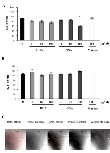
Considering that both HHA and GNA were non-toxic in the epithelial cell system, we further investigated the inhi-bition of HIV adsorption on the apical side of HEC-1A by the compounds. Cells were thus incubated with HIV-1Ba-L in the presence or absence of different concentrations of HHA or GNA. As depicted in Figure 2A, HHA faintly decreased the attachment of HIV-1Ba-L on epithelial cells (range: 12–17%). Interestingly, GNA afforded a 33% decrease of HIV adsorption only at a concentration of 100
µg/ml. In order to evaluate whether the observed effects of the plant lectins are linked to molecules implicated in HIV attachment towards genital epithelial cells, such as gly-cosaminoglycans (GAG) heparan sulfates, we assessed the role of plant lectins on the attachment of X4-tropic viruses, known to interact very efficiently with heparan sulfate [11] and data not shown]. Thus, cells were incu-bated with HIV-1NDK (Fig. 2B) in the presence or absence of different concentrations of HHA and GNA. As depicted in Figure 2B, none of the plant lectins, whatever the con-centration, inhibited HIV-1NDK attachment on HEC-1A cells. These data suggested that HHA and GNA did not interact with HIV recognition sites on GAG. In addition, mannan (100 µg/ml) did not decrease HIV adsorption on HEC-1A cells whatever the viral strain used (Fig. 2A and Fig. 2B). To investigate the efficiency of each lectin to interact with epithelial cell's receptors, labeled HHA and GNA were incubated with epithelial cells. As depicted in Figure 2C, only HHA but not GNA interacted with recep-tors expressed at the surface of HEC-1. Moreover, the pre-incubation of GNA with epithelial cells followed with several washes did not inhibit viral attachment whereas the preincubation of GNA with R5 viral particles induced an inhibition of 35 ± 5% of R5 HIV-1 attachment on epi-thelial cells (data not shown), similarly to the experimen-tation made without preincubation. Altogether these results suggest that GNA interrupts the virus attachment process by interfering with the virus envelope glycopro-tein.
(iv) Efficacy of plant lectins on HIV-1 transcytosis through a tight epithelial cells monolayer
To evaluate the capacity of each plant lectin to inhibit in vitro HIV-1 transcytosis through genital epithelial cells, such as HEC-1A cells, we used a dual-chamber model in which the apical chamber consisted of a confluent mon-olayer of HEC-1A cells, and the basal chamber contained fresh medium. Cell-free virus (HIV-1J-RCSF) was deposited on the apical surface of HEC-1A cells with increasing
con-centrations of the plant lectins. The HIV-1J-RCSF strain was used in this experiment due to the low transcytosis ability of HIV-1Ba-L strain [8]. As shown in Figure 3A, HHA inhib-ited transcytosis of cell-free HIV-1 in a dose-dependent manner. Similarly to mannan (100 µg/ml) that inhibited HIV transcytosis up to 41%, increasing concentrations of HHA (range 1-10-100 µg/ml) afforded a 17%, 42% and 54% decrease of HIV-1JRCSF transcytosis, respectively (Fig. 3A). In contrast, GNA did not inhibit the transcytosis of HIV-1 cell-free particles (Fig. 3B). Altogether, these results suggest that HHA inhibited transcytosis by interacting specifically with HEC-1A receptors involved in the HIV transcytosis process [12].
Ability of lectins to inhibit attachment of HIV-1 cell-free par-ticles on epithelial cells
Figure 2
Ability of lectins to inhibit attachment of HIV-1 cell-free par-ticles on epithelial cells. HEC-1A cells were co-incubated with non-toxic concentrations of each lectins or mannan (100 µg/ml) and 5 ng of HIV-1Ba-L (A) or HIV-1NDK (B) virus
were added for 1 h. Incubation with mannan was used as control. The quantity of attached virus was evaluated by measurement of p24 antigen by ELISA. Means of three inde-pendent experiments are presented ; error bars represent standard deviations. (C) HEC-1A cells were incubated with or without each labelled-lectin at 4°C. Cells were then ana-lyzed by confocal microscopy. * < 0.05.
A
B
0 20 40 60 80 100 120 140
1 10 100 100 HHA GNA
1 10 100
p24
(p
g/ml
)
(µµg/ml)
0
Mannan
140
1 10 100 HHA GNA 1 10 100
p24
(p
g/ml
)
0 20 40 60 80 100 120
(µµg/ml)
Mannan 100
0
*
C
[image:5.612.324.536.107.400.2]HHA and GNA activities in monocyte-derived dendritic cells
(i) Toxicity
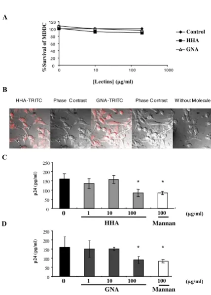
Cellular viability was determined for MDDC by culturing for 24 h in the presence of serial dilutions of the plant lectins. Plant lectin concentrations that gave culture via-bilities of more than 80% compared to control cultures were considered to be non-toxic (Fig. 4A).
(ii) Efficacy of lectins against attachment of HIV-1 on MDDC
Intraepithelial and submucosal dendritic cells (DCs) and CD4+ T lymphocytes are the predominant cell popula-tions firstly targeted by HIV-1 [13]. Indeed, successful
[image:6.612.326.540.100.399.2]transfer of virus across epithelial barriers would result in HIV-1 capture by DC and subsequent transmission to nearby CD4+ T cells or dissemination to draining lymph nodes. We first investigated whether the plant lectins are able to interact with receptors expressed at the surface of monocyte-derived dendritic cells (MDDC). As depicted in Figure 4B, both HHA and GNA interacted with receptors expressed at the surface of MDDC, suggesting that these (A). Lack of toxicity of HHA and GNA towards dendritic cells (MDDC)
Figure 4
(A). Lack of toxicity of HHA and GNA towards dendritic cells (MDDC). MDDC were cultured with concentrations of products for 24 h. After washing, culture viability was deter-mined by using the MTT cytotoxicity assay. The values given are the percentage of viability. (B). Interactions of plant lectins with surface receptors of dendritic cells. Dendritic cells (MDDC) were incubated with or without each labelled-lectin at 4°C. Cells were then analyzed by confocal micros-copy. (C and D). Ability of lectins to inhibit uptake of HIV-1Ba-L cell-free particles on dendritic cells. MDDC were co-incubated with non-toxic concentrations of HHA (C), or GNA (D) or mannan (100 µg/ml) and 1 ng p24 antigen of HIV-1Ba-L were added for 1 h. The quantity of attached-virus was evaluated by measurement of p24 antigen by ELISA. Means of three independent experiments are presented ; error bars represent standard deviations. *< 0.05
%S
urvival
of
M
DDC
B
C
Control HHA GNA
A
[Lectins] (µµg/ml)
0 50 100 150 200 250
1 10 100
HHA
(µµg/ml)
100
Mannan 0
p24 (
pg/
ml
)
* *
0 50 100 150 200 250
p24 (
pg/
ml
)
* *
D
HHA-TRITC Phase Contrast GNA-TRITC Phase Contrast W ithout Molecules
1 10 100 100 (µµg/ml)
Mannan 0
GNA 0
20 40 60 80 100 120
0 10 100 1000
Ability of lectins to inhibit transcytosis of HIV-1J-RCSF free par-ticles through a tight monolayer of endometrial epithelial cell (HEC-1A)
Figure 3
Ability of lectins to inhibit transcytosis of HIV-1J-RCSF free par-ticles through a tight monolayer of endometrial epithelial cell (HEC-1A). HIV-1J-RCSF (5 ng p24 antigen) was co-incubated with a non-toxic concentrations of each lectins or mannan (100 µg/ml) on the HEC-1A apical pole cultured in dual-chamber by the transwell system for 1 h. Results of an exper-iment performed in duplicate are expressed as the quantity of virus recovered in the basal chamber in the presence of HHA (A) and GNA (B), or in the absence of lectins. Means of three independent experiments are presented ; error bars represent standard deviations. *< 0.05, **< 0.01.
A
1 10 100
HHA
(µg/ml)
p24
(p
g/ml
)
0 5 10 15 20 25 B
p24
(p
g/ml)
0 5 10 15 20 25
Mannan 100
0
*
* ** *
1 10 100
GNA
(µg/ml)
Mannan 100
Journal of Translational Medicine 2007, 5:28 http://www.translational-medicine.com/content/5/1/28
Page 7 of 9
(page number not for citation purposes)
plant lectins would interrupt the virus attachment process by interfering both with the virus envelope glycoprotein and with the receptors expressed at the surface of MDDC. Then, we evaluated the effect of plant lectins on HIV-1 uptake by MDDC. Therefore, MDDC were incubated with HIV-1Ba-L in the presence of increasing doses of each plant lectin (Fig. 4C and Fig. 4D). Similarly to mannan (100 µg/ ml) that induced a 46% decrease of HIV adsorption on MDDC, both HHA and GNA (100 µg/ml) inhibited the virus attachment on MDDC, of 47.2 ± 5.5% and 43.1 ± 5.8%, respectively. No inhibition of HIV adsorption on MDDC was observed for both lectins at lower concentra-tions (1 and 10 µg/ml).
(iii) Efficacy of the plant lectins on HIV-1 transmission to T cell blasts by MDDC
We finally evaluated whether inhibition of attachment on MDDC resulted in subsequent inhibition of HIV-1 trans-mission to autologous T lymphocytes. MDDC were incu-bated with HIV-1Ba-L in the presence of increasing doses of each plant lectin. Free viral particles and plant lectins were removed and autologous PBL were added. The pretreat-ment of MDDC by each plant lectin resulted in a partially reduced infection of autologous PBL by HIV-1 (Fig. 5). We observed 26.9 ± 0.5% of inhibition of HIV transfer at a concentration of 100 µg/ml of HHA and 15.3 ± 1.3% of inhibition at a concentration of 100 µg/ml of GNA. In these same experiments, we used blocking anti-DC-SIGN mAb (clone 507) as positive control of inhibition of trans-fer [14]. The addition of anti-DC-SIGN mAb inhibited more than 43.8 ± 5.7% both R5- and X4- HIV-1 transfer from MDDC to T cells (Fig. 5).
Discussion
The plan lectins HHA and GNA constitute two promising microbicide molecules of great interest, capable to effi-ciently inhibit the infection of T lymphocytes and PBMC by a broad range of HIV strains [4]. This effect is mostly due to direct interactions between the plant lectins and mannose residues at the gp120 of the HIV-1 envelope and thus inhibiting fusion rather adsorption of HIV [9]. How-ever, anti-HIV effects resulting of interactions between plant lectins and cellular HIV receptors were not pro-foundly investigated. In addition, no direct interaction between the plant lectins and CD4, CCR5, CXCR4 and DC-SIGN was observed [4]. In the present study, we thus aimed at characterizing whether interactions exist between mannose-specific plant lectins and manno-sylated HIV-interacting molecules expressed at the cell surface. Indeed, HIV was described to interact with a large number of molecules onto the cell surface during the adsorption phase [2,15-19]. If some of these receptors are mannosylated close to the HIV-binding sites, the plant lectins may recognize such mannosylated residues, and may mask the HIV binding site by steric hindrance, thus
limiting HIV attachment onto target cells. Therefore, we particularly focused on the initial phase of virus adsorp-tion.
We evaluated the effect of the plant lectins on HIV adsorp-tion on genital epithelial cells. We found that HHA faintly inhibited attachment of the R5-tropic HIV-1Ba-L strain, in a dose-dependent manner, whereas GNA moderately inhibited HIV adsorption in the same context, and only at high drug doses. HSPG are known to be highly expressed on HEC-1 cells [12], and are required for HIV-1 attach-ment to epithelial cells [20]. HSPG interact with positively charged-V3 loop of X4-tropîc HIV-1 gp120, and weakly with less positively charged R5-tropic strains on epithelial cells [12]. In our hands, GNA did not inhibit the attach-ment of X4-tropic HIV-1 on epithelial cells, and did not interact with epithelial cell surface. Thus, the preincuba-tion of GNA with epithelial cells did not inhibit viral attachment whereas the preincubation of GNA with R5-tropic HIV-1 particles induced the inhibition of HIV-1 attachment on epithelial cells, suggesting that the antiviral activity of GNA is likely related to its interaction with virus particles. Indeed, altogether our results suggest that HIV-1Ba-L interact with (a) membrane molecule(s) expressed on apical side of epithelial cells during the attachment phase which is (or are) (i) not an HIV-interacting GAG, as indicated by our observations in the presence of X4-tropic viruses, (ii) not C-type lectin(s), considering the absence Ability of plant lectins to inhibit transfer of HIV-1Ba-L free par-ticles by dendritic cells to autologous T lymphocytes
Figure 5
Ability of plant lectins to inhibit transfer of HIV-1Ba-L free par-ticles by dendritic cells to autologous T lymphocytes. Den-dritic cells (MDDC) were incubated with non-toxic
concentrations of each plant lectin and 0.5 ng p24 antigen of virus were added for 3 h. The HIV-production by T cells was evaluated on the 6th day of co-culture by measurement of
p24 antigen by ELISA. Means of three independent experi-ments are presented; error bars represent standard devia-tions. *< 0.05, **< 0.01.
10
p24
(pg/ml)
1 10 100 1 10 100
0 0 2000 4000 6000 8000 10000 12000 14000
*
* *
-HHA GNA
of effect of mannan, (iii) α(1–3) mannosylated close to HIV-binding site, as indicated both by the lack of inhibi-tory effect on HIV adsorption in the presence of HHA and by the interaction of HHA with epithelial cells receptors. The identification of this (or these) molecule(s) remains to be performed.
We further assessed the role of both plant lectins on HIV transcytosis. Hocini and al. have previously demonstrated a partial inhibitory effect of mannan in transcytosis [8], thus involving a C-type lectin, conversely to adsorption phase. Only HHA, but not GNA, inhibited HIV-1JR-CSF transcytosis in a dose-dependent manner. Whereas GNA has a specificity for terminal α(1–3)-linked mannose res-idues, HHA recognizes both terminal and internal α(1–3) and α(1–6)-linked mannose residues [9]. Whether this additional specificity of HHA is the reason for the effect of this plant lectin on transcytosis remains to be demon-strated. Since HHA inhibited specifically transcytosis and faint to inhibit HIV-1 adsorption, we confirm herein that adsorption and transcytosis of HIV are likely two distinct phenomenons involving in the latter one (a) C-type lec-tin(s) and HHA-interacting mannosylated molecule(s) [12]. Involvement of a single HIV-interacting molecule exhibiting mannosylation close to the C-type lectin region (CLR domain) within the transcytosis phase is also possi-ble. HEC-1 epithelial cells used in our model do not express mannose receptor [19] nor DC-SIGN molecule [15], which could be involved in cell-free HIV-1 particles endocytosis (data not shown).
We evaluated the role of lectins on HIV adsorption on MDDC, representing one of the primary HIV target cells in the course of sexual transmission [13]. Similarly to MDM, HIV is adsorbed on MDDC via C-type lectins [15], synde-cans [20], and to some extent CD4 [21]. However, the diversity of C-type lectins expressed on MDDC is broader to that expressed on MDM, as illustrated by the restricted expression of DC-SIGN or DEC-205 on MDDC [22,23]. Nevertheless, if some C-type lectins interact with HIV, recent data suggest a specific role for each HIV-interacting C-type lectins. Thus, the mannose receptor seems to be more involved in HIV attachment and internalization whereas DC-SIGN seems to be more involved in the trans-fer of infectious viruses to susceptible CD4+ T cells [24]. In that context, our results indicated that both plant lectins partially inhibited HIV attachment on MDDC at the high-est concentration thigh-ested (100 µg/ml), similarly to man-nan. Therefore, these results strongly suggest that : (i) both HHA and GNA interact with MDDC surface proteins exhibiting terminal α(1–3)-mannosylation ; (ii) some of these mannosylated proteins interact with HIV in a man-ner which could be inhibited by plant lectin interaction, and (iii) that some of these mannosylated proteins could be C-type lectins, considering the blocking effect of
man-nan and the relative role of those lectins, syndecans and CD4 in HIV adsorption on MDDC. Interestingly, a recent report has described a selective tissus-specific mannosyla-tion of the mannose receptor on macrophages which could react in some cases with GNA [25]. Thus, it is also tempting to suggest that the mannose receptor could be mannosylated close to the HIV binding site when expressed on MDDC, conversely to an unmannosylated form of the mannose receptor expressed on MDM, there-fore explaining the observed difference between the plant-lectin effects on MDDC and on epithelial cells.
Finally, we assessed the role of HIV-lectins pre-treatment on MDDC to T cells transfer of HIV. Our results showed a modest decrease of HIV transfer between MDDC and PBL. In addition, this down-modulation was inferior to that observed for HIV adsorption on MDDC in the presence of lectins. In the system we used, unbound plant lectins were eliminated by washing treated-MDDC before the addition of T cells, in order to focus exclusively on the effect of adsorbed plant-lectin on MDDC. Our observations sug-gest that plant lectins act on MDDC more specifically on adsorption than on the transfer phase. This weak effi-ciency of plant lectins to inhibit HIV-1 transfer was also reported in a recent published study which was performed using cell lines (Raji/DC-SIGN cell system and C8166 T cell line [5]. Recently, it was reported that dendritic cells expressed a large array of receptors implicated on the HIV-1 attachment and/or transfer [2]. In particular, Turville and colleagues reported that dendritic cells do not express DC-SIGN but mannosylated receptors in tissues-where HIV-1 is captured by dendritic cells-, whereas in lymph nodes-where transfer to T cells occurs- dendritic cells express largely DC-SIGN, suggesting that receptors involved in attachment and transfer are different [2]. In the present study, HHA inhibited as efficiently as GNA HIV-1 adsorption on dendritic cells, but only HHA inhib-ited HIV-1 transfer from MDDC to T cells, suggesting that HIV-1 receptors invloved in HIV attachment and in HIV-1 transfer may be different, and that HHA and GNA may interact with different(s) receptor(s).
Journal of Translational Medicine 2007, 5:28 http://www.translational-medicine.com/content/5/1/28
Page 9 of 9
(page number not for citation purposes)
transport of HIV through the mucosa. Combination prod-ucts could in principle provide a greater degree of protec-tion than single agents, a broader spectrum of activity against various pathogens and a lower risk of adverse reac-tions. Since high concentration of HHA are necessary to inhibit the adsorption on the HIV target cells, the combi-nation of HHA derivates with one or two other compo-nent(s) would allow a lower dose of this component.
Competing interests
The author(s) declare that they have no competing inter-ests.
Authors' contributions
HSand NNcarried out differentiation and infection of dendritic cells, isolation of T cells, HIV-1 transfer assays, cytotoxicity assay, evaluation of the epithelial monolayer integrity and ELISA.
MAJ carried out cytotoxicity assay, transcytosis assays, HIV-1 attachment assays.
ML andCK carried out the confocal microscopy assays and helped draft the manuscript.
JBand DS: provided plant lectins, participated in the design of the study, and helped draft the manuscript.
LB and HS: conceived the study, participated in its design and coordination, and helped draft the manuscript.
All authors read and approved the final manuscript.
Acknowledgements
We gratefully acknowledge Christophe Klein for technical assistance and Dr Cédric Carbonneil for helpful discussions. This work was supported by a grant from the European Commission (VIth framework, project EMPRO Contract no. 503558) and by a grant from the ANRS (MultiMicro Project). H.S was recipient of grant from EMPRO.
References
1. Stone A: Potential of vaginal microbicides in HIV control. J R Soc Med 2004, 97(3):158.
2. Turville SG, Cameron PU, Handley A, Lin G, Pohlmann S, Doms RW, Cunningham AL: Diversity of receptors binding HIV on den-dritic cell subsets. Nat Immunol 2002, 3(10):975-983.
3. Van Damme EJM A. K. Allen, and W. J. Peumans.: Related man-nose-specific lectins from different species of the family Amaryllidaceae. Plant Physiol 1988, 73:52-57.:.
4. Balzarini J, Hatse S, Vermeire K, Princen K, Aquaro S, Perno CF, De Clercq E, Egberink H, Vanden Mooter G, Peumans W, Van Damme E, Schols D: Mannose-specific plant lectins from the Amarylli-daceae family qualify as efficient microbicides for prevention
of human immunodeficiency virus infection. Antimicrob Agents
Chemother 2004, 48(10):3858-3870.
5. Balzarini J, Van Herrewege Y, Vermeire K, Vanham G, Schols D: Car-bohydrate-binding agents efficiently prevent dendritic cell-specific intercellular adhesion molecule-3-grabbing nonin-tegrin (DC-SIGN)-directed HIV-1 transmission to T lym-phocytes. Mol Pharmacol 2007, 71(1):3-11.
6. Stafford MK, Ward H, Flanagan A, Rosenstein IJ, Taylor-Robinson D, Smith JR, Weber J, Kitchen VS: Safety study of nonoxynol-9 as a
vaginal microbicide: evidence of adverse effects. J Acquir
Immune Defic Syndr Hum Retrovirol 1998, 17(4):327-331.
7. Lorin C, Saidi H, Belaid A, Zairi A, Baleux F, Hocini H, Belec L, Hani K, Tangy F: The antimicrobial peptide dermaseptin S4 inhibits HIV-1 infectivity in vitro. Virology 2005, 334(2):264-275. 8. Hocini H, Becquart P, Bouhlal H, Chomont N, Ancuta P, Kazatchkine
MD, Belec L: Active and selective transcytosis of cell-free human immunodeficiency virus through a tight polarized
monolayer of human endometrial cells. J Virol 2001,
75(11):5370-5374.
9. Balzarini J, Schols D, Neyts J, Van Damme E, Peumans W, De Clercq
E: Alpha-(1-3)- and alpha-(1-6)-D-mannose-specific plant
lectins are markedly inhibitory to human immunodeficiency
virus and cytomegalovirus infections in vitro. Antimicrob Agents
Chemother 1991, 35(3):410-416.
10. Bomsel M: Transcytosis of infectious human immunodefi-ciency virus across a tight human epithelial cell line barrier.
Nat Med 1997, 3(1):42-47.
11. Vives RR, Imberty A, Sattentau QJ, Lortat-Jacob H: Heparan sulfate targets the HIV-1 envelope glycoprotein gp120 coreceptor binding site. J Biol Chem 2005, 280(22):21353-21357.
12. Saidi H, Magri G, Nasreddine N, Requena M, Belec L: R5- and X4-HIV-1 use differentially the endometrial epithelial cells HEC-1A to ensure their own spread: Implication for mechanisms of sexual transmission. Virology 2007, 358(1):55-68.
13. Hladik F, Sakchalathorn P, Ballweber L, Lentz G, Fialkow M, Eschen-bach D, McElrath MJ: Initial events in establishing vaginal entry and infection by human immunodeficiency virus Type-1.
Immunity 2007, 26(2):257-270.
14. Saidi H, Magri G, Carbonneil C, Nasreddine N, Requena M, Belec L:
IFN-{gamma}-activated monocytes weakly produce HIV-1 but induce the recruitment of HIV-sensitive T cells and
enhance the viral production by these recruited T cells. J
Leu-koc Biol 2006.
15. Geijtenbeek TB, Kwon DS, Torensma R, van Vliet SJ, van Duijnhoven GC, Middel J, Cornelissen IL, Nottet HS, KewalRamani VN, Littman DR, Figdor CG, van Kooyk Y: DC-SIGN, a dendritic cell-specific HIV-1-binding protein that enhances trans-infection of T cells. Cell 2000, 100(5):587-597.
16. Haywood AM: Virus receptors: binding, adhesion strengthen-ing, and changes in viral structure. J Virol 1994, 68(1):1-5. 17. McDougal JS, Maddon PJ, Orloff G, Clapham PR, Dalgleish AG, Jamal
S, Weiss RA, Axel RA: Role of CD4 in the penetration of cells by HIV. Adv Exp Med Biol 1991, 300:145-54; discussion 155-8. 18. Mondor I, Ugolini S, Sattentau QJ: Human immunodeficiency
virus type 1 attachment to HeLa CD4 cells is CD4 independ-ent and gp120 dependindepend-ent and requires cell surface heparans.
J Virol 1998, 72(5):3623-3634.
19. Nguyen DG, Hildreth JE: Involvement of macrophage mannose receptor in the binding and transmission of HIV by macro-phages. Eur J Immunol 2003, 33(2):483-493.
20. Saphire AC, Bobardt MD, Zhang Z, David G, Gallay PA: Syndecans serve as attachment receptors for human immunodeficiency virus type 1 on macrophages. J Virol 2001, 75(19):9187-9200. 21. Vermeire K, Schols D: Cyclotriazadisulfonamides: promising
new CD4-targeted anti-HIV drugs. J Antimicrob Chemother 2005,
56(2):270-272.
22. McGreal EP, Miller JL, Gordon S: Ligand recognition by
antigen-presenting cell C-type lectin receptors. Curr Opin Immunol
2005, 17(1):18-24.
23. McGreal EP, Martinez-Pomares L, Gordon S: Divergent roles for C-type lectins expressed by cells of the innate immune sys-tem. Mol Immunol 2004, 41(11):1109-1121.
24. Chehimi J, Luo Q, Azzoni L, Shawver L, Ngoubilly N, June R, Jerandi G, Farabaugh M, Montaner LJ: HIV-1 transmission and cytokine-induced expression of DC-SIGN in human monocyte-derived macrophages. J Leukoc Biol 2003, 74(5):757-763. 25. Su Y, Bakker T, Harris J, Tsang C, Brown GD, Wormald MR, Gordon