R E S E A R C H
Open Access
The development of sexual stage malaria
gametocytes in a Wave Bioreactor
Corine G. Demanga
1, Jenny W. L. Eng
1, Donald L. Gardiner
2,3, Alison Roth
4, Alice Butterworth
2, John H. Adams
5,
Katharine R. Trenholme
2,5and John P. Dalton
1,6*Abstract
Background:Blocking malaria gametocyte development in RBCs or their fertilization in the mosquito gut can prevent infection of the mosquito vector and passage of disease to the human host. A‘transmission blocking’ strategy is a component of future malaria control. However, the lack of robust culture systems for producing large amounts ofPlasmodium falciparumgametocytes has limited our understanding of sexual-stage malaria biology and made vaccine or chemotherapeutic discoveries more difficult.
Methods:The Wave BioreactorTM20/50 EHT culture system was used to develop a convenient and low-maintenance
protocol for inducing commitment ofP. falciparumparasites to gametocytogenesis. Culture conditions were optimised to obtain mature stage V gametocytes within 2 weeks in a large-scale culture of up to a 1 l.
Results:We report a simple method for the induction of gametocytogenesis with N-acetylglucosamine (10 mM) within a Wave Bioreactor. By maintaining the culture for 14–16 days as many as 100 million gametocytes (stage V) were produced in a 1 l culture. Gametocytes isolated using magnetic activated cell sorting (MACS) columns were frozen in aliquots for storage. These were revitalised by thawing and shown to retain their ability to exflagellate and infect mosquitoes (Anopheles stephansi).
Conclusions:The production of gametocytes in the Wave Bioreactor under GMP-compliant conditions will not only facilitate cellular, developmental and molecular studies of gametocytes, but also the high-throughput screening for new anti-malarial drugs and, possibly, the development of whole-cell gametocyte or sporozoite-based vaccines.
Keywords:Plasmodium falciparum, Gametocytes, In vitro culture, Wave Bioreactor, Malaria, Drug discovery, Vaccines
Background
According to the World Malaria Report 2014 there are an estimated 3.3 billion people at risk of malaria. In 2013, there were between 124 and 283 million cases of malaria and 367–755,000 deaths. Malaria is caused by protozoan parasites of the genus Plasmodium and among the five species that cause malaria in humansP. falciparumis the deadliest. Greater than 90% of malaria deaths occur in sub-Saharan Africa, predominantly in children below the age of five, where this species is most prevalent [1].
The life-cycle of P. falciparum parasites is complex and occurs within two hosts, the human and the female Anopheles mosquito [2]. In the human host, the para-sites grow and multiply asexually within red blood cells (RBCs), destroying these when they emerge to invade new cells. As the number of infected RBCs increase, some parasites develop into sexual forms, the micro-(male) and macro-(female) gametocytes. Gametocytes develop through five distinct stages, with only mature stage V parasites able to undergo sexual reproduction when ingested by a feeding mosquito [3–5]. The micro-gametes and macromicro-gametes of P. falciparum emerge from the ingested RBCs in the mosquito gut where fertilization occurs and results in the formation of a mo-tile ookinete. The ookinete develops into an oocyst con-taining sporozoites that subsequently migrate to the mosquito salivary glands where they become fully
* Correspondence:j.dalton@qub.ac.uk
1
Institute of Parasitology, McGill University, 21111 Lakeshore Road, Sainte-Anne-de-Bellevue, Québec H9X 3 V9, Canada
6School of Biological Sciences, Medical Biology Centre, Queen’s University of Belfast, 97 Lisburn Road, BT9 7BL Northern Ireland, UK
Full list of author information is available at the end of the article
mature after about 10 days. These mature sporozoites infect the next human host when the mosquito takes a blood meal [2, 5, 6].
Inhibiting gametocyte development in RBCs or venting their fertilization in the mosquito gut can pre-vent infection of the vector and, therefore, passage of disease to the human host. To achieve the ambitious goal of malaria eradication it is envisaged that the strat-egy of ‘transmission blocking’ will have a vital role to play [7–10]. This approach would reduce or interrupt the spread of malaria disease in endemic regions and will be employed alongside vector control and case manage-ment by chemotherapy host. The Malaria Eradication Research Agenda (malERA) initiative has identified the development of new safe and effective drugs that block the infectivity of mature P. falciparumgametocytes as a priority research area. However, identification of com-pounds with activity against late stage P falciparum ga-metocytes is challenging as this stage is relatively metabolically inert and in vitro culture methods to pro-duce gametocytes are much less straightforward than for asexual stage parasites. The 8-aminoquinoline prima-quine is currently the only licensed antimalarial drug ef-fective against late stage gametocytes, but known side effects of primaquine limit its usefulness [6, 8, 11, 12]. In addition to the discovery of new drugs that kill gameto-cytes, there has been a long-term interest in developing a vaccine that specifically targets the sexual stages and interrupts malaria transmission [7–10].
While studies on the asexual stages of P falciparum were greatly facilitated by the development of an in vitro culture system [13] the lack of a robust culture system for sexual stages has limited our understanding of P falcip-arum gametocyte biology and made studies with these stages much more difficult. The most commonly used methods for gametocyte production start with cultures of asexual stage parasites, which are then“induced’to differ-entiate into early stage gametocytes that develop to matur-ity in a stable culture period of 10–14 days [14]. The mechanism of commitment to sexual development ofP. falciparum is poorly understood, although this switch is suggested to occur in the generation preceding gametocy-togenesis [15] and are dependent on genetic, epigenetic and transcriptional factors [6]. The stimuli leading to commitment act on the merozoite and/or the early ring stage parasite [6, 15–17] and factors such as host immun-ity and presence of antimalarial drugs may increase game-tocytogenesis in vivo, while erythrocyte lysates, host immune sera, hypoxanthine, conditioned medium, high asexual parasite density and cAMP inducing pathway have been exploited, with variable results, to trigger or enhance gametocytogenesis in vitro [3, 5, 6, 15–20].
Several methodologies for small-scale gametocyte cul-ture have been published but regardless of the method
employed, the production of gametocytes in vitro is tech-nically challenging, time consuming, costly, and lacks sta-bility and reproducista-bility [4, 21–24]. Even in parasite lines with relatively high conversion rates, gametocytes usually represent less that 1% of the parasitized cells within a given culture [6, 15] and, consequently, the amount of gametocyte material that can be isolated from in vitro sys-tems is not conducive to large or high-throughput studies. Recently, Delves et al. [25] and Duffy et al. [26] reported detailed protocols for the routine culture of gametocytes in volumes between 0.2 and 200 ml.
We described the large-scale production of asexual blood stage P. falciparumcultures in the Wave Bioreac-torTM 20/50 EHT system [27], which is a closed sterile plastic bioreactor that rocks on a heated platform. Here, we report use of the Wave BioreactorTMfor the develop-ment of a convenient and low-maintenance protocol for producing gametocytes and subsequent maintenance of these to obtain mature stage V gametocytes in a large-scale culture of up to a 1 l. As many as 100 million ma-ture P. falciparum gametocytes can be produced and purified from each culture and these can be cryopre-served and resurrected for in vitro studies. The resur-rected gametocytes were shown to successfully infect mosquitoes.
Methods
Asexual parasite culture
ThePlasmodium falciparumstrains 3D7 and FCR3 were selected for this study. The incomplete culture medium was prepared from RPMI 1640 powder with L-Glutamine (Gibco/Invitrogen, Montreal, Canada) at 10.5 g/l, buffered with 24 mM sodium bicarbonate (NaHCO3), 35 mM HEPES and Hypoxanthine (Sigma-Aldrich, Montreal, Canada). Prior to use, Albumax-I (0.5% w/v) and 25μM of gentamycin (Gibco/Invitrogen) were added to the medium to give complete culture medium. Medium used for experiments performed at QIMRB was identical to that described above with the exception that Albumax-1 was replaced by 10% human serum. Fresh human erythro-cytes preserved in CPDA-1 (citrate-phosphate-dextrose-adeninine) anticoagulant solution were obtained once a month from Interstate Blood Bank, Memphis, Tennessee, USA, or from the Australian Red Cross Blood Service, Brisbane, Australia. They were washed twice with 10 vol-umes of RPMI-1640 and suspended at 50% (vol./vol.) with complete RPMI medium.
Canada) regulated with a gaseous environment of 3% O2, 5% CO2 and 92% N. To synchronize the cultures, sorbitol treatment was performed on parasites ring stage and repeated after 48 h [28, 29].
The Wave Bioreactor system forP. falciparumblood stage culture
The Wave BioreactorTM20/50 EHT system (GE Health-care, Montreal, Canada) was used for this study. This system is composed of a presterilized transparent inflat-able plastic bag, or Cellbag, which is the disposinflat-able culti-vation chamber. Each Cellbag has integrated inlet and outlet air filters and ports for adding medium and extracting samples (Additional file 1: Figure S1), for this study a 2 l Cellbag, recommended for cell culture vol-umes of 0.1 to 1 l was used. The Cellbag is placed on a platform that sits on a rocking unit, which is designed to inflate and rock the bioreactor for rapid gas transfer. Using a touch-screen connected to the rocking unit, the motion (speed and angle) and temperature of the plat-form and bioreactor is tightly controlled. The volume and rate of gas entering the bioreactor is also regulated. The platform of the Wave BioreactorTM20/50 EHT sys-tem contains two separate heating panels, allowing sin-gle or dual operation, i.e. one or two 2 l bioreactor chambers can be run at a given time. Several parameters within the bioreactor chamber can be controlled re-motely by a WavePODTMcontroller, including dissolved oxygen (disO2), pH, and O2/CO2 gas mixing. Although this controller is an optional unit, we employed it in the present study to monitor disO2 during culture of
Plas-modium in the Cellbag. We did not use the
Wave-PODTM controller to create the gas mix within the bioreactor; instead, an analyzed gas mix of 3% O2, 5% CO2and 92% N was directly fed from a cylinder into the bioreactor via the rocking unit. The protocol for culture of asexual P. falciparum blood stage in the Wave Bio-reactor is described in Additional file 1: Figure S1.
To optimise gametocyte production in the Wave Bio-reactor, several experiments with both synchronous or asynchronous blood stage cultures were performed inde-pendently at the McGill University in Canada and QIMR Berghofer Medical Research Institute (QIMRB) in Australia. Therefore, we have developed a straightfor-ward protocol to produce P. falciparum gametocytes in the Wave Bioreactor that is described in Fig. 1 approxi-mately 10–15 days before initiation of gametocyte pro-duction in the Wave Bioreactor, P. falciparum aliquots were thawed and cultured as described. Schizont enrich-ment by flotation on 1% (wt./vol.) gelatin (Sigma-Al-drich) was performed to replace old RBCs in the culture with fresh ones [28]. Parasites where synchronized as de-scribed for inoculation in the Cellbag [28, 29]. The start-ing volume in the Cellbag was between 350 and 500 ml.
Once induction of gametocytogenesis had occurred N-acetylglucosamine (GlcNAc, Sigma-Aldrich) was added to the Cellbag at a final concentration of 10 mM to pre-vent further proliferation of asexual stage parasites. Ali-quots of GlcNAc stock solution were prepared at 1 M, stored at -20 °C and thawed immediately prior to use. Culture samples (2–5 ml) were harvested daily from the Cellbag through the multi-use port via a 10 ml luer-lock syringe and using to monitor parasitemia and gametocy-taemia, as well as to measure glucose consumption, lac-tate production and pH levels. Glucose and laclac-tate measurement was performed at the Montreal General Hospital, Canada, with the blood gas analyzer ABL825 Flex (Radiometer, Diamond Diagnostic-USA). Parasite cell counts and assessment of specific developmental stages were performed with light microscope on Giemsa-stained thin smears. The parasitemia and game-tocytaemia were determined over the examination of be-tween 2000 and 12,000 RBCs. The morphological criteria of stage II-V gametocytes described elsewhere [3] were used to evaluate the maturity of gametocytes on Giemsa-stained thin smears. The sex of mature stage V gametocytes was differentiated by morphology as depicted by Mitri et al. [30].
Gametocyte purification
During the production of gametocytes in the Wave Bio-reactor, mature gametocytes can be purified over four consecutive days starting at day 10 of the culture. Sixteen hours prior to the purification, the speed and the angle of the rocking unit were adjusted to 2 rpm and 3°, respect-ively, to allow the RBCs to settle in the Cellbag. Since gametocyte infected RBCs tend to float as the parasites mature, they were collected by gently removing the super-natant via the multi-used port. From each 1 l culture bag, 200–400 ml of supernatant was removed each day and re-placed with fresh complete RPMI medium. The bioreactor was then returned to 8 rpm through an angle of 6° to allow mixing of the remaining culture. The isolation of ga-metocytes from the cells harvested in the supernatant was performed by magnet-activated cell sorting (MACS) as previously described [4]. Gametocyte numbers were quan-titated by haemocytometer.
Cryopreservation of gametocytes and procedure for thawing
washed with 10 volume of RPMI. The glycerin-based solution Glycerolyte 57 (Fenwall Inc., Lake Zurich, Il, USA) was added to the cell pellet to yield a final con-centration of 85% (v/v) in two steps. First, 1/5 of the pellet volume of Glycerolyte 57 was added drop wise to the cells and after about 2 min at room temperature, the remaining volume was added and the cell suspension was distributed in cryotubes. The cryotubes were placed at -80 °C for 24 h and then transferred to liquid nitrogen for storage.
[image:4.595.57.537.81.584.2]suspension. The cell suspension was centrifuged at 700× gfor 4 min, and the pellet was suspended in a 0.9% NaCl warmed solution equal to 10 vol. of the original vial con-tents, then centrifuged as before. Samples of the pellet were taken to prepare a thin smear for Giemsa-stain or for exflagellation test and the remaining was suspended in complete RPMI for time development study.
Assessment of gametocytes viability after freezing
Giemsa-stained thin smears made with samples of frozen cells prepared at different time after thawing, were ex-amined by microscopy for morphological evaluation of the different gametocytes stages. For the in vitro exfla-gellation assay, the exflaexfla-gellation-inducing medium con-taining 100μM of xanthurenic acid (XA, Sigma-Aldrich) was prepared as described by Bhattacharyya and Kumar [32]. All the components of the exflagellation test where kept at room temperature. Cell samples obtained after thawing, or freshly collected from the Bioreactor were centrifuged as above and the pellet was suspended at 25% hematocrit in exflagellation-inducing medium. To examine exflagellation, an aliquot of the cell suspension was collected after 10–15 min and transferred on a coverslip attached with silicone on cell chamber. The sample was diluted with exflagellation-inducing medium, to allow visualization of single cells, and analysed by confocal differential interference microscopy (DIC).
Assessment gametocyte infectivity
The cryopreserved gametocytes were shipped on dry ice to the University of South Florida where the mosquito membrane feeding assays (MFA) were performed. After rapid thawing by the procedure described above using graded salt solutions gametocytes were added to a warmed mixture of human serum and RBCs at a ratio of 1:1.6. Fifty female Anopheles stephensi mosquitoes starved for 4 h were fed with small volumes of gameto-cytes samples (500–1000μl) through the Hemotek 5 W1 membrane feeding system (Discovery Workshop, Eng-land) at 37 °C, or at room temperature to prevent rapid desiccation, although mosquitoes fed poorly on the un-heated samples. Then the mosquitoes were transferred in chamber set at 26 °C and 80% relative humidity and feed with 10% sugar solution on alternate days. The in-fected mosquitoes were dissected to determine midgut oocysts on day 8–12 and salivary gland sporozoites on days 16–20 post-blood meal.
Results
Protocol design for the large-scale culture ofP. falciparum gametocytes in the Wave Bioreactor
In our original protocol developed for the large-scale culture of asexual stages in a Wave Bioreactor we re-ported that the Cellbag in which the parasites were
cultured must be removed daily from the heated rocking platform and taken to a laminar flow hood for medium change [27]. To simplify and streamline this culture process a reservoir bag filled with medium was con-nected to the Cellbag via the extended inlet tubing creat-ing a completely closed system (Additional file 1: Figure S1). Medium can be removed from the Cellbag via the multi-port unit while it remains in situ on the platform and fresh medium added by opening the connection be-tween the reservoir and the Cellbag. In this modified protocol, set-up of a 1 l culture is completed in less than 2 h and, thereafter, the Cellbag is not removed from the heated rocking platform for the entire process. This pre-vents parasites being exposed to temperature fluctua-tions that occurred in our earlier protocol.
Using the protocol set out in Additional file 1: Figure S1, asexualP. falciparumparasites,3D7andFCR3, grow very favourably (Additional file 2: Figure S2a). Measure-ments of glucose consumption in the parasite cultures showed that this nutrient is maintained within optimal limits for growth (8 to 10 mM) (Additional file 2: Figure S2b). Furthermore, lactate build-up, with the consequent reduction of pH, is controlled so that these do not reach levels that are detrimental to the parasite, i.e. lactate above 12 mM and pH below 7 (Additional file 2: Figure S2b, c). The ability to control conditions within the pa-rameters optimal for P. falciparum parasite growth by replenishing medium daily ensured that a parasitemia of 5–6% was routinely obtained over four to 5 days in 1 l cultures.
P. falciparumgametocyte induction in the Wave Bioreactor
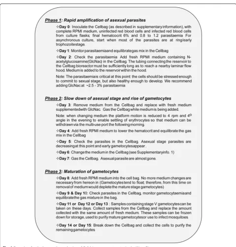
Our long-term culture ofP. falciparumwithin the Cell-bag bioreactor paved the way for the development of a protocol for the reliable large-scale production of the sexual stages. In our large-scale gametocyte production protocol (Fig. 1), sexual stage induction occurs during Phase 1 and Phase 2 leading to development of mature Stage V gametocytes ready for mosquito infection in phase 3. Gametocyte development is susceptible to temperature fluctuations that significantly deviate from normal body temperature and adversely affecting their ability to reach maturity. Thus, the addition of the medium reservoir to the system so that there is no need to remove the Cellbag from the heated platform was greatly beneficial (the temperature within the Cellbag never dropped below 35 °C).
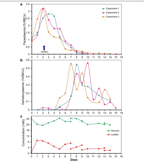
Therefore, to enhance production of gametocytes in the Wave Bioreactor, asexual stage cultures were established at a starting parasitemia of between 0.8 and 1.1% and allowed to multiply rapidly such that at day 2 the para-sitaemia was almost triple the starting parapara-sitaemia (Fig. 2a). During this initial period, the culture medium in the Wave Bioreactor was intentionally not changed to stress the parasites and induce gametocytogenesis [19, 33]. As alluded to above, when asexual parasites multi-ply, they rapidly deplete the glucose in the medium and produce lactic acid (Fig. 2c), which results in a rapid drop of pH to levels that are detrimental to gametocyte development. For this reason, we used N-acetylglucosamine (GlcNac) to halt the growth the asex-ual parasites before their density became too high [34].
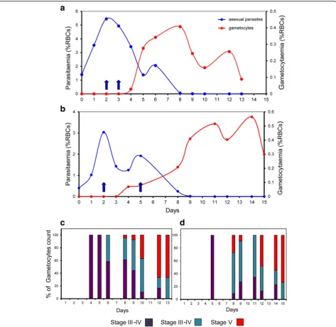
N-acetylglucosamine (GlcNac), at a concentration of 50 mM, was previously shown to stop the multiplication of asexual parasites over a 72 h period without harm to gametocyte development [26]. In our system, several ini-tial tests with asynchronous and synchronous cultures allowed us to determine that GlcNac at a concentration of 20 mM for 48 h successfully inhibited asexual parasite growth in the Cellbag (Fig. 1). GlcNac was added to the Cellbag 2 days after cultures were established when the parasitemia was between 2.5 and 3.5% for asynchronous culture and between 3 and 6% for synchronous culture. This timing was optimal as it allows the asexual parasit-aemia to be sufficiently high to allow for the generation of large numbers of gametocytes, but not so high as to overly stress the culture (Figs. 2a and 4a, b). The parasit-aemia dropped abruptly during the 48 h following the addition of GlcNac to the Cellbag, then the remaining number of asexual stage parasites slowly diminished be-fore completely disappearing over the following five to 7 days (Fig. 2a). The same growth pattern was observed with synchronous cultures in the experiments performed in two different research laboratories (McGill University, Canada, and QIMR Berghofer Medical Research Insti-tute, Australia) (Fig. 4a, b). The number of gametocytes started to rise soon after the addition of GlcNac, day 3 to 4 from the initiation of the culture, and developed to become mature stage V gametocytes over the following days (Figs. 2, 3 and 4).
The development ofP. falciparumgametocytes in large-scale culture
Sexual stage cultures produced in the Wave Bioreactor from asynchronous blood stage cultures were character-ized by the occurrence of successive “waves”of gameto-cytes (Fig. 2b). The development of gametogameto-cytes initiated with cultures containing synchronous asexual stage parasites exhibited more synchronicity (Fig. 4a, b). We observed that RBCs containing mature gametocytes tend to float (Fig. 3b) which posed a problem when
medium needed to be changed (it is necessary to slow down the rocking platform to allow erythrocytes to settle so that medium can be drawn from the top, Additional file 1: Figure S1). However, we discovered that it was not necessary to change the medium in the cultures after day 7 (Fig. 2). Indeed, from day 8 onwards the lactic acid level and pH of the culture medium remained stable, most likely because gametocytes consume far less glu-cose than do asexual blood stage parasites and therefore produce less lactic acid (see Fig. 2c).
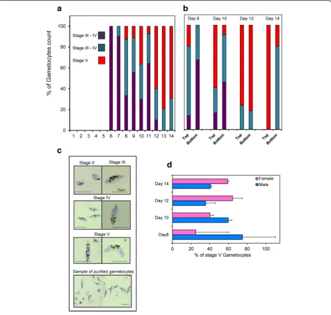
Gametocyte maturation is divided into five develop-mental stages (stages I to V) as distinguished on Giemsa-stained smears (Fig. 3c). Indeed, stage I gameto-cytes are morphological identical to young asexual trophozoite and are very difficult to identify on Giemsa stained blood film [5]. Mature stage V gametocytes first appeared on day 8 of the culture in the Wave Bioreactor and the number progressively increased thereafter to day 15 (Figs. 3a and 4c, d). During the development of game-tocytes to full maturity we observed a predominance of male gametocytes from day 8 to day 11+/−1 before the females became more abundant (Fig. 3d). The observa-tion that male gametocytes reach maturity earlier, or have a shorter lifespan, than female gametocytes which results in a greater abundance of female gametocytes in the final culture has also been reported by others [30].
Viability, infectivity and cryopreservation of gametocytes produced in the Wave Bioreactor
Plasmodium falciparum 3D7 gametocytes produced in the bioreactor were isolated using magnet-activated cell sorting (MACS) as previously described [4]. This repre-sents a logistical bottleneck for large-scale purification since a 10 ml MACS column is recommended for the isolation of parasites from about 1 ml PCV of erythrocytes (in the Wave Bioreactor, we routinely have 25 to 30 ml PCV). Nevertheless, the isolation of gametocyte can be spread over several days; we recommend taking samples from the Cellbag from day 10 to day 13 (see below).
An alternative means of isolating gametocytes takes advantage of the fact that gametocytes are more buoyant than uninfected cells and tend to float. Therefore, when the rocking motion of the platform was slowed, unin-fected erythrocytes settled to the bottom and the more buoyant gametocyte-infected RBCs remained at the top and could be drawn off via the exit port. These could then be further purified using a MACS column. In our studies, we could isolate up to 100 to 110 million game-tocytes from a 1 l culture.
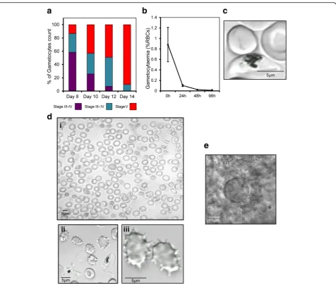
Peatey et al. [14]. We derived these reported methods to successfully cryopreserve and retrieve gametocytes at all stages of development (Fig. 5a). Gametocytes isolated from the Cellbag were frozen on day 10–13; these days
were judged to be the best time to prepare mature stage V gametocytes with a good ratio of male and female forms (Figs. 3a, d and 4c, d). Samples of the cryopreserved para-sites were thawed and examined in exflagellation tests. Fig. 2Time course for the development ofP. falciparumgametocytes in the Wave Bioreactor starting from asynchronous blood stage culture.a
Proliferation of asexual parasites and gametocyte induction in three representative experiments,arrowrepresents the time of addition of GlcNAc.
[image:7.595.60.538.85.618.2]The exflagellation of male gametocytes post-thawing was observed after incubation at 25 °C for 30 to 45 min in the presence of xanthurenic acid (Fig. 5d).
Tests were also performed to determine if resurrected parasite could infect mosquitoes. These tests were carried out on the same day as thawing since a sharp decline in via-bility was observed when gametocytes were cultured at 37 ° C (a reduction of > 50% during the first 24 h, Fig. 5b).P.
fal-ciparum gametocytes remained infectious after
cryopreservation as we observed 2–4 oocysts in 6 of 25 laboratory-rearedAn. stephensimosquitoes after membrane feeding (Fig. 5e). However, no sporozoites could be detected at 16–20 days after membrane feeding due to the small quantity of mosquitoes dissected and low infection rate.
Discussion
Here we describe methods for large scale in vitro culture of P. falciparum blood stages in a Wave Bioreactor Fig. 3The development ofP. falciparumgametocytes in the Wave Bioreactor.aGametocyte maturity over the time. Eachbarrepresents the percentage of a specific gametocyte stage over the total number of gametocytes (averaged over the three experimentsshown in Fig. 2).b
Comparison of Gametocyte maturity stages from samples collected in the Cellbag while rocking (“bottom”) or in the supernatant after the rocker was stopped for several hours (“top”). Bar represents 10μm.cPictures of Giemsa stained thin smears representing the different gametocyte maturity stages obtained during the culture in the Wave Bioreactor and sample of mature gametocytes harvested after purification.d
[image:8.595.61.538.88.542.2]system. Because parasites cultured in this system are kept in suspension by the slow rocking of the media, nu-trients are equally available to all cells, metabolites (in-cluding lactic acid) are quickly dispersed and the pH throughout the culture is balanced. As a result, malaria parasites exhibit improved grow rates than in static cul-tures, parasite cell synchronicity is preserved and the number of multiple-infected erythrocytes is much re-duced [27]. The addition of a reservoir for holding medium has made the operation of the system more
convenient for changing medium and because the cul-ture remains continuously on the heated rocking plat-form parasites do not experience temperature or gas fluctuations. Moreover, since the system is completely closed, the risk of contamination from the environment is minimal.
[image:9.595.63.537.87.548.2]constant temperature, good gas exchange, and regular medium exchange in the bioreactor are more conducive for the development of gametocytes than in static cultures. We can now reproducibly generate and purify approximately 100 million gametocytes from a 1 l culture. Samples of par-asites of sufficient quantities can be readily taken at any time-points during the culture to allow stage-specific stud-ies and batches of maturing gametocytes can be collected from the same Cellbag culture over several days.
Exflagellation tests verified that gametocytes produced in our system are viable, and can be stored frozen in small ali-quots and re-vitalized in vitro when needed. We have
described protocols for the freezing and thawing of gameto-cytes from stage III to V, although we found that after thawing gametocyte viability drops abruptly within the first 24 h of culture. Most importantly, our results provide clear evidence that mature gametocytes thawed from storage can exflagellate on the same day as thawing.
[image:10.595.61.538.85.489.2]from the same culture for studies of stage development, organelle isolation and other analyses requiring larger quantities of parasite products (e.g. antigens for trans-mission blocking vaccine, studying of gametocytes com-mitment and development, proteomics etc.). Greater efficiency and experimental reproducibility can be achieved since multiple aliquots of gametocytes can be frozen from the same culture preparation (potentially hundreds of identical aliquots) for use in controlled stan-dardized assays and other assays requiring reference para-sites. For example, thousands of anti-gametocyte drug tests could be potentially performed using parasites from a 1 l culture in high throughput drug discovery assays.
Despite the recent advances in the development of mo-lecular vaccines for malaria whole-cell malaria vaccines could still hold great promise particularly because they can overcome issues of antigenic variation and poly-morphism [7–10]. However, one of the major drawbacks is the lack of large-scale parasite production methods. The quantities of blood-stage malaria parasites (asexuals and gametocytes) that can be obtained from cultures in the Wave Bioreactor now make the manufacture of whole-cell vaccines under GMP-compliant procedures feasible. As well, if optimised, infection of mosquitoes from frozen ga-metocytes could facilitate the production of large quantity of sporozoites for vaccine purposes.
Conclusion
The largescale production of asexual [27] and gametocyte stages ofP. falciparum in the Wave Bioreactor should fa-cilitate cellular, developmental and molecular studies of this major human parasites. The quantities of parasites that can be readily obtained under GMP-conditions could also sim-plify the pipeline for high-throughput screening for new anti-malarial drugs and, possibly, the development of whole-cell asexual, gametocyte or sporozoite-based vac-cines. Furthermore, since we have shown that cryopre-served aliquots of gametocytes taken from these large-scale cultures can be transported to other laboratories on dry ice for functional studies it is possibility that a single laboratory could act as a service provider of frozen gametocytes.
Additional files
Additional file 1: Figure S1.Protocol to produce asexual blood stages ofP. falciparumin a Wave Bioreactor. (PPTX 29 kb)
Additional file 2: Figure S2.The proliferation of asexual blood stage malaria parasite in the Wave Bioreactor.aParasitemia curves of Plasmodium falciparum 3D7andFCR3strains cultured in 3–5 days;bTime course of glucose consumption and lactate production;cpH fluctuations over period of culture. (PPTX 23 kb)
Abbreviations
GlcNac:N-acetylglucosamine; GMP: Good manufacturing process; MACS: Magnet-activated cell sorting; MFA: Membrane feeding assays;
PCV: Packed cell volume; RBC: Red blood cell; RPMI 1640: Royal Park Memorial Institute 1640; XA: Xanthurenic acid
Acknowledgements
JPD is a Canada Institute of Health Research (CIHR) Canada Research Chair (Tier 1) in Infectious Diseases. JPD was recipient of the 2012 Bancroft Fellow-in-Residence Award, Queensland Institute for Medical Research (QIMR)/Ber-ghofer Medical Research Institute, Brisbane, Australia. We would like to thank Dr. Tina Skinner-Adams and Dr. Katherine Andrews, Queensland Institute for Medical Research (QIMR)/Berghofer Medical Research Institute, for assistance with culture of malaria parasites.
Funding
This project and CGD was supported by funding obtained from CIHR and Canada Foundation for Innovation (CFI). KRT and DLG were supported by the National Health & Medical Research Council (NHMRC), Australia. JHA was supported by funding from the US National Institutes of Health (R01AI094973).
Availability of data and materials
Data are available from the authors.
Authors’contributions
JPD, CGD, DLG, JHA and KRT conceived and designed the study and analysed and interpretated the results. JPD, CGD, DLG, AR, AB and KRT performed experiments. JPD, CGD, DLG, KT and JHA wrote and proofread the manuscript. All authors read and approved the final manuscript.
Competing interests
The authors declare no competing interests.
Consent for publication
Not applicable.
Ethics approval and consent to participate
Not applicable.
Publisher’s Note
Springer Nature remains neutral with regard to jurisdictional claims in published maps and institutional affiliations.
Author details
1Institute of Parasitology, McGill University, 21111 Lakeshore Road, Sainte-Anne-de-Bellevue, Québec H9X 3 V9, Canada.2Malaria Biology Laboratory, QIMR Berghofer Medical Research Institute, 300 Herston Rd, Herston, Brisbane, Australia.3School of Medicine, University of Queensland, St Lucia 4072, QLD, Australia.4Department of Global Health, College of Public Health, University of South Florida, Tampa 33612, FL, USA.5School of Biomolecular and Physical Sciences, Griffith University, Nathan 4111, QLD, Australia.6School of Biological Sciences, Medical Biology Centre, Queen’s University of Belfast, 97 Lisburn Road, BT9 7BL Northern Ireland, UK.
Received: 23 March 2016 Accepted: 25 April 2017
References
1. World Health Organisation World Malaria Report 2016. www.who.int/ malaria/publications/world_malaria_report_2016/en/
2. Sinden RE. The cell biology of malaria infection of mosquito: advances and opportunities. Cell Microbiol. 2015;17(4):451–66.
3. Carter R, Miller LH. Evidence for environmental modulation of gametocytogenesis inPlasmodium falciparumcontinuous culture. Bull World Health Organ. 1979;57:37–52.
4. Fivelman QL, Mcrobert L, Sharp S, Taylor CJ, Saeed M, Swales CA, et al. Improved synchronous production ofPlasmodium falciparumgametocytes in vitro. Mol Biochem Parasitol. 2007;154:119–23.
5. Baker DA. Malaria gametocytogenesis. Mol Biochem Parasitol. 2010;172:57–65. 6. Josling GA, Llinás M. Sexual development inPlasmodiumparasites: knowing
when it’s time to commit. Nat Rev Microbiol. 2015;13(9):573–87. 7. Kapulu MC, Da DF, Miura K, Li Y, Blagborough AM, Churcher TS, et al.
8. Nilsson SK, Childs LM, Buckee C, Marti M. Targeting human transmission biology for malaria elimination. Plos Pathog. 2015;11(6):e1004871. 9. Hoffman SL, Vekemans J, Richie TL, Duffy PE. The march toward malaria
vaccines. Vaccine. 2015;33 Suppl 4:D13–23.
10. Wu Y, Sinden RE, Churcher TS, Tsuboi T, Yusibov V. Development of malaria transmission-blocking vaccines: from concept to product. Adv Parasitol. 2015;89: 109–52.
11. Jeffery GM, Young MD, Eyles DE. The treatment ofPlasmodium falciparum infection with chloroquine, with a note on infectivity to mosquitoes of primaquine- and pyrimethamine-treated cases. Am J Trop Med Hyg. 1956;64:1–11. 12. Vale N, Nogueira F, do Rosario VE, Gomes P, Moreira R. Primaquine dipeptide
derivatives bearing an imidazolidin-4-one moiety at the N-terminus as potential antimalarial prodrugs. Eur J Med Chem. 2009;44:2506–16. 13. Trager W, Jensen JB. Human malaria parasites in continuous culture.
Science. 1976;193:673–5.
14. Peatey CL, Spicer TP, Hodder PS, Trenholme KR, Gardiner DL. A high-throughput assay for the identification of drugs against late-stagePlasmodium falciparumgametocytes. Mol Biochem Parasitol. 2011;180(2):127–31. 15. Bruce MC, Alano P, Duthie S, Carter R. Commitment of the malaria parasite
Plasmodium falciparumto sexual and asexual development. Parasitology. 1990;100(2):191–200.
16. Dyer M, Day KP. Commitment to gametocytogenesis inPlasmodium falciparum. Parasitol Today. 2007;16(3):102–7.
17. Alano P. The sound of sexual commitment breaks the silencing of malaria parasites. Trends Parasitol. 2014;30(11):509–10.
18. Maswoswe SM, Peters W, Warhurst DC. Corticosteroid stimulation of the growth ofPlasmodium falciparumgametocytes in vitro. Ann Trop Med Parasitol. 1985;79(6):607–16.
19. Willians JL. Stimulation ofPlasmodium falciparumgametocytogenesis by conditioned medium from parasite cultures. Am J Trop Med Hyg. 1999;60(1):7–13. 20. Dixon MW, Peatey CL, Gardiner DL, Trenholme KR. A green fluorescent
protein-based assay for determining gametocyte production inPlasmodium falciparum. Mol Biochem Parasitol. 2008;163(2):123–6.
21. Kaushal DC, Carter R, Miller LH, Krishna G. Gametocytogenesis by malaria parasites in continuous culture. Nature. 1980;286(5772):490–2. 22. Ifediba T, Vanderberg JP. Complete in vitro maturation ofPlasmodium
falciparumgametocytes. Nature. 1981;294:364–6.
23. Ponnudurai T, Lensen AH, Leeuwenberg AD, Meuwissen JH. Cultivation of fertilePlasmodium falciparumgametocytes in semi-automated systems. 1. Static cultures. Trans R Soc Trop Med Hyg. 1982;76:812–8.
24. Ponnudurai T, Meuwissen JH, Leeuwenberg AD, Verhave JP, Lensen AH. The production of mature gametocytes ofPlasmodium falciparumin continuous cultures of different isolates infective to mosquitoes. Trans R Soc Trop Med Hyg. 1982;76:242–50.
25. Delves MJ, Straschil U, Ruecker A, Miguel-Blanco C, Marques S, Baum J, Sinden RE. Routine in vitro culture ofP. falciparumgametocytes to evaluate novel transmission-blocking interventions. Nat Protoc. 2016;11:1668–80. 26. Duffy S, Loganathan S, Holleran JP, Avery VM. Large-scale production of
Plasmodium falciparumgametocytes for malaria drug discovery. Nat Protoc. 2016;11(5):976–92.
27. Dalton JP, Demanga CG, Reiling SJ, Wunderlich J, Eng JW, Rohrbach P. Large-scale growth of thePlasmodium falciparummalaria parasite in a wave bioreactor. Int J Parasitol. 2012;42:215–20.
28. Jensen JB. In vitro culture ofPlasmodiumparasites. Methods Mol Med. 2002;72: 477–88.
29. Lambros C, Vanderberg JP. Synchronization ofPlasmodium falciparum erythrocytic stages in culture. J Parasitol. 1979;65:418–20.
30. Mitri C, Thiery I, Bourgouin C, Paul RE. Density-dependent impact of the human malaria parasitePlasmodium falciparumgametocyte sex ratio on mosquito infection rates. Proc Biol Sci. 2009;276(1673):3721–6. 31. Keister DB, Kaslow DC. Cryopreservation ofPlasmodium falciparum
gametocytes. Exp Parasitol. 1994;78(1):118–9.
32. Bhattacharyya MK, Kumar N. Effect of xanthurenic acid on infectivity of Plasmodium falciparum to Anopheles stephensi. Int J Parasitol. 2001;31(10):1129–33. 33. Ponnudurai T, Lensen AH, Meis JF, Meuwissen JH. Synchronization of
Plasmodium falciparumgametocytes using an automated suspension culture system. Parasitology. 1986;93(2):263–74.
34. Gupta SK, Schulman S, Vanderberg JP. Stage-dependent toxicity of N-acetyl-glucosamine toPlasmodium falciparum. J Protozool. 1985;32(1):91–5.
• We accept pre-submission inquiries
• Our selector tool helps you to find the most relevant journal
• We provide round the clock customer support
• Convenient online submission
• Thorough peer review
• Inclusion in PubMed and all major indexing services
• Maximum visibility for your research
Submit your manuscript at www.biomedcentral.com/submit