This is a repository copy of
Unfolding dynamics of proteins under applied force
.
White Rose Research Online URL for this paper:
http://eprints.whiterose.ac.uk/609/
Article:
Smith, D.A., Brockwell, D.J., Zinober, R.C. et al. (4 more authors) (2003) Unfolding
dynamics of proteins under applied force. Philosophical Transactions Of The Royal Society
Of London Series A - Mathematical Physical and Engineering Sciences, 361 (1805). 713
-730. ISSN 1471-2962
https://doi.org/10.1098/rsta.2002.1160
eprints@whiterose.ac.uk https://eprints.whiterose.ac.uk/ Reuse
See Attached
Takedown
If you consider content in White Rose Research Online to be in breach of UK law, please notify us by
Unfolding dynamics of proteins
under applied force
By D. A l as ta i r S m i t h1
, D avid J. B r o c k w e l l2 , R e b e c c a C. Z i n o b e r1;2
, A n t h o n y W. B l a k e2 , G o d f r e y S. B e d d a r d3
, P e t e r D. O l m s t e d1 a n d S h e e n a E. R a d f o r d2
1
Department of Physics and Astronomy (d.a.m.smith@leeds.ac.uk), 2
School of Biochemistry and Molecular Biology, 3
School of Chemistry, University of Leeds, Leeds LS2 9JT, UK
Published online 28 February 2003
Understanding the mechanisms of protein folding is a major challenge that is being addressed e¬ectively by collaboration between researchers in the physical and life sciences. Recently, it has become possible to mechanically unfold proteins by pulling on their two termini using local force probes such as the atomic force microscope. Here, we present data from experiments in which synthetic protein polymers designed to mimic naturally occurring polyproteins have been mechanically unfolded. For many years protein folding dynamics have been studied using chemical denaturation, and we therefore rstly discuss our mechanical unfolding data in the context of such experiments and show that the two unfolding mechanisms are not the same, at least for the proteins studied here. We also report unexpected observations that indicate a history e¬ect in the observed unfolding forces of polymeric proteins and explain this in terms of the changing number of domains remaining to unfold and the increasing compliance of the lengthening unstructured polypeptide chain produced each time a domain unfolds.
Keywords: protein folding; atomic force microscop e; mechanical unfolding; force; single molecule; mechanical resistance
1. Introduction
The rst experiments involving the mechanical unfolding of a protein were per-formed in Ikai’s laboratory using a strategy that involved chemical derivitiza-tion of the tip and substrate (Mitsui et al. 1996). Since then proteins have been mechanically unfolded using laser tweezers (Kellermayer et al. 1997; Tskhovrebova et al. 1997) and the atomic force microscope (AFM) (Rief et al. 1997; Carrion-Vazquez et al. 1999a; Best et al. 2001; Brockwell et al. 2002). The AFM com-prises a cantilever of known sti¬ness, the de®ection of which under applied force is measured with angstrom accuracy using an optical lever (see, for example,
http://www.tmmicro.com/spmguide/contents.htm). Mechanical unfolding experi-ments typically record the applied force, calculated using the spring constant of the cantilever and the position of the cantilever tip.
One contribution of 14 to a Discussion Meeting `Slow dynamics in soft matter’.
Mechanical protein-unfolding experiments have been most successfully applied to polymeric proteins, i.e. proteins that comprise a linear sequence of, sometimes dif-ferent, domains. The rst polymeric protein to be mechanically unfolded was the giant muscle protein titin (Tskhovrebovaet al. 1997; Rief et al. 1997). This protein consists of ca. 300 immunoglobulin (Ig) and bronectin type-III domains as well as a 163{2174 residue-disordered region rich in P, E, V and K amino acids (Labeit & Kolmerer 1995), thought to be critically important to the mechanical properties of the polymer (Linke et al. 1998; Li et al. 2001, 2002). These experiments showed that individual domains could be observed to unfold abruptly at a critical `unfolding force’ in the range 50{300 pN, dependent on the pulling speed. The major drawback of studying natural polyproteins lies in their heterogeneity; interpretation of the unfolding data is limited by the presence of hundreds of di¬erent protein domains in the polymer. Thus, synthetic polyproteins, or concatamers, have been developed, which contain a controlled sequence of one type or a few di¬erent types of domain joined by amino-acid linkers (Carrion-Vazquezet al. 1999a; Bestet al. 2001; Brock-wellet al. 2002) or disulphide bridges (Yanget al. 2000). Several groups have chosen to study the 27th Ig domain of titin (I27), comprising 89 amino acids, and this is now by far the most extensively studied protein by mechanical unfolding experiments and theoretical studies (Lu et al. 1998; Fisher et al. 2000). The method of mechanical unfolding has also been applied to several other naturally occurring modular `beads on a string’ proteins: tenascin (Oberhauseret al. 1998), spectrin (Rief et al. 1999), bronectin (Oberdorferet al. 2000) and abalone shell protein (Smith et al. 1999).
This handful of studies has yielded some interesting and important results, not all of which are in agreement. The predicted pulling-speed dependence of the unbind-ing force of ligand:receptors (Merkel et al. 1999) has been shown to be applica-ble to forced protein unfolding. The measured dependence of unfolding force on pulling speed has also allowed the height of the unfolding and folding energy barriers (¢Gu and ¢Gf) and the position of the mechanical unfolding transition state (rela-tive to the na(rela-tive state) to be determined. Interestingly, the intrinsic unfolding rate constants of I27 obtained by chemical denaturation and mechanical unfolding were reported to be very similar (4:9£10¡4s¡1 and 3:3
£10¡4s¡1, respectively), and to occur with a transition-state with a similar placement along the reaction coordinate (ca. 10% from the native state), implying that mechanical and chemical denaturation probe the same unfolding process (Carrion-Vazquezet al. 1999a).
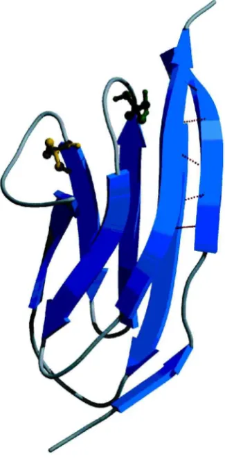
Figure 1. Nuclear magnetic resonance solution structure of monomeric I27. C47 and C63 are
shown in a ball-and-stick representation, and the hydrogen bonds between the A0 and G
strands are shown as dashed lines. The ¯gure was drawn using Molscript (Kraulis 1991)
andRaster3D(Merritt & Murphy 1994) using the coordinates from the protein data base ¯le 1TIT (Improtaet al. 1996). Individual-strands are labelled A to G.
2. Materials and methods
In the experiments to be described here, a pentameric I27 concatamer consisting either of four copies of the double C47S,C63S mutant and a single copy of the single C63S I27 mutant as the central domain (denoted (I27)¤
5) or ve copies of the double mutant (denoted (C47S,C63S I27)5) were studied. Both of these mutations have been shown to severely destabilize the protein in chemical unfolding experiments (Brockwell et al. 2002) but they do not a¬ect the hydrogen-bond network between the A0 and G strands and would therefore not be expected to a¬ect the observed unfolding forces.
inter-actions. The sequence of the linkers was designed to be as similar as possible to the natural I26{I27 and I27{I28 linkers (linker choice was constrained by restric-tion site sequence). Mechanical unfolding experiments were performed using a com-mercially available mechanical force probe (MFP-SA, Asylum Research Inc., USA). Coated unsharpened microlevers (MLCT-AUNM) were obtained from Veeco Metrol-ogy (Santa Barbara, USA). The spring-constant of each cantilever was calculated under phosphate-bu¬ered saline (PBS) using the thermal method (Florinet al. 1995) and was typically found to be ca. 51§5 pN nm¡1
. Protein (0.05 mg) was reconsti-tuted to 0.1 mg ml¡1
in sterile PBS and centrifuged (13 000 rpm, MSE, MicroCen-taur). Typically, 50mL of PBS was dropped onto a recently cleaved template stripped gold surface. 20mL of protein solution was then added and the two solutions allowed to mix. At this protein concentration the probability of attaching a molecule to the tip is relatively low (typically 4%). However, under these conditions ca. 50% of the traces result in the attachment of a single molecule and four or more clear unfolding peaks. Mechanical unfolding experiments were performed using the AFM at pulling speeds varying from 70 nm s¡1to 4000 nm s¡1at a room temperature of 23:3
§1¯C over a distance of 400{600 nm.
Kinetic chemical unfolding experiments were performed using an Applied Photo-physics SX.18 MV stopped-®ow ®uorimeter. The temperature was regulated using an external probe placed near the cuvette and maintained at 25¯C using a Nes-lab RTE-300 circulating water bath. Tryptophan ®uorescence was excited at 280 nm with a 10 nm bandwidth, and the emitted ®uorescence was monitored at more than 320 nm. Unfolding experiments were performed by manual mix. Protein (ca. 50mM) in native bu¬er (20 mM Na2HPO4/NaH2PO4pH 7.3, 1 mM EDTA and 2 mM DTT, or PBS, 1 mM EDTA and 2 mM DTT) was diluted 1:9 into solutions containing GnHCl. The decrease in ®uorescence at 315 nm (excitation 280 nm) was monitored in a 1 cm path-length cuvette for 600 s. Kinetic transients (of the monomeric I27 pro-tein) were tted to a three-parameter single exponential equation usingSigmaplot (SPSS Inc.).
A two-state model was used to perform Monte Carlo simulations of the forced extension of the I27 constructs (Rief et al. 1998). Each domain of the molecule was initially assumed to be in the lowest energy state and therefore folded. The folding and unfolding rate constants at applied force F were calculated using ai;F =a0i;Fexp(§F xi=kBT), where i= f or u for the folding and unfolding events,
respectively, and the negative sign is associated with folding. (So as to di¬erentiate between the intrinsic rate constants for chemical and forced unfolding, the notation k0
u ;GnHCl or a 0
u ;F, respectively, is used). The constants xf andxu represent the
dis-tance from the folded and unfolded well to the barrier, respectively (this reaction coordinate is assumed to be parallel to the stretch axis). The protein was extended with speeds from 10 to 10 000 nm s¡1
and with di¬erent values of xf, xu , a0 f;F and
a0
u ;F, which are the rate constants for folding and unfolding, respectively, in the
absence of applied force. The simplied worm-like chain (Bustamante et al. 1994) was used to calculate the force applied at any extensionxas
F = kBT p
· 1
4(1¡x=L)2 ¡ 1 4+
x L ¸
;
each of length Lu , where n is the total number of domains within the concatamer. At each extension the probability of folding, unfolding or extending the chain is calculated. If unfolding (folding) occurs, the chain lengthLis increased (decreased), as described above, the cantilever extension incremented, and the probability of folding, unfolding or extending the protein re-calculated. The sequence of domain unfolding is random. As a consequence, the rst domain to unfold, corresponding to the rst pulling event, can be any one of those in the construct and not necessarily the rst or last in the chain. The procedure is continued until all domains are unfolded. The whole calculation is then repeated 10 000 times.
Experimental force-extension data take the form of a sawtooth pattern from which the unfolding force can be measured for each unfolding event observed. For a given pulling speed, the observed unfolding forces are plotted in a force{frequency his-togram and the mean unfolding forceF is obtained from this histogram. This process is repeated at several pulling speeds which span the dynamic range of the instru-ment. The unfolding force at each speed is used to construct a graph of F against log(v) and this is compared with the results of the Monte Carlo simulation. The simulation parameters were varied until the experimental data were matched. Thus, the intrinsic unfolding rate constant of the monomeric speciesa0
u ;F and the distance
to the transition state from the native statexu can be obtained.
3. Results
(a) A comparison of mechanical and chemical unfolding
The results of chemical and mechanical unfolding experiments on I27 are shown in gure 2.
The chemical unfolding experiments were performed using both monomeric and polymeric protein to ensure that the behaviour of the protein is not a¬ected by its placement in the polymer (Brockwell et al. 2002). Since this concatamer comprises four double-cysteine-mutant domains and a single-cysteine-mutant domain, it is not surprising that the unfolding kinetics t well to a bi-exponential function with 80% of the amplitude corresponding to the decay noted for the double-cysteine mutant in monomeric form and 20% to that of the single-cysteine-mutant monomer. The intrinsic chemical unfolding rate constant of the concatamer (for the phase account-ing for 80% of the amplitude) (i.e. the mean unfoldaccount-ing rate constant obtained by extrapolation to zero GnHCl concentration) is 10:6§0:7£10¡3s¡1.
ln
kob
s
GnHCl conc/M
x
u
n
fo
ld
in
g
f
o
rc
e
(
p
N
)
50 100 150 200 250 300 350
×
×
×
×
×
×
10 100 1000 10 000
pulling speed (nm s-1)
8
6
4
2
-4 -2 0
-6
-8
-10
0 2 4 6
(a)
[image:7.449.99.351.46.422.2](b)
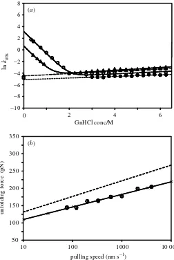
Figure 2. Chemical and mechanical unfolding of I27. (a) Chemical unfolding rate pro¯le of
I27 monomers and the (I27)¤
5 concatamer as a function of urea concentration. Open triangles,
monomeric C47S,C63S I27; open circles, monomeric C63S I27. Solid lines are ¯ts to a two-state model (Brockwellet al. 2002). Closed triangles, observed rate constant for the faster phase of (I27)¤
5 unfolding; closed circles, observed rate constant for the slower phase of (I27)¤5 unfolding.
Dashed lines show the best linear least squares ¯t for the fast and slow phases of unfolding
of the concatamer. The open triangle and circle on the abscissa are the extrapolated k0u for
C47S,C63S I27 and C63S I27, respectively. (b) The pulling speed dependence of observed unfold-ing force. Mutant (I27)¤
5 measured (open circles) unfolds at a lower force than wild-type (I27)8
(dashed line) (data taken from Carrion-Vazquezet al. (1999a)). All error bars are standard error of the mean. The solid line is a linear least-squares ¯t through (I27)¤
5 data. Crosses show the
best ¯t Monte Carlo simulation with parameters¬0
u ;F = 2:0£10¡ 3
s¡ 1
andxu = 0:29 nm.
experiments is not a valid test of the similarity of chemical and mechanical unfolding processes since one describes a monomeric species and the other describes a con-catamer. Thus, Monte Carlo simulations of the mechanical unfolding experiment, which have the monomeric intrinsic unfolding rate constant ¬0
Table 1.Comparison of the results of chemical and mechanical unfolding experiments
(The position of the transition state along the reaction coordinate in chemical unfolding
exper-iments (P) was determined for the monomer in both the mutant and wild-type cases. Cysteine
mutant data taken from Brockwellet al. (2002). Wild-type (I278) W T
data taken from Carrion-Vazquezet al. (1999a).)
mechanical unfolding chemical unfolding
z }| { z }| {
¬0 u ;F (s¡
1
) xu (nm) k
0
u ;G n H C l(s¡ 1
) P (%)
(I27)¤
5 2:0§0:2£10¡ 3
0:29§0:02 10:6§0:7£10¡ 3
6
(I27)W T
8 3:3£10¡ 4
0.25 4:9£10¡ 4
10
to the transition state from the native state xu as parameters (see x2), are used to t the data in gure 2b. This approach yields an intrinsic forced unfolding rate constant of 2:0§0:2£10¡3
s¡1
, more than ve times slower than the intrinsic chem-ical unfolding rate constant. The results of the chemchem-ical and mechanchem-ical unfolding experiments on the mutant and wild-type I27 proteins are summarized in table 1.
The signicant di¬erence between the chemical and mechanical intrinsic unfolding rate constants that partly parametrize the unfolding energy landscapes for the two processes suggests that they occur with di¬erent unfolding mechanisms in contradic-tion to earlier work (Carrion-Vazquezet al. 1999a). The transition state for unfolding of the double mutant described here and the wild-type I27 (Carrion-Vazquez et al. 1999a) occursca. 10% of the distance along the `reaction coordinate’ from the native state in both chemical and mechanical unfolding experiments. The relevance of this similarity is di¯cult to assess, however, due to the di¬ering nature of the reaction coordinates. In classical chemical denaturation this coordinate is usually the acces-sible surface area exposed to solvent (Myerset al. 1995). The reaction coordinate in mechanical unfolding is end-to-end concatamer extension. The physical meaning of this one-dimensional quantity in terms of the actual deformation of protein struc-ture is di¯cult to interpret. Comparison in structural terms of the transition state placement as measured by the two techniques is therefore not meaningful.
Whether the chemical and mechanical unfolding pathways are the same or not is an important issue, since there is a considerable body of literature reporting on chemical unfolding/refolding of proteins, and it is interesting to discuss the relatively new eld of mechanical unfolding in the context of these results. It is relatively simple to test the hypothesis (that mechanical and chemical unfolding are related processes) by destabilizing/stabilizing the native state of the protein and testing whether this has a similar e¬ect on the observed unfolding rate constants in the two experiments. For example, gure 3 shows the e¬ect on the two unfolding pathways of the addition of sodium sulphate which is known to stabilize compact (native) states in chemical unfolding experiments.
fl u o re sc en c e in te n si ty (r e la ti v e to u n fo ld e d s ta te ) 1 2 3 4
0 200 400 600
77 nm s-1
700 nm s-1
fo rc e ( p N )
77 nm s-1
distance from surface (nm) (i)
(ii)
time (s)
(i) (ii) 150 0 150 0 150 0 150 0
0 50 100 150
(a)
(b)
[image:9.449.107.348.43.380.2]700 nm s-1
Figure 3. Comparison of chemical and mechanical unfolding of (C47S, C63S I27)5 in the
pres-ence (i) and abspres-ence (ii) of sodium sulphate. (a) Chemical unfolding data in 25 mM sodium
phosphate pH 7.3, 2 mM DTT, 1 mM EDTA and 3.5 M guanidine hydrochloride with and with-out 0.4 M sodium sulphate. The rate of denaturation after a 1:9 dilution of native protein into guanidine hydrochloride containing bu®er was monitored by a change in °uorescence emission at 320 nm. The black curves show single exponential ¯ts that were used to extract the unfolding rate constants from the data. The unfolding rate constant is around three times slower with 0.4 M sodium sulphate. (b) Mechanical unfolding traces at two di®erent retract speeds (77 and 700 nm s¡ 1
, upper and lower traces, respectively) in sodium phosphate pH 7.3 with and without 0.4 M sodium sulphate. Detailed analysis of many such unfolding experiments reveals that the unfolding forces are unchanged with sodium sulphate present.
(¢¢GUN), while the transition states for unfolding determined mechanically and chemically are destabilized byca. 5 kJ mol¡1
and 8 kJ mol¡1
, respectively.
Consequently, it would appear that chemical and mechanical unfolding pathways are di¬erent, at least for the I27 polymer studied here, and a similar observation was reported recently for the enzyme barnase (Best et al. 2001). A further important conclusion can be drawn from analysis of these data. The wild-type protein unfolds at a higher force at a given pulling speed than the mutant proteins (Carrion-Vazquez et al. 1999a; Brockwellet al. 2002), suggesting that the mutations have a¬ected the mechanical sensitivity. However, as well as changing the barrier height for mechanical unfolding, the mutation also a¬ects the parameter xu (using Monte Carlo methods xWT
u = 0:33§0:05 nm (Best et al. 2002) andx¤u = 0:29§0:03 nm (Brockwellet al. 2002)). It is the productF xu that equates to the energy required to reduce the barrier height su¯ciently to allow crossing by thermal ®uctuations. This product, rather than the absolute value of unfolding force, is the most accurate measure of the protein’s sensitivity to force. It can therefore be seen that at a pulling speed of 600 nm s¡1 the barrier for mechanical unfolding of wild-type I27 domains is reduced by 31 kJ mol¡1 and by 29 kJ mol¡1 for the mutant. Thus it is clear that these mutations have not in fact a¬ected the mechanical stability, supporting the existing hypothesis that mechanical resistance is a locally endowed property of the protein, in the case of I27 is due to the hydrogen-bond clamp region, and not a¬ected by mutations to the protein core.
4. The e®ects of unfolding history and supramolecular sca®old
It has been widely assumed that in a hetero-polyprotein the domain with the fastest ¬0
u ;F must unfold rst under an applied load (Li et al. 2000b) and in a
homo-polyprotein all unfolding forces are equivalent within the limits of thermal ®uctua-tions (Carrion-Vazquezet al. 1999a; Yanget al. 2000; Bestet al. 2001; Brockwellet al. 2002). During our Monte Carlo simulations of the forced unfolding of the mutant described above it became clear that these two assumptions are not always valid. We observed rst in simulation, then in experiment, that for a homopolymer (in our case constructed of copies of a double mutant C47SC63S; seex2), the lowest unfolding force was not necessarily the rst unfolding event and that a more complex descrip-tion involving the number of domains remaining folded and the length of unfolded polypeptide chain in the system was required.
0 100 200 300 400 500
fo
rc
e
(p
N
)
#1 #2 #3
#4 #5
detachment
20 40 60 80 100 120
u
n
fo
ld
in
g
f
o
rc
e
(
p
N
)
extension (nm)
#1 #2
#3 #4
#5
0 50
extension (nm)
0 50 100 150 200 250
100 150 200
(a)
Figure 4. (a) A typical mechanical unfolding force-extension dataset of (C47S, C63S I27)5 by
experiment (main curve) and simulation (inset). Both the experimental and simulated data were obtained with cantilevers of spring constantkc = 50 pN nm¡
1
and at a pulling speed of 700 nm s¡ 1
. The Monte Carlo data are obtained with an unfolded domain contour length of
Lu = 28 nm identical to that of the I27 domain (Carrion-Vazquezet al. 1999a; Brockwellet al.
2002). Other parameters in the simulations were:¬0
u ;F = 2£10¡ 3
s¡ 1
, persistence length of the unfolded domainsp= 0:39 nm andxu = 0:29 nm.
[image:11.449.90.365.49.310.2]16 18 20 22 24 26 28 30 0 2 4 6 8 10 12 14
10 13 16 19 22 25 28
fr e q u e n c y
normalized force (%)
#1
10 13 16 19 22 25 28
#3
event number # (b)
0 5 10 15 20 25
normalized force (%)
n o rm al iz ed u n fo ld in g f o rc e ( % )
1 2 3 4 5
Figure 4. (Cont.) (b) Monte Carlo simulation (circles) and experimental data (squares) for
unfolding forces of (C47S, C63S I27)5 as a function of the unfolding event number # (see
¯gure 1). Data are expressed as a fraction of the sum of the unfolding forces in order to combine
many experimental datasets. Experimental data are expressed as a weighted mean§weighted
standard error of the mean. Inset shows relative force frequency histograms for #1 and #3 for all datasets (n= 104). Solid lines are ¯ts to a single (#3) or a double (#1) Gaussian function. The points (£) and (4) are the modes obtained from the ¯t to the distribution for #1.
What is the origin of this history e¬ect in the mechanical unfolding of polypro-teins? The reason that the minimum force in the unfolding sequence is not neces-sarily observed for the rst unfolding event is that the number of folded domains remaining at any given time and the length of the domains already unfolded have competing e¬ects on the unfolding force of the next domain to unfold. A decrease in the number of folded domains reduces the number of unfolding attempts at any given extension which decreases the unfolding probability. Thus, the measured mean unfolding force should rise monotonically as more domains unfold. How-ever, as each domain is unfolded, the total length of unfolded polypeptide present increases, which increases the overall compliance (or reduces the e¬ective spring constant) of the system. In a system with a higher compliance, J, the loading rate df =dt = J¡1¸, where ¸ is velocity, is reduced, resulting in more thermally driven unfolding attempts per unit time at each extension, which would result in a grad-ual lowering of the unfolding force. The net result of these competing e¬ects is the observed minimum in the unfolding force as a function of the unfolding event num-ber.
[image:12.449.96.361.48.307.2]polypep-150 155 160 165 170 175 180 185 190
L
u = 15 nm
L
u = 28 nm
t = 45 nm
150 155 160 165 175 180 185 190
k
c = 100 000 pN nm-1 170
for
c
e
(p
N
)
event number #
1 2 3 4 5
for
ce
(
pN
)
k
c = 50 pN nm-1
k
[image:13.449.95.361.42.525.2]c = 10 pN nm-1
Figure 5. Monte Carlo simulations showing the e®ect of varying (a) the unfolded contour length
Lu and (b) the AFM cantilever spring constantkc upon the observed unfolding forces at each
event number. In (a)kc= 50 pN nm¡ 1
and in (b)Lu = 28 nm.
An increase in the length of the unfolded domain to 45 nm from the value of 28 nm (I27) causes the minimum in the unfolding force to become more apparent, because in the early events the fractional contribution to the change in compliance by unfold-ing a larger protein is greater (gure 5a). This e¬ect is more clearly seen when a very sti¬ cantilever is used, since the contribution of the polypeptide chain length is a more signicant factor than when a very compliant cantilever with low spring constant is employed (gure 5b). Our observations have important implications for understanding the mechanical properties of heteropolymers that have evolved nat-urally to resist forcein vivo. The passive e¬ect of unstructured polymers acting as an `entropic spring’ is well known (for instance the PEVK domain of titin (Linkeet al. 1998) and the selectin cell{surface carbohydrate interaction (Fritz et al. 1998)). We have now shown that both the superstructure, or sca¬old, in which the polymer is held and the number and length of unfolded domains in®uence the mechanical resistance of the remaining folded domains. Thus, e¬ects such as the compliance of the surrounding tissue and the lengths of unstructured regions will play a key role in tailoring the mechanical resistance of folded domains in polyproteins. These obser-vations add another level of complexity to any valid description of the mechanical properties of naturally occurring polyproteins and reveal the wide range of param-eters available in biology for tuning the resistance of proteins to applied force for specic mechanical roles.
5. Summary and outlook
Mechanical unfolding studies of I27 from several laboratories indicate that the num-ber and geometry of interstrand hydrogen bonds and a transition state unusually close to the native state maximize the mechanical resistance of the protein. Steered molecular-dynamics simulations have suggested that the mechanical stability of -sheet proteins depends critically on the topology of the protein. Proteins with parallel N- and C-terminal strands exhibit the largest mechanical unfolding forces because all of the interstrand hydrogen bonds must be simultaneously broken for the pro-tein to unfold (Lu & Schulten 2000). Propro-teins with anti-parallel terminal -strands unfold at relatively low forces, possibly because the force is applied parallel to the hydrogen bond and results in the sequential `zipper-like’ rupture of these bonds with relatively low force (Rohset al. 1999). This parallel -strand secondary struc-ture and hydrogen-bond clamp region give I27 the largest mechanical strength of any protein studied to date (ca. 200 pN for the wild-type protein). In comparison tenascin (FNIII domains), barnase, T4 lysozyme, the C2 domain of synaptotagmin I and spectrin unfold at 140 pN (Oberhauser et al. 1998), 65 pN (Best et al. 2001), 64 pN (Yang et al. 2000), 60 pN (Carrion-Vazquez et al. 2000) and 30 pN (Rief et al. 1999), respectively. Calmodulin unfolded at too small a force to be measured (Carrion-Vazquez et al. 2000). These data suggest therefore that proteins with -sheet secondary structure are mechanically most stable, while¬-helical proteins are relatively mechanically unstable. Proteins with mixed¬= topologies fall in between these two extremes.
di¬erent insofar as the intrinsic unfolding rate constants are signicantly di¬erent and that altering the stability of the native state by mutagenesis or addition of sodium sulphate a¬ects the intrinsic unfolding rate constants of the I27 mutant studied here di¬erently. An unexpected dependence of the unfolding force of a given unfolding event on its position in the sequence of unfolding events of the entire concatamer was observed. This is due to the e¬ect of the number of folded domains remaining to be unfolded changing and the increased compliance of the mechanical system as domains unfold. The implication of these observations is that both the length of the unfolded domain and the compliance of the surrounding tissue will a¬ect the mechanical resistance of a folded protein in its biological context. The mechanical resistance of a protein domain is modulated by these e¬ects and therefore cannot be simply regarded as a property endowed by aspects of local secondary structure.
One particularly important aspect of the mechanical unfolding experiment is that the direction in which the force is applied can, in principle at least, be controlled. In contrast to other unfolding experiments, in which a low-molecular-weight denaturant (chemical unfolding), temperature or pH is used, and the unfolding reaction coordi-nate with respect to protein structure coordicoordi-nates is not under experimental control. We are currently conducting studies in which a protein is mechanically unfolded using force applied in well-dened and di¬erent directions in separate experiments. Our preliminary results indicate that the unfolding forces observed vary more than 10-fold, depending on the direction of the applied force relative to a hydrogen-bond clamp. Such experiments open the way for a detailed mapping of the mechanical unfolding energy landscape and an extensive comparison of unfolding experiments with molecular-dynamics simulations.
The authors are grateful to David Salt and Phil Williams for helpful discussions. We are partic-ularly grateful to Richard Perham for the inspiration for new experiments that permit proteins to be unfolded in di®erent directions and for the plasmids to conduct these experiments. We acknowledge the University of Leeds, BBSRC, EPSRC and The Wellcome Trust for ¯nancial support. S.E.R. is a BBSRC Professorial Research fellow. The manuscript is a contribution from the Astbury Centre for Structural Molecular Biology, which is part of the North of England Structural Biology Centre (NESBIC) and is funded by the BBSRC.
References
Best, R. B., Li, B., Steward, A., Daggett, V. & Clarke, J. 2001 Can non-mechanical proteins withstand force? Stretching Barnase by atomic force microscopy and molecular dynamics simulation.Biophys. J.81, 2344{2356.
Best, R. B., Fowler, S. B., Toca-Herrera, J. L. & Clarke, J. 2002 A simple method for probing the mechanical unfolding pathway of proteins in detail.Proc. Natl Acad. Sci. USA99, 12 143{ 12 148.
Brockwell, D. J., Beddard, G. S., Clarkson, J., Zinober, R. C., Blake, A. W., Trinick, J., Olmsted, P. D., Smith, D. A. & Radford, S. E. 2002 The e®ect of core destabilisation on the mechanical resistance of I27. Biophys. J.83, 458{472.
Bustamante, C., Marko, J. F., Siggia, E. D. & Smith, S. 1994 Entropic elasticity of lambda
phage DNA.Science 256, 1599{1600.
Carrion-Vazquez, M., Oberhauser, A. F., Fowler, S. B., Marszalek, P. E., Broedel, S. E., Clarke, J. & Fernandez, J. M. 1999aMechanical and chemical unfolding of a single protein: a
Carrion-Vazquez, M., Marszalek, P. E., Oberhauser, A. F. & Fernandez, J. M. 1999b Atomic
force microscopy captures length phenotypes in single proteins. Proc. Natl Acad. Sci. USA
96, 11288{11 292.
Carrion-Vazquez, M., Oberhauser, A. F., Fisher, T. E., Marszalek, P. E., Li, H. & Fernandez, J. M. 2000 Mechanical design of proteins studied by single-molecule force spectroscopy and protein engineering.Prog. Biophys. Molec. Biol.74, 63{91.
Fisher, T. E., Marszalek, P. E. & Fernandez, J. M. 2000 Stretching single molecules into novel conformations using the atomic force microscope. Nature Struct. Biol.7, 719{724.
Florin, E. L., Rief, M., Lehmann, H., Ludwig, M., Dornmair, C., Moy, V. T. & Gaub, H. E. 1995 Sensing speci¯c molecular interactions with the atomic force microscope.Biosens. Bioelectron. 10, 895{901.
Fowler, S. B. & Clarke, J. 2001 Mapping the folding pathway of an immunoglobulin domain: structural detail from phi value analysis and movement of the transition state.Structure 9, 1{12.
Fritz, J., Katopodis, A. G., Kolbinger, F. & Anselmetti, D. 1998 Force-mediated kinetics of
single P-selectin/ligand complexes observed by atomic force microscopy. Proc. Natl Acad.
Sci. USA95, 12283{12 288.
Improta, S., Politou, A. S. & Pastore, A. 1996 Immunoglobulin-like modules from titin I-band: extensible components of muscle elasticity.Structure 4, 323{337.
Kellermayer, M. S. Z., Smith, S. B., Granzier, H. L. & Bustamante, C. 1997 Folding-unfolding transitions in single titin molecules characterized with laser tweezers.Science276, 1112{1116.
Kraulis, P. J. 1991 Molscript: a program to produce both detailed and schematic plots of
protein structures.J. Appl. Cryst.24, 946{950.
Labeit, S. & Kolmerer, B. 1995 Titins: giant proteins in charge of muscle ultrastructure and elasticity.Science 270, 293{296.
Li, H., Carrion-Vasquez, M., Oberhauser, A. F., Marszalek, P. E. & Fernandez, J. M. 2000a
Point mutations alter the mechanical stability of immunoglobulin modules. Nature Struct.
Biol.7, 1117{1120.
Li, H., Oberhauser, A. F., Fowler, S. B., Clarke, J. & Fernandez, J. M. 2000b Atomic force
microscopy reveals the mechanical design of a modular protein. Proc. Natl Acad. Sci. USA
97, 6527{6531.
Li, H., Oberhauser, A. F., Redick, S. D., Carrion-Vazquez, M., Erickson, H. P. & Fernandez, J. M. 2001 Multiple conformations of PEVK proteins detected by single-molecule techniques.
Proc. Natl Acad. Sci. USA98, 10682{10 686.
Li, H. B., Linke, W. A., Oberhauser, A. F., Carrion-Vazquez, M., Kerkviliet, J. G., Lu, H., Marszalek, P. E. & Fernandez, J. M. 2002 Reverse engineering of the giant muscle protein titin.Nature418, 998{1002.
Linke, W. A., Ivemeyer, M., Mundel, P., Stockmeier, M. R. & Kolmerer, B. 1998 Nature of
PEVK-titin elasticity in skeletal muscle.Proc. Natl Acad. Sci. USA95, 8052{8057.
Lu, H. & Schulten, K. 2000 The key event in force-induced unfolding of titin’s immunoglobulin domains.Biophys. J.79, 51{65.
Lu, H., Isralewitz, B., Krammer, A., Vogel, V. & Schulten, K. 1998 Unfolding of titin
immunoglobulin domains by steered molecular dynamics simulation. Biophys. J. 75, 662{
671.
Merkel, R., Nassoy, P., Leung, A., Ritchie, K. & Evans, E. 1999 Energy landscapes of receptor{
ligand bonds explored with dynamic force spectroscopy.Nature 397, 50{53.
Merritt, E. A. & Murphy, M. E. P. 1994Raster3Dversion 2.0: a program for photorealistic
molecular graphics.Acta Crystallogr.D50, 869{873.
Mitsui, K., Hara, M. & Ikai, A. 1996 Mechanical unfolding of¬2-macroglobulin molecules with
Myers, J. K., Pace, C. N. & Scholtz, J. M. 1995 Denaturantmvalues and heat capacity changes: relation to changes in accessible surface areas of protein unfolding.Protein Sci.4, 2138{2148.
Oberdorfer, Y., Fuchs, H. & Jansho®, A. 2000 Conformational analysis of native ¯bronectin by
means of force spectroscopy. Langmuir 16, 9955{9958.
Oberhauser, A. F., Marszalek, P. E., Erickson, H. P. & Fernandez, J. M. 1998 The molecular elasticity of the extracellular matrix protein tenascin.Nature 393, 181{185.
Paci, E. & Karplus, M. 2000 Unfolding proteins by external forces and temperature: the
impor-tance of topology and energetics.Proc. Natl Acad. Sci. USA97, 6521{6526.
Rief, M., Gautel, M., Oesterhelt, F., Fernandez, J. M. & Gaub, H. E. 1997 Reversible unfolding
of individual titin immunoglobulin domains by AFM. Science 276, 1109{1111.
Rief, M., Fernandez, J. M. & Gaub, H. E. 1998 Elastically coupled two-level systems as a model for biopolymer extensibility.Phys. Rev. Lett.81, 4764{4767.
Rief, M., Pascual, J., Saraste, M. & Gaub, H. E. 1999 Single molecule force spectroscopy of spectrin repeats: low unfolding forces in helix bundles.J. Mol. Biol.286, 553{561.
Rohs, R., Etchebest, C. & Lavery, R. 1999 Unraveling proteins: a molecular mechanics study.
Biophys. J.76, 2760{2768.
Smith, B. L., Scha®er, T. E., Viani, M., Thompson, J. B., Frederick, N. A., Kindt, J., Belcher, A., Stucky, G. D., Morse, D. E. & Hansma, P. K. 1999 Molecular mechanistic origin of the
origin of the toughness of natural adhesives, ¯bres and composites. Nature399, 761{763.
Tskhovrebova, L., Trinick, J., Sleep, J. A. & Simmons, R. M. 1997 Elasticity and unfolding of single molecules of the giant muscle protein titin. Nature387, 308{312.
Yang, G., Cecconi, C., Baase, W. A., Vetter, I. R., Breyer, W. A., Haack, J. A., Matthews, B. W., Dahlquist, F. W. & Bustamante, C. 2000 Solid-state synthesis and mechanical unfolding of
polymers of T4 lysozyme. Proc. Natl Acad. Sci. USA97, 139{144.
Zinober, R. C., Brockwell, D. J., Beddard, G. S., Blake, A. W., Olmsted, P. D., Radford, S. E. & Smith, D. A. 2002 Mechanically unfolding proteins: the e®ect of unfolding history and the supramolecular sca®old.Protein Sci.11, 2759{2765.
Discussion
W. T. Coffey (School of Engineering, Trinity College Dublin, Ireland). You use transition state theory to describe the reaction rate which is based on equilibrium considerations. Kramers (1940) made a major improvement in transition-state theory by taking into account the coupling to the heat bath. This allowed him to connect the reaction rate to the Langevin equation describing the motion of a reacting particle, thus non-equilibrium e¬ects could be included in the reaction rate. Moreover, the dependence of the reaction rate on the parameters of the Langevin equation, inertia, friction, etc., could be determined. Subsequently, this theory was generalized to a reacting system ofndegrees of freedom by Langer (1969), whichinter aliaconstitutes a general theory of the decay of metastable states. It seems to me that Langer’s treatment would provide a useful basis for the theoretical discussion of your problem.
about the nature of other degrees of freedom (which undoubtedly are there but are probably quite fast and not accessible in our experiments) analysis in this manner is not possible.
E. Sackmann(Faculty of Physics, Technical University of Munich, Garching, Ger-many). You appear to be able to measure the unfolding of a protein under applied force with exquisite accuracy. Could similar techniques be used to measure the inter-action potential between two particles: two proteins for example?
D. A. Smith.That is indeed the case, although current instrumentation has a prac-tical force resolution (due to Brownian motion of the cantilever) ofca. 15 pN, which clearly limits the range of `particles’ that one might be able to study. However, devel-opments in instrumentation (see, for example, Aoki et al. 1997) permit at least an order of magnitude increase in force sensitivity, opening the possibility for experi-ments to study the interaction potentials in a wider range of cases.
B. U. Felderhof(RWTH, Aachen, Germany). The discussion in terms of energy landscape is only valid in the adiabatic limit. It seems to me that the dependence on the rate at which you are pulling, or on frequency in an oscillating experiment, necessarily involves hydrodynamic e¬ects due to friction with the ambient ®uid.
D. A. Smith. You are correct that the Kramers calculation assumes adiabaticity, i.e. the pulling speed is slow compared with the rate at which the pulled molecule explores its energy landscape (internal degrees of freedom). This treatment is incor-rect at pulling speeds well beyond AFM capabilities. Hydrodynamic friction e¬ects are quite small. For example, the friction due to the cantilever at a pulling speed of 500 nm s¡1 is only a few pN (assuming Stokes drag), while that on the individual domains is much smaller and negligible on the scale of the measured unfolding forces (of the order of 100{200 pN).
M. Maaloum(Institut Charles Sadron, Strasbourg, France). The force values you measure depend on the precision of the spring constant. How do you calibrate the cantilever? Do you stretch the same molecule or di¬erent molecules in each experi-ment? Is the force prole reversible?
D. A. Smith. The spring constant of each cantilever was calibrated under PBS using the thermal noise method (Florin et al. 1995) and was typically found to be ca. 51§5 pN nm¡1
. In general, di¬erent molecules are stretched each time the experiment is performed, mainly due to thermal drift of the sample below the AFM tip. However, with care it is perfectly possible to pull the same molecule repeatedly. The force prole isrepeatable but notreversible. This is because the protein will not refold under even the slightest applied force. Thus, the tip with the protein attached must be returned to the substrate to release all tension in the system before refolding will occur (see, for example, Carrion-Vazquez et al. 1999a).
D. A. Smith. If multiple proteins are picked up and extended together then it is very clear because the extension between the sawtooth peaks does not correlate to the expected extension due to the length of an unfolded domain.
D. S. F. Crothers(Department of Applied Mathematics and Theoretical Physics, Queen’s University, Belfast, UK). In your Monte Carlo simulations, which variables are randomized?
D. A. Smith.We calculated a transition probability as a function of applied exten-sion (using the Kramers result for the rst passage time as a function of force applied to a potential). A random number was then chosen; if this number was less than the calculated transition probability then the event was accepted and unfolding occurred. Our Monte Carlo simulations are based on the treatment by Riefet al. (1998). The intrinsic unfolding rate constant and the distance to the transition state from the native state are used as t parameters to achieve the best t to the experimental data (see Brockwellet al. (2002) for details).
P. Bartlett(School of Chemistry, University of Bristol, UK). Have you thought about the possibility of doing the experiment at constant force rather than at con-stant rate of pulling? It might be possible, for instance, to access the signicance of ®uctuations more readily by sitting at the top of the free energy barrier.
D. A. Smith.That is a very nice experiment which we have not done but has been done in Paul Hansma’s laboratory (Oberhauser et al. 2001). One can observe the rate of occurrence of unfolding events and obtain the unfolding rate constants at di¬erent applied loads. In principle one could poise the system close to the transition state and study the e¬ect of ®uctuations on the unfolding rate constant. However, the dominant process could be the Brownian motion of the cantilever in a standard AFM (see my earlier response to Dr Sackmann).
Additional references
Aoki, T., Hiroshima, M., Kitamura, K., Tokunaga, M. & Yanagida, T. 1997 Ultramicroscopy
70, 45{55.
Bell, G. I. 1978Science 200, 618{627.
Evans, E. & Ritchie, K. 1997Biophys. J.72, 1541{1555.
Kramers, H. A. 1940Physica7, 284.
Langer, J. S. 1969Ann. Phys. NY 54, 258.
Oberhauser, A. F., Hansma, P. K., Carrion-Vazquez, M. & Fernandez, J. M. 2001Proc. Natl