The temporal profile of activity-dependent
presynaptic phospho-signalling reveals
long-lasting patterns of poststimulus regulation
Kasper Engholm-KellerID1,2,3☯, Ashley J. WaardenbergID4,5☯, Johannes A. Mu¨ ller6, Jesse R. Wark1, Rowena N. Fernando1, Jonathan W. ArthurID4, Phillip J. RobinsonID3,
Dirk Dietrich7, Susanne SchochID6, Mark E. GrahamID1*
1 Synapse Proteomics, Children’s Medical Research Institute, The University of Sydney, Westmead, Australia, 2 Department of Biochemistry and Molecular Biology, University of Southern Denmark, Odense, Denmark, 3 Cell Signalling Unit, Children’s Medical Research Institute, The University of Sydney, Westmead, Australia, 4 Bioinformatics Unit, Children’s Medical Research Institute, The University of Sydney, Westmead, Australia, 5 Genome Biology Unit, European Molecular Biology Laboratory, Heidelberg, Germany, 6 Institute of Neuropathology, University of Bonn Medical Center, Bonn, Germany, 7 Department of Neurosurgery, University of Bonn Medical Center, Bonn, Germany
☯These authors contributed equally to this work.
Abstract
Depolarization of presynaptic terminals stimulates calcium influx, which evokes neurotrans-mitter release and activates phosphorylation-based signalling. Here, we present the first global temporal profile of presynaptic activity-dependent phospho-signalling, which includes two KCl stimulation levels and analysis of the poststimulus period. We profiled 1,917 regu-lated phosphopeptides and bioinformatically identified six temporal patterns of co-reguregu-lated proteins. The presynaptic proteins with large changes in phospho-status were again promi-nently regulated in the analysis of 7,070 activity-dependent phosphopeptides from KCl-stim-ulated cultured hippocampal neurons. Active zone scaffold proteins showed a high level of activity-dependent phospho-regulation that far exceeded the response from postsynaptic density scaffold proteins. Accordingly, bassoon was identified as the major target of neuro-nal phospho-signeuro-nalling. We developed a probabilistic computationeuro-nal method, KinSwing, which matched protein kinase substrate motifs to regulated phosphorylation sites to reveal underlying protein kinase activity. This approach allowed us to link protein kinases to profiles of co-regulated presynaptic protein networks. Ca2+- and calmodulin-dependent protein kinase IIα(CaMKIIα) responded rapidly, scaled with stimulus strength, and had long-lasting activity. Mitogen-activated protein kinase (MAPK)/extracellular signal–regulated kinase (ERK) was the main protein kinase predicted to control a distinct and significant pattern of poststimulus up-regulation of phosphorylation. This work provides a unique resource of activity-dependent phosphorylation sites of synaptosomes and neurons, the vast majority of which have not been investigated with regard to their functional impact. This resource will enable detailed characterization of the phospho-regulated mechanisms impacting the plas-ticity of neurotransmitter release.
a1111111111 a1111111111 a1111111111 a1111111111 a1111111111 OPEN ACCESS
Citation: Engholm-Keller K, Waardenberg AJ,
Mu¨ller JA, Wark JR, Fernando RN, Arthur JW, et al. (2019) The temporal profile of activity-dependent presynaptic phospho-signalling reveals long-lasting patterns of poststimulus regulation. PLoS Biol 17(3): e3000170.https://doi.org/10.1371/ journal.pbio.3000170
Academic Editor: Thomas C Su¨dhof, Stanford
University School of Medicine, UNITED STATES
Published: March 1, 2019
Copyright:©2019 Engholm-Keller et al. This is an open access article distributed under the terms of theCreative Commons Attribution License, which permits unrestricted use, distribution, and reproduction in any medium, provided the original author and source are credited.
Data Availability Statement: Highly curated and
processed phosphoproteomics data are available in the Supporting Information files. All unprocessed mass spectrometry data files are available from the PRIDE database (accession number PXD010007). All other relevant data are within the paper and its Supporting Information files.
Funding: KEK was funded by the Lundbeck
Author summary
Neurobiological processes are altered by linking neuronal activity to regulated changes in protein phosphorylation levels that influence protein function. Although some of the major targets of activity-dependent phospho-signalling have been identified, a large num-ber of substrates remain unknown. Here, we have screened systematically for these sub-strates and extended the list from hundreds to thousands of phosphorylation sites, thereby providing a new depth of understanding. We monitored phospho-signalling for 15 min after the stimulation, which to our knowledge had not been attempted at a large scale. We focused on presynaptic protein substrates of phospho-signalling by isolating the pre-synaptic terminal. We also stimulated hippocampal neurons but did not monitor the post-stimulus. Although the phospho-signalling is immensely complex, the findings could be simplified through data exploration. We identified distinct patterns of presynaptic phos-pho-regulation across the time course that may constitute co-regulated protein networks. In addition, we found a subset of proteins that had many more phosphorylation sites than the average and high-magnitude responses, implying major signalling or functional roles for these proteins. We also determined the likely protein kinases with the strongest responses to the stimulus at different times using KinSwing, a computational tool that we developed. This resource reveals a new depth of activity-dependent phospho-signalling and identifies major signalling targets, major protein kinases, and co-regulated phospho-protein networks.
Introduction
Depolarization of the presynaptic plasma membrane stimulates the opening of voltage-gated Ca2+channels, and Ca2+rapidly enters at the active zone. Increased Ca2+triggers neurotrans-mitter release by binding to Ca2+sensors such as synaptotagmin 1, which facilitates fast syn-chronous fusion of docked synaptic vesicles [1–3]. Docking and priming of synaptic vesicles, as well as Ca2+channel clustering [4,5], are coordinated by scaffold proteins at the active zone.
Ca2+channels are tethered in proximity to synaptic vesicles directly by Rab3-interacting
mole-cules (RIMs) [6,7] and/or by a complex including bassoon and RIM-binding protein [8]. As
such, the protein composition of the active zone scaffold not only is important for vesicle release but also exerts influence on release probability and presynaptic homeostatic plasticity [9].
The influx of Ca2+following depolarization also stimulates phosphorylation-based
signal-ling. Phosphorylation and dephosphorylation of presynaptic proteins at the active zone are strongly coupled to Ca2+influx in the vicinity of Ca2+channels, rather than the cytosolic Ca2+
concentration [10]. Ca2+binds calmodulin, and this complex activates downstream
phospho-signalling pathways that directly affect release properties. For example, Ca2+- and calmodulin-dependent protein kinase II (CaMKII) phosphorylation of synapsin 1 has a role in changing the availability of synaptic vesicles for release [11]. Ca2+/calmodulin also activates the phospha-tase calcineurin (protein phosphaphospha-tase 2B) and downstream phosphaphospha-tases. Three synapsin 1 sites—S62, S67, and S549—are dephosphorylated by calcineurin and may contribute to increased availability of synaptic vesicles [12]. Calcineurin has also been shown to directly reg-ulate synaptic vesicle endocytosis [13,14]. The main protein kinases mediating presynaptic
phospho-signalling and plasticity are protein kinase C (PKC) [15,16], protein kinase A (PKA)
[9], cyclin-dependent kinase 5 (CDK5) [17], and extracellular signal–regulated kinase 1/2 Brorsons Rejselegat, Beckett-fonden, Etly og
Jørgen Stjerngrens Fond, Dagmar Marshalls Fond, Grosserer L. F. Foghts Fond, and Brødrene Hartmanns Fond. MEG was funded by the Rebecca L Cooper Medical Research Foundation. MEG and PJR were funded by National Health and Medical Research Project Grants (1079160, 1077989, 1052494, and 571070). Additional funding was by the Brain Foundation, Australian Cancer Research Foundation, Cancer Institute New South Wales, Zero Childhood Cancer, Ramaciotti Foundation, Honda Foundation, and Bruce Wall Estate. SS and DD were funded by the German Research Council (DFG, SFB1089, SPP1757, SCHO 820/6-1, SCHO 820/4-1, DI853/3-2, DI853/7-1) and local funding (BONFOR). The funders had no role in study design, data collection and analysis, decision to publish, or preparation of the manuscript.
Competing interests: The authors have declared
that no competing interests exist.
Abbreviations: AGC, protein kinase A, G, and C;
AKT, protein kinase B; CaMK, Ca2+- and calmodulin-dependent protein kinase; CaMKL, CaMK-like; Caskin1, Ca2+- and calmodulin-dependent serine protein kinase-interacting protein 1; CDC7, cell division cycle 7-related; CDK, cyclin-dependent kinase; CGMC, CDK, GSK3, MAPK, and CLK; CK2, casein kinase 2; Clasp2, cytoplasmic linker–associated protein 2; DAPK, death-associated protein kinase; Dlgap4, disks large-associated protein 4; DMPK, myotonic dystrophy protein kinase; DYRK, dual-specificity tyrosine-regulated kinases; ELKS, protein rich in E, L, K, and S; ERK, extracellular signal–regulated kinase; Exoc7, exocyst complex component 7; GRK, G protein–coupled receptor kinase; GSK3, glycogen synthase kinase; GTPase, guanosine
triphosphatase; IKK, I kappa B kinase; IRSp53, insulin receptor substrate p53; Kcnma1, calcium-activated potassium channel subunit alpha-1; KS score, KinSwing score; LRRK, leucine-rich repeat kinase; MAP1B, microtubule-associated protein 1B; MAPK, mitogen-activated protein kinase; MAPKAPK, MAPK-activated protein kinase; MTOR, mechanistic target of rapamycin; PCA, principle component analysis; PDK1, pyruvate dehydrogenase kinase 1; PIKK,
(ERK)/mitogen-activated protein kinase 1/3 (MAPK1/3) [18]. However, the identity of the presumably large number of substrates, the timing and interdependence of protein kinase activation or inactivation, and mechanistic consequences remain largely unresolved, as the complex activity-driven network of presynaptic kinases has previously not been explored sys-tematically, to our knowledge.
There are few global studies of activity-dependent neuronal phospho-signalling. The post-synaptic density has been enriched to reveal postpost-synaptic signalling [19,20]. Here, we use a subcellular compartment enrichment strategy to focus on presynaptic proteins. Sucrose homogenates of brain tissue produce a particulate fraction that contains both pre- and post-synaptic components but is primarily enriched in isolated prepost-synaptic terminals, i.e., synap-tosomes [21,22]. Synaptosomes contain synaptic vesicles [22], are metabolically active,
generate adenosine triphosphate [23], and maintain a membrane potential [24] and calcium
homeostasis [25]. Thus, synaptosomes are a highly functional model for studying presynaptic
phospho-signalling in isolation. Depolarization of synaptosomes with KCl stimulates con-centration-dependent Ca2+influx [25,26]. A subset of the targets of presynaptic phospho-signalling, which are dependent on the initial Ca2+influx, has been identified [27]. However, the broader and functionally important question of how synaptic phospho-signalling devel-ops following a period of neuronal activity remains to be addressed. The poststimulus period has only been examined with very limited scope for a few well-characterized phosphorylation
sites in synapsin 1 and MAPK [28,29]. Such transient and persistent changes in the
presyn-aptic phosphoproteome following activity are of crucial importance, as they are likely tightly linked to several forms of synaptic plasticity, which ensure that activity in neuronal networks remains within physiological limits and provide the basis for information storage, learning, and memory.
Here, we used quantitative phosphoproteomics to extensively define the targets and regula-tors of presynaptic phospho-signalling by examining both the stimulus and poststimulus peri-ods. Exploration of the data revealed distinct temporal patterns of regulation. The relevance of our data is further underscored by the finding that cultured hippocampal neurons exhibited a similar activity-dependent response and confirmed the identity of several predominantly pre-synaptic phospho-signalling integrator proteins. A new computational method, KinSwing, was used to determine the profile of relative protein kinase activity, based on protein kinase sub-strate prediction. The inferred protein kinase activity was matched to the patterns of presynap-tic phospho-regulation, revealing that a poststimulus up-regulation of phosphorylation was likely to be substantially mediated by MAPK/ERK. Proteins regulating vesicle release were prominent substrates of poststimulus up-regulation of phosphorylation.
Results
Phosphoproteomic analysis of depolarized and repolarized synaptosomes
and depolarized hippocampal neurons
Synaptosomes were stimulated with 20 mM KCl or 76 mM KCl (half of total monovalent salt) —or mock treated by keeping 4.7 mM KCl—for 10 s and subsequently returned to 4.7 mM KCl to enable repolarization. We confirmed the acute nature of this stimulation protocol by showing that the depolarization only acted during the 10-s period (S1 Fig,S1 Data). The synap-tosomes were lysed at specific times to monitor changes to the phosphoproteome—i.e., at the end of the depolarization (10 s) and at three poststimulus time points (90, 300, and 900 s) (Fig
1A). Changes in phosphorylation levels relative to the mock stimulation over time were
deter-mined using a global quantitative phosphoproteomics workflow [30] (Fig 1AandMaterials
and methods). syntaphilin; STE, sterile; Sybu, syntabulin; TKL,
Fig 1. Quantitative analysis of activity-dependent phospho-signalling in synaptosomes and hippocampal neurons. (A) The
The synaptosome data were processed and interrogated using a robust bioinformatics
approach. Only high-confidence phosphopeptides with a probability score�0.75 for
phos-phorylation site assignment detected in at least three of the six biological replicates were used
for further analyses. Our statistical workflow (Fig 1AandMaterials and methods) involved
normalization, missing value imputation, and correction for nonbiological sources of
varia-tion, as described [31,32]. Unsupervised principle component analysis (PCA) of all
phospho-peptides separated the time points across the first principle component, indicating that
phosphorylation of peptides was largely occurring in a time-dependent manner (Fig 1B). A
total of 5,715 unique phosphopeptides were quantified in the intersection of 20 and 76 mM KCl stimulation experiments from 1,825 proteins (not counting isoforms and multiprotein identifications) (Fig 1CandS1 Table). A total of 1,917 phosphopeptides were significantly up-/down-regulated over time and detected in both the 20 mM and 76 mM KCl conditions. Significance across time was determined using a moderated F-statistic adjusted for multiple hypothesis testing.P<0.05 was required for inclusion in the set of 1,917 phosphopeptides.
The phosphoproteome was perturbed more by 76 mM KCl stimulation, when compared to 20 mM KCl, and at earlier time points. At 10 s, there were approximately double the number
of significant changes resulting from 76 mM KCl stimulation compared to 20 mM KCl (S2A–
S2C Fig; a moderated t-statistic adjusted for multiple hypothesis testing was used to determine the significance of single time points,P<0.05). This supports the view of a graded response of the presynaptic phosphoproteome to different levels of stimulation. At 300 and 900 s, signifi-cant phospho-signalling persisted, but the difference in number of signifisignifi-cantly regulated
phosphorylation sites between 20 mM and 76 mM KCl stimulation was smaller (S2A–S2C
Fig). Thus, perturbation of phospho-signalling was found to be long-lasting as a consequence
of acute stimulation.
To provide a resource of broad utility, we also stimulated cultured hippocampal neurons with 76 mM KCl for 10 s and quantified changes in phospho-signalling using our
bioinformat-ics approach (Fig 1A). This resulted in the confident identification of 22,063 unique
phospho-peptides (Fig 1DandS1 Table), of which 7,070 were significantly regulated by the stimulation (moderated t-statistic adjusted for multiple hypothesis testing,P<0.05). Only 4.6% of these activity-dependent sites have a known function or regulatory role, and only four of the top 100
largest magnitude significant changes have been explored up to now (PhosphoSitePlus, [33]).
A similarly small fraction of activity-independent phosphorylation sites (4.8%) identified have a known function or regulatory role. This indicates a very large gap in the knowledge of signal-ling mechanisms dependent on neuronal activity.
A total of 3,549 phosphopeptides were identified in both neurons and synaptosomes (intersection). An intersection of 195 phosphopeptides from 123 proteins were significantly regulated after 10 s of 76 mM KCl stimulation in synaptosomes and neurons. These phospho-peptide signals were well correlated (R2= 0.51,S2D Fig,S1 Data). To avoid comparison of phosphoproteins that occur in multiple subneuronal compartments, we focused on active zone scaffold and synaptic vesicle–associated proteins, which are highly specific to the nerve
terminal and key components of the release machinery (listed inS1 Table), thereby excluding
postsynaptic proteins by default. This presynaptic-focused correlation analysis is shown inFig
2A(S1 Data). Active zone scaffold proteins (bassoon; piccolo; RIM1; liprin-α3; protein rich in
E, L, K, and S 1 [ELKS1]; corresponding to gene names:Bsn,Pclo,Rims1,Ppfia3, andErc1),
The cultured hippocampal neuron data are from three independent experiments. HILIC, hydrophilic interaction liquid chromatography; HPLC, high-performance liquid chromatography; IMAC, immobilized metal affinity chromatography; LC-MS/ MS, liquid chromatography–tandem mass spectrometry; TMT, tandem mass tag.
Fig 2. Comparison of activity-dependent changes in synaptosomes and cultured neurons identifies bassoon as a major target of phospho-signalling. (A) Plot of log2(76 mM KCl 10-s-stimulated intensity/control intensity) for selected phosphopeptides from
cultured hippocampal neurons versus whole-brain synaptosomes. The phosphopeptides compared were required to be from well-established presynaptic proteins. That is, they were exclusively active zone scaffold proteins or synaptic vesicle–associated proteins. Thus, by default, all postsynaptic and ubiquitous proteins were excluded. Solid circles are phosphopeptides significantly regulated in both types of samples. Open circles are phosphopeptides significantly regulated in at least one sample. Selected solid circles have a red outline and are labelled with a gene name. Two specific synapsin 1 phosphorylation sites are featured. Underlying data for this figure can be found inS1 Data. (B) Heat map of bassoon log2(stimulated intensity/control intensity) values across time for significantly regulated
phosphopeptides from synaptosomes and hippocampal neurons using the indicated colour scale. Note that some log2fold changes are
synaptic vesicle endocytosis-specific clathrin uncoating protein auxilin 1 (Dnajc6), and presyn-aptic-specific cytoskeletal organizing protein tau (Mapt) were among those proteins with highly correlated activity-dependent phospho-signalling (R2= 0.56,Fig 2A,S1 Data). The high correlation between the activity-induced phospho-signalling in synaptosomes and cultured hippocampal neurons indicates that signalling pathways in synaptosomes were well preserved.
Eight of the top 100 largest changes from hippocampal neurons were sites from bassoon (Fig 2B). Bassoon appears to be the major target of neuronal activity-dependent phospho-sig-nalling and is targeted for phosphorylation and dephosphorylation along its entire lengthy sequence (Fig 2B). In contrast, the postsynaptic density proteins and glutamate receptors had a
relatively modest response to stimulation (Fig 2C). A formal comparison of the number of
sig-nificantly regulated phosphorylation sites against protein length revealed that bassoon did indeed have one of the highest numbers of regulated phosphorylation sites per amino acid resi-due. There was no correlation between number of regulated phosphorylation sites and length (Fig 2D,S1 Data). The same lack of correlation was observed for the regulated phosphorylation
sites from hippocampal neurons (Fig 2D). Presynaptic substrates, such as RIM1 and
microtu-bule-associated protein 1B (MAP1B; gene names:Rims1andMap1b) featured among the
high-magnitude changes (S1 TableandS1 Data), indicating that activity-dependent
phospho-signalling has a pronounced effect on a subset of presynaptic substrates.
Validation of activity-dependent phospho-signalling
The vesicle-tethering protein synapsin 1 is the only presynaptic protein that has been substan-tially examined during the poststimulus period [28]. We identified 26 significantly regulated
synapsin 1 phosphopeptides from synaptosomes. Synapsin 1 S566, a CaMKIIαsubstrate site
from in vitro studies [35], was the most up-regulated site at 10 s (Fig 3A). Another CaMKIIα
substrate and activity-dependent site, S603 [12], was also up-regulated at 10 s (Fig 3A), and
this was confirmed by western blot (Fig 3B,S1 Data) using 76 mM KCl stimulation.
Phosphor-ylation of these D domain sites is expected to promote the dispersion of the recently identified
synapsin liquid phase and associated vesicles [36]. Phosphorylation was generally up-regulated
for the vesicle binding A domain and the E domain of synapsin 1. The phospho-regulation pat-terns were complex for domains B and D that contain evolutionarily conserved low complexity and disordered sequences, as identified by the Pfam (protein families) database [34] (Fig 3C).
In agreement with a previous report [28], we observed bidirectional regulation of S549 and
S62+S67 in our synaptosome data for both 20 and 76 mM KCl stimulations (Fig 3A). Another
well-studied presynaptic substrate, dynamin 1 [27,37,38], was down-regulated at major
activ-ity-dependent phosphosites S774 and S778 at 10 s (Fig 3A and 3B). Thus, our synaptosome
phosphoproteomics data reproduce known stimulus and poststimulus phospho-specific pro-files, verifying the validity of our stimulation and repolarization paradigm.
domain structure of UniProt accession O88778, obtained from Pfam [34], is shown with accurate phosphorylation site positions. Synaptosome data were required to have significant up-/down-regulation at one or more time points. For hippocampal neurons, “ns” means not significantly regulated. Phosphorylation site positions are from bassoon accessions O8878 and G3V984 (seeS1 Table). (C) A protein localization and interaction network for selected protein components of the active zone scaffold, synaptic vesicles, postsynaptic density, and glutamate receptor proteins. The log2intensities for phosphopeptides after 10 s of 76 mM KCl stimulation of hippocampal
cultured neurons were summed for each protein. This value was used to scale the letter size of the gene name in the protein network. Experimentally verified protein interactions (STRING) are shown as red edges. (D) Graphs of the number of significantly regulated phosphorylation sites against the protein length (amino acid residues) for synaptosomes (upper) and hippocampal neurons (lower). Proteins with relative high numbers of regulated phosphorylation sites are labelled by their gene name. Proteins proposed to be presynaptic signal integrators have blue labels. There was no linear correlation between the parameters in either graph of synaptosome (R2= 0.06) or neuronal data (R2= 0.04). The synaptosome data are the result of six independent experiments for each stimulation condition (20 mM and 76 mM KCl). The cultured hippocampal neuron data are from three independent experiments. Underlying data for this figure can be found inS1 Data.
Fig 3. Validation of stimulus and poststimulus phospho-signalling. (A) Heat map of significantly regulated
phosphorylation sites for syn1 (upper) and dyn1 (lower) from 20 mM and 76 mM KCl stimulated synaptosomes (left,
n= 6) and a single 10-s time point of 76 mM KCl stimulated hippocampal neurons (right, “ns” means not significantly regulated,n= 3). The log2(stimulated intensity/control intensity) is shown using the same colour scale asFig 2B. Note:
phospho-S662 in syn1 was detected with differential regulation from 1a and 1b isoforms. The synaptosome data are the result of six independent experiments for each stimulation condition (20 mM and 76 mM KCl). The cultured hippocampal neuron data are from three independent experiments. (B) Representative western blots of syn1-pS603, syn1-pS62+S67, dyn1-pS774, andβ-actin loading control for 76 mM KCl depolarized and repolarized synaptosomes. Bar graphs of the densitometry of the western blots, after correction for loading, are shown (below). The intensities were normalized to the control (“Cont.”)/mock stimulation. The bar graphs show the mean and standard deviation of 3 (syn1 pS603), 4 (dyn1 S774), or 5 (syn1 S62+S67) independent experiments. Statistical significance was determined by one-way analysis of variance with Dunnett’s post hoc test;�P<0.05;��P<0.01;���P<0.001; in (B), “ns” means
“not significant”.P= 0.0005 using Studentttest to compare dyn1-pS774 intensity at 10 s versus control andP= 0.17 when adjusted for multiple comparisons (time points). The heat map rows in (A) for the sites examined in (B) are boxed. Underlying data for this figure can be found inS1 Data. (C) Domain structure of Syn1 using A-E domain naming [39] and Pfam evolutionarily conserved domains [34] with the activity-dependent phosphorylation site positions indicated. dyn1, dynamin 1; syn1, synapsin 1.
Furthermore, the majority of synapsin 1 phosphorylation sites had the same direction of regulation after 76 mM KCl stimulation in both synaptosomes and hippocampal neurons (16
of 19 significantly regulated at 10 s,Fig 3A). Western blotting confirmed up-regulated
phos-phorylation at S603 and down-regulated S62+S67 phosphos-phorylation (Fig 3A, boxed), which
were highly correlated phosphorylation sites inFig 2A(red circles).
Data clustering and enrichment analysis
To determine which groups of proteins and/or biological processes had similar activity-depen-dent temporal phospho-signalling, the 1,917 significantly regulated phosphopeptides from
synaptosomes were k-means clustered across time for both concentrations of KCl (Fig 4Aand
S1 Table). Phospho-regulated postsynaptic proteins were not filtered from the data prior to the clustering, or for any analysis in this work, except where stated (Fig 2A). We rely on the knowl-edge that, although small intact postsynaptic compartments are present in synaptosome prepa-rations, the predominant component is the isolated presynaptic terminal [22]. Six clusters
were determined as optimal for enrichment analysis (seeS3A and S3B Fig,S1 Data). Clusters
were summarized as (1) high-magnitude up-regulated, (2) high-magnitude down-regulated, (3) poststimulus down-regulated, (4) poststimulus up-regulated, (5) low-magnitude down-reg-ulated, and (6) low-magnitude up-regdown-reg-ulated, represented by a small stylized line graph
adja-cent to the heat map inFig 4A. These line graphs are representative of the overall 20 mM and
76 mM KCl stimulated patterns.Fig 4B and 4C(S1 Data) show the sum of log2(stimulated
intensity/control intensity) for each cluster versus time for the 20 and 76 mM KCl stimulations and are provided to allow a comparison of the cluster trends using accurate line graphs.
Cluster 4 phosphorylation was, overall, slightly down-regulated at 10 s and highly
up-regu-lated at 300–900 s after 20 mM KCl (Fig 4A–4C). After 76 mM KCl, the down-regulation at 10
s was greater in magnitude, and there was less up-regulation at 300–900 s. Cluster 2 phospho-regulation also exhibited an overall slight bidirectionality; a change in direction occurred for
some phosphopeptides from 90 to 300 s after 20 mM KCl (Fig 4A–4C). The pattern changed
to down-regulation at all time points after 76 mM KCl. Cluster 1 and 2 had high-magnitude initial regulation that continued into the poststimulus, and the regulation extended further
after 76 mM KCl (Fig 4A–4C). Cluster 1 up-regulation outlasted cluster 2 down-regulation for
at least 10 min of the time course after 20 mM KCl (Fig 4A and 4B). Cluster 3 phosphorylation
sites mainly failed to respond at 10 s but were down-regulated throughout the poststimulus,
and this was independent of the strength of stimulus (Fig 4A–4C). This implies a temporally
controlled signalling mechanism that specifically changes the poststimulus balance of
phos-phorylation and dephosphos-phorylation independent of the level of Ca2+influx. Cluster 5 was
simi-larly disconnected from the stimulus strength in the poststimulus but at a relatively low level of down-regulation. Low-magnitude up-regulated cluster 6 had more phosphopeptides up-regu-lated at 300 s after 76 mM KCl (Fig 4A–4C). Thus, clustering enabled the identification of six
specific patterns of phosphopeptide regulation, including high-magnitude up-regulation>5
min post stimulus, and some were highly influenced by the stimulus strength.
Each cluster was subsequently investigated for gene ontology enrichment. The cellular com-ponent term “presynaptic active zone” was most enriched in cluster 2, followed by clusters 1, 4, and 5. There was a lack of enrichment in cluster 3 (Fig 5A,S1 Data). Clusters showing
enrichment contained active zone proteins such as bassoon, piccolo, RIM1, liprin-α3, ELKS1,
and ELKS2 [5]. High-magnitude down-regulated cluster 2 was also enriched with the “synaptic
vesicle” term (Fig 5A) and contained synapsin 1 and endocytic proteins known to be substrates
of calcineurin [12,13]. Multiple members of the microtubule-binding collapsin response
“hydrolase activity, acting on carbon-nitrogen (but not peptide) bonds” molecular function
term inFig 5B(S1 Data). The “protein serine/threonine kinase activity” molecular function
term was enriched for clusters 1 and 4. The “protein kinase binding” term was enriched for up-regulated clusters 1 and 6 and somewhat enriched for down-regulated clusters 2 and 5 (Fig 5B). Poststimulus up-regulated cluster 4 was enriched with the molecular function term
“microtubule binding” (Fig 5B). Low-magnitude down-regulated cluster 5 was most enriched
[image:10.612.178.566.64.509.2]for “GTPase activator activity”. Low-magnitude up-regulated cluster 6 was the only cluster enriched for “voltage-gated potassium channel activity” (Fig 5B) and the biological process Fig 4. Clustering of temporally profiled phosphopeptides from synaptosomes stimulated at two KCl concentrations. (A) Heat map of k-means clustering of all 1,917 phosphopeptides for both 20 and 76 mM KCl
experiments with at least one significant change at any time point. Log2(stimulated intensity/control intensity) is
shown using the same colour scale as inFig 2B. Dendrogram shown on the left. Representative line graph of each cluster shown on the right. (B-C) Graphs accurately depicting the sum of log2(stimulated intensity/control intensity) versus time for each of the six clusters are shown for 20 mM (B) and 76 mM (C) KCl stimulations. The data are the result of six independent experiments for each stimulation condition (20 mM and 76 mM KCl). Underlying data for this figure can be found inS1 Data.
Fig 5. Enrichment of gene ontology terms for clusters of temporally regulated phosphopeptides. Heat maps are
shown for the probability of (A) cellular component, (B) molecular function, and (C) biological process term enrichment for each cluster, using the indicated colour scale. Underlying data for this figure can be found inS1 Data. (D) Heat map and fold enrichment of phosphorylation site regulation ontology terms from PhosphoSitePlus [33] for each cluster (�P<0.05). GTPase, guanosine triphosphatase; reg., regulated; Ser/Thr, serine/threonine.
term “potassium ion transmembrane transport” (Fig 5C,S1 Data). The term “neurotransmit-ter secretion” was enriched for clus“neurotransmit-ters 1, 2, 4, and 6. Clus“neurotransmit-ters 2 and 6 were enriched for
“exo-cytosis” (Fig 5C). Cluster 1 was enriched for the “microtubule cytoskeleton organization” term
(Fig 5C). Cluster 3 lacked significant enrichment for most terms, except “cytoskeletal organiza-tion” (Fig 5C). In general, higher-magnitude clusters 1, 2, and 4 revealed phospho-signalling that targeted active zone scaffold components, protein kinases, the collapsin response mediator protein family, and vesicular and cytoskeletal-related proteins, which may influence neuro-transmitter release, the synaptic vesicle cycle, and regulation of the cytoskeleton/microtubules.
The enriched gene ontology terms lacked cluster specificity (Fig 5A–5C). The enrichment
analysis may have been undermined by proteins participating in multiple distinct signalling pathways. These proteins contained phosphorylation sites spread across multiple clusters.
Pic-colo, bassoon, and MAP1B (gene names:Pclo,Bsn, andMap1b) had phosphopeptides
repre-sented in all six clusters. Cytoplasmic linker–associated protein 2 (Clasp2) as well as synapsin 1 and 3 were represented in five of six clusters. The number of regulated phosphorylation sites for each protein was plotted against the number of clusters of which each protein was a member in S4A Fig. This enabled the identification of proteins highly represented in greater than four clus-ters. Further separation of proteins was achieved by using the largest magnitude change of log2
intensity as a multiplier of cluster number (S4B Fig,S1 Data). Tau (Mapt), SNAP25-interacting protein (Srcin1), and RIM1 (Rims1) were identified as proteins represented in four of six clus-ters, with relatively high-magnitude responses and numbers of significantly regulated phos-phorylation sites (S4A and S4B Fig,S1 Data). In contrast, spectrin beta chain (273 kDa,Sptbn1) had 14 regulated phosphopeptide signals that were present in only two clusters, 13 of which
were in lower-magnitude cluster 5 (S1 Table). As shown inFig 2D(blue-coloured gene names),
the same proteins we singled out for representation in multiple clusters (S5A–S5F Fig) had higher numbers of regulated sites per length but were not required to be large proteins. Repre-sentation in many clusters suggests participation in multiple phospho-signalling pathways, which can potentially be independently modulated and impact multiple downstream functions. These highly phospho-regulated proteins are important presynaptic signal integrators and indi-cate that presynaptic protein functions can be subject to diverse phospho-signalling pathways.
Since phosphorylation site functions do not relate directly to ontology terms at the gene level (Fig 5A–5C), we investigated ontology at the level of phosphorylation sites. Publicly avail-able curated descriptions of the regulatory role of specific phosphorylation sites are availavail-able
from PhosphoSitePlus [33]. We determined the enrichment of these phospho-regulation
terms for each cluster (Fig 5D, seeMaterials and methods). Most clusters exhibited a different pattern of enrichment. However, the low number of phosphorylation sites associated with phospho-regulation terms (<5%, seeS1 Table) limited the power of this analysis. Significant fold enrichment was determined in poststimulus up-regulated cluster 4 for “intracellular local-ization” and “cytoskeletal reorganlocal-ization” terms (P<0.05,Fig 5D). The process of cytoskeletal reorganization was collectively regulated by specific phosphorylation sites in tau (Mapt),
phos-phatidylinositol 4-phosphate 5-kinase type-1γ(Pip5k1c), insulin receptor substrate p53
(IRSp53;Baiap2), Septin 7 (Sept7), and exocyst complex component 7 (Exoc7) (S1 Table).
Thus, poststimulus phospho-regulation of the cytoskeleton was a significant process identified in the pattern of cluster 4.
Unbiased assessment of protein kinase contribution over time using
KinSwing
The clusters of phosphopeptides inFig 4Awere first analyzed for protein kinase substrate
enriched for cluster 1 (S6A Fig). Proline-directed motifs, with proline in the +1 position rela-tive to the phosphorylation site, were mainly enriched for down-regulated clusters. The PXSP motif associated with MAPK substrates [41] was enriched in cluster 4 (S6A Fig). These results indicate that particular families or classes of protein kinases may be associated with the clusters of regulated phosphorylation sites. However, this approach lacks the sophistication required to narrow the prediction to specific protein kinases.
Ideally, we would identify and determine the contribution of the major protein kinases at specific time points. To achieve this, we developed a computational method named KinSwing. KinSwing is a statistical approach that integrates known protein kinase substrate motifs, prob-abilistic matching to substrate sequences, significance of phospho-regulation, and direction of regulation over time to infer the contribution of specific protein kinases to phospho-signalling (Fig 6A, seeMaterials and methods). KinSwing does not require the data to be clus-tered a priori. Instead, KinSwing determines protein kinase contribution at each time point, which can be subsequently used for clustering. First, position weight matrices, representing the frequency of occurrence for each amino acid residue, at and adjacent to the phosphoryla-tion site, were determined for 355 protein kinases using experimentally verified substrate sequences described in the PhosphoSitePlus database [33]. The probability of a significant motif match for each phosphopeptide in our study was then determined and used to identify substrate sequences. The substrates that either significantly increased or decreased in phos-phorylation, at each time point, were assigned a positive or negative “swing” (Fig 6A). An over-all sum of “swings” was calculated, with consideration for the number of substrate sequence matches, such that the sum for each protein kinase was weighted and transformed into a
z-score, enabling relative comparisons between protein kinases and across time (seeMaterials
and methods). The z-score will henceforth be referred to as the KinSwing score (KS score). KinSwing aims to predict the positive or negative inferred activity for a specific protein kinase. Eighty-seven KS score profiles were clustered (Fig 6B) after removing protein kinases
based on consensus sequences with limited and highly variable scores (seeMaterials and
methodsandS1 Table). Clusters are represented as line graphs of averaged KS scores inFig 6C (S1 Data). It is important to appreciate that protein kinase activity should be considered in light of opposing protein phosphatase activity. Since substrate motifs for protein phosphatases lack specificity relative to protein kinase substrates and protein kinases have a more diverse impact on cellular pathways [42], we take a protein kinase–centric view of activity. Also, Kin-Swing cannot resolve the activities of protein kinases and protein phosphatases at the subcellu-lar level when applied to phosphoproteomics data from whole cells.
KS profiles allowed the identification of the major regulatory protein kinases and their
pat-terns of regulation. CaMKIIα(CAMK2A) clustered alone (Fig 6B and 6C, cluster vii) and was
the only protein kinase predicted to have increased activity after 76 mM KCl stimulation, rela-tive to 20 mM KCl stimulation. Many protein kinases contain regulatory phosphorylation sites, which could be utilized to validate activity inferred by KinSwing. There were 23 and 68 significantly perturbed protein kinase regulatory sites identified from synaptosomes and hip-pocampal neurons, respectively (S1 Table, regulatory sites listed). The T286 autoactivation site
and the T306 inhibitory site for CaMKIIαwere up-regulated and down-regulated, respectively,
at 10 s in hippocampal neurons but were not detected in synaptosomes using mass
spectrome-try (S7A Fig). Western blotting demonstrated that T286 was indeed significantly up-regulated
at 10–300 s in synaptosomes (S7A Fig,S1 Data). In support of increased protein kinase activity, the CaMKIIαS275 putative autoactivation site [43] was significantly up-regulated at all time
points in synaptosomes by mass spectrometry (S7A Fig).
CaMKIIαbinds the postsynaptic density and to synaptic vesicles. InS7B Fig(S1 Data), we
Fig 6. Determination of the inferred protein kinase activity across time using KinSwing. (A) Schematic of the process for generating KS scores. The
substrate motif matches for the pre- and postsynaptic substrates inFig 2C. Disks large-associ-ated protein 4 (Dlgap4) and SH3 and multiple ankyrin repeat domains protein 3 (Shank3) had
the largest up-regulation of those predicted as CaMKIIαsubstrates by KinSwing (S7B Fig).
Known CaMKIIαsubstrate glutamate receptor 1 S849 (S831 in mature protein, gene name:
Gria1) was weakly up-regulated in hippocampal neurons (S1 Table) but was above the
proba-bility threshold for prediction as a CaMKIIαsubstrate in the KinSwing analysis. Of the
presyn-aptic proteins inFig 2C, RIM1, piccolo, and known substrate synapsin 1 (S5A, S5D and S5E
Fig) were supported by probability as CaMKIIαsubstrates (S7B Fig). Bassoon had many
long-lasting up-regulated phosphorylation sites, which can be considered as putative CaMKIIα
sub-strates (Fig 2B, synaptosome data), but only S2845 and S1126 were supported by probability as
CaMKIIαsubstrates (P<0.05). Likely, many more substrates exist in nonlinear motifs, defy-ing prediction. In these cases, the pattern of regulation may be useful for predictdefy-ing kinase
sub-strate relationships. Not all putative CaMKIIαsubstrates were up-regulated or regulated in the
same direction on the same protein, indicating incorrect prediction or site-specific effects of
CaMKIIαor opposing protein phosphatase activity that depended on localization or other
biological factors. Nevertheless, correlation of protein kinase activity patterns, via KinSwing and regulatory sites, with substrate phosphorylation patterns allowed for the identification of
approximately 20 putative CaMKIIαsubstrates.
Clusters vii, viii, and ix were exclusively made up of CaMK and protein kinase A, G, and C (AGC) family members and were generally up-regulated at 10 s and 300 s after 20 mM KCl (Fig 6B and 6C). The PKAαsubunit (PRKACA) was the most up-regulated at 300 s after 20
mM KCl, but unlike CaMKIIα, its inferred activity was much reduced with increased
stimula-tion strength (76 mM KCl). PKCα(PRKCA) in cluster ix underwent the most rapid reversal
from positive to negative KS score from 10 s to 90 s after 20 mM KCl. Another pattern was that the PKA, CaMK-like (CaMKL), sterile 20 (STE20), and ribosomal S6 kinase 1 (RSK) clas-ses of protein kinaclas-ses had positive inferred activity after 20 mM KCl stimulation that was
diminished or ablated after 76 mM KCl stimulation (S6B Fig). The PKC class separated into its
own cluster (S6B Fig) and was not as affected by the stimulation level as PKCαindividually (Fig 6B). PKCz(PRKCZ) had a stimulation level–independent negative KS score for all
post-stimulus time points (Fig 6B and 6Ccluster v), which most resembled the down-regulated
phosphopeptide clusters 3 and 5 inFig 4A. Thus, the inferred activity of some subclasses of the CaMK and AGC families were up-regulated at particular times and were dependent on the strength of the stimulus.
The CDK, glycogen synthase kinase 3 (GSK3), MAPK, and CLK (CGMC) family members
CDK1/2/5, GSK3α/β, and MAPK1/3/8/14 in clusters i and ii (Fig 6B and 6C) had the most
negative KS scores at 10 s, implying rapid deactivation of CMGC kinases and/or
dephosphory-lation of the putative CGMC substrates. GSK3βS9 up-regulation was detected at 10 s in
point independently. (B) Left: Heat map of KS scores across time for 20 mM and 76 mM KCl for a filtered set of protein kinases arranged by hierarchical clustering and simplified to nine clusters (i to ix). The Ser/Thr PK families and classes are indicated. Right: Heat map of KS scores (meeting the criteria inMaterials and methods) for cultured hippocampal neurons after 10 s of 76 mM KCl. (C) Graph of the average KS score versus time for each of the nine clusters for 20 mM and 76 mM KCl. Underlying data for this figure can be found inS1 Data. (D) Plot of KS scores after 10 s of 76 mM KCl for neurons versus synaptosomes (S1 Table). Selected protein kinases are shown in red. The synaptosome data are the result of six independent experiments for each stimulation condition (20 mM and 76 mM KCl). The cultured hippocampal neuron data are from three independent experiments. AGC, protein kinase A, G, and C; AKT, protein kinase B; CaMKIIα, Ca2+- and calmodulin-dependent protein kinase; CaMKL, CaMK-like; CDC7, cell division cycle 7-related; CDK, cyclin-dependent kinase; CGMC, CDK, GSK3, MAPK, and CLK; CK, casein kinase; DAPK, death-associated protein kinase; DMPK, myotonic dystrophy protein kinase; DYRK, dual-specificity tyrosine-regulated kinases; GRK, G protein–coupled receptor kinase; GSK, glycogen synthase kinase; IKK, I kappa B kinase; KS score, KinSwing score; LRRK, leucine-rich repeat kinase; MAPK, mitogen-activated protein kinase; MAPKAPK, MAPK-activated protein kinase; MTOR, mechanistic target of rapamycin; PDK1, pyruvate dehydrogenase kinase 1; PIKK, phosphatidylinositol 3-kinase-related kinase; PK, protein kinase; PLK, polo-like kinase; RSK, ribosomal S6 kinase 1; Ser/Thr, serine/threonine; STE, sterile; TKL, tyrosine kinase-like.
hippocampal neurons, which inhibits GSK3β[44]. Protein kinase Bα(AKT1), which targets
GSK3βS9, also had corresponding up-regulated phosphorylation at S129, which induces
activ-ity (S1 Table) in agreement with a positive AKT1 KS score at 10 s (Fig 6B). However, GSK3β
KS scores were negative at all times after 76 mM KCl, and this did not correlate with increased AKT1 KS scores. Higher protein phosphatase activation may have had a greater effect on
GSK3βsubstrates than AKT1-mediated GSK3βinactivation after 76 mM KCl. At 300 s after 20
mM KCl, MAPK1 had the highest inferred activity of cluster ii (Fig 6B). In contrast, at 300 s after 76 mM KCl stimulation, the positive KS score was ablated, indicating dominance of pro-tein phosphatases in both stimulus and poststimulus periods following 76 mM KCl stimula-tion. KS profiles implicate cluster ii protein kinases, particularly MAPK1 in the poststimulus up-regulated cluster 4 pattern inFig 4A.
In summary, several patterns of inferred protein kinase activity emerged. CaMKIIαwas
active under all conditions, aligning with the high-magnitude up-regulated cluster 1 inFig 4A.
A subset of CMGC protein kinases, including MAPKs, CDKs, and GSK3α/β, had negative KS
scores at 10 s (Fig 6B), aligning with the high-magnitude down-regulated pattern ofFig 4A
cluster 2. Poststimulus activity of MAPKs, PKA, and other protein kinases could contribute to the recovery of cluster 2 substrates or the poststimulus up-regulated pattern of cluster 4 inFig
4A. Many protein kinase contributions were ablated after stronger stimulus. Thus, our newly
developed KinSwing analysis identified protein kinases potentially contributing to the patterns of regulated phospho-signalling.
Using our activity-dependent phosphoproteome of hippocampal cultured neurons, we independently calculated KS scores for each protein kinase (Fig 6B). The response at 10 s after
76 mM KCl stimulation was highly correlated with that of synaptosomes (Fig 6D, R2= 0.81).
Casein kinase 2α(CK2α;CSNK2A1) was the only protein kinase with substantial deviation
between synaptosomes and hippocampal neurons (Fig 6D). CK2αinferred activity also
devi-ated greatly between 20 mM and 76 mM stimulations, resulting in a unique cluster (cluster iii, Fig 6B and 6C). Interpretation of the inferred CK2αactivity requires further experiments. Overall, the high correlation supports the concept that the synaptosomes and hippocampal neurons had a similar signalling network.
Networks of perturbed phospho-signalling at the presynaptic terminal
To guide our spatial understanding of phospho-signalling at the site of vesicle release and visu-alize activity-dependent protein targets in the stimulus and poststimulus periods, we generated curated protein interaction networks of the active zone and periphery for each of the clusters
determined inFig 4A. InFig 7A and 7B, we mapped the protein networks of clusters 1 and 2.
Proteins were represented by their gene names, and the letter size was scaled by the highest up- or down-regulated change at any time point to emphasize strongly responding proteins.
The blue letters indicate proteins with�3 represented phosphorylation sites. Active zone
scaf-fold proteins, synapsins, and cytoskeletal and endocytic proteins were rapidly responding tar-gets of activity-dependent phospho-signalling in clusters 1 and 2 (Fig 7A and 7B), of which a subset were identified previously as activity-dependent [27]. Up-regulated cluster 1 also included strongly responding protein kinases. This included activity-inducing
phosphoryla-tion of CaMKIIα(S7A Fig) and MAPK1 (Fig 7A).
To allow a comparison of the proteins affected by each cluster, substrates identified in
down-regulated cluster 3 (S7C Fig), low-magnitude down-regulated cluster 5 (S8A Fig), and
low-magnitude up-regulated cluster 6 (S8B Fig) were also presented as presynaptic protein
Fig 7. Cluster 1 high-magnitude up-regulated phosphorylation and cluster 2 high-magnitude down-regulated phosphorylation substrates within a network of presynaptic proteins. A word cloud visualization of the synaptosome phosphoproteomics data using
gene names anchored to cellular component ontology (subcellular localization). The size of the gene name is scaled to the absolute value of the largest change in the log2intensities at any time point or level of stimulation for (A) cluster 1 and (B) cluster 2 phosphopeptides.
scale the gene name letter size. Activity-dependent vesicle and mitochondrion transport pro-teins syntabulin (Sybu) [45] and syntaphilin (Snph) [46] featured in the cluster 3 network as
possessing multiple down-regulated phosphorylation sites (S7C Fig). Multiple voltage-gated
Ca2+channel subunits had down-regulated phosphorylation sites in cluster 5 (S8A Fig), and
down-regulated phosphorylation was frequent on many cytoskeletal proteins. Voltage-gated
K+channel subunits and synaptic vesicle and exocytosis proteins had up-regulated
phosphory-lation in cluster 6 (S8B Fig), accounting for the enrichment of related gene ontology terms in Fig 5A–5C.
A potential role for MAPK in poststimulus up-regulated phosphorylation
of a network of presynaptic proteins
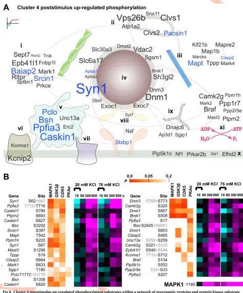
The poststimulus up-regulation observed for cluster 4 is of particular interest because the pat-tern of regulation implies a late but significant change in phospho-regulated protein function (Fig 5D). Cluster 4 substrates are displayed on the curated protein interaction network inFig
8A. Letter size was mapped to the largest difference in magnitude from 10 s to 300 or 900 s
after 20 mM KCl to identify the largest up-regulated phosphorylation relative to early time points. Motif analysis (S6A Fig) and KinSwing (Fig 6B) predicted that MAPK is likely to have phosphorylated cluster 4 substrates. In addition, a MAPK1 activation site was up-regulated at all poststimulus time points (Fig 8B, bottom). Thus, we compared the probability of a
confi-dent match for cluster 4 substrates to the MAPK1 substrate motif, as a heat map inFig 8B(S1
Data), using the first step of the KinSwing process to generate probabilities (Fig 6B). Alongside,
we also compared GSK3βand CDK5, which co-clustered with MAPK1, and PKAα, which was
the most up-regulated at 300 s after 20 mM KCl (Figs6Band8B). Thus, we present the
phos-pho-signalling network for cluster 4 and identify known and potential substrates of MAPK1 and other protein kinases.
Phosphorylation of (the protein expressed from)Pip5k1cat S552 (Fig 8B) promotes its role
in the formation of focal adhesions [47]. B-Raf (Braf) phosphorylation at S134 is known to be
up-regulated in a feedback loop within the MAPK pathway [48] and was up-regulated in
clus-ter 4 (Fig 8B). Phosphorylation of two sites on IRSp53 (Baiap2) by a CaMKL family protein
kinase Par1b (Mark2), itself a cluster 4 member, may negatively regulate cell polarity and
spreading [49]. The calcium-activated potassium channel subunit alpha-1 (Kcnma1) was
up-regulated from 90 s at S712, which is known to cause reduced probability of channel opening
and is expected to be a substrate of PKC [50]. These phosphorylation sites with known
func-tions in diverse processes can now be put into the context of neuronal activity through deter-mination of their patterns of phospho-signalling.
In contrast toFig 7A, in which all synapsins were prominent, only synapsin 1 had cluster 4
phosphorylation sites (Fig 8A). Synapsin 1 domain B sites S62 and S67 (Fig 8B) and S549 in
domain D (Fig 3C) are known MAPK substrates [12,28]. MAPK phosphorylation of synapsin
1 limits neurotransmitter release [18]. Active zone scaffold proteins were major targets within
the rapidly responding clusters 1 and 2 (Fig 7A and 7B) and also within the poststimulus
up-regulated phosphorylation of cluster 4, having four proteins with at least three represented
phosphopeptides (Fig 8A). However, a different set of proteins and phosphorylation sites were
clathrin-coated pit, and (x) presynaptic membrane and membrane raft. (xi) Protein kinases and protein phosphatases, including regulatory subunits, with no specific localization are also shown. Gene names in blue letters have three or more responding phosphorylation sites. Experimentally verified protein interactions (STRING) are shown as red edges, i.e., connecting lines. The data are the result of six independent experiments for each stimulation condition (20 mM and 76 mM KCl). SNARE, soluble N-ethylmaleimide-sensitive factor attachment protein receptor.
Fig 8. Cluster 4 poststimulus up-regulated phosphorylation substrates within a network of presynaptic proteins and protein kinase substrate probabilities. (A) A word cloud visualization of the synaptosome phosphoproteomics data using gene names anchored to cellular component
ontology (subcellular localization). The size of the gene name is scaled to the absolute value of the difference between the log2intensities after 20 mM
prominent in cluster 4: liprin-α3 (Ppfia3), Munc13-1 (Unc13a), and Ca2+- and calmodulin-dependent serine protein kinase-interacting protein 1 (Caskin1). This indicates that a post-stimulus phospho-signalling pathway targets the active zone scaffold using a distinct subset of protein components and activity-dependent sites. Phosphorylation of S2845 on bassoon pro-motes 14-3-3 binding and changes bassoon molecular exchange rates with the proposed effect of dissociation from the cytomatrix at the active zone [51]. S2845 is an in vitro RSK family sub-strate [51], and the RSK family had a pattern of inferred regulation that was consistent with
cluster 4 (S6B Fig). Dynamin 1 was poststimulus up-regulated at S857, a phosphorylation site
that is de-enriched at presynaptic terminals [38]. The up-regulation of dynamin 1
phosphory-lation is inhibitory for synaptic vesicle supply [17], which could promote a depression of
vesi-cle release. The dynamin 1 phospho-dependent binding partner syndapin 1 (Pacsin1) [52] was
among a group of endosomal proteins that were prominent in cluster 4 (Fig 8A) but not cluster
1 and 2 (Fig 7A and 7B). The phosphorylation of Munc18-1 (Stxbp1) on Y473 has been shown
to inhibit neurotransmitter release [53] and was one of three Munc18-1 sites with poststimulus
up-regulation. Two other cluster 4 proteins were associated with the vesicle fusion machinery (Fig 8A,Syt7andNsf). Thus, MAPK1 and other protein kinases are implicated as regulators of cluster 4. MAPK1 may be upstream of protein kinases in cluster 4, but this is currently only
supported by motif prediction (Fig 8B). Many substrate sites inFig 8Bhave not been
function-ally investigated; however; the known roles of bassoon, dynamin 1, synapsin 1, and Munc18-1 implicate the poststimulus up-regulation of cluster 4 in influencing the synaptic vesicle cycle.
Discussion
This first study of the temporal dynamics of the presynaptic phosphoproteome has led to a num-ber of new insights and provides a valuable resource for future analyses. Firstly, it is now appar-ent that activity-dependappar-ent poststimulus phospho-signalling is complex and long-lasting and targets distinct protein domains and networks of phosphoproteins across time. Secondly, initial activity-dependent phospho-signalling in synaptosomes closely correlates with signalling in cul-tured hippocampal neurons, despite the more numerous compartments harvested with the lat-ter. Thirdly, we have identified specific protein kinases that mediate the initial and poststimulus response, which we have analyzed at unprecedented depth. This achievement was facilitated by our development of a new computational method, KinSwing. These insights greatly advance our knowledge about the stimulus-coupled dynamics of phospho-signalling at the nerve terminal.
A key feature of the KinSwing method was to enable a comparison of the inferred protein kinase activity at each time point and condition. The underlying protein phosphatase activity was also indicated. For example, MAPK1 was highly activated by the stimulus, but its sub-strates were mainly dephosphorylated prior to 300 s. Although KinSwing weights kinase activ-ity by the number of substrates used to build a kinase model, the kinase substrate matching and the extent of representation of the kinome in the output is limited by the availability of protein kinase substrate data. This can be important when considering substrates that are tar-geted by closely related protein kinases with limited a priori specificity data [33]. In addition,
more responding phosphorylation sites. Experimentally verified protein interactions (STRING) are shown as red edges, i.e., connecting lines. (B) The phosphorylation sites from (A) withP<0.1 prediction as substrates of MAPK1, GSK3β, CDK5, and PKAα, by KinSwing, shown as a heat map, with the indicated colour scale alongside the heat map of log2(stimulated intensity/control intensity) using the same colour scale as inFig 2B. For data
derived from multisite phosphorylated peptides, the nonrelevant phosphorylation site is shown in grey lettering. The up-regulation across time of an activity-inducing MAPK1 phosphorylation site is also shown. The data are the result of six independent experiments for each stimulation condition (20 mM and 76 mM KCl). Underlying data for this figure can be found inS1 Data. CDK5, calcium-dependent kinase 5; GSK3β, glycogen synthase kinaseβ; MAPK1, mitogen-activated protein kinase 1; PKAα, protein kinase Aα; SNARE, soluble N-ethylmaleimide-sensitive factor attachment protein receptor.
KinSwing best leverages large-scale experimental data that are rich in repeated patterns of phospho-regulation. However, the value of KinSwing and similar approaches will scale well as the size of public phosphorylation site databases increases. Thus, KinSwing can be applied to any phosphoproteomics study, including any neuronal stimulation paradigm amenable to bio-chemical analysis, and demonstrates the value of using and developing probabilistic methods for future phosphoproteomics studies.
KinSwing identified CaMKIIαas a protein kinase with a large change in inferred activity
across the time course that scaled with stimulation strength and CaMKIIαactivity correlated
with the profile of high-magnitude up-regulated phosphorylation (Fig 4A, cluster 1). CaMKIIα
activation is known to enhance neurotransmitter release [54], but very few presynaptic
sub-strates and mechanisms are known. The putative subsub-strates identified here in the active zone
scaffold, including bassoon, piccolo, liprin-α3, and RIM1, might participate in
phospho-regu-lated mechanisms that promote vesicle release and presynaptic plasticity.
Calcineurin is the primary candidate for the observed high-magnitude dephosphorylation (Fig 4A, cluster 2), since known substrates associated with synaptic vesicles [12,13] were
enriched in this phospho-signalling pattern (Fig 5A). Protein phosphatase 2A (PP2A) is also a
candidate, since it can undergo Ca2+-dependent activation [55]. The high-magnitude
up-regu-lation (cluster 1) lasted longer than the high-magnitude dephosphoryup-regu-lation (cluster 2,Fig 4A–
4C). This observation fits with the knowledge that calcineurin or PP2A are dependent on
ele-vated calcium, but CaMKIIαbecomes autoactivated [56]. Stronger stimulation increased the
magnitude of dephosphorylation for the initially down-regulated patterns (Fig 4A–4C, clusters
2 and 4). This resulted in a delayed return to prestimulus levels and a dampened poststimulus up-regulation for the bidirectional pattern (Fig 4A–4C, cluster 4). Mechanistically, the former could be achieved by sustained inactivation of protein kinases (cluster 2), such as the inhibition
of GSK3β[44]. For the latter, initial stronger down-regulation could have dampened
up-regu-lation (cluster 4); however, an alternative mechanism could be the increased activation of protein phosphatases. Phosphatases could have feasibly kept up-regulation low at 900 s after
76 mM KCl while MAPK1 activation was high (Fig 8B). Protein phosphatase 1 (PP1) acts
downstream of calcineurin [57,58]. PP1 and PP2A account for the vast majority of basal
phos-phatase activity in presynaptic terminals [57] and are implicated in the regulation of neuro-transmitter release [59–61].
Poststimulus up-regulated phosphorylation (cluster 4,Fig 8A) was associated with the
increased activity of MAPK1 (Fig 6BandS6A Fig). KinSwing predicted that PKAα, CaMKL,
STE20, and RSK classes of protein kinases also contributed to poststimulus up-regulation. These predictions were supported by evidence from the literature [49–51]. Substrates of post-stimulus up-regulation included synapsin 1, Munc18-1, and distinct components of active
zone scaffold proteins. Endocytosis and endosomal proteins were also implicated (Fig 8A).
MAPK1/3 activation and targeting of synapsin 1 was found by others to have a negative effect
on posttetanic enhancement [18]. In this context, the poststimulus up-regulation may be a
homeostatic plasticity mechanism that dampens the effects of strong stimuli. The up-regulated phosphorylation we identified involves a possible MAPK-signalling pathway that has many
more components beyond synapsin 1 (Fig 8B).
Activity-dependent phosphorylation in synaptosomes and hippocampal neurons was highly correlated for presynaptic proteins at the common 10-s time point (Fig 2A). Active zone scaf-fold proteins and synapsins were more highly targeted than postsynaptic scafscaf-fold proteins and neurotransmitter receptors (Fig 2C). This indicates that the active zone scaffold proteins may be more finely tuned to respond to Ca2+influx, despite vastly greater postsynaptic abundance of
Ca2+-sensitive proteins such as CaMKIIα. We have identified bassoon as the major presynaptic
hippocampal neurons. Piccolo, RIM1, microtubule-regulating MAP1B, and tau, as well as SNA-P25-interacting protein, were also identified as candidate signal integrators. Thus, phospho-sig-nalling in synaptosomes and intact neurons was similar, and several presynaptic proteins were identified as potential signal integrators, of which bassoon was most prominent.
Our 10-s stimulation with KCl would have caused a large calcium influx, which likely exceeds that produced by electrophysiological stimulation. Thus, our results likely include some nonphysiological phospho-signalling, necessitating validation of our work with
electrophysiological stimulation paradigms. However, the strength and duration of stimulation may be more important from a physiology prospective than the method of depolarization. A
10-s high-frequency (�40 Hz) electrical stimulation, combined with phospho-specific
anti-body detection, produced virtually identical phospho-signalling responses for the proteins
dynamin 1, Akt/PKB, and GSK3β[62]. High-frequency firing rates within the brain are not
typical but occur naturally in specific neurons during tasks such as spatial navigation [63]. Obtaining meaningful biochemical measurements at low frequencies will be challenging. In this work, we could have missed regulated phosphorylation sites because of undersampling but also because the change in phosphorylation was too low to measure (despite functional sig-nificance). In general, our lower potassium concentration produced robust phospho-signal-ling, particularly during the poststimulus (S2B and S2C Fig), indicating that high potassium concentrations can potentially be avoided. Nevertheless, elevated potassium stimulation is highly physiologically relevant to understanding the molecular consequences of traumatic
brain injury [64] and in the understanding of homeostatic plasticity mechanisms, since
pre-treatment with elevated potassium was shown to be neuroprotective in a model of excitotoxi-city [65]. Our 10-s stimulation was relatively acute, compared to the previous sustained depolarization [27], and allowed for the first examination of the poststimulus response.
Stimu-lating synaptosomes at a high and low level increased our ability to discern Ca2+
concentra-tion–dependent patterns of temporal regulation. Future work will require a combination of synaptic vesicle turnover and electrophysiological measurements in neurons to obtain a clearer view of the functional consequences of our stimulations and phosphoproteome dynamics.
The value of information on activity-dependent phosphorylation is in knowing which pro-tein domains and functions are mechanistically linked to neuronal activity, while ruling out phosphorylation that is independent of activity. Our studies revealed that the vast majority of phospho-regulated functions remain unidentified. The lack of association with activity indi-cates that neuroscience-relevant signalling mechanisms have not yet been a significant focus
for research, which might be explained by known biases in research effort [66]. Here, we have
highlighted the activity-dependent phosphorylation site data relevant to presynaptic mecha-nisms and neurotransmitter release. The sensitivity of presynaptic phospho-signalling, if trans-lated into major functional changes, could have implications for the pharmacological targeting of protein kinase/phosphatase activity within neurons. Other important targets of phospho-signalling that regulate diverse cellular functions are contained within this resource for neuro-scientists. Therefore, this resource, and our highly developed analysis approach, will allow others to further decipher the functional significance of synaptosomal and neuronal activity-dependent phosphorylation.
Materials and methods
Ethics statement
Isolation of presynaptic terminals (synaptosomes)
Eight- to 20-wk-old Sprague-Dawley male rats were humanely killed by decapitation. The whole brain was extracted. P2 fraction synaptosomes were isolated and prepared as described previously [67], with minor modifications. Briefly, the S1 fraction was centrifuged at 948gfor 10 min. All centrifugation steps were performed at 4 ˚C. The supernatant was discarded, and the P2 fraction was resuspended in a Krebs-like buffer solution (118 mM NaCl, 4.7 mM KCl, 20 mM 4-[2-hydroxyethyl]-1-piperazineethanesulfonic acid [HEPES]/trisaminomethane
[Tris] [pH 7.4], 25 mM NaHCO3, 1.18 mM MgSO4, 1.2 mM CaCl2, 0.1 mM Na2HPO4, 1.85 g/
L glucose), which had previously been bubbled with carbogen (95% O2, 5% CO2) for 45 min to
produce a final pH of 7.3–7.5. The solution was centrifuged at 13,800gfor 10 min, and the
resulting pellet was resuspended in 10 mL Krebs-like solution and centrifuged at 948gfor 10
min to obtain a P2 synaptosome pellet. The pellet was resuspended in 1.75 mL of Krebs-like solution, rested at 37 ˚C for 45 min, and then placed on ice until stimulated. The number of biological replicates is reported for each experiment. For the mass spectrometry and the west-ern blotting, each biological replicate was produced using the brain tissue of an individual rat.
Stimulation of presynaptic terminals for phosphorylation analysis
Prior to stimulation, the synaptosomes were warmed to 37 ˚C for 5 min. To depolarize the
syn-aptosomes, aliquots of 300μL of synaptosomes were mixed with an equal volume of solution
such that the final KCl concentration was 20 or 76 mM, and the sodium concentration was lowered by a similar amount such that the monovalent salt and osmotic concentration was constant. The depolarization continued for 10 s only before centrifugation at 13,500 rpm in a CM-50 MP (ELMI, Riga, Latvia) benchtop centrifuge. To repolarize the synaptosomes, they were resuspended in Krebs solution and incubated for up to 15 min. Samples were collected after 10 s of mock treatment or depolarization and at 90 s, 300 s, and 900 s in the repolarization solution. Prior to lysis, the synaptosomes were pelleted by centrifugation and the supernatant
removed. The synaptosomes were lysed in a 300μL solution and briefly agitated with a
bench-top vortex. The lysis solution consisted of 2% SDS, 25 mM HEPES/Tris (pH 7.4), 1 mM
ethyl-enediaminetetraacetic acid (EDTA), 1 mM ethylene glycol-bis(β-aminoethyl ether)-N,N,N0,
N0-tetraacetic acid (EGTA), 1x Roche Complete protease inhibitor cocktail, and 1x
Calbio-chem Phosphatase inhibitor cocktail II. The samples were incubated at 85 ˚C for 10 min to ensure inactivation of proteases and phosphatases.
Comparison of the effect of elevated KCl concentration on suspended
versus pelleted synaptosomes
Synaptosomes were prepared as above. Synaptosomes were stimulated with 76 mM KCl or mock stimulated with 4.7 mM KCl for 10 s, centrifuged to remove the liquid, and then lysed, as above. In a third condition, synaptosomes were centrifuged, and then KCl was added to achieve an isotonic solution of 76 mM KCl, as in the stimulated condition, but without dis-turbing the pellet. After 45 s, the supernatant was removed, and the sample was lysed. Equal amounts of each sample were applied to SDS-PAGE and examined by western blot with anti-synapsin 1 pS603 (see “SDS-PAGE and western blotting” section below).
Preparation of synaptosome samples for large-scale phosphoproteomics
using dimethylation
mM DTT was added to quench excess iodoacetamide. The protein content of each sample was
then precipitated using methanol/chloroform precipitation [68]. The dry protein pellets were
redissolved in 90μL 8 M urea and 10μL 1 M triethylammonium bicarbonate (TEAB) and
digested for 2 h at 23 ˚C using 0.15 U endoproteinase Lys-C per sample. The samples were
then diluted to 1 M urea with 50 mM TEAB followed by digestion with 20μg TrypZean trypsin
(Sigma-Aldrich, St. Louis, MI, United States) per sample. After 4 h, another 20μg was added
and the samples digested for another 4 h (both steps performed at 23 ˚C). Digested samples were then subjected to in-solution reductive dimethylation [69] essentially according to
Boer-sema and colleagues [70]. Labelling was performed according to the below scheme (note:
con-trol is mock stimulated for 10 s):
1. Replicates 1 and 2:
a. 20 mM KCl set 1: Control (light), 10 s (medium), 90 s (heavy)
b. 20 mM KCl set 2: Control (light), 300 s (medium), 900 s (heavy)
c. 76 mM KCl set 1: Control (light), 10 s (medium), 90 s (heavy)
d. 76 mM KCl set 2: Control (light), 300 s (medium), 900 s (heavy)
2. Replicates 3 and 4:
a. 20 mM KCl set 1: Control (medium), 10 s (heavy), 90 s (light)
b. 20 mM KCl set 2: Control (medium), 300 s (heavy), 900 s (light)
c. 76 mM KCl set 1: Control (medium), 10 s (heavy), 90 s (light)
d. 76 mM KCl set 2: Control (medium), 300 s (heavy), 900 s (light)
3. Replicates 5 and 6:
a. 20 mM KCl set 1: Control (heavy), 10 s (light), 90 s (medium)
b. 20 mM KCl set 2: Control (heavy), 300 s (light), 900 s (medium)
c. 76 mM KCl set 1: Control (heavy), 10 s (light), 90 s (medium)
d. 76 mM KCl set 2: Control (heavy), 300 s (light), 900 s (medium)
After dimethyl labelling, each set of light, medium, and heavy samples were mixed and acid-ified to 0.2% trifluoroacetic acid (TFA). Insoluble material was removed by centrifugation.
Phosphopeptide enrichment and peptide fractionation
Samples were then subjected to phosphopeptide enrichment and fractionation by TiSH (TiO2,
sequential elution from immobilized metal affinity chromatography [SIMAC] and hydrophilic
interaction liquid chromatography [HI