Downregulating Mitochondrial Trifunctional-Protein Expression
Yutaka Amako,a*Tsubasa Munakata,bMichinori Kohara,bAleem Siddiqui,dChris Peers,cMark Harrisa
School of Molecular and Cellular Biology, Faculty of Biological Sciences,aand Faculty of Medicine and Health,cUniversity of Leeds, Leeds, United Kingdom; Department of Microbiology and Cell Biology, Tokyo Metropolitan Institute of Medical Science, Tokyo, Japanb; Department of Medicine, Division of Infectious Diseases, University of California, San Diego, California, USAd
ABSTRACT
The course of hepatitis C virus (HCV) infection and disease progression involves alterations in lipid metabolism, leading to symptoms such as hypocholesterolemia and steatosis. Steatosis can be induced by multiple mechanisms, including increases in lipid biosynthesis and uptake, impaired lipoprotein secretion, and/or attenuation of lipid-oxidation. However, little is known about the effects of HCV on lipid-oxidation. A previous proteomics study revealed that HCV interacted with both the␣- and
-subunits of the mitochondrial trifunctional protein (MTP), an enzyme complex which catalyzes the last 3 steps of mitochon-drial lipid-oxidation for cellular energy production. Here we show that in HCV-infected Huh7.5 cells, lipid-oxidation was significantly attenuated. Consistently with this, MTP protein and mRNA levels were suppressed by HCV infection. A loss-of-function study showed that MTP depletion rendered cells less responsive to alpha interferon (IFN-␣) treatment by impairing IFN-stimulated gene expression. These aspects of host-virus interaction explain how HCV alters host energy homeostasis and how it may also contribute to the establishment of persistent infection in the liver.
IMPORTANCE
HCV infection triggers metabolic alterations, which lead to significant disease outcomes, such as fatty liver (steatosis). This study revealed that HCV impairs mitochondrial lipid-oxidation, which results in low lipid combustion. On the other hand, the HCV-induced defects in metabolic status played an important role in the control of the type I interferon system. Under the con-ditions of impaired lipid-oxidation, host cells were less responsive to the ability of exogenously added IFN-␣to suppress HCV replication. This suggests that interference with lipid-oxidation may assist the virus in the establishment of a long-term, per-sistent infection. Further understanding of this aspect of virus-host interaction may lead to improvements in the current stan-dard therapy.
H
epatitis C virus (HCV) is a member of genusHepacivirusinthe family Flaviviridae. Its genome consists of up to 9,600
bases of single-stranded RNA with positive polarity. Genomic RNA contains a single open reading frame encoding a polyprotein
(⬃3,000 amino acids), which is processed into 10 mature proteins
by both cellular and viral proteases. The 10 mature proteins con-sist of three structural proteins (core, E1, and E2), p7, and the nonstructural (NS) proteins NS2, NS3, NS4A, NS4B, NS5A, and
NS5B (1). HCV is a global health problem; it chronically infects
⬃170 million people and is a leading cause of liver
transplanta-tion. Because of the lack of a prophylactic vaccine and limitation of effective therapeutic options, persistent HCV infection leads patients to chronic hepatitis, fatty liver (steatosis), cirrhosis, and
liver cancer (2,3).
Until recently, the standard of care was 24 to 48 weeks of ther-apy with pegylated interferon (IFN) and ribavirin. Success rates of this therapy differed greatly, from 6% to 84%, depending on the patient’s situation (viral load and genotype, severity of liver dis-ease, etc.). Direct-acting antivirals, which include viral protease (NS3), polymerase (NS5B), and NS5A inhibitors, are now
ap-proved for clinical treatment (4). Although a combination of these
inhibitors will soon replace current standard therapy, the poten-tial for the emergence of resistant virus is high, due to the high rate of turnover of virus and the high error rate of the NS5B
polymer-ase (5).
A body of evidence shows that the course of disease
progres-sion involves metabolic alteration of lipid biogenesis and its
ho-meostasis in the liver (6). Furthermore, viral replication and
egress have been reported to correlate with very-low-density
lipo-protein (VLDL) biogenesis and the secretion pathway (7). It has
been shown that viral genomic replication takes place on altered endoplasmic reticulum and is tightly associated with cytosolic
lipid droplets (8). The following stages of viral maturation/release
have been reported to be coupled with VLDL secretion, so that infectious viral particles appear to associate with certain
lipopro-teins to form lipo-viro-particles (LVPs) (7,9–12). In this regard,
Received6 June 2014Accepted4 February 2015
Accepted manuscript posted online11 February 2015
CitationAmako Y, Munakata T, Kohara M, Siddiqui A, Peers C, Harris M. 2015. Hepatitis C virus attenuates mitochondrial lipid-oxidation by downregulating mitochondrial trifunctional-protein expression. J Virol 89:4092– 4101. doi:10.1128/JVI.01653-14.
Editor:M. S. Diamond
Address correspondence to Yutaka Amako, [email protected], or Mark Harris, [email protected].
* Present address: Yutaka Amako, Department of Microbiology and Cell Biology, Tokyo Metropolitan Institute of Medical Science, Tokyo, Japan.
Copyright © 2015, Amako et al. This is an open-access article distributed under the terms of theCreative Commons Attribution 3.0 Unported license. doi:10.1128/JVI.01653-14
on November 7, 2019 by guest
http://jvi.asm.org/
HCV hijacks the VLDL biogenesis/secretion pathway (13). Since the liver is the most important organ for lipid transpor-tation and storage, understanding why the entire viral life cycle is so tightly associated with lipid biogenesis and transport will be key to an understanding of the mechanisms of viral invasion and pathogenesis.
While HCV utilizes VLDL biogenesis for its dissemination, vi-ral infection causes fatty liver (steatosis), which potentially wors-ens chronic inflammation (steatohepatitis), most likely due to
re-active oxygen species (ROS) in the liver (14,15). Furthermore,
fatty liver is a common risk factor for the development of insulin
resistance (16). Chronic HCV infection can thus be regarded as a
metabolic syndrome. Although many studies have investigated
the upregulation of lipogenesis by HCV (17,18), little is known
about the effect of virus infection on mitochondrial lipid
-oxi-dation, apart from one study suggesting that HCV might impair
this processin vivo(19). Since mitochondrial lipid-oxidation
yields a greater amount of ATP than carbohydrate or protein con-sumption, host energy metabolism may be unbalanced when it is negatively impacted by viral infection. Indeed, recent studies sug-gest that HCV infection triggers a shift in the energy expenditure
profile in terms of glucose consumption and production (20–22).
Our previous characterization of the protein interactome of the HCV nonstructural protein NS5A suggested that it interacts
with both the␣- and-subunits of mitochondrial trifunctional
protein (MTP) (23). The MTP complex, which is an octamer of 4
␣-subunits and 4-subunits, has three enzymatic activities, which
catalyze the last stages in mitochondrial lipid -oxidation:
3-hydroxyl– coenzyme A (CoA) dehydrogenase, 3-ketoacyl–CoA
thiolase, and enoyl-CoA hydratase. Interestingly, MTP␣
hetero-zygous (MTP␣⫹/–) transgenic mice have been shown to develop
fatty liver and insulin resistance (24). In this study, we aim to
dissect the molecular mechanism underpinning the effect of viral
infection on lipid-oxidation and the possible impact of this on
the innate immune response to HCV.
MATERIALS AND METHODS
Plasmids.pJFH1 was provided by Takaji Wakita (National Institute of Infectious Disease, Japan) and described previously (25). The pJc1 con-struct was a gift from R. Bartenschlager (University of Heidelberg) and used to construct the pJc1-p7NLuc2A reporter virus construct (26). pJc1-p7NLuc2A and pJc1-pJc1-p7NLuc2A/GND were constructed by conventional molecular cloning methods, and nucleotide sequences are available upon request. pISRE-Luc was purchased from Agilent Technologies. pRL-TK was from Promega.
Cell culture and virus infection.Huh7.5 cells were a gift from C. Rice (Rockefeller University). The 293FT cell line is from Life Technologies and was used for lentiviral packaging. Huh7.5 cells were maintained in high-glucose Dulbecco’s modified Eagle medium supplemented with 10% fetal bovine serum, 100 U/ml penicillin, 100g/ml streptomycin, 1 mM nonessential amino acids, and 2 mM GlutaMAX (Life Technologies). To generate viruses, vectors were linearized by XbaI digestion followed by mung bean nuclease treatment to blunt the digested termini and used as template DNAs for T7 promoter-drivenin vitrotranscription using the T7 RiboMAX large-scale RNA production system (Promega, Madison, MA). RNA transfection was performed as described previously (25). Cultured virus titers were determined as described in reference28.
Lentiviral vectors for expressing shRNA.pCS-RfA-EG and a set of packaging plasmid vectors, including pCMV-VSV-G-RSV-Rev and pCAG-HIVgp, were provided by Hiroyuki Miyoshi (Riken BioResorce Center, Japan). To construct short-hairpin RNAs (shRNAs) expressing lentiviral vectors, a pair of 65-mer oligonucleotides were inserted into
BglII and XbaI sites of pENTR4-H1. Oligonucleotide sequences for con-structing luciferase mRNA-specific shRNA (shLuc), short-hairpin green fluorescent protein (shGFP), shLacZ, shMTP␣, and shMTPare available upon request. The shRNA-encoding insert was confirmed by DNA se-quencing. The shRNA expression cassette of pENTR4 was inserted into pCS-RfA-EG by Gateway LR Clonase II (Invitrogen)-directed DNA re-combination as per the manufacturer’s recommendations. Lentiviral par-ticles were obtained as cultured supernatants from packaging transfec-tions performed using Fugene 6 (Promega), as per the manufacturer’s recommendations.
NanoLuc reporter virus and replication assay.T7 RNA transcription and electroporation for pJc1-p7NLuc2A were performed as described above. Reporter virus titers in cultured supernatants were determined by focus-forming unit (FFU) assay. Naive Huh7.5 cells were infected at a defined multiplicity of infection (MOI) of 0.05 to initiate virus culture. The nanoluciferase (NanoLuc) assay was performed using the Nano-Glo luciferase assay system according to the manufacturer’s instructions.
Fatty acid-oxidation assay.Huh7.5 cells were infected with JFH-1 virus at an MOI of 0.3. [3H]palmitic acid was conjugated to fatty-acid-free
bovine serum albumin (BSA). HCV-infected cells were incubated with Krebs’s buffer containing [3H]palmitic acid-BSA for 2 h at 37°C. The
amount of released3H
2O was measured using an LSC-6100 scintillation
counter (Aloka) as described previously (29). Etomoxir andL-carnitine were used at 20M and 1 mM, respectively.
Real-time PCR.Quantification of HCV RNA and GAPDH (glyceral-dehyde-3-phosphate dehydrogenase) mRNA was performed as described previously (30). Primer pairs for quantification of MTPs,-actin, and proliferator-activated receptor␥coactivator 1␣(PGC-1␣) mRNA are available upon request. Real-time PCR was performed using Superscript III (Life Technologies) for first-strand synthesis and QuantiFast SYBR green (Qiagen) according to the manufacturers’ instructions. Mitochon-drial DNA (mtDNA) quantitation was performed as described previously (31).
Interferon promoter/reporter assay. Plasmids pISRE-Luc and pRL-TK (20:1, wt/wt, ratio) were transfected into Huh7.5 cells on a 12-well plate with Fugene 6 transfection reagent. Transfected cells were split into a 96-well plate at 24 h posttransfection. Cells were further incubated for 48 h, and during the last 5 h, they were exposed to IFN-␣at the concentrations indicated inFig. 6. Measurement of luciferase activity was performed using the dual-luciferase reporter assay system for pISRE-Luc and pRL-TK.
Protein assay.Western blotting was performed using the Odyssey Sa infrared imaging system (LI-COR). Detected protein band intensities were analyzed by Image Studio software (LI-COR).
Antibodies and reagents. Human IFN-␣ was purchased from Mochida Seiyaku (Japan). Commercially obtained antibodies are anti-MTP␣and -MTP(Santa Cruz Biotechnology), anti-protein disulfide isomerase (PDI), and-actin (Sigma-Aldrich).
RESULTS
HCV infection attenuates lipid-oxidation.It was previously shown that HCV infection led to a reduction in ATP production in
tissue culture (32). It has also been shown that HCV proteins
interact with mitochondria to negatively impact mitochondrial functions and/or modulate innate immune signaling cascades,
which utilize mitochondrial membranes as a platform (14,15,33,
34). We previously showed that NS5A interacts with MTP␣and
MTP(also known as 3-hydroxyl-CoA
dehydrogenase/3-keto-acyl-CoA thiolase/enoyl-CoA hydratase, alpha and beta subunits
[HADHA and HADHB], respectively). MTPwas also proposed
to be involved in HCV-induced metabolic changes within infected
cells following another proteomic analysis (18). These data
sug-gest that the major mitochondrial lipid -oxidation pathway
might be affected by HCV infection, which might in turn cause
HCV Attenuates Mitochondrial Lipid-Oxidation
on November 7, 2019 by guest
http://jvi.asm.org/
impaired ATP production and alter the balance of energy
homeo-stasis. To assess this possibility, we performed a lipid-oxidation
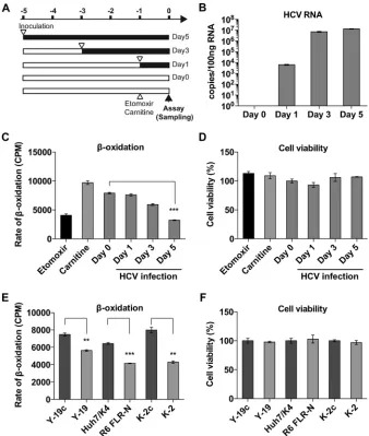
assay using [3H]palmitate as a tracer. The results clearly indicated
that HCV infection significantly attenuated lipid -oxidation
(Fig. 1C) in a time-dependent manner that correlated with the progress of HCV infection, with inhibition reaching 59.3% at day 5 (Fig. 1C). Treatment of uninfected Huh7 cells with either
eto-moxir orL-carnitine (as a known inhibitor or enhancer of
mito-chondrial lipid-oxidation, respectively) confirmed the
appro-priate sensitivity of the assay to changes in-oxidation. The lipid
-oxidation assay was also performed for three pairs of replicon
cell lines and the corresponding IFN-cured cell lines. In both sub-genomic-replicon (SGR) cell lines for genotype 2a (Y-19) and
ge-notype 1b (R6 FLR-N), SGR RNA replication caused impairment
of lipid-oxidation by approximately 24.5% and 35.6%
(com-pared to lipid-oxidation in the cured cell lines), respectively. A
full-length genomic replicon of genotype 2a (K-2) inhibited lipid
-oxidation more dramatically (46.0%) than in the cured cell
control, implying that the additionally expressed viral proteins (core, E1, E2, p7, and NS2) might have caused additional
inhibi-tion of lipid-oxidation. Although lipid-oxidation was
signifi-cantly impaired, it was not associated with a loss in cell viability, as confirmed by using a Cell Counting Kit-8 (CCK-8; Dojindo,
Ja-pan) assay (Fig. 1DandF).
To investigate the mechanism of viral intervention with lipid
-oxidation, we measured the protein expression levels of MTP␣
FIG 1HCV infection attenuates lipid-oxidation. (A) Schematic of experiment. Huh7.5 cells were plated at 1.6⫻104cells per well in a 24-well plate and then
infected with HCV JFH-1 at different times (MOI of 0.3 FFU/cell), so that the lipid-oxidation assay could be performed for all samples simultaneously. (B) Cellular HCV RNA was measured by real-time quantitative RT-PCR. (C) Huh7.5 cells were fed with [3H]palmitate, and3H
2O was assayed as a measure of lipid
-oxidation. The rate of lipid-oxidation decreased in a time-dependent manner. Huh7.5 cells were also treated with etomoxir (20 mM) orL-carnitine (1 mM) as a known inhibitor or enhancer of mitochondrial lipid-oxidation, respectively. (D) Cell viabilities were unaffected and confirmed by using a CCK-8 assay. (E) The same lipid-oxidation assay was performed for HCV replicon cells. Y-19 is a stable genotype 2a JFH-1 SGR-harboring cell line, and Y-19c is the corresponding IFN-cured cell line. R6 FLR-N is a genotype 1b SGR-harboring cell line, and Huh7/K4 is the corresponding cured cell line. K-2 is a JFH-1 full-length replicon (FLR)-harboring cell line, and K-2c is the corresponding cured cell line. (F) Replicon RNA replications did not affect cell viabilities. Column error bars represent the standard errors of the means (SEM). Unpaired Studentttests were performed in order to calculate statistical significance between indicated columns. Asterisks indicatePvalues as follows: *,⬍0.05; **,⬍0.001; and ***,⬍0.0001.
on November 7, 2019 by guest
http://jvi.asm.org/
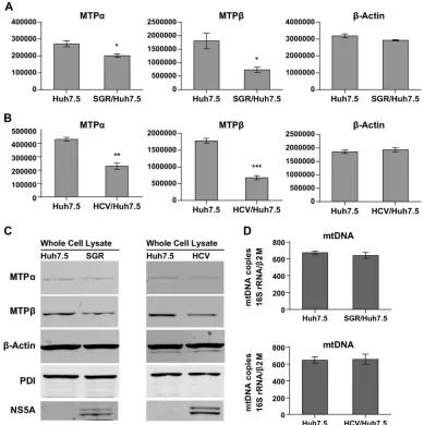
[image:3.585.123.461.66.465.2]and MTP. Whole-cell lysates from Huh7.5, SGR-harboring, or HCV-infected cells were analyzed by quantitative infrared West-ern blotting. The results show that both SGR replication and HCV
infection downregulated levels of both MTP␣and MTP(Fig. 2A
toC) compared to levels in the-actin and PDI controls. In the
case of HCV-infected Huh7.5 cells, levels of MTP␣and -
pro-teins were reduced by 46.6% and 61.9%, respectively (Fig. 2B).
Considering that ablation of MTP␣in mice caused embryonic
lethality, a 50% reduction of MTP protein expression and enzyme activity may cause severe outcomes in terms of cellular physiology
and pathology (35).
A recent study revealed that HCV infection promotes mi-tophagy, a selective degradation of mitochondria by autophagy
(34). In light of this finding, we speculated that HCV infection
might cause a decline in the numbers of mitochondria per cell; however, quantitative PCR for mitochondrial DNA (16S RNA
gene borne in mtDNA) revealed that neither HCV infection nor
SGR replication caused a change in the levels of mtDNA (Fig. 2D).
This implies that attenuation of lipid-oxidation was not caused
by a decrease in the mitochondrial content within the cells. Al-though little is known about how mitochondrial biogenesis can be affected by HCV infection, HCV-induced mitophagy might be compensated for by upregulation of mitochondrial biogenesis to maintain the overall mitochondrial number. To further investi-gate how MTP gene expression may be affected in the course of HCV infection, we performed a series of quantitative, real-time reverse transcription (RT)-PCR experiments.
HCV attenuates MTP expression at the transcriptional level.
To further investigate the mechanism of the HCV effect on lipid
-oxidation, MTP␣and MTPmRNAs were quantified by
quan-titative RT-PCR. Huh7.5 cells were infected with HCV (JFH-1,
MOI⫽0.05), and then mRNA levels for MTP␣, MTP, and
-ac-FIG 2HCV replication causes a decrease in MTP␣and -protein expression. (A to C) Whole-cell lysates from naive Huh7.5 cells, SGR-JFH-1-harboring cells, or HCV-infected cells (Jc1 MOI of 0.05 FFU/cell, day 7 postinfection) were analyzed by Western blotting by using conjugated infrared secondary antibodies. (A and B) Quantitative measurements of band intensities were taken using a LI-COR Odyssey Sa infrared imaging system and calculated by image studio software (n⫽4). They-axis scales are arbitrary units, representing protein band intensities. Asterisks indicatePvalues as follows: *,⬍0.05; **,⬍0.001; and ***,⬍0.0001. (C) Whole-cell lysates were analyzed to compare levels of expression of MTP␣, MTP, and cellular markers. Lysates (3g protein) were analyzed by infrared Western blotting.-Actin is an internal loading control. PDI is an endoplasmic reticulum- and mitochondrion-associated membrane protein. (D) mtDNA quantitations were performed for Huh7.5 SGR-harboring cells (upper panel) or HCV-infected cells (lower panel).
HCV Attenuates Mitochondrial Lipid-Oxidation
on November 7, 2019 by guest
http://jvi.asm.org/
[image:4.585.97.486.67.457.2]tin, together with levels of HCV RNA, were monitored for 5 days.
By day 5 postinfection, MTP␣and MTPmRNAs were
down-regulated by approximately 35% and 38%, respectively (Fig. 3C
andD, respectively), while HCV RNA reached a plateau at day 5
(Fig. 3A).-Actin mRNA levels were unaffected throughout this
period (Fig. 3B). Transcription of MTP␣and MTPwas inhibited
in parallel. This may be because these two genes share a promoter
region; both the MTP␣and MTPgenes are located on
chromo-some 2p23, where they are transcribed in opposite directions from
a shared bidirectional promoter of 350 bases (36). It is also
note-worthy that the level of transcriptional attenuation was compara-ble to those of protein and enzymatic downregulation, as de-scribed above. These data demonstrate that HCV regulation of MTP expression most likely occurs at the transcriptional level. We also assessed the level of peroxisome proliferator-activated
recep-tor␥coactivator 1␣(PGC-1␣) mRNA as a marker of
mitochon-drial biogenesis (37). Interestingly, the level of PGC-1␣was
ele-vated at day 5 after HCV infection (Fig. 3E). This implies that as
well as reducing the transcription of MTP␣and MTP, HCV
increased mitochondrial biogenesis, perhaps to compensate for the loss of MTP. Consistently with this, there was no reduction in the levels of mtDNA (and therefore mitochondrial numbers) over the period of HCV infection.
Inhibition of MTP expression impairs overall lipid -oxida-tion activity.In order to study how MTP function may relate to
HCV replication, we constructed lentiviral vectors expressing
shRNA targeted to either MTP␣or MTP, along with control
lentiviruses targeting luciferase (Luc), GFP, and-galactosidase
(LacZ). Three lentiviruses each for MTP␣and MTPwere
con-structed, and 5 of these 6 shMTPs effectively downregulated
pro-tein expression. The exception was shMTP␣number 1 (Fig. 4A),
although this shRNA did achieve a 50% knockdown of mRNA
levels (Fig. 4B). RNA interference triggered coincident ablation of
both MTP␣and MTP, and where shRNA mediated ablation of
either subunit, it also ablated the other. This was expected
be-cause, as mentioned above, transcription of MTP␣and MTP
mRNAs are tightly correlated, a relationship that seems to be ex-tended to posttranscriptional processing and translation, until they form a mature octamer complex. At the level of mRNA ex-pression, each shMTP construct specifically downregulated its
cognate target mRNA, except for shMTPnumber 2, which also
caused inhibition of MTP␣transcription (Fig. 4B). None of the
shRNAs tested in this study affected GAPDH mRNA levels (Fig.
4C) or GAPDH expression (Fig. 4A). Furthermore, we confirmed
that the shRNA-mediated knockdown of MTP genes led to the
impairment of overall lipid-oxidation activity (Fig. 4D),
with-out affecting their viabilities (Fig. 4E). We proceeded to test
MTP-depleted cells for effects on HCV replication.
A reporter virus construct carrying a novel luciferase gene
(NanoLuc) was generated for this experiment (Fig. 5A). This
re-FIG 3HCV attenuates MTP protein expression at the transcriptional level. The abundances of HCV RNA (A),-actin mRNA (B), MTP␣(C), MTP(D), and PGC-1␣mRNA (E) were examined by real-time RT-PCR in HCV-infected cells. ***,P⬍0.001. (F) mtDNA was quantitated by real-time PCR.
on November 7, 2019 by guest
http://jvi.asm.org/
[image:5.585.125.461.65.411.2]porter virus construct, designated Jc1-p7NLuc2A, was con-structed in a fashion analogous to that used to construct
p7-Rluc2A (described in reference38), and it replicated efficiently in
Huh7.5 cells upon RNA transfection (Fig. 5B). Reporter virus
par-ticles were very effectively produced by transfecting RNA into
Huh7.5 cells (Fig. 5CandD) and used to infect MTP-depleted
cells; however, the results showed that MTP-depleted cells
sup-ported HCV replication as well as control cells did (Fig. 6A),
ex-cept with shMTPnumber 2, which reduced HCV replication at a
late time point (day 4). Interestingly, this shRNA also reduced
MTP␣transcription (Fig. 4B), suggesting that depletion of both
MTP subunits was detrimental to HCV replication or that this
shRNA might have an off-target effect. For this reason, shMTP
number 2 was not used in the subsequent IFN experiments. These data are consistent with the fact that HCV infection itself
down-regulates MTP protein expression and lipid-oxidation so that
the effect of shRNA-mediated knockdown can be overshadowed by this autonomous reduction in MTPs by HCV. We also
con-structed lentivirus vectors to overexpress either MTP␣, MTP, or
both MTP subunits. However, although transduction of Huh7.5 cells with these vectors was able to increase levels of MTP
expres-sion, there was no effect on levels of lipid-oxidation (data not
shown), suggesting that other enzymes in the pathway were rate limiting or, alternatively, that merely overexpressing MTP does not result in an increase in the transport of these proteins to the mitochondria and the assembly of a functional enzymatic com-plex.
MTP protein depletion leads to deregulated responses to
IFN-␣.HCV has been shown to alter the functions of
mitochon-dria (39). Mitochondria not only are important for energy
pro-duction but also function in the innate immune response against
intracellular microbe infection, including viruses (40). HCV has a
specific strategy to evade this function, as the protease NS3 cleaves the mitochondrial antiviral signaling protein (MAVS) to prevent the association of this adaptor with the mitochondrial membrane,
thereby blocking activation of downstream effectors (e.g., NF-B
FIG 4shRNA-mediated knockdown of MTP␣and MTP. Lentiviral-vector-mediated shRNAs as indicated were used to downregulate gene expression. Huh7.5 cells were transduced with the indicated lentiviral shRNA vectors. After 2 days of incubation, efficient gene transduction was confirmed by fluorescence microscopy using GFP as a marker. Cells were harvested at day 4 after lentiviral infection. (A) Western blot analysis for expression of MTP␣, MTP, and GAPDH. Quantitative RT-PCR analysis of MTP␣and MTP(B) and GAPDH (C) mRNA levels. (D) Huh7.5 cells were fed with [3H]palmitate, and3H
2O was assayed as
a measure of lipid-oxidation. (E) Cell viability was measured by the WST-8 assay.
HCV Attenuates Mitochondrial Lipid-Oxidation
on November 7, 2019 by guest
http://jvi.asm.org/
[image:6.585.126.462.62.459.2]and IRF3). This effect is important in the determination of host cell permissiveness to HCV infection. In light of these facts, we
speculated that impaired mitochondrial lipid-oxidation might
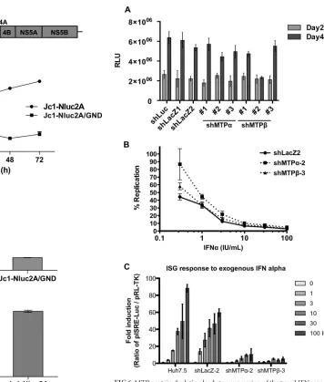
have an effect on the interferon response pathway. First, we in-fected MTP-depleted cells with the NanoLuc reporter virus and tested the ability of exogenously added interferon to control HCV infection. In comparison with the control cell line transduced with the LacZ targeting lentivirus (shLacZ), MTP-depleted cells were
less able to suppress viral replication in response to exogenously
added IFN-␣(Fig. 6B). The lack of responsiveness to IFN-␣was
more evident when cells were treated with lower doses of IFN-␣
(from 0.3 to 3 IU/ml). At higher concentrations, this effect was less evident. Paralleling the lack of effect of MTP overexpression on
lipid-oxidation, transduction with lentivirus vectors expressing
FIG 5Characterization of the NanoLuc reporter virus (Jc1-p7NLuc2A). (A) Schematic illustration of Jc1-p7NLuc2A. Nanoluciferase is the smallest lucif-erase reporter gene; thus, this construct is the shortest monocistronic, full-genomic reporter HCV construct. (B) The replication kinetics of Jc1-p7NLuc2A was evaluated by the nanoluciferase Glo assay following RNA electroporation into Huh7.5 cells. The GND derivative is a polymerase-inac-tive negapolymerase-inac-tive control, unable to replicate. (C) Cultured supernatants collected from day 3 of the experiment shown in panel B were overlaid onto 104cells in
96-well plates. After 2 days of incubation, infected cells were tested by the Nano-Glo luciferase assay to confirm reporter gene transduction via virus infection. (D) Comparison of the infectivities of Jc1-p7NLuc2A and the pa-rental Jc1 virus following electroporation of the corresponding RNAs into Huh7.5 cells.
FIG 6MTP protein depletion leads to suppression of the type I IFN response. MTP depletion does not directly affect HCV replication but suppresses the type I IFN response. (A) Huh7.5 cells were transduced with the indicated lentiviral shRNA vectors, and after 24 h of incubation, cells were infected with HCV Jc1-p7NLuc2A (MOI⫽0.05 FFU/cell) (Fig. 5). Infected cells were lysed at days 2 and 4 after HCV infection and assayed by the nanoluciferase A Glo assay to assess HCV replication. (B) MTP depletion renders host cells less responsive to exogenous IFN-␣to suppress HCV replication. Huh7.5 cells were transduced with the indicated lentiviral shRNA vectors. Two days later, cells were subsequently infected with HCV (MOI⫽0.05 FFU/cell), incubated for 4 days, and plated in a 96-well plate at 1⫻104cells per well for IFN-␣
treatment for a further 2 days. The levels of viral replication were assayed using Nano-Glo luciferase assay reagent as per the manufacturer’s instruction (n⫽ 6). (C) MTP depletion reduces the ISG response. Lentiviral-vector-mediated shRNAs as indicated were used to downregulate MTP gene expression. Trans-duced cells were transfected with pISRE-Luc and pRL-TK and then stimulated with IFN-␣at the indicated concentrations for 5 h prior to analysis by the dual-luciferase reporter assay (n⫽4). Error bars represent SEM.
on November 7, 2019 by guest
http://jvi.asm.org/
[image:7.585.173.529.62.481.2] [image:7.585.46.263.73.486.2]either MTP␣, MTP, or both MTP subunits had no significant
effect on IFN-␣responsiveness (data not shown).
To investigate the lack of IFN responsiveness following shRNA ablation of MTP expression, we analyzed transcription of inter-feron-stimulated genes (ISG) following exogenous addition of
IFN-␣. We used a reporter construct (pISRE-Luc) in which
lucif-erase expression was controlled by the interferon-stimulated
re-sponse element (ISRE). As shown inFig. 6C, ISRE responses to
IFN-␣were dramatically suppressed when MTP expression was
ablated by shRNA. These data help to explain why MTP depletion
may cause a lack of responsiveness to exogenous IFN-␣. However,
it is noteworthy that the effect on ISRE transcription was seen at all IFN concentrations, suggesting that there must be other effects of MTP depletion.
DISCUSSION
Mitochondrial lipid-oxidation is a major bioenergetic pathway
within the liver for the production of ATP from fatty acids. Met-abolic profiling studies have revealed that the cellular ATP
con-centration is significantly lowered by HCV infection (32,41), and
HCV infection alters the host energy expenditure profile in many
ways (20–22). HCV is known to interact with mitochondria to
induce reactive oxygen production, which is also thought to lead
to impairment of lipid-oxidation (39). Furthermore, a recent
clinical study showed that under fasting conditions, the total ke-tone body concentration was significantly lower for chronic HCV
patients than for their healthy uninfected counterparts (19). This
indicates that mitochondrial lipid-oxidation is impaired in the
livers of HCV-infected patients, because ketogenesis is a liver-specific metabolism that occurs in mitochondria and is directly
coupled to mitochondrial lipid -oxidation. Combining these
data with the data in this study, it is very likely that HCV
sup-presses mitochondrial lipid-oxidation to alter cellular
bioener-getics. This situation may be specific to HCV as, in contrast to our
observations, the related Dengue virus (also a member of the
Fla-viviridae) has been shown to upregulate-oxidation to facilitate
lipid consumption and ATP production by infected cells (29). The
phenotype of impaired mitochondrial lipid-oxidation has been
well studied and documented in the case of MTP gene deficiency.
Genetic defects in mitochondrial lipid-oxidation are recessively
inherited and cause pediatric and maternal morbidity and mor-tality. Children with this disorder suffer nonketotic hypoglycemia,
steatohepatitis, and skeletal and cardiac myopathy (42).
Consis-tently with this phenotype in humans, MTP␣knockout mice are
embryonic lethal (35), and heterozygous mice develop hepatic
steatosis (24). HCV infection attenuated lipid -oxidation by
more than 50% (Fig. 1), and such a degree of-oxidation
inhibi-tion may severely impact the overall health of the host, providing clues as to the molecular mechanisms underpinning the patho-genesis of HCV infection.
The mitochondrial lipid-oxidation pathway comprises
mi-tochondrial trifunctional proteins and other enzymes, which in-clude very-long-, long-, medium-, and short-chain acyl-CoA de-hydrogenases (CADs). Interestingly, a recent report showed that medium and short-chain CAD expression are also suppressed by
HCV (27). Phenotypes of these gene defects are marked with
in-tolerance to cold and fasting and an inability to convert fatty acids
to energy (ATP). HCV-induced impairment of lipid-oxidation
can cause the accumulation of lipids inside liver cells, leading to steatosis, although it is not yet fully confirmed to what degree this
can cause severe fatty liver, along with increased lipogenesis and impaired VLDL secretion.
To discuss possible mechanisms for how HCV attenuates
mitochondrial lipid-oxidation, we need to consider multiple
factors. In this study, we observed that HCV attenuates the tran-scription of both MTP genes. Data obtained by an MTP promoter/ reporter assay revealed that inflammatory cytokines, such as
tu-mor necrosis factor alpha (TNF-␣), interleukin 1(IL-1), and
IFN-␣inhibited the transcription of both MTP genes (data not
shown). HCV infection is capable of inducing the expression of these cytokines, and this may therefore drive a negative-feedback
loop for the suppression of lipid-oxidation. To the best of our
knowledge, Sp1 is the only transcription factor which has been
identified to bind to the MTP promoter region (36). Further
stud-ies are therefore needed to reveal how MTP promoter activity might be modulated by HCV infection. Intriguingly, we also
ob-served a physical interaction between HCV NS5A and both MTP␣
and MTP(data not shown); indeed, this was the observation that
first drew our attention to the-oxidation pathway. It is possible
therefore that HCV has an impact on-oxidation at multiple
levels, and it would be intriguing to determine whether NS5A in some way modulates the activity of the MTP complex.
In the last section of this study, we described that inhibition of
mitochondrial lipid-oxidation correlated with a concomitant
reduction in the transcriptional response to IFN-␣(Fig. 6C). This
is perhaps unsurprising, as mitochondria orchestrate a diverse
cellular response in survival situations (reviewed in reference43),
and indeed cell-fate-decisive signaling cascades take place on the surfaces of mitochondria. Although the mechanism by which
-oxidation may be linked to IFN responsiveness remains
ob-scure, a number of other observations may provide some clues: HCV protein expression (of either core, NS3, or NS5A) can sup-press type I IFN responses by disrupting STAT1 phosphorylation
and activation (44–46). Phosphorylation activation of STAT1 and
a subsequent nuclear location of p-STAT1 are required for ISRE-mediated gene transcription. Interestingly, core, NS3, and NS5A are all known to interact with mitochondria, and recent studies suggest that some STATs also potentially localize to mitochondria
(47). It will be interesting to see how localization and activation of
STATs may be affected by HCV infection-mediated attenuation of
lipid-oxidation or when MTP proteins are depleted by shRNA.
In summary, it is apparent that HCV interacts with mitochon-dria in multiple ways to modulate host energy metabolism and antiviral defense. Our evidence points to a link between mito-chondrial bioenergetics and the type I IFN response. Further study of this aspect of the host-virus interaction will lead to a better understanding of HCV biology and how current standard therapy can be helped by regaining mitochondrial functions.
ACKNOWLEDGMENTS
This study was supported by a grant to M.H. and C.P. from the Wellcome Trust (grant 090805). Y.A. was the recipient of a Marie Curie Career Integration Grant (304072 HCVFAO FP7-PEOPLE-2011-CIG) from the European Commission.
We thank Takaji Wakita (National Institute of Infectious Disease, Ja-pan) for pJFH1, Ralf Bartenschlager (University of Heidelberg) for pJc1, Charles Rice (Rockefeller University) for Huh7.5 cells, and Hiroyuki Mi-yoshi (RIKEN Institute) for the lentiviral vector system.
HCV Attenuates Mitochondrial Lipid-Oxidation
on November 7, 2019 by guest
http://jvi.asm.org/
REFERENCES
1.Lindenbach BD, Rice CM.2005. Unravelling hepatitis C virus replication from genome to function. Nature436:933–938.http://dx.doi.org/10.1038 /nature04077.
2.Lavanchy D.2009. The global burden of hepatitis C. Liver Int29(Suppl 1):S74 –S81.http://dx.doi.org/10.1111/j.1478-3231.2008.01934.x. 3.Pawlotsky JM.2004. Pathophysiology of hepatitis C virus infection and
related liver disease. Trends Microbiol12:96 –102.http://dx.doi.org/10 .1016/j.tim.2003.12.005.
4.Manns MP, von Hahn T.2013. Novel therapies for hepatitis C— one pill fits all? Nat Rev Drug Discov 12:595– 610. http://dx.doi.org/10.1038 /nrd4050.
5.Gelman MA, Glenn JS. 2011. Mixing the right hepatitis C inhibitor cocktail. Trends Mol Med17:34 – 46.http://dx.doi.org/10.1016/j.molmed .2010.10.005.
6.Negro F.2010. Abnormalities of lipid metabolism in hepatitis C virus infection. Gut59:1279 –1287.http://dx.doi.org/10.1136/gut.2009.192732. 7.Jiang J, Luo G.2009. Apolipoprotein E but not B is required for the formation of infectious hepatitis C virus particles. J Virol83:12680 – 12691.http://dx.doi.org/10.1128/JVI.01476-09.
8.Miyanari Y, Atsuzawa K, Usuda N, Watashi K, Hishiki T, Zayas M, Bartenschlager R, Wakita T, Hijikata M, Shimotohno K.2007. The lipid droplet is an important organelle for hepatitis C virus production. Nat Cell Biol9:1089 –1097.http://dx.doi.org/10.1038/ncb1631.
9.Chang KS, Jiang J, Cai Z, Luo G.2007. Human apolipoprotein E is required for infectivity and production of hepatitis C virus in cell culture. J Virol81:13783–13793.http://dx.doi.org/10.1128/JVI.01091-07. 10. Dreux M, Boson B, Ricard-Blum S, Molle J, Lavillette D, Bartosch B,
Pecheur EI, Cosset FL.2007. The exchangeable apolipoprotein ApoC-I promotes membrane fusion of hepatitis C virus. J Biol Chem282:32357– 32369.http://dx.doi.org/10.1074/jbc.M705358200.
11. Gastaminza P, Dryden KA, Boyd B, Wood MR, Law M, Yeager M, Chisari FV.2010. Ultrastructural and biophysical characterization of hep-atitis C virus particles produced in cell culture. J Virol84:10999 –11009.
http://dx.doi.org/10.1128/JVI.00526-10.
12. Merz A, Long G, Hiet MS, Brugger B, Chlanda P, Andre P, Wieland F, Krijnse-Locker J, Bartenschlager R.2011. Biochemical and morpholog-ical properties of hepatitis C virus particles and determination of their lipidome. J Biol Chem 286:3018 –3032. http://dx.doi.org/10.1074/jbc .M110.175018.
13. Syed GH, Amako Y, Siddiqui A.2010. Hepatitis C virus hijacks host lipid metabolism. Trends Endocrinol Metab 21:33– 40.http://dx.doi.org/10 .1016/j.tem.2009.07.005.
14. Korenaga M, Wang T, Li Y, Showalter LA, Chan T, Sun J, Weinman SA.
2005. Hepatitis C virus core protein inhibits mitochondrial electron trans-port and increases reactive oxygen species (ROS) production. J Biol Chem
280:37481–37488.http://dx.doi.org/10.1074/jbc.M506412200.
15. Piccoli C, Scrima R, Quarato G, D’Aprile A, Ripoli M, Lecce L, Boffoli D, Moradpour D, Capitanio N.2007. Hepatitis C virus protein expres-sion causes calcium-mediated mitochondrial bioenergetic dysfunction and nitro-oxidative stress. Hepatology 46:58 – 65.http://dx.doi.org/10 .1002/hep.21679.
16. Sanyal AJ.2011. Role of insulin resistance and hepatic steatosis in the pro-gression of fibrosis and response to treatment in hepatitis C. Liver Int
31(Suppl 1):S23–S28.http://dx.doi.org/10.1111/j.1478-3231.2010.02397.x. 17. Su AI, Pezacki JP, Wodicka L, Brideau AD, Supekova L, Thimme R,
Wieland S, Bukh J, Purcell RH, Schultz PG, Chisari FV.2002. Genomic analysis of the host response to hepatitis C virus infection. Proc Natl Acad Sci U S A99:15669 –15674.http://dx.doi.org/10.1073/pnas.202608199. 18. Diamond DL, Syder AJ, Jacobs JM, Sorensen CM, Walters KA, Proll
SC, McDermott JE, Gritsenko MA, Zhang Q, Zhao R, Metz TO, Camp DG, II, Waters KM, Smith RD, Rice CM, Katze MG.2010. Temporal proteome and lipidome profiles reveal hepatitis C virus-associated repro-gramming of hepatocellular metabolism and bioenergetics. PLoS Pathog
6:e1000719.http://dx.doi.org/10.1371/journal.ppat.1000719.
19. Sato C, Saito T, Misawa K, Katsumi T, Tomita K, Ishii R, Haga H, Okumoto K, Nishise Y, Watanabe H, Ueno Y, Kawata S.2013. Impaired mitochondrial beta-oxidation in patients with chronic hepatitis C: rela-tion with viral load and insulin resistance. BMC Gastroenterol13:112.
http://dx.doi.org/10.1186/1471-230X-13-112.
20. Ripoli M, D’Aprile A, Quarato G, Sarasin-Filipowicz M, Gouttenoire J, Scrima R, Cela O, Boffoli D, Heim MH, Moradpour D, Capitanio N,
Piccoli C.2010. Hepatitis C virus-linked mitochondrial dysfunction pro-motes hypoxia-inducible factor 1 alpha-mediated glycolytic adaptation. J Virol84:647– 660.http://dx.doi.org/10.1128/JVI.00769-09.
21. Ramiere C, Rodriguez J, Enache LS, Lotteau V, Andre P, Diaz O.
2014. Activity of hexokinase is increased by its interaction with hepa-titis C virus protein NS5A. J Virol88:3246 –3254.http://dx.doi.org/10 .1128/JVI.02862-13.
22. Deng L, Shoji I, Ogawa W, Kaneda S, Soga T, Jiang DP, Ide YH, Hotta H.2011. Hepatitis C virus infection promotes hepatic gluconeogenesis through an NS5A-mediated, FoxO1-dependent pathway. J Virol85:
8556 – 8568.http://dx.doi.org/10.1128/JVI.00146-11.
23. Amako Y, Sarkeshik A, Hotta H, Yates J, III, Siddiqui A.2009. Role of oxysterol binding protein in hepatitis C virus infection. J Virol83:9237– 9246.http://dx.doi.org/10.1128/JVI.00958-09.
24. Ibdah JA, Perlegas P, Zhao Y, Angdisen J, Borgerink H, Shadoan MK, Wagner JD, Matern D, Rinaldo P, Cline JM.2005. Mice heterozygous for a defect in mitochondrial trifunctional protein develop hepatic steato-sis and insulin resteato-sistance. Gastroenterology128:1381–1390.http://dx.doi .org/10.1053/j.gastro.2005.02.001.
25. Wakita T, Pietschmann T, Kato T, Date T, Miyamoto M, Zhao Z, Murthy K, Habermann A, Krausslich HG, Mizokami M, Bartenschlager R, Liang TJ.2005. Production of infectious hepatitis C virus in tissue culture from a cloned viral genome. Nat Med11:791–796.http://dx.doi .org/10.1038/nm1268.
26. Pietschmann T, Kaul A, Koutsoudakis G, Shavinskaya A, Kallis S, Steinmann E, Abid K, Negro F, Dreux M, Cosset FL, Bartenschlager R.
2006. Construction and characterization of infectious intragenotypic and intergenotypic hepatitis C virus chimeras. Proc Natl Acad Sci U S A103:
7408 –7413.http://dx.doi.org/10.1073/pnas.0504877103.
27. Bose SK, Kim H, Meyer K, Wolins N, Davidson NO, Ray R. 2014. Forkhead box transcription factor regulation and lipid accumulation by hepatitis C virus. J Virol88:4195– 4203.http://dx.doi.org/10.1128/JVI .03327-13.
28. Zhong J, Gastaminza P, Cheng G, Kapadia S, Kato T, Burton DR, Wieland SF, Uprichard SL, Wakita T, Chisari FV.2005. Robust hepatitis C virus infection in vitro. Proc Natl Acad Sci U S A102:9294 –9299.http: //dx.doi.org/10.1073/pnas.0503596102.
29. Heaton NS, Randall G.2010. Dengue virus-induced autophagy regulates lipid metabolism. Cell Host Microbe 8:422– 432.http://dx.doi.org/10 .1016/j.chom.2010.10.006.
30. Takeuchi T, Katsume A, Tanaka T, Abe A, Inoue K, Tsukiyama-Kohara K, Kawaguchi R, Tanaka S, Kohara M.1999. Real-time detection system for quantification of hepatitis C virus genome. Gastroenterology116:636 – 642.http://dx.doi.org/10.1016/S0016-5085(99)70185-X.
31. Venegas V, Wang J, Dimmock D, Wong LJ.2011. Real-time quantitative PCR analysis of mitochondrial DNA content. Curr Protoc Hum Genet Chapter 19:Unit 19.7.
32. Ando T, Imamura H, Suzuki R, Aizaki H, Watanabe T, Wakita T, Suzuki T.2012. Visualization and measurement of ATP levels in living cells replicating hepatitis C virus genome RNA. PLoS Pathog8:e1002561.
http://dx.doi.org/10.1371/journal.ppat.1002561.
33. Li XD, Sun L, Seth RB, Pineda G, Chen ZJ.2005. Hepatitis C virus protease NS3/4A cleaves mitochondrial antiviral signaling protein off the mitochondria to evade innate immunity. Proc Natl Acad Sci U S A102:
17717–17722.http://dx.doi.org/10.1073/pnas.0508531102.
34. Kim SJ, Syed GH, Siddiqui A.2013. Hepatitis C virus induces the mito-chondrial translocation of Parkin and subsequent mitophagy. PLoS Pathog9:e1003285.http://dx.doi.org/10.1371/journal.ppat.1003285. 35. Ibdah JA, Paul H, Zhao Y, Binford S, Salleng K, Cline M, Matern D,
Bennett MJ, Rinaldo P, Strauss AW.2001. Lack of mitochondrial tri-functional protein in mice causes neonatal hypoglycemia and sudden death. J Clin Invest107:1403–1409.http://dx.doi.org/10.1172/JCI12590. 36. Orii KE, Orii KO, Souri M, Orii T, Kondo N, Hashimoto T, Aoyama T.
1999. Genes for the human mitochondrial trifunctional protein alpha-and beta-subunits are divergently transcribed from a common promoter region. J Biol Chem274:8077– 8084.http://dx.doi.org/10.1074/jbc.274.12 .8077.
37. Wu Z, Puigserver P, Andersson U, Zhang C, Adelmant G, Mootha V, Troy A, Cinti S, Lowell B, Scarpulla RC, Spiegelman BM.1999. Mech-anisms controlling mitochondrial biogenesis and respiration through the thermogenic coactivator PGC-1. Cell98:115–124.http://dx.doi.org/10 .1016/S0092-8674(00)80611-X.
38. Jones CT, Murray CL, Eastman DK, Tassello J, Rice CM.2007. Hepatitis
on November 7, 2019 by guest
http://jvi.asm.org/
C virus p7 and NS2 proteins are essential for production of infectious virus. J Virol81:8374 – 8383.http://dx.doi.org/10.1128/JVI.00690-07. 39. Piccoli C, Quarato G, Ripoli M, D’Aprile A, Scrima R, Cela O, Boffoli
D, Moradpour D, Capitanio N.2009. HCV infection induces mitochon-drial bioenergetic unbalance: causes and effects. Biochim Biophys Acta
1787:539 –546.http://dx.doi.org/10.1016/j.bbabio.2008.11.008. 40. Cloonan SM, Choi AM.2013. Mitochondria: sensors and mediators of
innate immune receptor signaling. Curr Opin Microbiol 16:327–338.
http://dx.doi.org/10.1016/j.mib.2013.05.005.
41. Mankouri J, Tedbury PR, Gretton S, Hughes ME, Griffin SD, Dallas ML, Green KA, Hardie DG, Peers C, Harris M.2010. Enhanced hepatitis C virus genome replication and lipid accumulation mediated by inhibi-tion of AMP-activated protein kinase. Proc Natl Acad Sci U S A107:
11549 –11554.http://dx.doi.org/10.1073/pnas.0912426107.
42. Rector RS, Payne RM, Ibdah JA. 2008. Mitochondrial trifunctional protein defects: clinical implications and therapeutic approaches. Adv Drug Deliv Rev60:1488 –1496.http://dx.doi.org/10.1016/j.addr.2008.04 .014.
43. Galluzzi L, Kepp O, Kroemer G.2012. Mitochondria: master regulators
of danger signalling. Nat Rev Mol Cell Biol13:780 –788.http://dx.doi.org /10.1038/nrm3479.
44. Lin W, Kim SS, Yeung E, Kamegaya Y, Blackard JT, Kim KA, Holtzman MJ, Chung RT.2006. Hepatitis C virus core protein blocks interferon signaling by interaction with the STAT1 SH2 domain. J Virol80:9226 – 9235.http://dx.doi.org/10.1128/JVI.00459-06.
45. Bellecave P, Sarasin-Filipowicz M, Donze O, Kennel A, Gouttenoire J, Meylan E, Terracciano L, Tschopp J, Sarrazin C, Berg T, Moradpour D, Heim MH.2010. Cleavage of mitochondrial antiviral signaling protein in the liver of patients with chronic hepatitis C correlates with a reduced activation of the endogenous interferon system. Hepatology51:1127– 1136.http://dx.doi.org/10.1002/hep.23426.
46. Kumthip K, Chusri P, Jilg N, Zhao L, Fusco DN, Zhao H, Goto K, Cheng D, Schaefer EA, Zhang L, Pantip C, Thongsawat S, O’Brien A, Peng LF, Maneekarn N, Chung RT, Lin W.2012. Hepatitis C virus NS5A disrupts STAT1 phosphorylation and suppresses type I interferon signal-ing. J Virol86:8581– 8591.http://dx.doi.org/10.1128/JVI.00533-12. 47. Meier JA, Larner AC.2014. Toward a new STATe: the role of STATs in
mitochondrial function. Semin Immunol26:20 –28.http://dx.doi.org/10 .1016/j.smim.2013.12.005.
HCV Attenuates Mitochondrial Lipid-Oxidation