Vimentin Modulates Infectious
Internalization of Human Papillomavirus
16 Pseudovirions
Georgia Schäfer,
a,b,c,dLisa M. Graham,
a,b,c,dDirk M. Lang,
eMelissa J. Blumenthal,
a,b,c,dMartina Bergant Marušicˇ,
fArieh A. Katz
a,b,c,d Division of Medical Biochemistry and Structural Biology, Department of Integrative Biomedical Sciences, University of Cape Town, Cape Town, South Africaa; Institute of Infectious Disease and Molecular Medicine,University of Cape Town, Cape Town, South Africab; UCT Receptor Biology Research Unit, University of Cape
Town, Cape Town, South Africac; SA-MRC Gynecology Cancer Research Centre, Faculty of Health Sciences,
University of Cape Town, Cape Town, South Africad; Division of Physiological Sciences, Department of Human
Biology, University of Cape Town, Cape Town, South Africae; Laboratory for Environmental and Life Sciences,
University of Nova Gorica, Nova Gorica, Sloveniaf
ABSTRACT
Human papillomavirus (HPV) infection is the most common viral
infec-tion of the reproductive tract, with virtually all cases of cervical cancer being
attrib-utable to infection by oncogenic HPVs. However, the exact mechanism and
recep-tors used by HPV to infect epithelial cells are controversial. The current entry model
suggests that HPV initially attaches to heparan sulfate proteoglycans (HSPGs) at the
cell surface, followed by conformational changes, cleavage by furin convertase, and
subsequent transfer of the virus to an as-yet-unidentified high-affinity receptor. In
line with this model, we established an
in vitro
infection system using the
HSPG-deficient cell line pgsD677 together with HPV16 pseudovirions (HPV16-PsVs). While
pgsD677 cells were nonpermissive for untreated HPV16-PsVs, furin cleavage of the
particles led to a substantial increase in infection. Biochemical pulldown assays
fol-lowed by mass spectrometry analysis showed that furin-precleaved HPV16-PsVs
spe-cifically interacted with surface-expressed vimentin on pgsD677 cells. We further
demonstrated that both furin-precleaved and uncleaved HPV16-PsVs colocalized
with surface-expressed vimentin on pgsD677, HeLa, HaCaT, and NIKS cells, while
binding of incoming viral particles to soluble vimentin protein before infection led
to a substantial decrease in viral uptake. Interestingly, decreasing cell surface
vimen-tin by small interfering RNA (siRNA) knockdown in HeLa and NIKS cells significantly
increased HPV16-PsV infectious internalization, while overexpression of vimentin had
the opposite effect. The identification of vimentin as an HPV restriction factor
en-hances our understanding of the initial steps of HPV-host interaction and may lay
the basis for the design of novel antiviral drugs preventing HPV internalization into
epithelial cells.
IMPORTANCE
Despite HPV being a highly prevalent sexually transmitted virus
caus-ing significant disease burden worldwide, particularly cancer of the cervix, cell
sur-face events preceding oncogenic HPV internalization are poorly understood. We
herein describe the identification of surface-expressed vimentin as a novel molecule
not previously implicated in the infectious internalization of HPV16. Contrary to our
expectations, vimentin was found to act not as a receptor but rather as a restriction
factor dampening the initial steps of HPV16 infection. These results importantly
con-tribute to our current understanding of the molecular events during the infectious
internalization of HPV16 and open a new direction in the development of alternative
drugs to prevent HPV infection.
KEYWORDS
HPV16, vimentin, pseudovirions, infectious internalization
Received24 February 2017Accepted19 May
2017
Accepted manuscript posted online31 May
2017
CitationSchäfer G, Graham LM, Lang DM,
Blumenthal MJ, Bergant Marušicˇ M, Katz AA. 2017. Vimentin modulates infectious internalization of human papillomavirus 16 pseudovirions. J Virol 91:e00307-17.https://doi .org/10.1128/JVI.00307-17.
EditorSusan R. Ross, University of Illinois at
Chicago
Copyright© 2017 Schäfer et al. This is an
open-access article distributed under the terms of theCreative Commons Attribution 4.0 International license.
Address correspondence to Georgia Schäfer, [email protected].
crossm
on November 7, 2019 by guest
http://jvi.asm.org/
H
igh-risk human papillomaviruses (HPV) belong to the seven groups of viruses
known to date which have been recognized to be consistently associated with
various types of human cancer (1). HPV type 16 (HPV16), the most common oncogenic
genotype, is an obligatory intracellular virus which has been extensively studied with
regard to its entry mechanisms. While multiple possible receptors have been described,
the exact infectious uptake of HPV16 into epithelial cells remains controversial (2, 3).
The current 2-step entry model highlights the crucial role of heparan sulfate
proteogly-cans (HSPGs) as the initial attachment receptor situated within the plasma membrane
or the extracellular matrix via engagement of the HPV major capsid protein L1 (4–10).
In addition, laminin 332 secreted onto the extracellular matrix by keratinocytes has
been proposed as a high-affinity initial attachment molecule (11). Binding to HSPGs
induces conformational changes in the icosahedral capsid that facilitate proteolytic
cleavage of L1 by the secreted serine protease kallikrein 8 (KLK8) (12). Subsequent
interactions of the viral particle with the host cell chaperone cyclophilin B lead to the
exposure of the N-terminal portion of the minor capsid protein L2 which contains a
conserved consensus cleavage site for the host-encoded extracellular proprotein
con-vertase furin (13). Cleavage of L2 by furin was found to be an indispensable step for HPV
infection (14, 15). Although it is now accepted that furin cleavage of L2 plays a critical
role for endosomal escape at later stages during the infection process rather than being
a prerequisite at the early stages of binding and entry, it nevertheless leads to exposure
of a cell surface receptor binding site on L1 (16, 17). This, together with the described
changes in virion conformation, is postulated to reduce their affinity for HSPGs, thereby
facilitating their transfer to and engagement with a secondary non-HSPG entry
recep-tor(s), culminating in virions that are primed for infectious internalization (9, 18, 19).
Interestingly, furin-precleaved viral particles were found to bind directly to the
second-ary receptor(s), thereby bypassing the necessity of HSPG interaction (20). Several
secondary, predominantly L1-specific receptors have been proposed that may mediate
infectious uptake, such as
␣
6 integrin (21), epidermal growth factor receptor (EGFR),
keratinocyte growth factor receptor (KGFR) (22), and tetraspanins (23). Moreover, with
the identification of the annexin A2 heterotetramer (AIIt), a multifunctional protein
involved in diverse cellular processes (24), an L2-specific receptor has been suggested
(25) to regulate entry and intracellular trafficking of the virus (26). However, the precise
cellular components mediating HPV uptake into permissive cells remain unknown.
While viral attachment to the host cell is rather quick, internalization of HPV was
found to be an unusually slow and asynchronous process (27). Despite contradicting
observations due to different experimental systems used to study virus entry, it is
assumed that the endocytic uptake of HPV is dependent on actin dynamics but is
clathrin, caveolin, cholesterol, and dynamin independent, implying a noncanonical
internalization pathway related to macropinocytosis (27, 28). After internalization, the
virus traffics through the endosomal system, interacting with sorting nexin 17 in the
endosome (29, 30) as well as with retromer components (31) and
␥
-secretase in
the
trans
-Golgi network (32, 33). In order to initiate infection, the viral genome complexed
with L2 needs to escape from the endosomal compartment into the cytoplasm and
travel to the nucleus via dynein-mediated transport along microtubules (34, 35).
Effective translocation from the endosomes into the cytosol is dependent upon prior
furin cleavage of L2 at the cell surface (14, 36, 37), demonstrating again the critical role
of furin cleavage in HPV entry.
In an attempt to identify novel host molecules that interact with furin-precleaved
(FPC) HPV16 particles at the stage of cell entry, we performed biochemical pulldown
experiments and found vimentin expressed at the host cell surface to be a potential
candidate modulating viral uptake. Vimentin is a type III intermediate filament (IF)
protein and the major IF of mesenchymal cells and cells adapted to tissue culture as
well as transformed cell lines (38). Unlike microfilaments and microtubules, IFs are a
chemically heterogeneous group of structural proteins (39). Besides its traditionally
assigned functions in maintaining the mechanical and structural properties of cells,
vimentin also acts as a key organizer in the context of intracellular dynamics and
on November 7, 2019 by guest
http://jvi.asm.org/
architecture, such as cell adhesion, cell migration, and cell signaling (40). Interestingly,
several viruses exploit vimentin during different phases of their life cycle, ranging from
viral entry to replication, virus assembly, and egress (39). As one of the major
compo-nents of the cytoskeleton, vimentin is primarily located in the cytoplasm, especially
around the plasma membrane. However, vimentin has also been found to be expressed
on the cell surface and in the extracellular space in several cell types (40). While the
biological functions of surface-expressed vimentin still remain to be elucidated, an
increasing number of reports suggest a conserved role for cell surface vimentin as a
component of the attachment and uptake complex of several viruses (41–48). Also,
bacteria interact with surface vimentin to mediate cellular attachment (49), for example,
Escherichia coli
and group A streptococci (50, 51), while
Mycobacterium tuberculosis
-infected monocytes are recognized by natural killer cells through surface-expressed
vimentin (52).
Here we report the identification of surface-expressed vimentin for binding of both
furin-precleaved and uncleaved HPV16-PsVs modulating infectious internalization of
the incoming virions.
RESULTS
Identification of cell surface vimentin as a binding protein for HPV16-PsVs.
While the extracellular events preceding HPV uptake into host cells are relatively well
understood (4–13, 18–20), the nature of the specific entry receptor is still widely
debated. In an attempt to biochemically pull down cell surface molecules that interact
with and potentially mediate uptake of the virus, we established an experimental
system that bypasses the initial extracellular attachment steps necessary for
conforma-tional changes that ultimately lead to furin cleavage of L2 and viral uptake. We
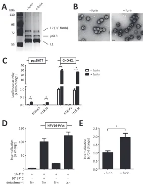
generated furin-precleaved HPV16 pseudovirions which expectedly contain some
(al-though not all [17]) L2 proteins with proteolytically processed N termini (Fig. 1A).
Importantly, no copurifications were visible by SDS-PAGE with silver staining, thereby
minimizing the possibility of furin targeting other proteins besides the virus capsids
(Fig. 1A).
Although we could not detect any obvious morphological differences between
un-cleaved and FPC HPV16-PsVs by negative electron microscopic (EM) staining (Fig. 1B),
furin cleavage had a substantial functional impact on infection of the HSPG-deficient
cell line pgsD677: while pgsD677 cells were practically noninfectible by HPV16-PsVs,
furin cleavage of the particles led to an approximately 40-fold increase in infection as
measured by luciferase reporter gene activity (Fig. 1C). Moreover, infection of CHO-K1
wild-type cells also resulted in a more robust (approximately 30-fold) increase of
infection in the presence of FPC particles, while neutralization with the
HPV16-neutralizing antibody H16.V5 (but not with the HPV18-HPV16-neutralizing antibody H18.J4)
abolished infectious uptake independently of furin pretreatment as expected (53) in
both cell types (Fig. 1C). These experiments not only demonstrated the impact of
furin treatment on HPV16-PsV infectivity but also confirmed the suitability of
pgsD677 cells together with FPC HPV16-PsVs as an HSPG-independent infection
system (17).
In order to study early steps in HPV infection involving quantification of virus
internalization, we tested the effect of trypsin-EDTA on the removal of surface-bound
but not internalized particles. When analyzed by flow cytometry, binding of Alexa Fluor
488 succinimidyl ester (AF488)-labeled HPV16-PsVs to pgsD677 cells for 1 h at 4°C was
found to be almost completely removed by treatment with trypsin-EDTA but not with
lidocaine hydrochloride-EDTA (Fig. 1D). However, internalization of the particles was
well detected when cells were subsequently shifted to 37°C for 30 min and treated with
trypsin-EDTA, almost reaching the levels seen when cells were only allowed to bind for
1 h at 4°C and lifted with lidocaine hydrochloride-EDTA (Fig. 1D). These results were
also confirmed with all other cell lines used in this study (data not shown) and
demonstrated the suitability of trypsin digestion for removal of surface-bound
HPV16-PsVs, allowing the quantification of their internalization. Interestingly, furin
on November 7, 2019 by guest
http://jvi.asm.org/
ment of the viral particles not only substantially affected infectivity of pgsD677 cells
(Fig. 1C) but also increased FPC HPV16-PsV internalization as measured by flow
cytom-etry using AF488-labeled virions (Fig. 1E). These data confirmed that FPC HPV16-PsVs
can bypass the requirement for HSPG engagement during infectious uptake, thereby
permitting direct binding to the still elusive secondary receptor (17).
A
C
Lu
ci
fe
ra
se
ac
v
it
y
(x
-f
o
ld
c
h
a
n
g
e
)
CHO-K1 130
95
72
55
L2 (+/- furin)
pGL3
L1
-0.0 0.5 1.0 10 20 30 40
+ furin - furin pgsD677
-E
0.0 0.5 1.0 1.5 2.0
2.5 *
In
te
rn
a
liza
o
n
(x
-f
o
ld
c
h
a
n
g
e
)
- furin + furin
* *
* *
- furin + furin
B
D
1h 4°C
HPV16-PsVs
30’ 37°C
detachment
+ + + +
+ -
-Trn Trn Trn Lcn
In
te
rn
a
liza
o
n
(%
c
h
a
n
g
e
)
0 50 100 150 kDa
FIG 1Establishing the experimental conditions for subsequent identification of HPV16-PsV-binding surface molecules. HPV16 pseudovirions encapsidating the luciferase reporter gene plasmid pGL3-control were treated with furin or left untreated. (A) Purity and successful furin treatment of the PsV
preparations (1.5g per lane) was assessed by SDS-PAGE and subsequent silver staining. Note that the
additional band at approximately 60 kDa is the silver-stained pGL3-control plasmid (70). The leftmost lane shows molecular mass markers, in kilodaltons. (B) Negative EM staining of furin-treated and untreated HPV16-PsVs. (C) HSPG-deficient pgsD677 cells and their corresponding wild-type CHO-K1 cells were infected with furin-treated or untreated HPV16-PsVs. In control experiments, the virions were preincubated with either of the neutralizing antibodies H16.V5 and H18.J4. Luciferase activity of the cell lysates was measured 48 h postinfection and normalized against total protein concentration and is presented as x-fold change relative to control infection of CHO-K1 cells with untreated HPV16-PsVs, which was set as 1. (D) Quantification of viral internalization was performed by flow cytometry of pgsD677 cells infected with Alexa Fluor 488 (AF488)-conjugated HPV16-PsVs for 1 h on ice followed by 30 min at 37°C. Cells were lifted with trypsin-EDTA (Trn) to remove surface-bound virions. In control experiments, cells were either not shifted to 37°C to avoid internalization or lifted with lidocaine hydrochloride-EDTA (Lcn), which does not remove surface-bound particles. Experiments were per-formed in triplicate, and results were quantified by quadrant analysis of the dot plot of three independent experiments and are presented as percent change relative to the mean fluorescence intensity of cells lifted with Trn, which was set as 100%. (E) Internalization of furin-pretreated or untreated AF488-conjugated HPV16-PsVs by pgsD677 cells as determined by flow cytometry as
described for panel D. Significance was calculated by means of the two-tailed Studentttest from
three independent experiments performed in triplicate, and aPvalue ofⱕ0.05 (*) was regarded as
statistically significant.
on November 7, 2019 by guest
http://jvi.asm.org/
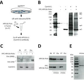
[image:4.585.59.348.69.443.2]We therefore performed immunoprecipitation (IP) assays of live pgsD677 cells
incubated with FPC HPV16-PsVs using the HPV16-L1-specific antibody CamVir1 (Fig. 2A).
Precipitated proteins were separated by SDS-PAGE followed by silver staining of the
gel, allowing visual comparison to appropriate controls (Fig. 2B). Candidate protein
bands were excised, processed for matrix-assisted laser desorption ionization–time of
flight mass spectrometry (MALDI-TOF) analysis, and identified using the Matrix Science
Database (MSDB) and searching the NCBI database. Among the molecules identified,
vimentin received the highest protein significance score, 139, and was considered an
attractive candidate involved in HPV recognition and binding due to its involvement in
the attachment and uptake of several other viruses and bacteria when expressed at the
cell surface (41–48, 50–52). In order to validate the involvement of pgsD677 cell surface
vimentin in FPC HPV16-PsV binding, Western blots based on the immunoprecipitation
experiment shown in Fig. 2B were performed. Interestingly, when probing with
anti-bodies raised against different vimentin epitopes (i.e., H84 versus V9), we found that the
presence of the viral particles led to a slight reduction in the size of the precipitated
vimentin protein (Fig. 2C). No such protein was precipitated in the absence of FPC
HPV16-PsVs. Unspecific binding of the vimentin V9 antibody to the HPV16-L1 protein
A
C
212
65 97
56
43
34
+ FPC-HPV16-PsVs:
CamVir1: - - + +
- - +
158
1
3
4
D
B
2
5 pgsD677
Li with lidocaine/EDTA
HPV16-PsVs (+ furin)
1h 4°C
Co-IP with HPV16-L1 (CamVir1) anbody
rhVim
180
70 100
55
40
35 130
E
IN FT Elu FT Elu
HPV16-PsVs
FPC-HPV16-PsVs
+rhVim -rhVim Vim (V9)
FPC-HPV16-PsVs: - + - + - +
IN FT Elu
CamVir1 Vim (H84)
25 kDa
kDa
FIG 2Identification of cell surface vimentin as a binding protein for HPV16-PsVs. (A) Schematic outline of the pulldown experiment to isolate FPC HPV16-PsVs-binding surface proteins. (B) Silver-stained SDS-PAGE gel of pgsD677 surface proteins bound to FPC HPV16-PsVs pulled down by the HPV16-L1-specific antibody CamVir1. Numbers on the right side refer to protein bands HPV16-L1-specific to FPC HPV16-PsV interaction, which were determined visually by comparing to control pgsD677 cells that were not incubated with HPV16-PsVs before immunoprecipitation. Candidate bands were excised from the gel and
identified by MALDI-TOF mass spectrometry analysis: 1, tetratricopeptide repeat protein 39B OS (Rattus
norvegicus) (protein significance score: 42); 2, guanine nucleotide-binding protein G(I)/G(S)/G(O) subunit
gamma-5-like (Mus musculus) (protein significance score: 36); 3, immunoglobulin heavy chain (Mus
musculus) (protein significance score: 40); 4, vimentin (Rattus norvegicus) (protein significance score: 139);
5, ␥-actin (Mus musculus) (protein significance score: 88). The leftmost lane shows molecular mass
markers, in kilodaltons. (C) Western blot of pulldown experiment (as in panel B), probed simultaneously with the antivimentin antibodies H84 and V9 (top) and reprobed with the antibody CamVir1 (bottom). Note that in the presence of HPV16-PsVs the size of the vimentin protein is slightly reduced. IN, input; FT, flowthrough; Elu, eluate. (D) Pulldown of HPV16-PsVs (with or without furin pretreatment) using immobilized recombinant human vimentin protein (rhVim). Proteins were detected by Western blotting
using the antibody CamVir1. (E) Silver-stained SDS-PAGE gel of 0.5g of recombinant human vimentin
protein. The leftmost lane shows molecular mass markers in kilodaltons.
on November 7, 2019 by guest
http://jvi.asm.org/
[image:5.585.62.349.74.347.2](both being of similar size) was excluded in appropriate control experiments (data not
shown). As it is possible that the observed different vimentin fragments were artifacts
caused by proteolysis during cell isolation (54, 55), we performed pulldown of the viral
particles in a cell-free context in the presence or absence of immobilized recombinant
human vimentin protein (Fig. 2D). The purity of the vimentin protein was confirmed by
silver staining following SDS-PAGE (Fig. 2E), thereby excluding virus binding to other
molecules besides vimentin. These experiments confirmed that HPV16-PsVs directly
interacted with vimentin. Interestingly, this interaction was found to be independent of
furin pretreatment of the viral particles.
Surface vimentin colocalizes with HPV16-PsVs and affects viral uptake by pgsD677
cells.
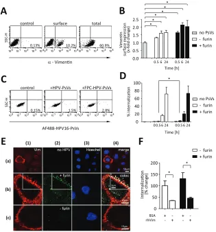
To determine if vimentin could act as a surface molecule involved in HPV uptake,
we initially characterized vimentin expression in pgsD677 cells and studied binding of
AF488-labeled HPV16-PsVs (both furin treated and untreated) by flow cytometry and
confocal microscopy. The expression pattern of surface vimentin agrees with the
literature, as only a small percentage of cells presented detectable vimentin at the
surface (Fig. 3A), which appeared polarized (punctate) at the cell membrane (44, 56),
with a striking localization of vimentin to filopodia near the substrate (Fig. 3E, column
1). Due to the lack of HSPGs on pgsD677 cells, total binding of the viral particles to the
cells was relatively low (Fig. 3C); however, individual cells expressing vimentin at the
surface showed impressively high binding of the viral particles, with the majority
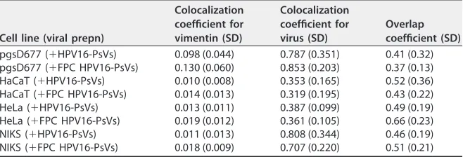
displaying colocalization with vimentin (Fig. 3E, rows b and c). Generally, increased
binding to pgsD677 cells was observed for FPC HPV16-PsVs compared to uncleaved
particles (Fig. 3C and E, rows b and c), although levels of colocalization with vimentin
were comparable: 85.3% (
⫾
20.3%) of FPC HPV16-PsVs and 78.7% (
⫾
35.1%) of
un-cleaved particles, respectively, colocalized with surface-expressed vimentin (Table 1).
Interestingly, vimentin surface levels slightly increased over time upon infection with
the viral particles; however, furin-precleaved and untreated particles had a comparable
effects (Fig. 3B). Although initial (30 min) internalization of FPC HPV16-PsVs was
increased compared to untreated particles (Fig. 1E), the two virus preparations were
internalized to similar extents over a time course of 24 h (Fig. 3D).
In order to study any functional involvement of vimentin for infectious HPV
inter-nalization, we preincubated both untreated and FPC HPV16-PsVs with soluble human
recombinant vimentin protein before infection. This led to a significant reduction in
viral internalization as determined by flow cytometry (Fig. 3F). However, infectivity as
measured by luciferase activity was not affected (data not shown), possibly due to
dissociation of the vimentin-virus complexes by the time luciferase activity was
mea-sured (48 h postinfection).
Vimentin modulates uptake of HPV16-PsVs in epithelial cells.
Although they
were found suitable in this study as a cell system to investigate HPV infection at the
immediate-uptake stage (by bypassing the initial attachment steps due to their lack of
HSPGs), pgsD677 cells are nonhuman and nonkeratinocytes and thereby rather
unre-lated to the natural infectious environment for HPV. We therefore set out to confirm our
initial observations using three cell lines representing physiologically relevant cell
culture models: the human cervical cancer cell line HeLa and the human immortalized
keratinocyte cell lines HaCaT and NIKS, two of the most widely accepted keratinocyte
models for studying HPV infection (57). Unlike other immortalized cell lines, NIKS cells
retain cell-type-specific growth requirements and differentiation properties, are
non-tumorigenic, and are virus free (58). Most importantly, NIKS cells were found to support
the complete HPV life cycle (59).
To verify cell surface expression of vimentin, we performed both flow cytometry and
immunofluorescence microscopy on HeLa, HaCaT, and NIKS cells. Similar to the results
for pgsD677 cells (Fig. 3A), only a small fraction of cells stained positive for surface
vimentin (Fig. 4A), which appeared unevenly scattered over the cell surface, with some
areas of high vimentin density (Fig. 4C). Unlike for pgsD677 cells (Fig. 3C), binding of
HPV16-PsVs to HeLa, HaCaT, and NIKS cells was found to be much higher over the
on November 7, 2019 by guest
http://jvi.asm.org/
entire cell population as observed by flow cytometry, as attachment of the viral
particles to HSPGs at the cell surface could occur (Fig. 4B). Interestingly, binding of
untreated and FPC HPV16-PsVs occurred at comparable extents for the individual cell
systems (Fig. 4B), with HeLa
⬎
HaCaT
⬎
NIKS. This cell line-specific trend was also
reflected at the level of luciferase activity being a readout for infectivity (57) (data not
shown), with furin treatment largely increasing infection in the individual cell system
A
10 0 10 1 10 2 10 3 10 4
10.2%
10 0 10 1 10 2 10 3 10 4 10 0 10 1 10 2 10 3 10 4
control surface total
α- Vimenn
0.13% 60.9%
E
AF488-HPV16-PsVs SSC -H *a b c
F
rhVim BSA 0 50 100 150 200 In te rn aliza o n (% c h a n g e ) + furin - furin + - - + + - + -* SSC -HC
control +HPV-PsVs +FPC-HPV-PsVs
B
D
0 20 40 60 80 100 % in te rn alizao n0.5 6 24
0.0 0.5 1.0 1.5 2.0 2.5 V im e n n su rfac e e x p re ssi o n (x -f o ld c h an g e ) + furin - furin no PsVs
0.5 6 24
Time [h]
0.5 6 24
Time [h] 0 00.5 6 24
+ furin - furin no PsVs
10 0 10 1 10 2 10 3 10 410 0 10 1 10 2 10 3 10 410 0 10 1 10 2 10 3 10 4
0.15% 1.5% 2.8%
** * * * * * (a) (b) (c)
(1) (2) (3) (4)
FIG 3Surface vimentin modulates uptake of PsVs by pgsD677 cells. AF488-conjugated HPV16-PsVs were used in all qualitative and quantitative assessments of infectious internalization by pgsD677
cells. Significance was calculated by means of the two-tailed Studentttest from three independent
experiments performed in triplicate;Pvalues ofⱕ0.05 (*) were regarded as statistically significant. (A) Cell
surface versus total vimentin expression was determined by flow cytometry and quantified using quadrant statistics of three independent experiments. Shown are representative dot plots with the average percentage of the mean fluorescence intensities of vimentin-positive cells in the lower right quadrant. (B) Quantification of vimentin surface expression after incubation of pgsD677 cells with HPV16-PsVs (with or without prior furin cleavage) for the indicated periods. (C) Flow cytometry analysis of pgsD677 cells with bound AF488-conjugated HPV16-PsVs (pretreated or not with furin). Quantification was done using quadrant statistics analysis. Shown are representative dot plots with the average percentage of the mean fluorescence intensities of AF488-positive cells in the lower right quadrant. (D) Quantification of internalization of HPV16-PsVs (with or without prior furin cleavage) for the indicated periods. (E) Confocal imaging of pgsD677 cells showing surface vimentin (column 1, red), which is present only in a subpopulation of cells (indicated by Hoechst nuclear stain, blue, column 3) (a) and colocalization analysis of cells stained for cell surface vimentin (column 1, red) and bound by AF488-conjugated HPV16-PsVs (with or without prior furin cleavage, column 2, green) for 1 h at 4°C (b and c). Nuclei were stained with Hoechst to indicate presence of cells. Signal overlays in column 4 indicate colocalization of the viral particles with cell surface vimentin as white pixels. High-magnification insets in columns 1, 2, and 4 show a detail of signal distribution and colocalization at the filopodial edge of the cell in a single optical section. Shown are representative images. (F) AF488-conjugated HPV16-PsVs (with or without prior furin cleavage) were pretreated with soluble recombinant human vimentin protein or BSA before infection of pgsD677 cells. Internalization of viral particles was assessed by flow cytometry and quantified using quadrant statistics and is presented as change in mean fluorescence intensity of AF488-positive cells compared to that of untreated particles, which was set as 100%.
on November 7, 2019 by guest
http://jvi.asm.org/
[image:7.585.49.364.72.410.2](Fig. 1C). However, individual vimentin-positive cells showed comparable
particle-binding patterns of both untreated and FPC HPV16-PsVs when analyzed by confocal
microscopy (Fig. 4C) in the three cell lines. Again, clear colocalization of AF488-labeled
viral particles to surface-expressed vimentin was observed on HeLa, HaCaT, and NIKS
cells (Fig. 4C), with no obvious difference between uncleaved and FPC HPV16-PsVs
(Table 1).
Having demonstrated that all three cell lines expressed vimentin at the cell surface
which colocalized with the viral particles (Fig. 4A and C), we next set out to investigate
whether it also affected HPV16-PsV internalization, leading to infection. We
preincu-bated the viral particles with soluble human recombinant vimentin protein before
infection. As already seen for pgsD677 cells (Fig. 3F), this treatment led to a substantial
reduction in viral internalization in all cell systems (Fig. 4D); however, infectivity was not
affected for the reasons mentioned above (data not shown). Furin pretreatment did not
influence virion internalization (Fig. 4D), unlike what was seen in pgsD677 cells (Fig. 1E),
which is consistent with expression of HSPG in these human cells.
The observed reduction in viral uptake upon preincubation of the virus particles
with soluble vimentin (Fig. 3F and 4D) could be explained either by competition for
surface-expressed vimentin (if vimentin functions as an uptake molecule), by a
seques-tering function vimentin employs toward infectious HPV particles, or by interference of
vimentin with viral binding to the still-unknown entry receptor. We therefore altered
vimentin expression in HeLa and NIKS cells by either small interfering RNA (siRNA)
knockdown or overexpression. Interestingly, reduced vimentin expression upon
knock-down by vimentin-specific siRNA (Fig. 5A and B) led to an increase in HPV16-PsV
binding and internalization which was independent of prior furin treatment (Fig. 5C
and D). Likewise, infection as measured by luciferase activity was significantly increased
when vimentin expression was reduced, which was more pronounced when the
particles were not pretreated with furin (Fig. 5C and D). Consistent with the observed
increase in viral internalization upon vimentin knockdown, increasing the amount of
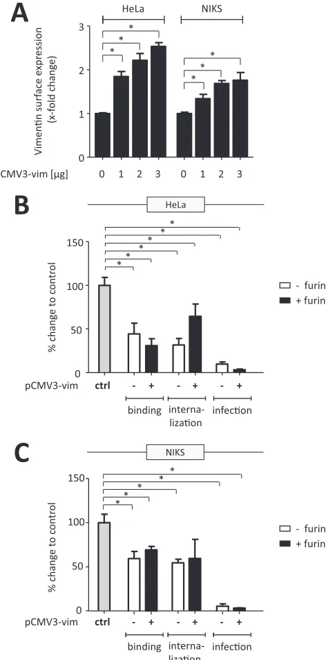
surface vimentin by transient overexpression (Fig. 6A) significantly decreased
HPV16-PsV binding and uptake independently of prior furin treatment (Fig. 6B and C). This was
also accompanied by significant decreases in luciferase activity in both cell systems (Fig.
6B and C).
DISCUSSION
[image:8.585.42.371.93.205.2]Engagement with host cell surface molecules is one of the initial requirements for
a successful viral infection; therefore, targeting viral receptors and identifying novel
molecules that prevent receptor engagement by the incoming viral particles are
promising strategies for the control of several viral infections. In the case of HPV, it has
been assumed that the virions are unable to bind to their entry receptor until they have
TABLE 1Quantification of HPV16-PsVs colocalization with surface vimentin on pgsD677,
HaCaT, HeLa, and NIKS cellsa
Cell line (viral prepn)
Colocalization coefficient for vimentin (SD)
Colocalization coefficient for virus (SD)
Overlap coefficient (SD)
pgsD677 (⫹HPV16-PsVs) 0.098 (0.044) 0.787 (0.351) 0.41 (0.32)
pgsD677 (⫹FPC HPV16-PsVs) 0.130 (0.060) 0.853 (0.203) 0.37 (0.13)
HaCaT (⫹HPV16-PsVs) 0.010 (0.008) 0.353 (0.165) 0.52 (0.36)
HaCaT (⫹FPC HPV16-PsVs) 0.014 (0.013) 0.319 (0.195) 0.43 (0.22)
HeLa (⫹HPV16-PsVs) 0.013 (0.011) 0.387 (0.099) 0.49 (0.19)
HeLa (⫹FPC HPV16-PsVs) 0.019 (0.012) 0.361 (0.105) 0.66 (0.23)
NIKS (⫹HPV16-PsVs) 0.011 (0.013) 0.808 (0.344) 0.46 (0.19)
NIKS (⫹FPC HPV16-PsVs) 0.018 (0.009) 0.707 (0.220) 0.51 (0.21)
aBinding of the indicated virus preparations (with or without furin treatment) to approximately 50 cells per
cell line was analyzed by confocal microscopy in two independent experiments. Manders colocalization coefficients for vimentin (i.e., amount of vimentin colocalizing with virus) and virus (i.e., amount of virus colocalizing with vimentin) are presented, along with Manders overlap coefficients; a value of 0 indicates no colocalization, and a value of 1 indicates maximal colocalization.
on November 7, 2019 by guest
http://jvi.asm.org/
10 0 10 1 10 2 10 3 10 410 0 10 1 10 2 10 3 10 410 0 10 1 10 2 10 3 10 4
10 0 10 1 10 2 10 3 10 4 0 0 10 1 10 2 10 3 10 4
10 0 10 1 10 2 10 3 10 4 10 0 10 1 10 2 10 3 10 4
Dat a. 013
10 0 10 1 10 2 10 3 10 4100 10 1 10 2 10 3 10 4
10 0 10 1 10 2 10 3 10 4 10 0 10 1 10 2 10 3 10 4
HeLa
SSC
-H
17.3% 0.05%
10 0 10 1 10 2 10 3 10 4
control
surface
10 0 10 1 10 2 10 3 10 4
16.5% 0.54%
α
- Vimenn
SSC
-H
10 0 10 1 10 2 10 3 10 4 100 10 1 10 2 10 3 10 4
0.44% 12.2%
SSC
-H
HeLa
AF488-HPV16-PsVs
47.9% 0.03%
14.6% 0%
SSC
-H
4.9% 0.13%
control
+ HPV16-PsVs
SSC
-H
NIKS HaCaT
A
+ FPC-HPV16-PsVs
35.7%
16.3%
HaCaT
NIKS
SSC
-H
3.1%
B
D
C
rhVim BSA
HeLa
HaCaT
In
te
rn
aliza
o
n
(%
ch
an
g
e
)
+
+
-NIKS
+
+
-+
+
-+
+
-+
+
-+
+
-+ furin
- furin
He
La
Ha
Ca
T
NI
KS
(1) (2) (3) (4)
FIG 4Vimentin expression and its role in HPV16-PsV infection of epithelial cells. (A) Cell surface vimentin expression of HeLa, HaCaT, and NIKS cells was determined by flow cytometry and quantified using quadrant statistics of three independent experiments. Shown are representative dot plots with the average percentage of the mean fluorescence intensities of vimentin-positive cells in the lower right quadrant. (B) Flow cytometry analysis of HeLa, HaCaT, and NIKS cells with bound AF488-conjugated HPV16-PsVs (with or without prior furin cleavage). Quantification was done using quadrant statistics analysis. Shown are representative dot plots with the average percentage of the mean fluorescence intensities of AF488-positive cells in the lower right quadrant. (C) Confocal imaging of HeLa, HaCaT, and NIKS cells stained for cell surface vimentin (column 1, red) and bound by AF488-conjugated HPV16-PsVs (with or without prior furin cleavage, column 2, green) for 1 h at 4°C. Nuclei were stained with Hoechst to indicate presence of cells (column 3, blue). Colocalization of the viral particles with cell surface vimentin is indicated by overlay of white pixels in the merged red and green channels (column 4). Shown are representative images. (D) AF488-conjugated HPV16-PsVs (with or without prior furin cleavage) were pretreated with soluble recombinant human vimentin protein or BSA before infection of HeLa, HaCaT, and NIKS cells. Internalization of viral particles was assessed by flow cytometry and is presented as percent change in mean fluorescence intensity of AF488-positive cells compared to that of untreated particles, which was set as 100%.
on November 7, 2019 by guest
http://jvi.asm.org/
[image:9.585.39.491.76.611.2]undergone an HSPG-dependent conformational change and subsequent furin cleavage
(8). While HSPGs are expressed at the cell surface and are used by HPV as cellular
attachment molecules
in vitro
, it is assumed that the conformational changes of the
HPV capsid required for infection occur HSPG dependently on the basement membrane
in vivo
prior to the transit of the virions to the host cell surface (7). In order to bypass
the need for these extracellular steps, we set up an experimental infection system using
the HSPG-deficient cell line pgsD677 together with furin-precleaved HPV16
pseudovi-rions (FPC HPV16-PsVs). This model allowed us to biochemically pull down cellular
surface molecules that the FPC particles directly interacted with, potentially including
the still-elusive non-HSPG secondary entry receptor (20). One of the most promising
candidate molecules that was precipitated together with FPC HPV16-PsVs was
identi-fied as vimentin, which, in addition to its function as a cytoskeletal filament protein,
plays important roles in entry of, infection with, and/or replication of several viruses
A
Vimenn
GAPDH - 10 25 50 75 100 vim siRNA [nM]:
B
C
D
vim-siRNA V im e n n s ur fa ce e x pr e ss io n (% c h an g e ) x-fo ld c h an g e t o c o n tr o l - +Vim-siRNA - +
- + HeLa NIKS 0 50 100 150 - + - + * * 0 1 2 3 0 1 2 2 4 6 8 ctrl * * * HeLa binding interna-lizaon infecon + furin - furin NIKS *
Vim-siRNA ctrl - + - + - + binding interna-lizaon infecon + furin - furin x-fo ld c h an g e t o c o n tr o l
FIG 5Decreased surface vimentin facilitates HPV16-PsV infectivity. HeLa and NIKS cells were reversely transfected with human vimentin siRNA for transient knockdown. (A) Western blot of HeLa cell lysates prepared 72 h after transfection with increasing concentrations of vimentin siRNA. Total vimentin was detected by the antivimentin antibody V9; glyceraldehyde-3-phosphate dehydrogenase (GAPDH) served as a loading control. (B) Cell surface expression of vimentin after knockdown with 100 nM siRNA for 72 h was determined by flow cytometry and quantified using quadrant statistics of three independent experiments and is presented as change in surface expression of the vimentin-positive cell population relative to cells transfected with scrambled (control) siRNA, which was set as 100%. (C and D) Quantification of viral binding and internalization was performed by flow cytometry of HeLa (C) and NIKS (D) cells transfected with 100 nM vimentin siRNA for 72 h and exposed to furin-pretreated or untreated AF488-conjugated HPV16-PsVs. Quadrant analysis of the dot plots of three indepen-dent experiments performed in triplicate is presented as x-fold change relative to the mean fluorescence intensity of cells transfected with control (scrambled) siRNA and infected with either furin-treated or untreated particles, as appropriate, which was set as 1 for all conditions. For quantification of infection, the cells were transfected with 100 nM vimentin siRNA for 72 h and infected with furin-pretreated or untreated HPV16-PsVs. Forty-eight hours postinfection, luciferase activity was measured and was normalized against total protein concentration and is presented as x-fold change relative to cells transfected with control (scrambled) siRNA and infected with either furin-treated or untreated particles, as appropriate, which was set as 1. Significance was calculated by means of the
two-tailed Studentttest, and aPvalue ofⱕ0.05 (*) was regarded as statistically significant.
on November 7, 2019 by guest
http://jvi.asm.org/
[image:10.585.42.406.72.399.2]V
im
e
n
n
su
rf
a
ce
e
x
p
re
ssi
o
n
(x
-f
o
ld
c
h
an
g
e
)
pCMV3-vim [μg] 0 1 2 3
A
B
C
0 1 2 3
HeLa NIKS
0 1 2 3
- + - + - +
pCMV3-vim
+ furin - furin *
* *
* *
*
0 50 100 150 0 50 100 150
%c
h
a
n
g
e
t
o
c
o
n
tr
o
l
ctrl
binding interna-lizaon
infecon
**
**
**
HeLa
%c
h
a
n
g
e
t
o
c
o
n
tr
o
l
- + - + - +
pCMV3-vim ctrl
binding interna-lizaon
infecon
+ furin - furin
NIKS
**
* *
*
FIG 6Surface vimentin acts as a viral restriction factor in HPV16-PsV infection. HeLa and NIKS cells were transiently transfected with plasmid pCMV3-vim for overexpression studies. (A) Cell surface expression of vimentin after transfection with increasing concentrations of plasmid pCMV3-vim for 14 h was deter-mined by flow cytometry and quantified using quadrant statistics of three independent experiments and is presented as x-fold change in surface expression relative to control cells, which was set as 1. (B and C) Quantification of viral binding and internalization was performed by flow cytometry of HeLa (B) and NIKS
(C) cells transfected with 1g of pCMV3-vim for 14 h and exposed to furin-pretreated or untreated
AF488-conjugated HPV16-PsVs. Quadrant analysis of the dot plots of three independent experiments performed in triplicate is presented as percent change relative to the mean fluorescence intensity of control cells infected with either furin-treated or untreated particles, as appropriate, which was set as
100% for all conditions. For quantification of infection, the cells were transfected with 1g of pCMV3-vim
for 14 h and infected with furin-pretreated or untreated HPV16-PsVs. Forty-eight hours postinfection, both floating and adherent cells were harvested to measure luciferase activity, which was normal-ized against total protein concentration and is presented as percent change relative to control cells infected with either furin-treated or untreated particles, as appropriate, which was set as 100%.
Significance was calculated by means of the two-tailed Studentttest, and aPvalue ofⱕ0.05 (*) was
regarded as statistically significant.
on November 7, 2019 by guest
http://jvi.asm.org/
[image:11.585.90.326.75.552.2](39). Although vimentin is primarily expressed intracellularly, we and others observed
surface expression on a subpopulation of cultured cells displaying a particular
punc-tuate expression pattern (40). Importantly, several viral pathogens, including
picorna-viruses, mammalian porcine reproductive and respiratory syndrome virus (PRRSV),
cowpea mosaic virus (CPMV), Japanese encephalitis virus (JEV), severe acute respiratory
syndrome coronavirus (SARS-CoV), enterovirus 71, Theiler’s virus, and human
immuno-deficiency virus 1 (HIV-1), use vimentin as a component of their cellular attachment
and/or receptor machinery (41–46, 48, 60).
The physical interaction between FPC HPV16-PsVs and vimentin initially observed by
immunoprecipitation was confirmed by immunofluorescence confocal microscopy as
colocalization of the viral particles with surface-expressed vimentin on pgsD677 cells
and on selected epithelial cell culture models, namely, HeLa, HaCaT, and NIKS, the last
being particularly physiologically relevant for HPV infection, as NIKS cells retain
cell-type-specific growth requirements and differentiation properties. Contrary to our
ex-pectation, however, the presence of vimentin was not found to aid in viral
internaliza-tion but rather funcinternaliza-tioned as an inhibitory molecule. Both preincubainternaliza-tion of the viral
particles with soluble recombinant vimentin protein and overexpression of vimentin on
the cell surface led to a substantial decrease in viral uptake and infection. In contrast,
diminishing surface-expressed vimentin by siRNA knockdown significantly increased
infectious internalization of the particles.
The functional significance of vimentin during natural HPV infection still needs to be
elucidated, as cell culture systems do not fully represent the series of changes the virus
undergoes
in vivo
after binding to the basement membrane prior to keratinocyte
infection. We hypothesize that cell surface vimentin binding to viral particles plays a
role in modulating the HPV entry process, possibly by interfering with the binding of
the viral particle with the (still unknown) viral binding and uptake molecules. Although
novel in the context of HPV infection, extracellular vimentin has been shown to be
involved in the response to pathogens through interactions with incoming bacteria and
soluble factors, thereby contributing to bacterial killing (54). Similar to the dual role of
lectins in virus biology—lectins can be membrane associated or soluble and are known
to be involved in either immune defense through neutralization and clearance of viral
infection or can be exploited as entry molecules to facilitate viral spread (61)—vimentin
may also exert dual effects on viral infection. While surface-expressed vimentin plays a
role in the attachment and uptake machinery of several viruses as mentioned above
(41–46, 48, 60), it appears to interfere with crucial steps in the HPV16 pseudovirus
infection process at the level of binding and internalization into epithelial cells, possibly
by either masking portions of the virus that interact with its receptor or by causing
steric hindrance, thereby destabilizing or preventing receptor-virus interaction
neces-sary for successful infection. Interestingly, vimentin is known to be associated with
several integrins, such as
␣
6 integrin (62), which has been proposed as a potential HPV
receptor (21) and whose function in HPV uptake might be influenced by the presence
of vimentin.
In this study, we focused on HPV16, the most common and best-characterized
oncogenic HPV genotype. It is possible that some of the entry steps, including the role
of vimentin, differ among HPV serotypes (2, 63). The participation of different receptors
and/or modulating molecules can therefore account for differences in pathogenic
profiles and differences in cell tropism. It is also interesting that HPV16 pseudovirus
binding to recombinant vimentin protein as well as internalization by epithelial cells
was little influenced by pretreatment of the viral particles with furin. While furin
cleavage of L2 is indispensable for successful infection, as demonstrated on
HSPG-deficient pgsD677 cells, the fact that untreated particles still enter the cells confirms the
current notion that furin cleavage is important for endosomal escape rather than at the
early stages of the infectious entry (14, 15). We therefore propose that the interaction
between HPV16-PsVs and vimentin is independent of furin cleavage. Moreover, as the
observed binding and internalization behavior of HPV16-PsVs did not greatly differ
on November 7, 2019 by guest
http://jvi.asm.org/
between the HSPG-expressing cell lines used in this study, we hypothesize that surface
vimentin may act as a general viral restriction factor rather than a cell-type-specific one.
Knowledge of the early events in HPV infection, such as cellular surface molecule
binding and its consequences for entry into susceptible cells, aids in the design of novel
strategies for blocking HPV infection. Although the prophylactic vaccines that target
certain HPV types show great efficacy (64), they are too costly for general use
world-wide, they protect against only a limited number of oncogenic genotypes, and there is
limited vaccine efficacy in people already infected by HPV. Clearly, there is a need to
identify other modalities and alternative cost-effective means for preventing and/or
treating HPV infection. With the identification of vimentin as a viral restriction factor
preventing HPV16-PsV infection, approaches in drug design based on molecule
struc-ture which can eventually be incorporated into microbicides or lubricants to prevent
HPV infection are conceivable (65).
MATERIALS AND METHODS
Cell culture. The spontaneously immortalized human keratinocyte cell line HaCaT, the human epithelioid cervix carcinoma cell line HeLa (ATCC CCL-2), and the virus packaging cell line 293TT (established from primary embryonal human kidney cells transformed with modified human adenovirus) (66, 67) were grown and maintained in Dulbecco modified Eagle medium (DMEM; Life Technologies) supplemented with 10% heat-inactivated fetal calf serum (Biochrom), 100 U/ml of penicillin, and 100
g/ml of streptomycin. CHO-K1 (Chinese hamster ovary K1; ATCC CCL-61) and pgsD677 (heparan
sulfate-deficient cells derived from CHO-K1; ATCC CRL-2244) cells were grown and maintained in Ham’s F-12 K medium (Life Technologies) supplemented with 10% heat-inactivated fetal calf serum, 100U/ml of
penicillin, and 100g/ml of streptomycin. The spontaneously immortalized human keratinocyte cell line
NIKS (ATCC CRL-12191) (58, 68, 69) was grown and maintained in F medium (3:1 [vol/vol] Ham’s F-12 K
medium-DMEM, 5% heat-inactivated fetal calf serum, 0.4g/ml of hydrocortisone [Calbiochem], 5g/ml
of insulin [NovoRapid], 8.4 ng/ml of cholera toxin [Sigma], 10 ng/ml of recombinant human epidermal
growth factor [Life Technologies], 24g/ml of adenine [Sigma], 100 U/ml of penicillin, and 100g/ml of
streptomycin). All cells were grown at 37°C in a 5% CO2–95% air humidified atmosphere.
All cell lines have been authenticated by IDEXX BioResearch, Columbia, MO.
HPV16 pseudovirion preparation, labeling, and quality controls.HPV16-PsVs encapsidating the luciferase reporter gene plasmid pGL3-control (Promega) were produced in 293TT cells by cotransfection with plasmid pXULL, which encodes codon-optimized HPV16 L1 and L2, by following published proce-dures (67, 70). Where indicated, the virions were labeled with Alexa Fluor 488 succinimidyl ester (AF488; Life Technologies) before purification by CsCl density gradient centrifugation as described previously (30). To assess the purity of the virus stock, the purified preparations of HPV16-PsVs were subjected to SDS-PAGE and subsequent silver staining using a Pierce silver stain kit (Thermo Scientific). The protein content of the HPV16-PsV stock was quantified with a Pierce bicinchoninic acid (BCA) protein assay kit (Thermo Scientific). Furin-precleaved pseudovirions were produced after CsCl gradient centrifugation by
incubation of approximately 1g of HPV16-PsVs with 1 U of recombinant human furin (Sigma) at 37°C
for 16 to 20 h in the presence of 100 mM HEPES (pH 7.5), 0.5% Triton X-100, and 1 mM CaCl2.
Neutralization assays were performed by incubation of the HPV16-PsVs with the neutralizing antibody H16.V5 (positive control) or H18.J4 (negative control) at a final concentration of 1:1,000 for 1 h at 4°C (70). Neutralized HPV16-PsVs were then added to the cells, and infectivity was measured as luciferase activity as described below.
Negative-stain electron microscopy.Both uncleaved and FPC HPV16-PsVs at a final concentration
of 0.1g/l were prepared for electron microscopy by washing in HSB buffer (25 mM HEPES [pH 5.7],
0.5 M NaCl, 0.02% Brij 58, 1 mM MgCl2, 100M EDTA, 0.5% ethanol) and purification through an Amicon
Ultra-4 filter device (100,000-kDa molecular mass cutoff). A 3-l drop of sample solution was then
adsorbed to a glow-discharged carbon-coated copper grid (Agar Scientific) for 30 s, washed with 2 drops of deionized water, and stained with 2 drops of 2% aqueous uranyl acetate (SPI Supplies). The air-dried grid was then viewed with a Philips Tecnai F20 transmission electron microscope equipped with a field emission gun operating at 200 kV. Imaging was done with a Gatan US 4000 4kX4k charge-coupled-device (CCD) camera using the Digital Micrograph software suite.
Pseudovirus binding and internalization assay.For monitoring HPV16-PsV attachment and
up-take, pgsD677, HeLa, HaCaT, and NIKS cells were seeded in 6-well plates at a density of 2⫻105per well
and grown overnight unless otherwise specified for HeLa and NIKS cells (see “Transfection assays” below). AF488-labeled HPV16-PsVs (with or without prior furin treatment as indicated) were added to the cells at a density of approximately 4 pg/cell for 1 h at 4°C to synchronize virus attachment and to study viral binding. For virus binding assays, cells were rinsed with phosphate-buffered saline (PBS) and either prepared for confocal microscopy (see below) or lifted with 4 mg/ml of lidocaine hydrochloride (Sigma) in PBS supplemented with 10 mM EDTA. For internalization assays, cells were shifted to 37°C for 30 min unless otherwise indicated. Where indicated, virions were incubated with recombinant human vimentin protein (rhVim; Peprotech) or bovine serum albumin (BSA; Sigma) for 1 h at 4°C before addition to the
cells at a final protein concentration of 20g/ml. Cells were rinsed with PBS and lifted with 0.025%
trypsin– 0.01% EDTA in PBS to remove surface-bound virions, thereby allowing detection of internalized AF488-labeled viral particles by flow cytometry. Cells were washed in fluorescence-activated cell sorter
on November 7, 2019 by guest
http://jvi.asm.org/
(FACS) wash solution (0.5% BSA in PBS) and fixed with 1% (vol/vol) formaldehyde (Sigma) in PBS. The detection of AF488-positive cells was performed using a FACSCalibur (Becton Dickinson) together with the software CellQuest Pro. Quadrant statistics of three independent experiments performed in triplicate
were used to calculate the means⫾standard errors of the means (SEM), with the Studentttest used for
determination of statistical significance compared to controls.
Pseudovirus infection assay.For infections which were monitored by expression of the pseudoviral luciferase reporter gene, pgsD677, HeLa, HaCaT, and NIKS cells were seeded in 12-well plates at a density
of 5 ⫻ 104 per well and grown overnight unless otherwise specified for HeLa and NIKS cells (see
“Transfection assays” below). Furin-precleaved or uncleaved HPV16-PsVs were then added at a density of approximately 2 pg/cell at 37°C for 48 h, after which cells were washed with PBS and harvested and luciferase activity was measured using a luciferase assay system kit (Promega) with the Fluoroscan Ascent FL (Thermo Fisher Scientific) according to the manufacturer’s instructions. For assays of vimentin overexpression in HeLa and NIKS cells (see “Transfection assays” below), both adherent and floating cells from the supernatant were harvested and analyzed for luciferase activity. Raw luciferase data (relative light units [RLU]) were normalized against the total protein concentration of the lysates, determined with the Pierce BCA protein assay kit (Thermo Scientific). Data are presented as percentage of infection relative to control infections, which were set as 100% or 1, respectively.
IP and Western blot analysis. To isolate surface proteins that interact with FPC HPV16-PsVs, pgsD677 cells were grown to 80% confluence in T175 flasks, detached using lidocaine hydrochloride– EDTA in order to maintain surface protein expression, and incubated in 2 ml of complete Ham’s F-12 K
medium with 1g of furin-precleaved HPV16-PsVs per 40g of total protein for 1 to 2 h at 4°C to allow
particle binding but not internalization. Cells were then lysed and subjected to immunoprecipitation (IP) using the HPV16-L1-specific antibody CamVir1 (Abcam) together with the Pierce coimmunoprecipitation kit (Thermo Scientific) according to the manufacturer’s instructions (Fig. 2A). Precipitated proteins were separated by SDS-PAGE, visualized by silver staining using the Pierce silver stain kit (Thermo Scientific), and compared to appropriate control IPs as indicated. Individual protein bands specific to FPC HPV16-PsV interaction were determined visually, excised from the gel, and further processed for analysis by MALDI-TOF mass spectrometry at the Centre for Proteomic & Genomic Research (CPGR; Cape Town, South Africa). Proteins were identified from the mass spectrometry data using the Matrix Science database and searching the NCBI database.
Pulldown experiments were also performed in a cell-free context using 1g of HPV16-PsVs (with or
without furin pretreatment) per 10g of immobilized recombinant human vimentin protein (Peprotech)
together with the Pierce coimmunoprecipitation kit (Thermo Scientific).
For Western blots of either total proteins or immunoprecipitated proteins from cell lysates, 10g of
total protein or 10g of input (IN) and unbound (flowthrough [FT]) sample as well as the eluate (Elu) was
subjected to SDS-PAGE and subsequent transfer to nitrocellulose membranes according to conventional
protocols. When purified proteins were used in immunoprecipitation experiments, 0.1g of each IN and
FT sample as well as the eluate was loaded onto the gels. Proteins were detected using the antibodies CamVir1 (Abcam) against the HPV16-L1 protein as well as the antivimentin antibodies H84 and V9 (Santa Cruz) as indicated.
Flow cytometry.Analysis of total as well as cell surface vimentin expression was performed by flow cytometry as previously described (71). Briefly, pgsD677, HeLa, HaCaT, and NIKS cells were cultured in
6-well plates at a density of 2⫻105per well and grown overnight unless otherwise specified for HeLa
and NIKS cells (see “Transfection assays” below). Cells were incubated with approximately 4 pg/cell of HPV16-PsVs (with or without prior furin treatment) where indicated and lifted with 4 mg/ml of lidocaine hydrochloride (Sigma) in PBS supplemented with 10 mM EDTA and fixed with 1% (vol/vol) formaldehyde (Sigma) in PBS followed by blocking with 5% BSA (Sigma) in PBS containing 0.5% saponin (Sigma) to permeabilize cells (total vimentin expression) or not (surface vimentin expression). Rabbit antivimentin H84 antibody (Santa Cruz) together with R-phycoerythrin-conjugated donkey anti-rabbit IgG (Jackson ImmunoResearch Laboratories, Inc.) was used to detect total vimentin or surface vimentin expression relative to controls. Analysis was done on a FACSCalibur (Becton Dickinson) together with the software CellQuest Pro. Quadrant statistics of three independent experiments performed in triplicate were used
to calculate the means⫾standard deviations (SD), with the Studentttest used for determination of
statistical significance compared to controls.
Immunofluorescence and confocal microscopy.pgsD677, HeLa, HaCaT, and NIKS cells were seeded
on 10-mm sterile glass coverslips at a density of 1⫻105per well in 12-well plates and grown overnight
at 37°C. For cell surface labeling and colocalization experiments, 5.5 pg of AF488-labeled FPC HPV16-PsVs per cell was added and allowed to bind to cells for 1 h at 4°C, then rinsed with cold culture medium, and incubated with a goat antivimentin polyclonal antibody (Sigma; V4630; dilution of 1:100 in culture medium) for 1 h at 4°C. Cells were then rinsed with cold PBS and fixed with 4% (vol/vol) paraformal-dehyde in PBS for 10 min at room temperature, washed 3 times for 5 min with PBS, and blocked with 1% BSA in PBS (blocking solution) for 1 h at room temperature before being stained with a Cy3-conjugated donkey anti-goat antibody (Jackson ImmunoResearch Laboratories, Inc.; 1:1,000 in blocking solution) for 90 min at room temperature. Cells were then counterstained with Hoechst (Sigma; 1:2,000 in PBS), washed in PBS, and mounted on glass slides with Mowiol. Slides were visualized using a Zeiss LSM 880 Airyscan confocal microscope. Colocalization analysis was performed using Zen 2012 imaging software for each optical section in z-stacks of individual cells. Colocalized pixels derived from analysis of the three-dimensional (3D) data sets were then superimposed on maximum-intensity projections of cells for display. Quantification of colocalization was performed on approximately 50 cells (derived from 2 independent experiments) for each experimental condition tested, using the built-in colocalization
on November 7, 2019 by guest
http://jvi.asm.org/
function in the ZEN 2012 software. Colocalization coefficients for the fluorescence channels representing Cy3-labeled vimentin and AF488-labeled FPC HPV16-PsVs, respectively, as well as overlap coefficients were determined according to the method of Manders et al. (72).
Transfection assays.To alter protein expression, vimentin was either knocked down or
overex-pressed in HeLa and NIKS cells by means of transient transfection. For knockdown experiments, 1⫻105
cells per well of a 6-well plate were reverse transfected with 100 nM (unless otherwise indicated) predesigned small interfering RNA (siRNA) oligonucleotides against human vimentin (Silencer Select; Life Technologies) or 100 nM scrambled nontargeting control siRNA (Dharmacon) using Lipofectamine RNAiMax (Life Technologies) according to the manufacturer’s instructions.
To overexpress vimentin, HeLa and NIKS cells at a density of 1⫻105per well of a 6-well plate were
reverse transfected with 1g (unless otherwise indicated) of human vimentin cDNA cloned into pCMV3
(Sino Biological Inc.) using TransFectin lipid reagent (Bio-Rad) according to the manufacturer’s instruc-tions. Seventy-two hours posttransfection (for knockdown experiments) or 14 h posttransfection (for overexpression experiments), cells were analyzed for total vimentin expression by Western blotting or assessed for vimentin surface expression by flow cytometry, respectively. Moreover, transfected cells were infected with AF488-HPV16-PsVs (with or without prior furin treatment) to determine binding and infectious internalization by flow cytometry and luciferase infection assay.
ACKNOWLEDGMENTS
We thank Lawrence Banks (ICGEB Trieste, Italy) for provision of HaCaT, NIKS, and
293TT cells and plasmid pXULL. The neutralizing antibodies H16.V5 and H18.J4 were
generously provided by Neil D. Christensen (Pennsylvania State University College of
Medicine, USA). We further thank Mohamed Jaffer (Electron Microscopy Unit, Centre for
Imaging & Analysis, University of Cape Town, South Africa) for his assistance with
electron microscopy as well as Maré Vlok and Busiswa Kekana (Centre for Proteomic &
Genomic Research, Cape Town, South Africa) for their assistance with mass
spectrom-etry and analysis.
G.S. was funded by grants from the Poliomyelitis Research Foundation (PRF), the
Cancer Association of South Africa (CANSA), the South African Medical Research Council
(SA-MRC), and the National Research Foundation (NRF) of South Africa; L.M.G. was
supported by a postdoctoral bursary from the NRF; M.J.B. received MSc bursaries from
the NRF, the PRF, and UCT; and A.A.K. was funded by the SA-MRC, CANSA, and PRF. The
microscopy equipment used in this study was funded by a Wellcome Trust multiuser
equipment grant (grant no. 108473) to D.M.L.
REFERENCES
1. Schäfer G, Blumenthal MJ, Katz AA. 2015. Interaction of human tumor viruses with host cell surface receptors and cell entry. Viruses
7:2592–2617.https://doi.org/10.3390/v7052592.
2. Raff AB, Woodham AW, Raff LM, Skeate JG, Yan L, Da Silva DM, Schelhaas M, Kast WM. 2013. The evolving field of human papillomavirus receptor
research: a review of binding and entry. J Virol 87:6062– 6072.https://
doi.org/10.1128/JVI.00330-13.
3. Day PM, Schelhaas M. 2014. Concepts of papillomavirus entry into host cells.
Curr Opin Virol 4:24 –31.https://doi.org/10.1016/j.coviro.2013.11.002.
4. Abban CY, Meneses PI. 2010. Usage of heparan sulfate, integrins, and
FAK in HPV16 infection. Virology 403:1–16.https://doi.org/10.1016/j.virol
.2010.04.007.
5. Culp TD, Budgeon LR, Christensen ND. 2006. Human papillomaviruses bind a basal extracellular matrix component secreted by keratinocytes which is distinct from a membrane-associated receptor. Virology 347:
147–159.https://doi.org/10.1016/j.virol.2005.11.025.
6. Johnson KM, Kines RC, Roberts JN, Lowy DR, Schiller JT, Day PM. 2009. Role of heparan sulfate in attachment to and infection of the murine female genital tract by human papillomavirus. J Virol 83:2067–2074. https://doi.org/10.1128/JVI.02190-08.
7. Kines RC, Thompson CD, Lowy DR, Schiller JT, Day PM. 2009. The initial steps leading to papillomavirus infection occur on the basement mem-brane prior to cell surface binding. Proc Natl Acad Sci U S A 106:
20458 –20463.https://doi.org/10.1073/pnas.0908502106.
8. Schiller JT, Day PM, Kines RC. 2010. Current understanding of the
mechanism of HPV infection. Gynecol Oncol 118:S12–S17.https://doi
.org/10.1016/j.ygyno.2010.04.004.
9. Selinka HC, Florin L, Patel HD, Freitag K, Schmidtke M, Makarov VA, Sapp M. 2007. Inhibition of transfer to secondary receptors by heparan sulfate-binding drug or antibody induces noninfectious uptake of
hu-man papillomavirus. J Virol 81:10970 –10980.https://doi.org/10.1128/JVI
.00998-07.
10. Shafti-Keramat S, Handisurya A, Kriehuber E, Meneguzzi G, Slupetzky K, Kirnbauer R. 2003. Different heparan sulfate proteoglycans serve as cellular receptors for human papillomaviruses. J Virol 77:13125–13135. https://doi.org/10.1128/JVI.77.24.13125-13135.2003.
11. Culp TD, Budgeon LR, Marinkovich MP, Meneguzzi G, Christensen ND. 2006. Keratinocyte-secreted laminin 5 can function as a transient receptor for human papillomaviruses by binding virions and
trans-ferring them to adjacent cells. J Virol 80:8940 – 8950.https://doi.org/
10.1128/JVI.00724-06.
12. Cerqueira C, Samperio Ventayol P, Vogeley C, Schelhaas M. 2015. Kallikrein-8 proteolytically processes human papillomaviruses in the extracellular space to facilitate entry into host cells. J Virol 89:
7038 –7052.https://doi.org/10.1128/JVI.00234-15.
13. Bienkowska-Haba M, Patel HD, Sapp M. 2009. Target cell cyclophilins facilitate human papillomavirus type 16 infection. PLoS Pathog
5:e1000524.https://doi.org/10.1371/journal.ppat.1000524.
14. Richards RM, Lowy DR, Schiller JT, Day PM. 2006. Cleavage of the papillomavirus minor capsid protein, L2, at a furin consensus site is
necessary for infection. Proc Natl Acad Sci U S A 103:1522–1527.https://
doi.org/10.1073/pnas.0508815103.
15. Wiens ME, Smith JG. 2015. Alpha-defensin HD5 inhibits furin cleavage of human papillomavirus 16 L2 to block infection. J Virol 89:2866 –2874. https://doi.org/10.1128/JVI.02901-14.
16. Day PM, Gambhira R, Roden RB, Lowy DR, Schiller JT. 2008. Mechanisms of human papillomavirus type 16 neutralization by l2 cross-neutralizing
and l1 type-specific antibodies. J Virol 82:4638 – 4646.https://doi.org/10
.1128/JVI.00143-08.
17. Day PM, Lowy DR, Schiller JT. 2008. Heparan sulfate-independent cell